1
博士論文
Studies on the expression of interleukin-2 receptor alpha chain (IL-2rα)
and measurement of its soluble form in canine lymphoid tumours
(犬のリンパ系腫瘍におけるインターロイキン 2 受容体α鎖(IL-2rα)の
発現および血清中可溶性 IL-2rαの測定に関する研究)
Noriyuki Mizutani
2
CONTENTS
Page
General Introduction 3
Chapter 1 10
Clinical and histopathological evaluation of 16 dogs with T-zone
lymphoma
Chapter 2 36
Expression of interleukin-2 receptor alpha chain (IL-2rα) in
canine lymphoid tumour cells and its relationship with prognosis
Chapter 3 59
Measurement of the concentration of serum soluble interleukin-2
receptor alpha chain in dogs with lymphoma
Conclusion 89
Acknowledgements 94
References 96
3
4
Non-Hodgkin lymphomas are common in dogs, and their annual incidence has been estimated as 13–107 per 100,000 dogs per year (Dorn et al., 1968; Dobson et al., 2002; Mellanby et al., 2003). Lymphoma accounts for approximately 80% of hematopoietic malignancies in dogs (Dorn et al., 1968). In human medicine, World Health Organization (WHO) classification of lymphoid neoplasms has been used to distinguish the indolent subgroups from the major aggressive subgroups (The Non-Hodgkin's Lymphoma Classification Project., 1997). Clinical features in each patient are quite diverse; therefore, lymphoma is composed of multiple disease subtypes in humans. Therapeutic trials focusing on each subtype have been carried out to identify more effective treatment modalities (The Non-Hodgkin's Lymphoma Classification Project., 1997; Armitage., 2012). In veterinary medicine, canine lymphoma is also found to include diverse range of its subtypes and the World Health Organization (WHO) classification of hematopoietic tumours of domestic animals was published in 2002 (Valli et al., 2002). The clinical features of various lymphoma subtypes were found to be remarkably variable as with human lymphomas and a recent study on a large number of dogs with lymphoma showed the subtype categorization as well as their prognoses after treatment (Valli et al., 2013). Further efforts to determine the most effective therapeutic strategy in each subtype of lymphoma are considered to be necessary for obtaining better prognoses and outcomes in each subtype of canine lymphomas showing different histopathological characteristics.
5
Valli et al reported architectural features, immunophenotypes, and molecular clonality findings for canine indolent lymphoma and proposed a new subtype named nodal paracortical (T-zone) lymphoma (TZL), which showed clinically indolent behavior with long survival despite generalized lymphadenopathy and limited clinical response to chemotherapy (Valli et al., 2006). It was reported that the most common histopathological subtype in canine indolent lymphoma was TZL (62%), followed by marginal zone lymphoma (25%) in 60 dogs with indolent lymphoma (Flood-Knapik et al., 2013). Dogs with TZL had the longest median survival (622 days), although conceivably they were diagnosed in later stages of the disease because of the lack of signs associated with progression (Valli et al., 2013). Recently, Seelig et al. reported that immunophenotyping was an important tool for classification of canine lymphomas: all cases with CD45- T-cell lymphoma could be classified as TZL (Seelig et al., 2014). Immunophenotypic features in conjunction with the histopathological findings would facilitate the subtype classification and understanding its pathophysiology.
In humans, Interleukin (IL)-2 receptor alpha chain (CD25) expression has been found in various types of lymphoproliferative diseases such as adult T-cell leukaemia/lymphoma (ATL), hairy-cell leukaemia (HCL), chronic lymphocytic leukaemia (CLL), acute lymphoblastic leukaemia (ALL), Hodgkin’s lymphoma (HL), and non-Hodgkin’s lymphoma (NHL) (Sheibani et al., 1987; Tesch et al., 1993; Burton et al., 1994; Nakase et al., 1994a, b; Horiuchi et al., 1997). CD25 is a component of interleukin-2 receptor and has two other
6
subunits, beta and gamma chains. Heterotrimerization of these subunits leads to formation of a receptor with high-affinity binding to IL-2, which induces proliferation and maturation of lymphocytes (Lowenthal et al., 1985). Therefore, it was hypothesized that CD25 on the tumour cells played some biological role in tumorigenesis or resistance to chemotherapeutic drugs (Kuhn et al., 2005). Moreover, CD25 could be a new diagnostic or prognostic marker as well as the therapeutic target in lymphoid malignancies with CD25 expression (Waldmann et al., 2007; Olejniczak et al., 2008; Fujiwara et al., 2013). As shown in humans, canine CD25 is known as an important marker of activated lymphocytes (Helfand et al., 1992; Galkowska et al., 1996). Although expression of CD25 messenger RNA (mRNA) has also been reported in canine lymphoma and leukaemia (Dickerson et al., 2002), the relationships between its expression and pathophysiology or prognosis have not been well understood.
In general, dogs with lymphoma are treated by combination chemotherapy, because many of the canine lymphoma subtypes respond to chemotherapy (Garrett et al., 2002; Ettinger et al., 2003). CHOP-based protocols, using cyclophosphamide, doxorubicin (hydroxydaunorubicin), vincristine (oncovin), and prednisolone, have been used as a standard therapy for canine lymphoma, and the complete response (CR) rates and initial remission durations were reported to be 60–90% and 8–12 months, respectively (Keller et al., 1993; Myers et al., 1997; Zemann et al., 1998). Various CHOP-based protocols have been introduced and remarkable improvement in terms of the response rate, survival time, and
7
disease-free duration has been obtained since the CHOP-based protocol had been introduced for the treatment of canine lymphoma. In one of the standard CHOP-based protocols, a 6-month modified version of the University of Wisconsin (UW)-Madison chemotherapy protocol (UW25), remission rate as high as 94.2% and median survival time as long as 397 days were reported (Garrett et al., 2002). Many of the canine lymphoma patients can achieve remission after combination chemotherapy; however, they eventually relapse or acquire drug resistance (Garrett et al., 2002; Sorenmo et al., 2010). Therefore, new monitoring systems to predict the relapse or drug resistance are warranted.
In veterinary medicine, measurement of the sizes of lymph nodes and tumor masses after chemotherapy has been used to indicate the response to the treatment. An objective marker to evaluate the amount of tumor cells burden in the body has been required to precisely compare the efficacy of different chemotherapeutic protocols. Recently, a minimal residual disease (MRD) quantification system for canine lymphoma to detect residual malignant cells that have escaped from anti-tumor therapies has been reported (Yamazaki et al., 2008). It was demonstrated that an increase in MRD could be detected before clinical relapse in dogs with lymphoma (Sato et al., 2011); however, sequencing of the rearranged antigen receptor genes and synthesis of patient-specific primers and probe were needed to measure the MRD in these studies (Yamazaki et al., 2008; Sato et al., 2011)
8
directly from the surface of neoplastic cells constitutively expressing CD25 and its serum level has been found to reflect the prognosis. The level of soluble interleukin-2 receptor (sIL-2r) has been measured in serum samples from the patients with various type of lymphoid malignancies such as HL, NHL, HCL, and ATL (Rubin et al., 1990; Tesch et al., 1993; Nakase et al., 1994a). Several reports revealed the relation between serum sIL-2r concentration and the clinical outcome (Kono et al., 2000; Niitsu et al., 2001; Oki et al., 2008; Ennishi et al., 2009; Morito et al., 2009). Therefore, serum sIL-2r concentration can be used as a biological marker to indicate clinical behavior and the prognosis in human NHL. However, there has been no study to examine the presence of serum sIL-2r in dogs with lymphoma so far.
A series of studies in this thesis were carried out to investigate the expression of CD25 and measurement of sIL-2r in relation to the prognosis and clinical behavior in canine lymphoid tumours. The study in Chapter 1 was carried out to understand the clinical and histopathological features of TZL in conjunction with the expression of CD25. The study in Chapter 2 was aimed to evaluate CD25 expression in various canine lymphoid tumour cells and its relationship with their prognosis. Based on the results from Chapters 1 and 2, Chapter 3 was carried out to detect the presence of sIL-2r in canine serum and quantify its concentration using a newly developed sandwich enzyme-linked immunosorbent assay (ELISA) system especially in dogs with high-grade B-cell lymphoma that underwent a
9
10
Chapter 1
Clinical and histopathological evaluation of
16 dogs with T-zone lymphoma
11
Abstract
Clinical and histopathological characteristics of 16 dogs with nodal paracortical (T-zone) lymphoma (TZL) were evaluated. At initial examination, generalized lymphadenopathy was found in all dogs, and peripheral lymphocytosis was found in 10 of the 16 dogs. At initial diagnosis or during the disease course, 8 dogs (50%) were affected with demodicosis. Immunohistochemical analysis for CD3, CD20, and CD25 was performed for 6 dogs with TZL; the tumor cells were positive for CD3 and CD25 and negative for CD20. Median overall survival time was 938 days. A watchful waiting approach was adopted for 6 cases (38%), and 5 of the 6 dogs were still alive at the end of follow-up. The clinical course of TZL in dogs is generally indolent; however, many cases develop a variety of infectious and other neoplastic diseases after the diagnosis of TZL.
12
Introduction
Non-Hodgkin lymphomas are common in dogs, and their annual incidence has been estimated as 13–107 per 100,000 dogs per year (Dorn et al., 1968; Dobson et al., 2002; Mellanby et al., 2003). In 2002, the World Health Organization (WHO) classification of hematopoietic tumors of domestic animals was published (Valli et al., 2002). In 2006, Valli et al reported architectural features, immunophenotypes, and molecular clonality findings for 66
cases of canine indolent lymphoma and proposed a new subtype named nodal paracortical (T-zone) lymphoma (TZL), which showed clinically indolent behavior with long survival despite generalized lymphadenopathy and limited clinical response to chemotherapy (Valli et al., 2006).In the Kiel classification, the lymphoma subtype corresponding to TZL was shown to have cytological characteristics of small clear cell (Fournel-Fleury et al., 1997).
It was reported that the most common histopathological subtype in canine indolent lymphoma was TZL (61.7%), followed by marginal zone lymphoma (25%) in 60 dogs with indolent lymphoma (Flood-Knapik et al., 2013). Dogs with TZL had the longest median survival (622 days), although conceivably they were diagnosed in later stages of the disease because of the lack of signs associated with progression (Valli et al., 2013). Recently, immunophenotyping has been considered an important tool for classification of canine T-cell lymphoma (TCL), which is clinically and histologically heterogeneous. Seelig et al
13
demonstrated immunophenotyping assessment for dogs with TCL by flow cytometric analyses using several types of antibodies and reported that all cases with CD45- TCL could be classified as TZL (Seelig et al., 2014).
In human medicine, WHO classification of lymphoid neoplasms has been used to distinguish the indolent subgroups from the major aggressive subgroups (The Non-Hodgkin’s lymphoma Classification Project., 1997). The clinical features of various lymphoma subtypes were found to be remarkably variable. Therapeutic trials focusing on these specific subgroups have been carried out to identify more effective treatment options (The Non-Hodgkin’s lymphoma Classification Project., 1997; Armitage et al., 2012). In veterinary medicine, a recent study showed the subtype categorization of 456 dogs with lymphoma and reported their prognoses after a variety of treatment protocols (Valli et al., 2013). Further efforts to determine the most effective therapeutic strategy in each subtype of lymphoma are considered to be necessary for obtaining better prognoses and outcomes in canine lymphoma.
The aim of the present study is to understand the clinical and histopathological features in 16 dogs with TZL, which seems to have distinct clinical characteristics and disease course compared with the more common high-grade lymphomas in dogs.
14
Materials and methods
Criteria for selection of cases
A retrospective study was performed at the Veterinary Medical Center of the University of Tokyo (UTokyo-VMC) from May 2004 through November 2012. Medical records were reviewed to identify dogs with TZL that presented to the UTokyo-VMC during the period. Cases included in this study were those involving 25 dogs that were histologically diagnosed with multicentric TZL. Of the 25 cases, 9 were excluded for the following reasons: 6 cases for incomplete clinical data and 3 cases for inadequate biopsy procedures instead of the resection of a whole lymph node. All nodal biopsy specimens were reviewed by a single pathologist blinded to clinical information during the slide-review process. All cases were also required to have adequate follow-up information from initial diagnosis.
Immunological subtypes
To examine the clonal expansion of the cells and the cell lineage, polymerase chain reaction (PCR) for antigen receptor gene rearrangement (PARR) and immunohistochemistry were employed. For the detection of clonal rearrangement of IgH and TCRγ genes, PCR analyses were performed for 8 cases. DNA samples were extracted from primary lymphoma cells obtained by fine needle aspiration or biopsy. PARR was performed as described
15
previously (Burnet et al., 2003; Valli et al., 2006).
Immunohistochemistry for CD3 and CD20 was employed to determine the tumor phenotype for 16 cases in this study. Anti-CD3 (polyclonal rabbit anti-human A0452; DAKO, Glostrup, Denmark; 1:50 dilution) and anti-CD20 (polyclonal rabbit anti-human RB-9013-P; Thermo Fisher Scientific, Waltham, MA; 1:400 dilution) antibodies were used as primary antibodies. Moreover, immunohistochemical analysis using anti-CD25 (monoclonal mouse anti-human, clone 4C9; Thermo Fisher scientific, Waltham, MA; 1:40 dilution) antibody was performed for 6 dogs with TZL.
Routine protocols of immunohistochemistry were performed on 4-μm sections of formalin-fixed and paraffin-embedded tissues. These sections were dewaxed and rehydrated through graded alcohols to water, and antigen retrieval was performed in 1% citrate buffer solution (pH 6.0) in the autoclave at 120 °C for 15 min. Endogenous peroxidase was inactivated by 1% hydrogen peroxide in methanol for 30 min, and blocking was processed by 8% skim milk in Tris-buffered saline (TBS) for 40 min. The slides then were incubated overnight at 4 °C with a panel of primary antibodies directed to CD3, CD20, and CD25. Negative and positive control sections also were incubated at this stage. After all slides were washed for 15 min in TBS, EnVision + Dual Link System-HRP for CD3, CD20, and CD25 was applied for 45 min at room temperature. After the slides were washed for 15 min, binding of the antibody was visualized using 3,3′-diaminobenzidine (DAB) as a chromogen, and
16
slides were counterstained in Mayer’s hematoxylin.
Clinicopathological characteristics
Medical records of the cases were reviewed to study clinicopathological characteristics. Age, sex, breed, physical examination features, the complete blood cell count profile at the time of diagnosis, concomitant illness, and survival time were reviewed. Staging was based on the WHO clinical stage criteria for canine lymphomas (Withrow et al., 2007). In addition, dogs were assigned to substage categories ‘‘a’’ (without systemic signs of illness) or ‘‘b’’ (with systemic signs of illness) (Withrow et al., 2007).
Sizes of the peripheral lymph nodes were measured with slide calipers, and the sum of the longest diameter for the target lesions (up to 5 nodes) was calculated (Vail et al., 2010).All cases were divided into 4 groups according to treatment: dogs treated with (1) CHOP (cyclophosphamide, doxorubicin, vincristine and prednisolone)-based protocols; (2) MP (melphalan and prednisolone); (3) prednisolone alone; or (4) no chemotherapy. Owners or referring veterinarians were contacted when the information of the treatment or outcome in the medical record was incomplete.
Statistical analysis
17
calculated by Kaplan-Meier statistics for 16 cases with TZL. For both OST and LSST calculations, dogs were censored if they were still alive or lost to follow up. OST was measured from the day on which dogs were newly diagnosed as having TZL to the date of death as a result of any cause. LSST was defined as the time from the day on which dogs were newly diagnosed with TZL to the date of death from TZL. Dogs that died due to causes not likely associated with lymphoma (e.g., progression of concomitant malignant tumors, cardiac disease) were censored.
18
Results
Characteristics of the dogs with TZL
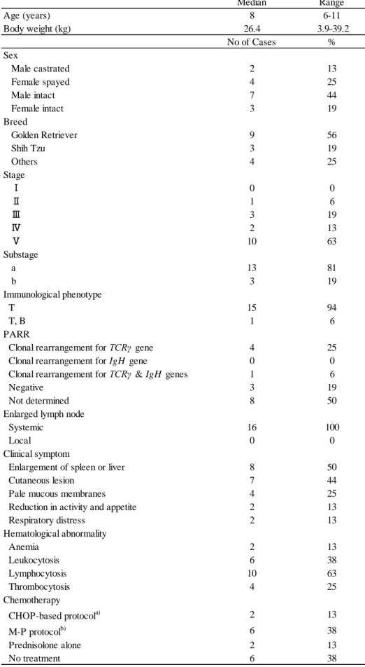
Sixteen dogs diagnosed with TZL included the following breeds: 9 Golden Retrievers (56%), 3 Shih Tzu (19%), and 1 case each of Cavalier King Charles Spaniel, Maltese, Pembroke Welsh Corgi, and mixed breed. The median age was 8 years (range: 6–11 years), and the sexes were as follows: 2 males, castrated; 4 females, spayed; 7 males, intact; and 3 females, intact.
Clinical conditions and complete blood cell count at diagnosis
All of the 16 dogs had generalized lymphadenopathy, primarily including submandibular, superficial cervical, axillary, inguinal, and popliteal lymph nodes. Splenomegaly and/or hepatomegaly were found in 8 of the 16 dogs. Cutaneous lesions were found in 7 dogs: 3 cases with demodicosis, 2 with pyoderma, 1 with lipoma, and 1 with otitis externa. Four dogs were found to have pale mucous membranes. Of the 16 cases, only 3 cases showed systemic symptoms such as a reduction in activity and appetite (2 cases), and weight loss (1 case). In the WHO clinical staging, 10 cases (63%) were shown to have a large number of typical “clear cells” (as observed in fine-needle aspiration [FNA] samples of the lymph node) in peripheral blood, and thus were classified into stage V. Of the 16 cases, 13 cases (81%) were
19
assigned into substage “a.” Consequently, WHO clinical stages and substages of the 16 dogs were IIa (n = 1), IIIa (n = 3), IVa (n = 1), IVb (n = 1), Va (n = 8), and Vb (n = 2).
In complete blood cell count at initial diagnosis, of the 16 dogs, 2 (13%) had mild anemia (hematocrit [Hct] 0.30, 0.34). Ten cases (63%) had lymphocytosis (5,230–19,200/μl) containing lymphoid cells characterized as “clear cells” in Kiel classification, in the peripheral blood (Fournel-Fleury et al., 1997). Four cases had thrombocytosis (511,000– 845,000/μl) above the laboratory’s reference range. Information regarding signalment, WHO clinical stages, substages, and clinical information is presented in Table 1–1.
Cytological and histopathological characteristics
The mature lymphoid cells similar to “clear cells” that increased in the peripheral blood in 10 cases had small- to medium-sized nuclei (an average size of 1 to 1.5 red blood cells) showing densely stained chromatin and relatively abundant clear cytoplasm (Fig 1–1. A, B). Such lymphoid cells were also found in the peripheral blood, even in cases without lymphocytosis, although these cases were not assigned to stage V in the WHO clinical staging system.
In the cytological specimens obtained from enlarged peripheral lymph nodes, TZL was composed of small- to medium-sized lymphocytes with clear cytoplasm sometimes showing extension to form hand-mirror or tear-drop image (Fig 1–2. A–D). The nuclei had little
20
internal detail and were small to medium in size, showed densely stained chromatin, and sometimes had sharp shallow indentations. Mitotic figures were quite rare.
Histopathologically, neoplastic cells were small to intermediate in size, and they expanded from the paracortex to the cortex and medullary cords. The capsule of the lymph node was at least focally thinned, although perinodal tissue was not involved. Fading follicles and germinal centers were found and tended to be peripheralized by the expanding population of neoplastic T cells (Fig 1–3. A, B).
PARR and immunohistochemistry
Of 16 dogs, 8 underwent genetic clonality analyses. Four of the 8 dogs with T-zone lymphoma showed monoclonal rearrangement of TCRγ. One dog showed clonal rearrangement of both IgH and TCRγ. In the other 3 dogs, clonal rearrangement of IgH or TCRγ was not detected by PARR.
Immunohistochemical analysis for CD3 and CD20 was performed for 16 dogs. In 15 dogs (94%), the majority of the lymphoid cells on the nodes were immunopositive for CD3. In 1 dog (6%), the tumor cells were double-positive for CD3 and CD20. The results regarding immunohistological phenotype and PARR are presented in Table 1–1.
Immunohistochemical analysis for the CD25 antigen was performed for 6 dogs with TZL that were positive for CD3, as confirmed by immunohistochemistry. In all 6 cases,
21
CD25-positive cells were found in accordance with CD3-positive and CD20-negative neoplastic cellular localization. Among the expanding population of neoplastic cells in the paracortical regions of nodes, almost all the cells were strongly immunopositive for CD25 (Fig 1–4. A−F).
Complications observed in the 16 dogs with TZL
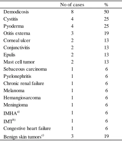
Eleven (69%) of 16 cases had complications during the disease course by follow-up (181–1,662 days; median, 841 days): demodicosis (8 cases), cystitis (4 cases), pyoderma (4 cases), otitis externa (3 cases), corneal ulcer (2 cases), conjunctivitis (2 cases), epulis (2 cases), mast cell tumor (2 cases), sebaceous carcinoma (1 case), pyelonephritis (1 case), chronic renal failure (1 case), melanoma (1 case), hemangiosarcoma (1 case), and meningioma (1 case). Three cases (19%) had a benign skin tumor (lipoma, papilloma, and cutaneous plasmacytoma) and there were no complications in 5 cases (31%) (Table 1–2).
Treatment and response to treatment
All cases were divided into 4 groups according to the treatment: dogs treated with [1] CHOP-based protocols (cyclophosphamide, doxorubicin, vincristine and prednisolone) (2 cases); [2] MP (melphalan and prednisolone) (6 cases); [3] prednisolone alone (2 cases); and [4] no chemotherapy (6 cases) (Table 1–1).
22
Chemotherapy with a CHOP-based protocol was conducted for 2 cases, because these dogs were found at the initial visit to have respiratory distress due to the lymphadenopathy. A follow-up of response to therapy was conducted for the 2 cases. After the treatment, 1 case attained a partial remission (PR); however, the other case did not respond to CHOP-based protocol.
Treatment with MP protocol was conducted for 6 cases, because of respiratory distress from enlargement of nodes (2 cases) and progressive lymphocytosis in the peripheral blood (4 cases). Of the 6 cases, 5 attained PR and 1 achieved CR. Prednisolone alone was administered for 2 cases. One case achieved PR, but the other did not show any response.
Watchful waiting was selected for 6 cases, because they did not show any symptoms caused by lymphoma during the follow-up period. Although these dogs did not receive any chemotherapy, the lymph node sizes were found to wax and wane for the observation period (10.7–37.1 months, median 26.2 months). One case developed diffuse large B-cell lymphoma (DLBCL) 37.1 months after the diagnosis of TZL.
Prognostic analyses
The median OST was 938 days, and the median LSST was not calculated because lymphoma-related death was found in only 3 dogs, and the number did not exceed half (Fig 1–5). One of the 3 dogs had respiratory distress from enlargement of nodes, and the other 2
23
dogs had disseminated intravascular coagulation (DIC) associated with progression of lymphoma. All 3 dogs that died of the progression of lymphoma received chemotherapy with CHOP-based protocol or MP protocol. Of the 16 dogs with TZL, 8 dogs died of any reason by the end of the follow-up duration. Cause of death other than lymphoma included metastasis of sebaceous carcinoma (1 dog), hemangiosarcoma (1), IMHA plus IMT (1), congestive heart failure (1) and melanoma (1).
A case that developed DLBCL after the onset of TZL died of the progression of DLBCL 45.8 months after the initial diagnosis of TZL. The dog was treated with CHOP protocol but was unresponsive to the chemotherapeutic agents. In the prognostic analyses, the case was included in the OST group and censored at onset of DLBCL.
24
Discussion
In this study, I evaluated the clinical and histological characteristics of 16 dogs with TZL and demonstrated that 10 cases with TZL were found to be in advanced clinical stage (stage V) at time of the initial diagnosis; however, 8 of these dogs retained good general conditions and 6 of 16 dogs with TZL showed long-term survival (10.7–37.1 months, median 26.2 months) even without any chemotherapy.
At the initial diagnosis or during the disease course, many cases were found to be affected with demodicosis or experienced the infectious diseases associated with mucocutaneous lesions. The peripheral lymph nodes biopsied from the 16 cases in this study showed characteristic histological appearances as reported by Valli et al (Valli et al., 2006; 2011). Moreover, immunohistochemistry revealed that the tumor cells of canine TZL were strongly positive for CD25.
Golden Retrievers and Shih Tzu accounted for three-fourths of all breeds evaluated, and other breeds included in the study were Cavalier King Charles Spaniel, Maltese, Pembroke Welsh Corgi, and mixed breed. It might be possible that TZL occurs with elevated frequency in Golden retrievers and Shih Tzu, as described in the previous reports (Flood-Knapik et al., 2013; Seelig et al., 2014). In this study, the median age at the time of diagnosis was 8 years (range: 6–11years). A previous study showed that the median age of 10 dogs with TZL was
25
8.9 years (Valli et al., 2006). Age of the dogs that developed TZL is conceivably similar or slightly older than that of dogs with lymphomas in general (Valli et al., 2013).
At the initial visit, generalized lymphadenopathies in more than 4 regions of the peripheral lymph nodes were found in all of the 16 cases in this study. Demodicosis was found frequently, occurring in 8 cases (50%) throughout the observation period. A previous report showed that almost 10% of canine cases with TZL had demodex infection at diagnosis (Flood-Knapik et al., 2013); however, many more cases with TZL in the study developed an infection of demodex at time of initial diagnosis (3 cases, 19 %) and during the treatment (8 cases, 50%). Demodex canis is normally present in dog skin but it has been assumed that immune dysfunctions and genetic disorders cause overgrowth of demodex in canine patients (Barriga et al., 1992; Sarkar et al., 2004). Humans undergoing chemotherapy have been shown to have an increased incidence of demodex infestation (Seyhan et al., 2004; Herron et al., 2005). Considering that many dogs with TZL developed demodicosis, canine TZL could be one of the important underlying diseases for the development of canine demodicosis. The immune abnormalities might be associated with the development of TZL and lead to a frequent occurrence of demoicosis. When peripheral lymphadenopathy is found in the presence of dermatologic problems, differentiation between hyperplastic/inflammatory and neoplastic changes should be made. Diagnosis as TZL should be made not just by cytology but by histopathological examination in order to discriminate accurately between neoplastic
26
proliferation and hyperplasia by cutaneous lesions. PARR also could be used for the demonstration of the clonal antigen of the lymphoma cells.
Immunohistochemically, the neoplastic cells of 6 cases with TZL examined were intensely positive for CD25. It has been known that heterotrimerization of interleukin-2 receptor α, β, and γ chains leads to high-affinity binding for IL-2 and IL-2/IL-2R complex and has an important role in controlling proliferation and differentiation of immune system cells (Lowenthal et al., 1985). In humans, previous reports have shown that CD25 expression is found in several types of leukemia or malignant lymphoma, including Hodgkin’s lymphoma (HL) and non-Hodgkin’s lymphoma (NHL), which express CD25 antigen in a great proportion of cases (Tesch et al., 1993; Nakase et al., 1994a). The complete biological role of IL-2Rα expression in tumors is unclear, although IL-2Rα can stimulate tumor cell proliferation (Strauchen et al., 1987; Peuchmaur et al., 1990; Tsilivakos et al., 1994). It has been reported that particular hematological tumors highly express the membranous CD25 forming an important phenotypic trait that could be a therapeutic target molecule (Sheibani et al., 1987; Waldmann et al., 1993; Horiuchi et al., 1997). A recent study revealed that canine TZL was associated with loss of CD45 expression, and CD45-negative cells expressed CD25 by flow cytometry analysis (Seelig et al., 2014). A large amount of CD25 expression on canine TZL cells might have some influence on proliferation of lymphoma cells or status of host immunity that leads to demodicosis in the dogs with TZL. It is important to learn more
27
about aberrant phenotypes in canine lymphomas, not only for accurate diagnostic information but also for the study of the pathophysiology of canine lymphoma.
In our study, of the 16 dogs, only 3 dogs died of lymphoma (TZL) at the end of the follow-up duration. All of them were treated with CHOP-based chemotherapy or MP protocol. Although the number of dogs treated with chemotherapy was small, response rates for dogs treated with CHOP-based chemotherapy or MP protocol were 50% and 100%, respectively. In dogs treated with prednisolone alone or those who did not undergo chemotherapy, there were no cases of death from progression of TZL. A previous report concluded that combination chemotherapy seemed to be less effective in dogs with TZL, compared with that of more aggressive lymphomas (Valli et al., 2007). It was also reported that systemic chemotherapy such as CHOP-based chemotherapy did not make a difference in improvement of outcome, and thus watchful waiting may be a reasonable approach in the early phase of the disease (Flood-Knapik et al., 2013). The findings from this study also concluded that therapeutic intervention based on aggressive chemotherapy was not necessarily required for all dogs with TZL. Moreover, MP protocol could be an effective and reasonable approach in terms of response rate and fewer side effects, if chemotherapy is required.
Clinical features associated with TZL were characterized in most cases by systemic generalized lymphadenopathy, splenomegaly and/or hepatomegaly, lymphocytosis, or reduction in activity. There have been no criteria for initiation of treatment and therapy
28
protocols, therefore chemotherapy was started for the cases with conceivable disease-related symptoms such as extreme fatigue, occurrence of anemia and/or thrombocytopenia, progressive lymphocytosis, or massive or progressive splenomegaly or lymphadenopathy in this study. This study included only a small number of cases. Therefore, further studies are required to clarify the optimal timing for the initiation of chemotherapy for canine TZL.
A watchful waiting approach was made for 6 (38%) of 16 cases in the study. During the clinical course, most cases retained a good health performance with repeated regression and swelling of lymph nodes for a long time, and no cases experienced the lymphoma-related death by the end of follow-up. It could be suggested that watchful waiting may be a reasonable approach for a proportion of dogs with TZL, especially for dogs without problems in general conditions and blood cytopenias.
In conclusion, TZL is a characteristic subgroup of lymphoma in dogs. Dogs with TZL show an indolent clinical course; however, it may be difficult to achieve a complete remission even if using chemotherapy. Special attention is needed to consider the diagnosis and treatment modalities when a dog is suspected to have TZL.
29
Fig 1–1. Neoplastic cells in PBMC.
(A) TZL cells are small or intermediate lymphocytes, and the cytoplasm is more abundant than that in normal lymphocytes and very lightly stained. × 1000.
(B) The nuclei are relatively uniform in size and about 1 to 1.5 red blood cells in diameter. The cytoplasm is abundant and has indentations. Wright-Giemsa stain, × 1000.
A
30
Fig 1–2. Neoplastic cells in lymph node.
(A) Cells with nuclei only slightly larger than red blood cells and sharp shallow nuclear indentation. The cytoplasm in most neoplastic cells is more abundant than normal small lymphocytes. × 1000.
(B) Cells with small nuclei and dense chromatin pattern. The cytoplasm is small or moderate in volume and clear. × 1000.
(C) Cells with dense chromatin pattern and highly extended cytoplasm. The nuclei have sharp shallow indentations and little internal detail. The nucleoli are inapparent. ×1000.
(D) Cells with small central nucleoli and abundant clear cytoplasm. Mitoses are not present in most fields. Wright-Giemsa stain,×1000.
C
D
31
Fig 1–3. The histological features of canine TZL.
(A) Neoplastic lymphocytes expand in the paracortex and are peripheralizing the fading lymphoid follicles. Hematoxylin and eosin (H&E) stain. × 40.
(B) Neoplastic cells are small to intermediate in size. The nuclei are small and have sharp shallow indentations. Some nuclei have small central nucleoli. H&E stain. × 1000.
A
32
Fig 1–4. The immunohistochemical features of canine TZL.
(A) Neoplastic T cells in the paracortex are uniformly immunopositive for CD3. Immunolabeling with anti-CD3. × 40.
(B) Intense labeling of neoplastic T cells with CD3. The neoplastic T cells are small or intermediate in size. Immunolabeling with anti-CD3. × 1000.
(C) B cells in fading lymphoid follicles are immunopositive for CD20. Follicular structures are pressed against the outer sinus by the expanding population of neoplastic T cells. Immunolabeling with anti-CD20. × 40.
(D) Neoplastic cells in the paracortex are negative for CD20. Immunolabeling with anti-CD20. × 1000.
(E) Neoplastic cells in the paracortex are strongly immunopositive for CD25. B cells in the lymphoid follicles are negative for CD25. Immunolabeling with anti-CD25. × 40.
(F) CD25-positive neoplastic cells are small to medium in size. Some cells have sharp shallow nuclear indentations and abundant cytoplasm. Immunolabeling with anti-CD25. × 1000.
A
B
C
D
33
Fig 1–5. Survival curves for overall survival and lymphoma-specific survival in 16 dogs with TZL.
34 Median Range Age (years) 8 6-11 Body weight (kg) 26.4 3.9-39.2 No of Cases % Sex Male castrated 2 13 Female spayed 4 25 Male intact 7 44 Female intact 3 19 Breed Golden Retriever 9 56 Shih Tzu 3 19 Others 4 25 Stage Ⅰ 0 0 Ⅱ 1 6 Ⅲ 3 19 Ⅳ 2 13 Ⅴ 10 63 Substage a 13 81 b 3 19 Immunological phenotype T 15 94 T, B 1 6 PARR
Clonal rearrangement for TCRγ gene 4 25 Clonal rearrangement for IgH gene 0 0 Clonal rearrangement for TCRγ & IgH genes 1 6
Negative 3 19
Not determined 8 50
Enlarged lymph node
Systemic 16 100
Local 0 0
Clinical symptom
Enlargement of spleen or liver 8 50
Cutaneous lesion 7 44
Pale mucous membranes 4 25 Reduction in activity and appetite 2 13 Respiratory distress 2 13 Hematological abnormality Anemia 2 13 Leukocytosis 6 38 Lymphocytosis 10 63 Thrombocytosis 4 25 Chemotherapy CHOP-based protocola) 2 13 M-P protocolb) 6 38 Prednisolone alone 2 13 No treatment 6 38
a) CHOP-based protocol: cyclophosphamide, doxorubicin, vincristine, and prednisolone. b) M-P protocol: melphalan and prednisolone.
35 No of cases % Demodicosis 8 50 Cystitis 4 25 Pyoderma 4 25 Otitis externa 3 19 Corneal ulcer 2 13 Conjunctivitis 2 13 Epulis 2 13
Mast cell tumor 2 13
Sebaceous carcinoma 1 6
Pyelonephritis 1 6
Chronic renal failure 1 6
Melanoma 1 6
Hemangiosarcoma 1 6
Meningioma 1 6
IMHAa) 1 6
IMTb) 1 6
Congestive heart failure 1 6 Benign skin tumorsc) 3 19 a) IMHA: immune-mediated hemolytic anemia.
b) IMT: immune-mediated thrombocytopenia.
c) Benign skin tumors: lipoma, papilloma, and plasmacytoma.
36
Chapter 2
Expression of interleukin-2 receptor alpha chain
(IL-2rα) in canine lymphoid tumour cells
37
Abstract
Interleukin-2 receptor alpha (CD25) expression has been reported in human lymphoid tumours and suggested to correlate with the prognosis. In this study, I evaluated the expression of CD25 in various canine lymphoid malignancies. Immunohistochemical analyses of the tissues from diffuse large B-cell lymphoma (DLBCL) (n=6), T-zone lymphoma (TZL) (n=5), and follicular lymphoma (FL) (n=2) revealed that intense CD25 expression was observed in accordance with lymphoma cell localization. CD25 expression was detected in all of the TZL and FL cases, while it was variable among DLBCL cases. Furthermore, I evaluated CD25 expression in tumour cells by flow cytometric analysis in 29 dogs with lymphoid malignancies, including high-grade B-cell lymphoma (n=17), TZL (n=5), FL (n=2), cutaneous lymphoma (n=2), and acute lymphoblastic leukaemia (ALL) (n=3). CD25 expression was significantly higher in dogs with high-grade B-cell lymphoma (n=17, mean±SD, 49.6±31.3%) or TZL (n=5, mean±SD, 80.2±10.0%), but significantly lower in dogs with ALL (n=3, mean±SD, 3.3±2.2%) than that in healthy dogs. In prognostic analysis of 15 cases with high-grade B-cell lymphoma, the progression-free survival was significantly shorter in CD25-high group than that in CD25-low group. The results obtained in this study are useful for subtype differentiation and prognostic analysis of canine lymphomas and future development of molecular-targeted therapy directed at CD25.
38
Introduction
Interleukin (IL)-2 receptor alpha chain (CD25) is a component of IL-2 receptor and has two other subunits, beta and gamma chains. Heterotrimerization of these subunits leads to high-affinity binding to IL-2, which induces proliferation and maturation of lymphocytes (Lowenthal et al., 1985).
In human medicine, CD25 expression has been reported in activated normal T cells and B- and T-cell neoplasms, whereas only <5% of unstimulated peripheral blood T cells were CD25-positive (Robb et al., 1981; Sheibani et al., 1987). Particularly intense expression of CD25 was observed in blast cells in adult T-cell leukaemia (ATL) or hairy-cell leukaemia patients (Sheibani et al., 1987; Horiuchi et al., 1997). Expression of CD25 has also been demonstrated in B-cell chronic lymphocytic leukaemia, acute lymphoblastic leukaemia (ALL) (Burton et al., 1994; Nakase et al., 1994a, b), Hodgkin’s lymphoma, and non-Hodgkin’s lymphoma (Tesch et al., 1993; Nakase et al., 1994a). Recently, it was reported that CD25 expression was associated with poor response rates and inferior progression-free survival (PFS) in diffuse large B-cell lymphoma (DLBCL) or follicular lymphoma (FL), and are expected to constitute a new prognostic marker (Fujiwara et al., 2013, 2014).
39
lymphocytes (Helfand et al., 1992; Galkowska et al., 1996). Although expression of CD25 messenger RNA (mRNA) has also been reported in canine lymphoma and leukaemia (Dickerson et al., 2002), the relationship between its expression level and the prognosis has not been investigated.
In the present study, I evaluated CD25 expression in various subtypes of canine lymphoma and ALL by using immunohistochemistry (IHC) and flow cytometry (FCM). Furthermore, I analysed the relationship between CD25 expression and the prognoses for dogs with high-grade B-cell lymphoma.
40
Materials and methods
Samples for IHC
Thirteen dogs with enlarged peripheral lymph nodes who were referred to the Veterinary Medical Center of the University of Tokyo, or whose tissue samples were subjected to histopathological examination at North Lab, were diagnosed as DLBCL (n=6), TZL (n=5), and FL (n=2) based on the histopathological findings of the biopsy specimens by referring World Health Organization (WHO) histological classification system for haematopoietic tumours of domestic animals (Valli et al., 2011). These tissue samples were further subjected to IHC to examine the expression of CD25. As controls, lymph node specimens from seven healthy dogs and six dogs with reactive lymphadenopathy were also subjected to this analysis.
IHC
Sections of formalin-fixed paraffin-embedded tissues (4 μm) were prepared using the following protocol. The sections were de-waxed, rehydrated, and subjected to antigen retrieval in 1% citrate buffer solution (pH 6.0) in an autoclave at 120°C for 15 min. Endogenous peroxidase was inactivated by 1% hydrogen peroxide in methanol for 30 min, and blocking was processed by 8% skim milk in Tris-buffered saline (TBS) for 40 min. The
41
slides were then incubated overnight at 4°C with the panel of anti-CD3 (polyclonal rabbit anti-human A0452; DAKO, Glostrup, Denmark; 1:50 dilution), anti-CD20 (polyclonal rabbit anti-human RB-9013-P; Thermo Fisher Scientific, Waltham, MA, USA; 1:400 dilution), or anti-CD25 (monoclonal mouse anti-human, clone 4C9; Thermo Fisher Scientific; 1:40 dilution). After washing for 15 min in TBS, slides underwent reactions with EnVision + Dual Link System-HRP (DAKO) for 45 min at room temperature, were washed for 15 min, and visualized using 3,3’-diaminobenzidine (DAB) as a chromogen. Slides were counterstained in Mayer’s hematoxylin.
In six DLBCL cases, the number of CD25-positive lymphoma cells was counted and classified into three groups: group 1 with <20% positive tumour cells, group 2 with 20–60% positive tumour cells, or group 3 with >60% positive tumour cells.
Samples for FCM analysis
Neoplastic cells were obtained from 29 dogs with lymphoid malignancies who were referred to the Veterinary Medical Center of the University of Tokyo. Samples were obtained from peripheral lymph nodes in 24 nodal lymphoma cases, cutaneous masses in two cutaneous lymphoma cases, and peripheral blood separated by Ficoll-Paque gradient centrifugation (Ficoll-Paque PLUS; GE Healthcare, Amersham Place, England) from three ALL cases at the initial diagnoses. Diagnosis was made based on clinical data and
42
cytological/histological examination of lymph-node or bone marrow in dogs with lymphoma and leukaemia, respectively. PCR for antigen-receptor gene rearrangement (PARR) was performed to determine the clonality and cell lineage of 26 lymphoma and three ALL cases. DNA samples of all cases were extracted from fresh samples from the lymph node, peripheral blood, or bone marrow. Primers and protocols for PCR analyses were described previously (Burnett et al., 2003; Valli et al., 2006). Twenty six dogs with lymphomas were further classified into subtypes of high-grade B-cell lymphoma (n=17), TZL (n=5), FL (n=2), or cutaneous lymphomas (n=2) according to the cytology and/or histopathology (Fournel-Fleury et al., 1997, 2002; Valli et al., 2006, 2011). For comparison, samples were also obtained from peripheral lymph nodes from seven healthy dogs and six dogs with reactive lymphadenopathy.
FCM
Single-color FCM was performed for the cell samples obtained from dogs with lymphoid malignancies. Cell suspensions were washed with staining medium (phosphate-buffered saline supplemented with 5% foetal calf serum) and stained with fluorescein isothyocyanate (FITC)-conjugated anti-canine CD25 mAb (P4A10; eBioscience, San Diego, CA, USA) for 30 min at 4°C. Cells were washed twice and analysed with a flow cytometer (FACSCalibur; BD Biosciences, San Jose, CA, USA). An FITC-conjugated mouse IgG1 was used to analyse
43
the background fluorescence intensity during each analysis. Lymphoma or leukaemic cells were gated from the forward- and side-scatter properties. A minimum of 10,000 events in the gated region was collected for each sample. The obtained data were analysed using FlowJo software (Tree Star, Ashland, OR, USA). The percentages of CD25-positive cells were calculated from the isotype-matched control for each sample.
Statistical analysis
The Mann-Whitney U test was used to compare the prognosis in dogs with DLBCL showing different CD25 expression. PFS and overall survival (OS) were calculated according to the criteria proposed by the veterinary cooperative oncology group (Vail et al., 2010). PFS was calculated from the date of initial treatment to disease progression or death from any cause. OS was calculated from the date of initial treatment to death from any cause. Dogs that were still alive or lost to follow-up were censored at the date on which they were last known to be alive. PFS and OS were estimated by the Kaplan-Meier method, and the groups were compared by the log-rank test. The p values were two-sided and regarded as significant if p < 0.05. Data were analysed using StatMate software version IV for windows (ATMS, Tokyo, Japan).
44
Results
IHC for CD25 expression
Six dogs with DLBCL, five dogs with TZL, two dogs with FL, six dogs with reactive lymphadenopathy, and seven healthy dogs were subjected to immunohistochemical analyses to examine CD25 expression.
In seven healthy dogs and six dogs with reactive lymphadenopathy, CD25 expression was generally weak in both CD3-positive and CD20-positive cells, regardless of lymph node regions (Fig 2–1. A–D). Less than 10% of the cells in the entire lymph node field were CD25-positive in these dog groups.
Next, CD25 expression was examined in the lymph nodes derived from dogs with TZL. In the lymph nodes from five dogs with TZL, neoplastic T cells, which are small or intermediate in size, expanded in the paracortex, pressing the atrophic germinal centres against the outer sinus. Majority of the neoplastic CD3-positive cells were shown to be strongly immunopositive for CD25 (Fig 2–2. A–D).
Further, I evaluated CD25 expression in the lymph nodes of FL cases. In the two FL cases included in this study, the majority of CD20-positive centrocytes in the follicles were shown to be strongly immunopositive for CD25 (Fig. 2–3. A–D).
45
neoplastic large B cells with uniformly large nuclei and scanty cytoplasm diffusely proliferated in the lymph nodes as a mixture of centroblastic cells with multiple nucleoli and immunoblastic cells with a single central prominent nucleolus. Unlike in TZL and FL, CD25 expression in the lymph nodes of DLBCL was variable. Two of the six cases showed relatively weak CD25 expression and were categorized as group 1 (<20% positive tumour cells; Fig 2–4. A, B). Two cases showed intermediate CD25 expression and were judged as group 2 (20–60% positive tumour cells; Fig 2–4. C, D), and the remaining two cases strongly expressed CD25 and were categorized as group 3 (>60% positive tumour cells; Fig 2–4. E, F). Morphological features were not different among the six DLBCL cases exhibiting various degrees of CD25 expression.
FCM for CD25 expression
Samples from 26 dogs with lymphoma, three dogs with ALL, six dogs with reactive lymphadenopathy, and seven healthy dogs were subjected to FCM. Immunological subtypes of tumour samples were determined by PARR. Information regarding signalment, staging at the initial diagnosis, and immunological phenotype is presented in Table 2–1.
Single-color FCM was performed to evaluate CD25 expression in the lymphoid neoplastic cells. The percentages (mean±SD) of lymph node-derived lymphocytes that were CD25-positive in healthy dogs and dogs with reactive lymphadenopathy were 9.8±2.8% (n=7,
46
range: 6.4–13.8%) and 13.7±5.0% (n=6, range: 8.3–22.2%), respectively. CD25 expression was 49.6±31.3% (n=17, range: 0.4–97.1%) in high-grade B-cell lymphoma, 80.2±10.0% (n=5, range: 64.6–95.4%) in TZL, 69.4±22.3% (n=2, range: 47.1–91.7%) in FL, 73.7±16.5% (n=2, range: 57.2–90.2%) in cutaneous lymphoma, and 3.3±2.2% (n=3, range: 1.5–6.4%) in ALL. The percentages of CD25-positive cells were significantly higher in high-grade B-cell lymphoma and TZL, but significantly lower in ALL than those in healthy dogs (Fig 2–5).
Therapeutic response and prognosis of dogs with high-grade B-cell lymphoma
In order to examine the relationship between CD25 expression and prognosis, the clinical course of 15 dogs with high-grade B-cell lymphoma, who received CHOP (cyclophosphamide, doxorubicin, vincristine, and prednisolone)-based protocols (Garret et al., 2002), were followed for up to 919 days. Since the mean value of CD25-positive tumours among the 15 cases was 58.9%, the cut-off value was set to 60% in this study. Fifteen dogs were divided into two groups: CD25-positive cells > 60% (CD25-high) (n=7) and <60% (CD25-low) (n=8). Three of seven CD25-high dogs (42.9%) responded to the treatment, while seven of eight CD25-low dogs (87.5%) showed the response to treatment. The response rate was not significantly different between the CD25-high and CD25-low dogs. The median value of PFS was 28 days in CD25-high dogs, which was significantly shorter than that observed for CD25-low dogs (median: 140 days; Fig 2–6. A). Kaplan-Meier curves also
47
showed an apparently inferior OS in the CD25-high group (median: 115 days) relative to the CD25-low group (median: 244 days). However, there was no significant difference between the two groups (Fig 2–6. B).
48
Discussion
IHC revealed that neoplastic lymphocytes strongly expressed CD25 in all cases of TZL and FL, while lymphocytes of healthy dogs and dogs with reactive lymphadenopathy weakly expressed CD25. However, CD25 expression was variable among DLBCL cases. In all of these types of lymphoma, CD25 expression was predominantly detected in malignant cells. The surrounding reactive lymphoid cells were negative or weakly immunopositive for CD25. In agreement with IHC results, FCM revealed that non-neoplastic lymphocytes of healthy dogs and dogs with reactive lymphadenopathy showed low CD25 expression as compared to lymphoma cells at the initial diagnosis.
In several types of leukaemia or malignant lymphoma in humans, CD25 was shown to be expressed mainly in tumour cells (Waldmann, 1986; Sheibani et al., 1987). Participation of the α subunit of the IL-2R complex was shown to be necessary for the formation of the high-affinity IL-2 receptor in human lymphocytes (Gutgsell et al., 1994; Cassell et al., 2002). Since it was reported that CD25-positive lymphoma cells also produce IL-2 (Sheibani et al., 1987; Peuchmaur et al., 1990), IL-2/IL-2R signalling might activate the proliferation of lymphoma cells (Olejniczak et al., 2008). In dogs, it was shown that multicentric lymphoma cells expressed mRNA of interleukin-2 receptor α, β, and γ chains (Dickerson et al., 2002). The resulting surface IL-2R on lymphoma cells had binding affinity for IL-2. Moreover, it
49
has been suggested that canine lymphoma cells expressing IL-2R might maintain their proliferative state through an IL-2-dependent autocrine-growth pathway (Helfand et al., 1995). The expression of CD25 in lymphoma cells shown in this study might result in a high-affinity binding to IL-2 leading to lymphoma cell proliferation; however, it remains unknown whether IL-2/IL-2R signalling on each type of canine lymphoma is functional.
Notably, neoplastic lymphocytes from three ALL cases (mean±SD: 3.3±2.2%) showed weak CD25 expression. In human ALL, CD25 expression has been demonstrated (Nakase et al., 1994c), with expression levels significantly lower in T-lineage ALL relative to B-lineage ALL (Nakase et al., 2007). In this study, the three cases of ALL were found to be of T-cell origin based on results of CD3 expression from FCM and clonal rearrangement of the TCRγ gene in PARR. Although the number of ALL dogs analysed in our study was only three, the results showing that canine T-ALL expressed low levels of CD25 was consistent with a previous study, which showed no expression of IL-2Rα mRNA in a dog with T-ALL (Dickerson et al., 2002). The result obtained in this study may indicate that expression of CD25 in canine T-ALL is different from that in canine T-cell lymphoma.
In agreement with IHC results, FCM analysis also demonstrated that neoplastic lymphocytes of TZL and FL cases showed high CD25 expression. Canine cutaneous lymphomas also expressed high levels of CD25. In humans, high CD25 expression in lymphoma cells was also reported in cutaneous T-cell lymphomas (CTCL) and FL (Talpur et
50
al., 2006; Fujiwara et al., 2014). Moreover, in human CTCL, CD25 expression was more commonly observed in lesions from advanced patients, and high CD25 expression was associated with clinical tumour-node metastasis stage and histologic grade (Talpur et al., 2006). In this study, all cases of TZL and FL were found to have systemic lymphadenopathy at their initial diagnosis. Therefore, CD25-postitivity might be associated with tumour-node metastasis stage, as is the case with human lymphoma.
However, CD25 expression levels were variable in high-grade B-cell lymphoma (mean±SD: 49.6±31.3%, range: 0.4–97.1%). In human DLBCL, the rate of CD25-positive neoplastic cells was significantly higher than that observed for reactive lymphadenopathy. Similar to our results, the rates of CD25-positive cells detected by single-color FCM varied among human DLBCL cases (n=123; mean±SD: 27.8±30.6%) (Fujiwara et al., 2013). In view of the prognosis, human DLBCL was shown to be a heterogeneous group based on the pattern of gene expression or particular protein expression (Alizadeh et al., 2000; Hans et al., 2004). As with human DLBCL, canine high-grade B-cell lymphoma might comprise several different subtypes of lymphoma. It is most likely that canine high-grade B-cell lymphoma could be subdivided on the basis of gene or protein expression pattern in tumour cells.
I investigated the relationship between CD25 expression and the prognosis in dogs with high-grade B-cell lymphoma. PFS was significantly shorter in the CD25-high group (median: 28 days) than that in the CD25-low group (median: 140 days). Although not significant, the
51
response rate tended to be higher in the CD25-low group than that in the CD25-high group. In human DLBCL patients with high CD25 expression, response rate and PFS were significantly inferior to those with low CD25 expression (Fujiwara et al., 2013). While the biological role of CD25 expression or IL-2/IL-2R signalling in canine lymphoma cells is not clear, the tumour cells express large amounts of CD25, which most likely induces proliferation through IL-2/IL-2R binding, as shown in human lymphoma. In canine high-grade B-cell lymphoma, strong CD25 expression might be associated with proliferation rates and result in a shorter PFS in CD25-high groups. Moreover, CD25 might be an important phenotypic trait and a novel therapeutic target molecule in particular lymphoma subtypes.
In summary, I demonstrated the expression of CD25 in canine lymphoid tumours and that its phenotype varied between subtypes. Moreover, CD25 expression was related with the prognoses in high-grade B-cell lymphoma cases. The results obtained in this study contribute to the subtype differentiation, prognostic analysis, and future development of molecular-targeted therapy directed at CD25.
52
A
B
C
D
Fig 2–1.
Immunohistochemistry to detect CD25 expression in the lymph nodes of (A, B) a healthy dog and (C, D) a dog with reactive lymphadenopathy. Magnification for A and C:×40; B and D: ×1,000.
53
Fig 2–2. Immunohistochemistry of a lymph node in a dog with T-zone lymphoma.
Immunolabeled with (A) anti-CD3, (B) anti-CD20, or (C, D) anti-CD25. Magnification for A–C: ×40; D: ×1,000.
A
B
54
Fig 2–3. Immunohistochemistry of a lymph node in a dog with follicular lymphoma.
Immunolabeled with (A) anti-CD3, (B) anti-CD20, or (C, D) anti-CD25. Magnification for A–C: ×40; D: ×1,000.
A
B
55
B-cell lymphoma for CD25.
Fig 2–4. Immunohistochemistry of three lymph node samples from dogs with diffuse large B-cell lymphoma.
(A) Group 1 (CD25-positive tumour cells < 20%), (B) Group 2 (expression 20–60%), and (C) Group 3 (expression > 60%). Magnification for A, C, and E: ×40; B, D, and F: ×1,000.
A
B
C
D
F
56
Fig 2–5. Flow cytometric analysis of the lymphoid cell samples from dogs with lymphoid tumours and reactive lymphadenopathy and healthy dogs for CD25.
Lymph-node aspirates were obtained from 17 dogs with high-grade B-cell lymphoma, five dogs with T-zone lymphoma (TZL), two dogs with follicular lymphoma (FL), six dogs with reactive lymphadenopathy, and seven healthy dogs.
Aspirates of the cutaneous masses were obtained from two dogs with cutaneous lymphoma. Peripheral blood mononuclear cells were obtained from three dogs with acute lymphoblastic leukaemia (ALL).
Horizontal barsindicate the mean value of each group.
*
CD25 expression is significantly higher as compared to healthy dogs. p < 0.05, Mann-Whitney U test.
57
(A)
(B)
Fig 2–6. Kaplan-Meier plot of (A) progression-free survival and (B) overall survival in the CD25-high and CD25-low groups of high-grade B-cell lymphoma.
Dogs with CD25-positive cells > 60% were categorized as the CD25-high group (n=7), and those with CD25-positive cells < 60% as the CD25-low group (n=8). The two groups were compared using the log-rank test.
58 Cases % Sex Male, castrated 7 27 Female, spayed 9 35 Male, intact 6 23 Female, intact 4 15 Breed
Pembroke Welsh Corgi 7 27
Miniature Dachshund 4 15 Shih Tzu 4 15 French Bulldog 2 8 Pomeranian 2 8 Mixed breed 2 8 Others 5 19
WHO clinical stage
Ⅰ 1 4 Ⅱ 0 0 Ⅲ 3 12 Ⅳ 2 8 Ⅴ 20 77 Substage a 19 73 b 7 27 Hitological/cytological subtype
High-grade B-cell lymphoma 17 65
TZLb 5 19 FLc 2 8 Cutaneous lymphoma 2 8 a FCM, flow cytometry. b TZL, T-zone lymphoma. c FL, follicular lymphoma.
59
Chapter 3
Measurement of the concentration of serum
soluble interleukin-2 receptor alpha chain
60
Abstract
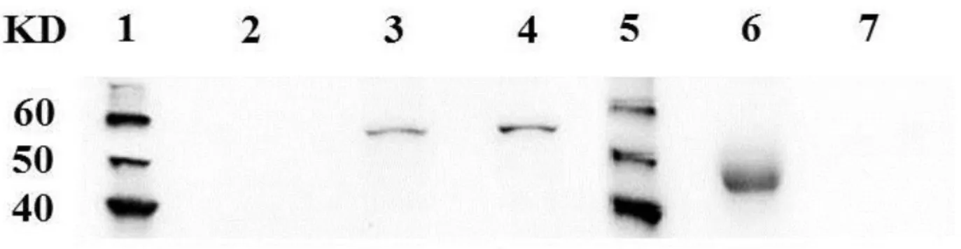
Soluble interleukin 2 receptor (sIL-2r) is released directly from the surface of lymphocytes expressing interleukin 2 receptor alpha chain (CD25) and its serum concentration has been found to reflect the prognosis in human lymphoproliferative malignancies. In this study, I demonstrated the existence of sIL-2r in canine serum and developed a specific sandwich enzyme-linked immunosorbent assay (ELISA) to quantify the concentration of canine serum sIL-2r. Using immunoprecipitation (IP) assay, CD25 protein weighing approximately 45 KDa was detected in canine serum, smaller than the membrane-bound CD25 (approximately 55 KDa). To measure the concentration of serum sIL-2r in dogs, ELISA system was developed. Serum sIL-2r level was significantly higher in dogs with multicentric high-grade B-cell lymphoma before therapy than that in healthy dogs and dogs with inflammatory diseases. Serum sIL-2r concentration was also found to be elevated in a proportion of dogs with other lymphoma types and immune-mediated diseases. Changes in serum sIL-2r levels generally paralleled with the changes of lymph node size in dogs with high-grade B-cell lymphoma. This study demonstrated that serum sIL-2r level would be a marker to monitor the tumor growth and regression in canine lymphoma.
61
Introduction
Interleukin (IL)-2 receptor alpha chain (CD25) is a component of IL-2 receptor (IL-2r) together with other subunits, beta and gamma chains. Heterotrimerization of these three subunits leads to high-affinity binding to IL-2, which induces proliferation and maturation of lymphocytes (Lowenthal et al., 1985). CD25 is shown to be a membrane-bound protein of approximately 55 kDa in humans and dogs (Leonard et al., 1983; Abrams et al., 2010). In humans, soluble IL-2 receptor (sIL-2r), which is smaller than its membrane-bound form (45 kDa vs. 55 kDa), can be detected in the serum of healthy individuals (Rubin et al. 1985, 1990). Elevation of the serum sIL-2r concentration has been shown in human patients with various lymphoid malignancies including Hodgkin’s lymphoma (HL), non-Hodgkin’s lymphoma (NHL), hairy cell leukemia (HCL) and adult T-cell leukemia/lymphoma (ATL) (Rubin et al., 1990; Tesch et al., 1993; Nakase et al., 1994a). Several reports demonstrated the relation between serum sIL-2r concentration and the clinical outcome in these diseases (Kono et al., 2000; Niitsu et al., 2001; Oki et al., 2008; Morito et al., 2009). Therefore, serum sIL-2r concentration has been used as a biological marker to indicate the growth of tumour cells as well as the prognosis in lymphoid malignancies in humans, especially in NHL.
62
per 100,000 dogs per year (Dorn et al., 1968; Dobson et al., 2002; Mellanby et al., 2003). Lymphoma accounts for approximately 80% of hematopoietic malignancies in dogs (Dorn et al., 1968). Similar to humans, CD25 is expressed on the cell surface of T-lymphocytes during activation and proliferation in dogs (Helfand et al., 1992; Galkowska et al., 1996). Expression of CD25 messenger RNA (mRNA) has also been detected in canine lymphoma and leukaemia cells (Dickerson et al., 2002). Moreover, it has been suggested that canine lymphoma cells expressing CD25 maintain their proliferative state through an IL-2-dependent autocrine growth pathway (Helfand et al., 1995). In Chapter 1 in this thesis, strong expression of CD25 was revealed in the tumour cells derived from T-zone lymphoma (TZL) in dogs. Next, in Chapter 2, CD25 expression was detected in the tumour cells derived from diffuse large B-cell lymphoma, follicular lymphoma (FL), and cutaneous lymphoma. However, there has been no study to evaluate the existence of sIL-2r in dogs so far.
In this chapter, I carried out a study to examine the presence of sIL-2r in canine serum and quantify its concentration in canine serum using a newly developed sandwich enzyme-linked immunosorbent assay (ELISA) system. Moreover, sequential change of the serum sIL-2r concentration was examined in 4 dogs with lymphoma that had relatively high serum sIL-2r concentrations before chemotherapy.
63
Materials and methods
Antibody production
A rabbit polyclonal antibody (PAb) directed to a 20-amino acid peptide, CISESQFPDDEELQASTDAP, corresponding to the extracellular domain of canine CD25 (Dickerson et al., 2002) was synthesized. Rabbits were immunized with the synthesized peptide conjugated to keyhole limpet hemocyanin together with complete Freund’s adjuvant (Sigma-Aldrich, St. Louis, MO). Antiserum was subjected to an affinity purification column (Sigma-Aldrich) with the peptide used for the immunization. The purified polyclonal antibody was biotinylated as indicated by the manufacturer (EZ-Link NHS-Biotin; Pierce, Rockford, IL) for sandwich ELISA.
Sandwich ELISA
For the sandwich ELISA, microplates (96-well) (Immuno Plate Maxisorp C96; Nunc, Roskilde, Denmark) were coated with 2 μg/ml mouse anti-canine CD25 mAb (P4A10; AbD Serotec, Kidlington, UK) diluted in 50mM carbonate buffer (pH 9.6) and incubated overnight at 4°C. The antibody-coated plates were then washed with PBS containing 0.05% (v/v) Tween 20 (PBS-T) three times. PBS containing 5% rabbit serum (Sigma-Aldrich), 2% block ace (DS Pharma Biomedical, Osaka, Japan) and 0.4 M NaCl was added to each well as a