St udi es on i nhi bi t or y ef f ec t of ool ong t ea
pol yphenol on oxi dat i ve s t r es s
その他のタイトル
(ウーロン茶ポリフェノールの酸化ストレス抑制効
果に関する研究)
著者
SU
KH
BO
LD
EN
KH
TSETSEG
学位名
博士(工学)
学位授与機関
北見工業大学
学位授与番号
10106甲第160号
研究科・専攻名
生産基盤工学専攻
学位授与年月日
2017- 09- 11
Doctoral Thesis
Studies on inhibitory effect of oolong tea
polyphenol on oxidative stress
(
ー
ン茶ポ
フ
ノー
の酸化スト
ス抑制効果に
関する研究
)
Kitami Institute of Technology
Manufacturing Engineering
SUKHBOLD ENKHTSETSEG
Contents
Abstract p1
Chapter 1 General introduction p3
Chapter 2 Radical scavenging capacity of oolonghomobisflavan A (OFA) in solution
2.1 Objectives p17 2.2 Materials and methods p17 2.2.1 Materials p17
2.2.2 DPPH radical scavenging assay p17
2.3 Results p17 2.3.1 DPPH radical scavenging capacity of OFA in solution p17
Chapter 3 Effect of OFA on low-density lipoprotein (LDL) oxidation by reactive oxygen species (ROS)
3.1 Objectives p20 3.2 Materials and methods p20 3.2.1 Materials p20
3.2.2 Isolation and purification of LDL p20
Chapter 4 Effect of OFA on LDL oxidation by reactive nitrogen species (RNS)
4.1 Objectives p31
4.2 Materials and methods p31
4.2.1 Materials p31
4.2.3 Peroxynitrite-mediated oxidation of LDL p31 4.2.4 Peroxynitrite-mediated nitrotyrosine analysis p31 4.3 Results p31 4.3.1 Effect of OFA on lipid peroxidation of LDL by RNS p32 4.3.2 Effect of OFA on protein oxidation of LDL by RNS p32
Chapter 5 Discussion p36
Chapter 6 Conclusions p40
References p41
1
Abstract
Oolong tea contains unique polymerized polyphenols including oolonghomobisflavan A (OFA) that is generated from green tea leaves during the semi-fermentation process. It has been reported that intake of oolong tea has beneficial effects on health, although the antioxidant effects of OFA remains unclear.
Oxidation of low-density lipoprotein (LDL) by reactive oxygen species (ROS) and reactive nitrogen species (RNS) has been suggested to be involved in the onset of atherosclerosis. We studied effect of OFA on LDL oxidation by ROS and RNS in vitro. LDL oxidation was induced by a peroxyl radical-generating reagent (AAPH), transition-metal ions (Cu2+), or a peroxynitrite generator (SIN-1) in the presence of OFA. Cholesterol ester hydroperoxide (CE-OOH) and thiobarbituric acid reactive substances (TBARS) were determined by HPLC as the indices of lipid peroxidation of LDL, while protein fragmentation, protein carbonyl formation, and nitrotyrosine formation were analyzed by SDS-PAGE and western blotting as the indices of oxidative modification of apolipoprotein B-100 (apo B-100) in LDL. OFA suppressed CE-OOH formation in LDL oxidized by AAPH or SIN-1, and formation of TBARS in LDL oxidized by Cu2+. In addition, OFA inhibited fragmentation, carbonylation, and nitration of apo B-100, in which the heparin-binding activity of apo B-100 was protected by OFA. These results suggest that OFA has antioxidant activity to suppress both lipid peroxidation and oxidative modification of apo B-100 in LDL oxidized by ROS and RNS. Polymerized polyphenols in oolong tea may contribute to prevent atherosclerosis by reducing oxidative stress.
Keywords
2
Abbreviations
3
Chapter 1
General introduction
Tea is one of the most widely consumed plant-based beverage for its specific aroma, taste and healthy effects. Freshly harvested tea leaves are processed differently to produce specific types of tea. Oolong is a semifermented tea prepared from leaves of Camellia
sinensis, while green tea is an unfermented tea. The fermentation of oolong tea is not
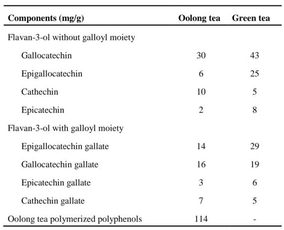
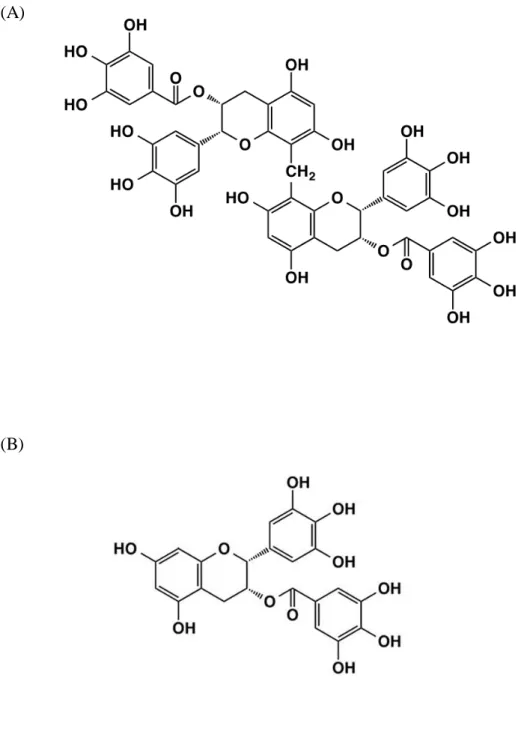
mediated by microbes, but instead, it is mediated by oxidative enzymes such as polyphenol oxidase and peroxidase in the leaves.1) The fermentation process induces browning and generates unique flavors and tastes. Depending on the method of preparation including degree of oxidation, various oolong teas are available in Asian countries. Several studies have been reported that oolong tea has a wide range of beneficial health effects including antihyperglycemic effects,2) mitigation of the risk of cardiovascular disease.3) The major components in oolong tea are alkaloids, saponins, polysaccharides, L-theanines, and polyphenols.4,5) The degree of oxidation in fermentation is affected to polyphenols of oolong tea. Oolong tea contains polymerized polyphenols derived from the oxidative polymerization of catechins such as oolonghomobisflavans.6) The polymerized polyphenols are not presented in green tea (Table 1.1).7) Oolonghomobisflavan A (OFA, Figure 1.1 A) are most abundant oolong tea polymerized polyphenol (OTPP), has a unique structure as a dimer of epigallocatechin gallate (EGCG, Figure 1.1 B).8) The biological effects of oolong tea polyphenols have been attracted considerable attention in recent years, although the mechanism remains unclear.
4
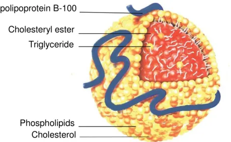
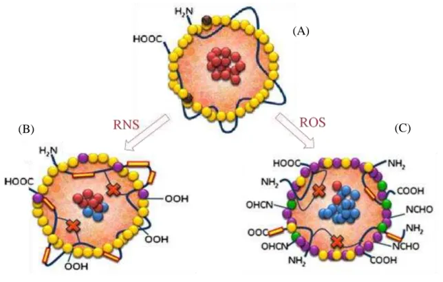
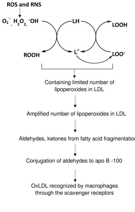
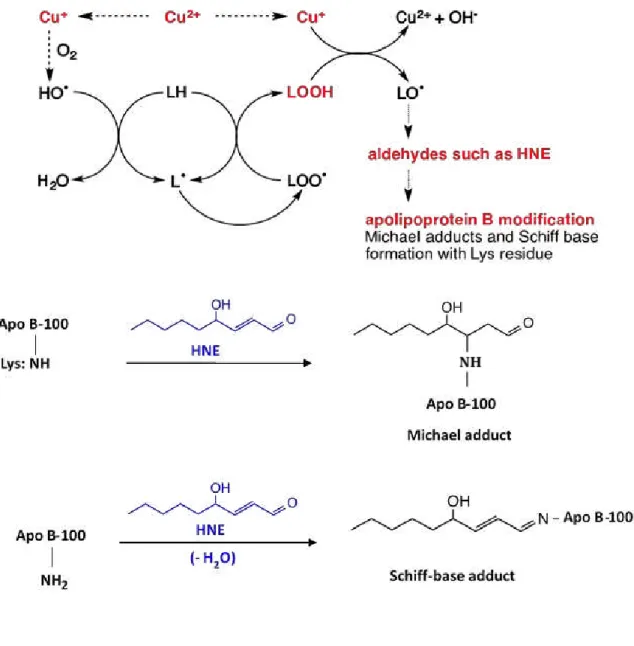
monolayer containing unesterified cholesterol and apolipoprotein B-100 (apo B-100) (Figure 1.2).11,12) LDL is major cholesterol transporters,9) it has been suggested that the increasing of LDL in blood and the oxidation of LDL are positively associated with the onset of cardiovascular diseases.13) A high level of reactive oxygen species (ROS) and reactive nitrogen species (RNS) in human body can inflict damage to the structure and function of LDL (Figure 1.3),14) which may lead to atherosclerosis.14-16) LDL oxidation involves lipid peroxidation and modification of apolipoprotein.17) Several studies indicate that the process of LDL oxidation in vitro occur in two main step, which is minimally oxidation and extensively oxidation of LDL (Figure 1.4).18,19) During the minimally oxidation of LDL occur little modification in apo B-100, which has low affinity to macrophages scavenger receptors.19) Subsequently, unsaturated fatty acid moieties of lipids and apo B-100 are further oxidized resulting to recognition by macrophages through the scavenger receptors.19) Unsaturated fatty acid moieties of lipids can be oxidized by ROS
such as hydroxyl radical (·OH) and RNS such as peroxynitrite (ONOO-), and generating
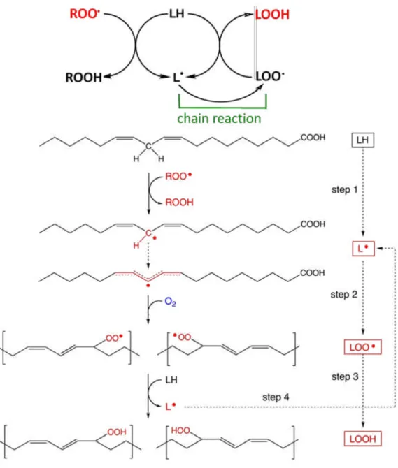
lipid hydroperoxides17,20) that process conducts by free radical chain reaction (Figure 1.5). Polyunsaturated fatty acid side chain (LH) containing multiple double bonds in between which lie methylene bridges reactive hydroxyl radical (·OH) (reaction 1).21) The resulting, lipid radical (L·) adds rapidly to molecular oxygen (O2) to generate a lipid peroxyl radical
(LOO·) (reaction 2).21)
That itself can propagate the chain by reacting with another polyunsaturated fatty acid to generate L· and lipid hydroperoxide (LOOH) (reaction 3).21) Reactive aldehydes and ketones are the end products of lipid hydroperoxide.22)
LH + ·OH L· + H2O (1)
L· + O2 LOO· (2)
LOO· + LH L· + LOOH (3)
5
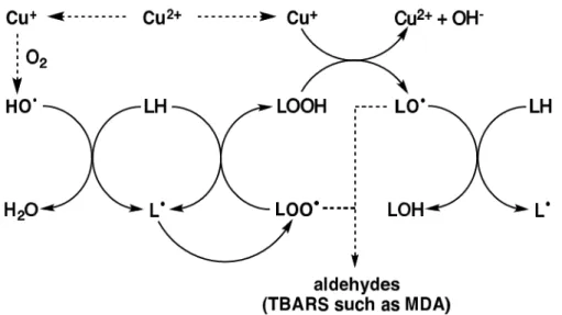
superoxide (O2·-) (reaction 4) or endogenous α-tocopherol (reaction 5).23) Cu+ is capable of
catalyzing the hydroxyl radicals (OH·) from hydrogen peroxide (H2O2) by Haber-Weiss
reaction.23)
Cu2+ + O2·- Cu+ + O2 (4)
Cu2++ α-TocOH Cu++ α-TocO· + H+ (5) Cu+ + H2O2 Cu2+ + OH- + OH· (6)
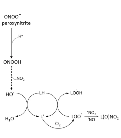
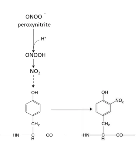
ONOO- is a powerful oxidant and nitrating agent, which is generated by the reaction between nitric oxide and superoxide anion (reaction 7).24) ONOO- can decompose to hydroxyl radicals by metal independent pathway at acid pH. ONOO- react with a hydrogen radical (H+) to generate the stable peroxynitrous acid (ONOOH).24) The hydroxyl radicals
(·OH) and free nitrogen species (NO2) is quickly formed from ONOOH (reaction 8).24)
·NO + O
2·- ONOO- (7)
ONOO- + H+ ONOOH ·OH + NO2 (8)
Consequent to the lipid peroxidation decomposition products such as aldehydes which can modify cysteine, lysine, histidine, and tyrosine residues of apo B-100 (Figure 1.8 and Figure 1.9).25-27) Oxidative modification of apo B-100 may deteriorate the function of apo B-100 as a ligand to LDL receptors. In the process of atherosclerosis, oxidized LDL (oxLDL) are phagocytosed by monocyte-derived macrophages through the scavenger receptor in the intima.28) Lipid-loaded macrophages became foam cells and accumulate to form fatty streak and fibrous plaque (Figure 1.10).29)
6
Table 1.1. Contents of polyphenols in oolong tea and green tea extracts.7)
Components (mg/g) Oolong tea Green tea
Flavan-3-ol without galloyl moiety Gallocatechin
Epigallocatechin Cathechin Epicatechin
Flavan-3-ol with galloyl moiety Epigallocatechin gallate Gallocatechin gallate Epicatechin gallate Cathechin gallate
Oolong tea polymerized polyphenols
7 (A)
(B)
8
Figure 1. 2. Structure of low density lipoprotein.11)
Low density lipoprotein consists of apolipoprotein B-100 and cholesterol esters, triacylglycerols, and phospholipid monolayer containing unesterified cholesterol.11)
Apolipoprotein B-100
Cholesteryl ester
Triglyceride
9
Figure 1. 3. Forms of oxidized low density lipoprotein.14)
Cholesteryl ester Oxidized cholesteryl ester Antioxidants Phospholipid
Oxidized phospholipid Lyso phospholipid Oxidized protein
(A) Native LDL with lysine residues of apo B-100 (B) Reactive nitrogen species (RNS) induced oxidized low density lipoprotein (oxLDL) with more apo B-100 and less lipid peroxidation (C) Reactive oxygen species (ROS) induced oxLDL with extensive apo B-100 and lipid peroxidation.
(B) (C)
10
Figure 1. 4. The mechanism of LDL oxidation.
Reactive species leading to initiation of lipid peroxidation in LDL. LDL oxidation involves lipid peroxidation and modification of apolipoprotein.
ROS and RNS
LOO
·
Containing limited number of lipoperoxides in LDL
Amplified number of lipoperoxides in LDL
Aldehydes, ketones from fatty acid fragmentation
Conjugation of aldehydes to apo B -100
OxLDL recognized by macrophages through the scavenger receptors
LOOH
O
2·
-,
H
2O
2,·
OH
11
Figure 1. 5. Peroxyl radical-mediated lipid peroxidation of polyunsaturated fatty acid.
Polyunsaturated fatty acid side chain (LH) containing active methylene hydrogen atom which is reactive with free radicals such as peroxyl radical (ROO·). The resulting, lipid
radical (L·) adds rapidly to molecular oxygen (O2) to generate a lipid peroxyl radical
12
Figure 1. 6. Cu2+ -catalyzed lipid peroxidation.
Cu2+ can be reduced to Cu+ in the presence of superoxide (O2·-) or endogenous
α-tocopherol. Cu+ is capable of catalyzing the hydroxyl radicals (OH·) from hydrogen
peroxide (H2O2) by Haber-Weiss reaction. Alkoxyl radical (LO·) is generated from lipid
13
Figure 1. 7. Peroxynitrite-mediated lipid peroxidation.24)
Peroxynitrite (ONOO-) at physiological pH to peroxynitrous acid (ONOOH) that
decomposes to hydroxyl radicals (·OH) and ·NO2. Decomposed hydroxyl radicals initiate
14
15
Figure 1. 9. Peroxynitrite-induced nitrotrysine formation.61)
16
(A) Lesion initiation (B) Fatty streak (C) Fibrous plaque (D) Thrombosis
Figure 1. 10. Atherosclerotic plaque progression.29)
17
Chapter 2
Radical scavenging capacity of oolonghomobisflavan A (OFA) in solution
2.1 ObjectivesThe biological effects of OTPP have attracted considerable attention over the past few years, but still the mechanisms remain unclear. First, we investigated the radical scavenging capacity of OFA.
2.2 Materials and Methods 2.2.1 Materials
OFA was purchased from Nagara Science Co. (Gifu, Japan). 2,2-Diphenyl-1-picrylhydrazyl (DPPH) was obtained from Wako Pure Chemical Industries (Osaka, Japan). 6-Hydroxy-2,5,7,8-tetramethylchroman-2-carboxylic acid (Trolox) was purchased from EMD Chemicals, Inc. (San Diego, CA, USA). All other chemicals were of reagent grade.
2.2.2 DPPH radical scavenging assay
Radical scavenging activity of OFA in solution was determined by DPPH method (Figure 2.1).30) Methanol solutions (400 μL) containing various concentrations of OFA (2-5
μM) were mixed with 400 μL of 200 μM DPPH in methanol. The absorbance of the reaction
mixture was measured at 517 nm after incubation at room temperature for 30 min in the dark. Trolox was used as a reference antioxidant.
2.3 Results
2.3.1 DPPH radical scavenging capacity of OFA in solution
OFA and Trolox dose-dependently scavenged DPPH radicals in methanol/water solution (Figure 2.2). The half maximal inhibitory concentration (IC50) of OFA and Trolox was 2.99
µM/mL and 23.7 µM/mL, respectively. The IC50 of OFA has a higher radical scavenging
18
Figure 2.1. Structure of 2,2-diphenyl-1-picrylhydrazyl (DPPH).
19
Figure 2.2. DPPH radical scavenging capacity of OFA.
DPPH was mixed with OFA and Trolox in methanol and incubated for 30 min at room temperature in the dark. The absorbance of the solutions was measured at 517 nm. Data are expressed as means ± SD (n = 3). OFA (■), Trolox (
●
).0 20 40 60 80 100
0 10 20 30 40
In
h
ib
it
io
n
㸦
%
㸧
20
Chapter 3
Effect of OFA on low-density lipoprotein (LDL) oxidation by reactive
oxygen species (ROS)
3.1 Objectives
The results in Chapter 2, OFA exhibited strong radical scavenging capacity. In this chapter, we investigated the effects of OFA on lipid peroxidation by peroxyl radicals and copper in vitro. Also, the inhibitory effect of OFA on apo B-100 modification in LDL oxidized by copper was analyzed in vitro.
3.2 Materials and Methods 3.2.1 Materials
2,4-Dinitrophenylhydrazine (DNPH), 2,2’-Azobis (2-amidinopropane) dihydrochloride (AAPH) were obtained from Wako Pure Chemical Industries (Osaka, Japan). All other chemicals were of reagent grade.
3.2.2 Isolation and purification of LDL
Human LDL was prepared by discontinuous density gradient ultracentrifugation using KBr.22) Human blood (200 mL) was collected from a healthy volunteer with informed consent after fasting overnight using 0.4% citric acid as an anti-coagulant, and blood plasma was separated by centrifugation at 1,200 × g at 4°C for 20 min. KBr solution (d = 1.019, 8 mL) was layered on 16 mL of the plasma and centrifuged at 100,000 × g at 16°C for 16 h. The top lipid layer was removed and density of the lower layer was adjusted to 1.063 by adding KBr. KBr solution (d = 1.063, 3.5 mL) was added to 21 mL of the lower fraction and centrifuged at 100,000 × g at 16°C for 20 h. The top layer including LDL fraction (d = 1.019 to 1.063) was then ultrafiltered and dialyzed against phosphate-buffered saline (PBS, pH 7.4) treated with chelating resin (Sigma-Aldrich Co., St. Louis, MO, USA)
at 4°C. Protein concentration of LDL was measured by Lowry’s method.31)
21
3.2.3 Peroxyl radical-mediated oxidation of LDL
LDL (200 μg protein/mL) was pre-incubated with OFA (0.5, 1, and 2 μM) in PBS
containing 10 mM diethylene triamine pentaacetic acid (DTPA) at 37°C for 5 min. Oxidation of LDL was induced by 5 mM of AAPH, an aqueous peroxyl radical generating reagent, at 37°C. Aliquots (50 μL) of the reaction mixture were mixed with 0.9 mL of 2 mM butyl hydroxyl anisole in methanol at intervals. n-Hexane (1.5 mL) was added to oxLDL in methanol to extract neutral lipids. After centrifugation at 1,200 × g at 4°C for 5 min, the upper layer was collected and extraction with n-hexane from the bottom layer was repeated again. The upper layer extracts were pooled and evaporated, and the residue was
dissolved in 100 μL of 2-propanol. Cholesteryl ester hydroperoxide (CE-OOH) in the
sample was analyzed by reversed-phase high-performance liquid chromatography (HPLC) using an InertSustian C8 column at 40°C (150 × 4.6 mm i.d.; GL Science Inc., Tokyo, Japan) with methanol as an eluent at a flow rate of 1.0 mL/min.32) The concentration of CE-OOH was detected at 235 nm and calculated using cholesteryl linoleate hydroperoxide (Cyaman Chemical Company, Ann Arbor, MI, USA) as an authentic standard.
3.2.4 Metal ion-catalyzed oxidation of LDL
LDL (200 μg protein/mL) was pre-incubated with OFA (0.5, 1, and 2 μM) in PBS at
37°C for 5 min. LDL oxidation was initiated by adding 5 μM CuCl2 at 37°C. Aliquots (50
μL) of the reaction mixture were mixed with 450 μL of 0.2% 2-thiobarbituric acid and
heated at 95°C for 60 min. After centrifugation at 20,000 × g at 4°C for 5 min, 2-thiobarbituric acid reactive substances (TBARS) in the supernatant were determined by reversed-phase HPLC with fluorescence detection (excitation at 515 nm, emission at 553 nm).33) The samples were eluted with acetonitrile/water (30:70, v/v) at 1.0 mL/min on InertSustain C18 column (150 × 4.6 mm i.d.; GL Science Inc.) at 40°C. 1,1,3,3-Tetramethoxypropane was used as a standard.
3.2.5 Apolipoprotein B-100 (apo B-100) analysis inoxidized LDL
22
of 2 μM OFA as described above. OxLDL was analyzed by sodium dodecyl
sulfate-polyacrylamide gel electrophoresis (SDS-PAGE) using an acrylamide slab gel system (AnykD TGX gel; Bio-Rad Laboratories, Hercules, CA, USA) according to Laemmli's method.34) The gel was stained with Coomassie Brilliant Blue (CBB). Proteins in the other gel were transferred to polyvinylidene difluoride membrane for Western blotting. The membrane was incubated with serum-free blocking buffer (Thermo Fisher Scientific Inc., Rockford, IL, USA) and washed with 0.05% Tween-Tris-buffered saline (TBS). The membrane was incubated with anti-human apo B-100 monoclonal (6H12) antibody (MP Biomedicals, Santa Ana, CA, USA) for 1 h at room temperature. After washing with 0.05% Tween-TBS, the membrane was incubated with goat anti-mouse IgG-Fc conjugated with horseradish peroxidase (Bethyl Laboratories, Inc., Montgomery, TX, USA) for 1 h at room temperature. The immunoreactive substances were detected with the enhanced chemiluminescence (ECL) reagent (GE Healthcare, Little Chalfont, UK).
3.2.6 Protein carbonyl analysis of oxidized LDL
LDL (200 μg protein/mL) was oxidized by 5 μM CuCl2 at 37°C for 3 h in the presence
of OFA (0.5, 1, and 2 μM). Protein carbonyls in oxLDL was derivatized with DNPH and
analyzed by SDS-PAGE and Western blotting as previously described.35) Briefly, rabbit anti-dinitrophenyl antisera (Dako Cytomation, Glostrup, Denmark) and goat anti-rabbit IgG-Fc conjugated with horseradish peroxidase (Bethyl Laboratories Inc.) were used as primary and secondary antibodies, respectively.
3.2.7 Heparin-binding activity of apo B-100 in oxidized LDL
LDL (200 μg protein/mL) was oxidized by 5 μM CuCl2 at 37°C for 3 h in the presence
of 2 μM OFA. OxLDL (20 μg protein/100 μL) was incubated with 100 μL of heparin
23
3.3 Results
3.3.1 Effect of OFA on lipid peroxidation of LDL by ROS
We measured CE-OOH as the major product of lipid peroxidation of LDL induced by AAPH.22) Figure 3.2 shows effect of OFA on peroxyl radical (AAPH)-mediated lipid peroxidation of LDL. CE-OOH concentration in the absence OFA (control) increased with time and attained 8.7 µM at 4 h. On the other hand, OFA dose-dependently suppressed CE-OOH accumulation, 2 µM of OFA exhibited strongest inhibition.
TBARS is a widely-used indicator to detect secondary oxidation products in transition metal ion-catalyzed lipid peroxidation. As shown in Figure 3.3, TBARS time-dependently accumulated up to 12.5 µM at 4 h in the absence of OFA (control). On the contrary, OFA dose-dependently inhibited TBARS formation; especially 2 µM OFA almost inhibited TBARS formation.
3.3.2 Effect of OFA on protein oxidation of LDL by ROS
Figure 3.4 illustrates the effect of OFA on oxidative modification of apoB-100 in LDL oxidized by Cu2+, which was analyzed by SDS-PAGE with CBB staining (Figure 3.4 A) and Western blot analysis using anti-human apoB-100 antibody (Figure 3.4 B). A single band with molecular weight greater than 250 kDa was observed in native LDL (lane 1 in Figure 3.4 A). However, the band was not detected in oxLDL (lane 2 in Figure 3.4 A), whereas a weak smear band was observed. Although the single band remained in oxLDL with 2 µM OFA (lane 3 in Figure 3.4 A). As in Figure 3.4 A, apo B-100 in Figure 3.4 B was observed as a single band with molecular weight more than 250 kDa in native LDL (lane 1). The oxidative modification of apoB-100 was detected as a ladder band at 100-150 kDa (lane 2 in Figure 3.4 B). Whereas, the oxidative modification of apoB-100 in oxLDL was suppressed by 2 µM OFA (lane 3 in Figure 3.4 B).
24
high molecular weight (lane 2). OFA dose-dependently inhibited protein carbonyl formation (lane 3, 4, and 5) in oxLDL, and 2 µM OFA mostly suppressed protein carbonyl formation.
25
Figure 3.1. Structure of 2,2’-azobis (2-amidinopropane) dihydrochloride (AAPH).
26
Figure 3.2. Effect of OFA on peroxyl radical-mediated lipid peroxidation of LDL.
LDL (200 μg protein/ml) oxidation was initiated by 5 mM AAPH at 37°C. CE-OOH was
determined by HPLC with UV detection. Data are expressed as means ± SD (n = 3). Control (
●
), 0.5 μM OFA (■), 1 μM OFA (▲), 2 μM OFA (◆).0 5 10
0 1 2 3 4
CE
-O
O
H
(
µ
M
)
27
Figure 3.3. Effect of OFA on transition metal ion-catalyzed lipid peroxidation of LDL.
LDL in PBS was oxidized by 5 μM Cu2+
at 37°C. TBARS was analyzed by HPLC with fluorescence detection. Data are expressed as means ± SD (n = 3). Control (
●
), 0.5 μMOFA (■), 1 μM OFA (▲), and 2 μM OFA (◆).
0 5 10 15
0 1 2 3 4
T
B
A
R
S
(
µ
M
)
28
Figure 3.4. Effect of OFA on apo B-100 modification in oxidized LDL.
LDL in PBS was oxidized by Cu2+ at 37°C for 3 h. (A) OxLDL was analyzed by
SDS-PAGE and CBB staining. (B) OxLDL was determined by Western blotting using anti-human apo B-100 antibody. Immunoreactive bands were detected with ECL reagent. Lane 1, native LDL; lane 2, LDL oxidized by Cu2+; lane 3, LDL oxidized by Cu2+ in the
29
Figure 3.5. Effect of OFA on protein carbonyl formation in oxidized LDL.
30
Figure 3.6. Effect of OFA on heparin-binding activity of apo B-100 in oxidized LDL.
LDL in PBS was oxidized by Cu2+ at 37°C for 3 h. OxLDL was incubated with 100 μL heparin Sepharose at 4°C for 3 h. After washing heparin Sepharose beads with Tween-TBS, proteins bound to the beads were subjected to SDS-PAGE and CBB staining. Lane 1, native LDL; lane 2, LDL oxidized by Cu2+; lane 3, LDL oxidized by Cu2+ in the presence
31
Chapter 4
Effect of OFA on LDL oxidation by reactive nitrogen species (RNS)
4.1 ObjectivesRNS such as peroxynitrite (ONOO-) is also involved in inducing oxidative stress in human body. Therefore, we evaluated the antioxidant effects of OFA on LDL oxidation induced by RNS in vitro; same as before both lipid peroxidation and oxidative modification of apo B-100 were analyzed as the oxidative markers.
4.2 Materials and Methods 4.2.1 Materials
3-(4-Morpholinyl) sydnonimine (SIN-1) was supplied from Dojindo Molecular Technologies Inc. (Kumamoto, Japan). All other chemicals were of reagent grade.
4.2.2 Peroxynitrite-mediated oxidation of LDL
LDL (200 μg protein/mL) was pre-incubated with OFA (2, 4, and 8 μM) in PBS
containing 10 mM DTPA at 37°C for 5 min. LDL was oxidized by 1 mM SIN-1, a water-soluble peroxynitrite generator, at 37°C. Determination of CE-OOH in oxLDL was performed as previously described.
4.2.3 Peroxynitrite-mediated nitrotyrosine analysis
LDL (200 μg protein/mL) was pre-incubated with 8 μM OFA in PBS containing 10
mM DTPA at 37°C for 5 min. LDL was oxidized by 1 mM SIN-1 at 37°C. Nitrotyrosine in oxLDL was analyzed by SDS-PAGE and Western blotting using primary mouse anti-nitrotyrosine monoclonal (CC.22.8C7.3) antibody (Cayman Chemical Company) and secondary goat anti-mouse IgG-Fc conjugated with horseradish peroxidase (Bethyl Laboratories Inc.). The immune reactive substances were detected with ECL reagent.
4.3 Results
32
Figure 4.2 shows the effect of OFA on peroxynitrite-mediated lipid peroxidation of LDL induced by SIN-1. SIN-1 generates peroxynitrite by simultaneously releasing nitric oxide and superoxide. Concentration of CE-OOH time-dependently increased in the absence of in OFA (control). OFA dose-dependently suppressed CE-OOH formation in LDL oxidized by peroxynitrite, in particular 8 µM OFA exhibited strong inhibition.
4.3.2 Effect of OFA on protein oxidation of LDL by RNS
33
Figure 4.1. Structure of 3-(4- morpholinyl)sydnonimine (SIN-1).
34
Figure 4.2. Effect of OFA on peroxynitrite-mediated lipid peroxidation of LDL.
LDL in PBS was oxidized by 1 mM SIN-1 at 37°C. CE-OOH was analyzed by HPLC with UV detection. Data are expressed as means ± SD (n = 3). Control (
●
), 2 μM OFA (■), 4μM OFA (▲), and 8 μM OFA (◆).
0 5 10 15
0 1 2 3 4 5 6
CE
-O
O
H
(
µ
M
)
35
Figure 4.3. Effect of OFA on nitrotyrosine formation in oxLDL.
36
Chapter 5
DiscussionThe polyphenols in oolong tea are different from those in green tea and black tea.38) Green tea leaves are rich in monomeric polyphenols known as flavan-3-ols, including
(−)-epicatechin-3-gallate (ECG), (−)-epigallocatechin (EGC), and (−)-epigallocatechin
gallate (EGCG).39,40) Black tea involves lower amounts of monomeric polyphenols and higher concentration of polymeric polyphenols such as theaflavins, thearubigins.40) The concentration of polymeric polyphenols in black tea is higher than that of oolong tea.41) Oolong tea contains numerous kinds of polymerized-polyphenols.38) During semi-fermentation to manufacture oolong tea, monomeric polyphenols such as catechins are converted to oligomeric flavan-3-ols by polyphenol oxidases.38) Several studies have reported the antioxidant effects of oolong tea. Xie et al.42) reported that oolong tea extract exerts higher antioxidative activity than black tea in vitro. Yen et al.43) showed that oolong tea extract exhibits stronger radical scavenging activity than that of green and black teas. However, the active compounds in oolong tea have not been identified. The contents of polymerized polyphenols in oolong tea leaves depend on the producing procedures such as fermentation. Komatsu et al.44) indicated that contents of polymerized polyphenols and EGCG are 68 mg/300 mL and 81 mg/300 mL in oolong tea prepared from 15 g of leaves, respectively. On the other hand, Toyoda-ono et al.7) showed that contents of polymerized polyphenols and EGCG in oolong tea extract are 114 mg/g and 6 mg/g, respectively. Zhu
et al.45) have reported that oolong tea water extracts had strong free radical scavenging
37
effects.47) Therefore, we made hypothesis that OFA exhibits antioxidant activity and might decrease oxidative stress in human body. As shown in Figure 2.2, OFA had very strong
radical scavenging activity than that of Trolox, α-tocopherol analog.
Lipid peroxidation of polyunsaturated fatty acids moieties of phospholipids, cholesteryl esters, and triacylglycerols in LDL is mediated by radical chain reaction, resulting in the generation of various highly reactive products including lipid hydroperoxide.48) Lipid hydroperoxide is decomposed to secondary products such as aldehydes in the presence of transition metal ions. These can react with free amino groups
of apolipoprotein, generating Schiff’s bases or Michael addition reactions, and making the
LDL particle more electronegative.49) We used agents such as peroxyl radicals (AAPH), coper ions for ROS generation, and peroxynitrite (SIN-1) for RNS generation to initiate LDL oxidation in vitro. As a major product of lipid peroxidation of LDL by AAPH, CE-OOH was measured.50) As shown in Figure 3.2, OFA seems to be a chain-breaking antioxidant against peroxyl radical-mediated lipid peroxidation of LDL.
There are several reports indicating that tea polyphenols inhibits oxLDL catalyzed by Cu2+, a transition metal ion.51) Cu2+ stimulates lipid peroxidation that can decompose lipid hydroperoxides to lipid alkoxyl and peroxyl radicals.52) During termination of lipid peroxidation secondary oxidation products such as lipofuscines, oxysterols, and aldehydes are generated.13) TBARS is a widely used indicator of metal ion catalyzed lipid peroxidation. In Figure 3.3, OFA shows potent inhibitory effect on Cu2+ catalyzed LDL oxidation, due to breaking of the chain reaction by scavenging radicals.
38
Carbonylated protein has been used as a marker of ROS mediated protein oxidation. The decomposed products of lipid hydroperoxides generated by transition metal ions can react with the lysine, histidine, threonine, and proline side-chain of proteins to form carbonyl compounds.55) As shown in Figure 3.5, OFA has also been shown to inhibit protein carbonyls of apoB-100 during Cu2+ oxidation. These results suggest that OFA protects apo B-100 from carbonylation in transition metal ion-catalyzed LDL oxidation, which may be due to preventing lipid peroxidation and its decomposition.
The interaction of heparin with apolipoprotein plays an important role in the regulation of normal physiological processes as well as LDL metabolism and cholesterol homeostasis.56) Heparin, a sulfated polysaccharide of the family of glycosaminoglycans, has the ability to bind to the apo B (LDL) receptor.56) Peptide analysis of apo B-100 demonstrated that positively charged amino acid residues such as lysine or arginine are responsible for specific binding to heparin.57) Oorni et al.58) suggested that reduction of the positive charge of lysine or arginine side chains by ROS results in loss of the heparin binding activity of apo B-100. The decomposed substance of lipid hydroperoxides induced by Cu2+ can modify of lysine residues. In our experiments, OFA showed an inhibitory effect on oxidative modification of the lysine residues of apo B-100 in LDL oxidized by transition metal ions.
39
40
Chapter 6
Conclusions
These results suggest that OFA, an oolong tea polymerized polyphenol, has a strong inhibitory effect on lipid peroxidation and modification of apoB-100 in LDL oxidized by ROS, such as peroxyl radicals and transition metal ions, and RNS, such as peroxynitrite. The antioxidant effects of OFA are attributable to its radical scavenging activity to reduce ROS and RNS. Oolong tea polyphenols may protect against atherosclerosis by reducing oxidative stress.
41
References
1) Sheibani E, Duncan SE, Kuhn DD, Dietrich AM, Newkirk JJ, O'Keefe SF. (2015). Changes in flavor volatile composition of oolong tea after panning during tea processing. Food Sci Nutr., 4(3):456-68.
2) Hosoda K, Wang MF, Liao ML, Chuang CK, Iha M, Clevidence B, Yamamoto S. (2003). Antihyperglycemic effect of oolong tea in type 2 diabetes. Diabetes Care.,
26(6):1714-8.
3) Yang TT, Koo MW. (1997). Hypocholesterolemic effects of Chinese tea. Pharmacol
Res., 35(6):505-12.
4) Weerawatanakorna M, Hungb WL, Panc MH, Lib Sh, Lid D, Wand X, Hob CT. (2016). Chemistry and health beneficial effects of oolong tea and theasinensins. Food
Science and Human Wellness 4(4):133-46.
5) Sae-tan S. (2016). Systematic Review: Hypolipidemic Activity of Oolong Tea Polymerized Polyphenols. J Health R., 30(6):451-9.
6) Yi D, Tan X, Zhao Z, Cai Y, Li Y, Lin X, Lu S, Chen Y, Zhang Q. (2014). Reduced risk of dyslipidaemia with oolong tea consumption: a population-based study in southern China. Br J Nutr., 111(8):1421-9.
7) Toyoda-Ono Y, Yoshimura M, Nakai M, Fukui Y, Asami S, Shibata H, Kiso Y, Ikeda I. (2007). Suppression of postprandial hypertriglyceridemia in rats and mice by oolong tea polymerized polyphenols. Biosci Biotechnol Biochem., 71(4):971-6.
8) Nakai M, Fuku Y, Asami S, Toyoda-Ono Y, Iwashita T, Shibata H, Mitsunaga T, Hashimoto F, Kiso Y. (2005). Inhibitory effects of oolong tea polyphenols on pancreatic lipase in vitro. J Agric Food Chem., 53(11):4593-8.
9) Catapano AL, Maggi FM, Tragni E. (2000). Low density lipoprotein oxidation, antioxidants, and atherosclerosis. Curr Opin Cardiol., 15(5):355-63.
10) Feingold KR, Grunfeld C. (2015). Introduction to Lipids and Lipoproteins. Endotext [Internet].
42
12) Morita SY. (2016). Metabolism and modification of apolipoprotein B-containing lipoproteins involved in dyslipidemia and atherosclerosis. Biol Pharm Bull.,39:1-24. 13) Salvayre R, Auge N, Benoist H, Negre-Salvayre A. (2002). Oxidized low-density
lipoprotein-induced apoptosis. Biochim Biophys Acta. 1585(2-3):213-21.
14) Jiang X1, Yang Z, Chandrakala AN, Pressley D, Parthasarathy S. (2011). Oxidized low density lipoproteins-do we know enough about them? Cardiovasc Drugs Ther.,
25(5):367-77.
15) McCall MR, Frei B. (1999). Can antioxidant vitamins materially reduce oxidative damage in humans? Free Rad Biol Med. 26(7-8):1034-53.
16) Rubbo H, O'Donnell V. (2005). Nitric oxide, peroxynitrite and lipoxygenase in atherogenesis: mechanistic insights. Toxicology. 208(2):305-17.
17) Esterbauer H, Gebicki J, Puhl H, Jürgens G. (1992). The role of lipid peroxidation and antioxidants in oxidative modification of LDL. Free Radic Biol Med., 13(4): 341-90. 18) Witztum JL. (1993). Role of oxidised low density lipoprotein in atherogenesis. Br
Heart J., 69(1): 12–18.
19) Yoshida H, Kisugi R. Mechanisms of LDL oxidation. Clin Chim Acta. (2010).
411(23-24):1875-82.
20) Darley-Usmar VM, Hogg N, VJ OL, Wilson MT, Mon-cada, S. (1992). The simultaneous generation of superoxideand nitric oxide can initiate lipid peroxidation in human low density lipoprotein. Free Radic Res Commun.,17(1):9–20.
21) Stocker R, Keaney JF Jr. (2004).Role of oxidative modifications in atherosclerosis.
Physiol Rev., 84(4):1381-478
22) Yang L, Kirikoshi J, Seikomoto S, Takasugi M, Fukunaga K, Hosomi R, Hishida A, Kawahara N, Yamagishi T, Arai H. (2015). Effect of Bean Extract of Yabumame (Amphicarpaea bracteata (L.) Fernald subsp. edgeworthii (Benth.) H. Ohashi) on Low-Density Lipoprotein Oxidation In Vitro. Food Sci Technol Res., 21(4):589-96. 23) Gaetke LM, Chow CK. (2003). Copper toxicity, oxidative stress, and antioxidant
nutrients. Toxicology., 189(1-2):147-63.
43
(2013). Reactive Oxygen Species and the Cardiovascular System. Oxidative Med Cell
Longev., 1:1-15.
25) Uchida K, Toyokuni S, Nishikawa K, Kawakishi S, Oda H, Hiai H, Stadtman ER. (1994). Michael addition-type 4-hydroxy-2-nonenal adducts in modified low-density lipoproteins: markers for atherosclerosis. Biochemistry. 33(41):12487-94.
26) Obama T, Kato R, Masuda, Y, Takahashi K, Aiuchi T, Itabe H. (2007). Analysis of modified apolipoprotein B-100 structures formed in oxidized low-density lipoprotein using LC-MS/MS. Proteomics., 7(13):2132-41.
27) Frijhoff J, Winyard PG, Zarkovic N, Davies SS, Stocker R, Cheng D, Knight AR, Taylor EL, Oettrich J, Ruskovska T, Gasparovic AC, Cuadrado A,1 Weber D1, Poulsen HE, Grune T, Schmidt HH, Ghezzi P. (2015). Clinical Relevance of Biomarkers of Oxidative Stress. Antioxid Redox Signal., 23(14):1144-70.
28) Moore KJ, Tabas I. (2011). Macrophages in the pathogenesis of atherosclerosis. Cell.,
145(3):341-355.
29) Steinl DC, Kaufmann BA. (2015). Ultrasound imaging for risk assessment in atherosclerosis. J. Mol. Sci.,16(5):9749-69.
30) Blois MS. (1958). Antioxidant determinations by the use of a stable free radical.
Nature., 181:1199-1200.
31) Lowry OH, Rosebrough NJ, Farr AL, Randall RS. (1951). Protein measurement with the Folin phenol reagent. J Biol Chem., 193:265-275.
32) Arai H, Terao J, Abdalla DS, Suzuki T, Takama K. (1996). Coulometric detection in high performance liquid chromatographic analysis of cholesteryl ester hydroperoxides.
Free Radic Biol Med., 20:365-371.
33) Fukunaga K, Yoshida M, Nakazono N. (1998). A simple, rapid, highly sensitive and reproducible quantification method for plasma malondialdehyde by high-performance liquid chromatography. Biomed Chromatogr., 12:300-303.
34) Laemmli UK. (1970). Cleavage of structural proteins during the assembly of the head of bacteriophage T4. Nature., 227:680-685
44
iron-mediated oxidation of low-density lipoprotein. Proc Natl Acad Sci USA.,
102:10472-10477.
36) Levine RL, Wehr N, Williams JA, Stadtman ER, Shacter E. (2000). Determination of carbonyl groups in oxidized proteins. Methods Mol Biol. 99:15-24.
37) Requena JR, Levine RL, Stadtman ER. (2003). Recent advances in the analysis of oxidized proteins. Amino Acids. 25(3-4):221-6.
38) Hsu TF, Kusumoto A, Abe K, Hosoda K, Kiso Y, Wang MF, Yamamoto S. (2006). Polyphenol-enriched oolong tea increases fecal lipid excretion. Eur J Clin Nutr.,
60(11):1330-6.
39) Graham HN. (1992). Green tea composition, consumption, and polyphenol chemistry.
Pre Med. 21(3):334-50.
40) Sharma V, Rao LJ. (2009). A thought on the biological activities of black tea. Crit Rev
Food Sci Nutr. 49(5):379–404.
41) Heber D, Zhang Y, Yang J, Ma JE, Henning SM, Li Z. (2014). Green tea, black tea, and oolong tea polyphenols reduce visceral fat and inflammation in mice fed high-fat, high-sucrose obesogenic diets. J Nutr.144:1385-93.
42) Xie B, Shi H, Chen Q, Ho CT. (1993). Antioxidant properties of fractions and polyphenol constituents from green, oolong and black teas. ProcNatl Sci Counc Repub
China B., 17:77-84.
43) Yen GC, Chen HY. (1995). Antioxidant activity of various tea extracts in relation to their antimutagenicity. J Agric Food Chem., 43:27-32.
44) Komatsu T, Nakamori M, Komatsu K, Hosoda K, Okamura M, Toyama K, Ishikura Y, Sakai T, Kunii D, Yamamoto S. (2003). Oolong tea increases energy metabolism in Japanese females. J Med Invest. 50(3-4):170–5.
45) Zhu QY, Hackman RM, Ensunsa JL, Holt RR, Keen CL. (2002). Antioxidative activities of oolong tea. J Agric Food Chem., 50:6929-6934.
46) Hashimoto F, Ono M, Masuoka C, Ito Y, Sakata Y, Shimizu K, Nonaka G, Nishioka I, Nohara T. (2003). Evaluation of the anti-oxidative effect (in vitro) of tea polyphenols.
45
47) Singh BN, Shankar S, Srivastava RK. (2011). Green tea catechin, epigallocatechin-3-gallate (EGCG): mechanisms, perspectives and clinical applications. Biochem Pharmacol., 82(12):1807-21.
48) Hevonoja T, Pentikäinen M O, Hyvönen M T, Kovanen PT, Ala-Korpela M. (2000) Structure of low density lipoprotein (LDL) particles: basis for understanding molecular changes in modified LDL. Biochim Biophys Acta., 1488(3):189–210.
49) Itakura K, Oya-Ito T, Osawa T, Yamada S, Toyokuni S, Shibata N. Kobayashi M, Uchida K. (2000). Detection of lipofuscin-like fluorophore in oxidized human low-density lipoprotein. 4-hydroxy-2-nonenal as a potential source of fluorescent chromophore. FEBS Lett., 473(2):249-53.
50) Ayala A, Muñoz MF, Arguelles S. (2014). Lipid peroxidation: production metabolism, and signaling mechanisms of malondialdehyde and 4-hydroxy-2-nonenal. Oxid Med
Cell Longev., 2014:360-438.
51) Miura S, Watanabe J, Tomita T, Sano M, Tomita I. (1994). The inhibitory effects of tea polyphenols (flavan-3-ol derivatives) on Cu2+ mediated oxidative modification of low density lipoprotein. Biol Pharm Bull., 17(12):1567-72.
52) Gutteridge JM. (1995). Lipid peroxidation and antioxidants as biomarkers of tissue damage. Clin Chem., 41(12):1819-28.
53) Itabe H, Jimi S, Kamimura S, Suzuki K, Uesugi N, Imanaka T, Shinjo H, Takano T. (1998). Appearance of cross linked proteins in human atheroma and rat pre-fibrotic liver detected by a new monoclonal antibody. Biochim Biophys Acta., 1406(1):28–39. 54) Stadtman ER, Levine RL. (2003). Free radical-mediated oxidation of free amino acids
and amino acid residues in proteins. Amino Acids., 25(3-4):207-18.
55) Gieseg SP, Pearson J, Firth CA. (2003). Protein hydroperoxides are a major product of low density lipoprotein oxidation during copper, peroxyl radical and macrophage-mediated oxidation. Free Radic Res., 37(9):983-91.
56) Capila I, Linhardt RJ. (2002). Heparin-protein interactions. Angew Chem Int Ed Engl.,
41(3):391-412.
46
Jurgens G. (2006). The effect of heparin on structural and functional properties of low density lipoproteins. Biophys Chem., 119(3):234-9.
58) Oorni K, Pentikainen MO, Annila A, Kovanen PT. (1997). Oxidation of low density lipoprotein particles decreases their ability to bind to human aortic proteoglycans. Dependence on oxidative modification of the lysine residues. J Biol Chem.,
272(34):21303-11.
59) Beckman JS, Beckman TW, Chen J, Marshall PA, Freeman BA. (1990). Apparent hydroxyl radical production by peroxynitrite: implications for endothelial injury from nitric oxide and superoxide. Proc Natl Acad Sci USA., 87(4):1620–4.
60) Guy RA, Maguire GF, Crandall I, Connelly PW, Kain KC. (2001). Characterization of peroxynitrite-oxidized lowdensity lipoprotein binding to human CD36. Atherosclerosis.
155(1):19-28.
61) Trostchansky A, Batthyany C, Botti H, Radi R, Denicola A, Rubbo H. (2001). Formation of Lipid-Protein Adducts in Low-Density Lipoprotein by Fluxes of Peroxynitrite and Its In-hibition by Nitric Oxide. Arch Biochem Biophys.,
47
Acknowledgment
Undertaking this PhD has been a truly life-changing experience for me. It would not have been possible without the support and guidance that I received from many people.
First and foremost, I would like to thank my supervisor Dr. Hirofumi Arai. I appreciate all his contributions, generous guidance and support, which made it possible for me to work on a topic that has been a great opportunity for me. I cannot express enough thanks to my supervisor for the learning opportunities provided.
My heartfelt thanks to Prof. Gereltu Borjihan, Prof. Jigmed Sukhdolgor, Dr. Takashi Yamagishi, Dr. Masayuki Hoshi, and Dr. Sarangowa for their help and encouragement. I would like to express my gratitude to Dr. Yang Lifeng, Mr. Harumi Hashimoto, Dr. Mikako Takasugi, Dr. Koji Yamada, Dr. Ryota Hosomi, Dr. Kenji Fukunaga for their kindly help. I also would like to thank my fellow labmates, especially Shogo Sekimoto, Emiko Watanabe, and Akane Yamazaki. Additionally, I would like to thank my committee members Dr. Takashi Yoshida, Dr. Toshitsugu Sato, Dr. Toru Kanno for their interest in my work.
I gratefully acknowledge the funding to undertake my PhD that I received through the Rotary Yoneyama Memorial Foundation, Kitami club.
I am also very grateful to the teachers and staff of the Kitami Institute of Technology for their kindly help.