Durability and Biological Response of a New Posterior Dynamic Stabilization System Using Polyethylene with Vitamin E
日本大学医学部整形外科学系整形外科学分野
松本 光司 申請年
2018
年 指導教員 德橋 泰明Research Article
Durability and Biological Response of a New Posterior Dynamic Stabilization System Using Polyethylene with Vitamin E
Koji Matsumoto and Yasuaki Tokuhashi
Department of Orthopaedic Surgery, Nihon University Itabashi Hospital, 30-1 Oyaguchikamimati Itabashi-ku, Tokyo 173-8610, Japan
Correspondence should be addressed to Koji Matsumoto; [email protected] Received 17 May 2018; Accepted 13 August 2018; Published 25 September 2018
Academic Editor: Shiro Imagama
Copyright © 2018 Koji Matsumoto and Yasuaki Tokuhashi. This is an open access article distributed under the Creative Commons Attribution License, which permits unrestricted use, distribution, and reproduction in any medium, provided the original work is properly cited.
Objective. The purpose of this study was to evaluate the durability and biological response of a new Posterior Dynamic Stabilization system using polyethylene with vitamin E on the sliding surface.Summary of Background Data. The use of polyethylene with vitamin E on the sliding surface in Posterior Dynamic Stabilization has not been reported previously.Methods. A developed pedicle screw- based Posterior Dynamic system consists of four parts: a set screw, a rod, a ball, and a pedicle screw. The rod is inserted into the through hole of the ball, and the ball is sandwiched by the set screw.(1) Fatigue Wear Test. Testing was conducted under a dynamic compressive load of 50N at a speed of 1 Hz for 1 million cycles. We examined the loss of polyethylene due to abrasion in 3 units.
(2) Biological Response in Pigs. In two pigs, a new pedicle screw and a conventional pedicle screw were inserted in L2 and L3/4, and L4 and L2/3, respectively. After breeding for 6 months, autopsies were performed. CT imaging was used to evaluate bone union of the facet joint. Abrasive specimens were prepared, and abrasion powder and inflammatory cell infiltration were evaluated microscopically.Results. The average loss of polyethylene due to abrasion was -0.01 mg. In all units, polyethylene showed a decrease of 0.1 mm or less at the contact point with the set screw. The facet joints between the conventional screws exhibited bone fusion, but the facet joint between the conventional and the new screw retained mobility with no bony fusion. No abrasion powder was found and inflammatory cell infiltration was only minimally observed.Conclusion. The new Posterior Dynamic Stabilization system exhibited a high level of durability and biological safety.
1. Introduction
At present, spinal fusion is widely performed. However, spinal fusion surgery increases the load and stress caused by movement in the adjacent segment. The occurrence of adjacent segment disease (ASD) after spinal fusion is reported as 4.8-92.2% [1] and is considered an inevitable complication after spinal fusion surgery. In order to avoid these complications, various Dynamic Stabilization systems have been developed [2–5]. Nonfusion stabilization has been reported as preventing ASD in two meta-analyses [6, 7].
However, these systems are problematic in terms of durability and biological reactions caused by abrasion powder [8–13].
Artificial joints for the knee and hip using polyethylene on the sliding surface have been developed and applied clinically and have achieved good results. We have developed a new pedicle screw system which features polyethylene on the sliding
surface. The system uses vitamin E-containing polyethylene, which has been used on the sliding surface for knee and hip artificial joint replacement surgery and is said to be resistant to oxidative stress and abrasion [10, 12]. Until now, no system using polyethylene with vitamin E on the sliding surface in Posterior Dynamic Stabilization has been reported.
The present study evaluates the durability and the biological response of this new system.
2. Materials and Methods
New Posterior Dynamic Stabilization system: a major feature of this new pedicle screw system is that the relationship between the vertebral body and the rod is not completely fixed but semi-fixed. It consists of four parts: a set screw, a rod, a ball, and a pedicle screw. The pedicle screw and set screw
Volume 2018, Article ID 5785708, 5 pages https://doi.org/10.1155/2018/5785708
2 BioMed Research International
① ②
③
④
①
②
④
③
Figure 1: A new Posterior Dynamic Stabilization system using polyethylene with vitamin E.AA set screw,Ba rod,Ca ball of polyethylene, andDa pedicle screw. The rod is inserted into the through hole of the ball and the ball is sandwiched by the set screw.
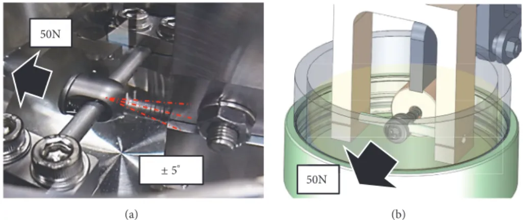
50N
±5∘ (a)
50N
(b)
Figure 2: (a) Photo of the durability testing machine: testing was conducted under a dynamic compressive load of 50 N at a speed of 1 Hz for 1 million cycles. (b) Pattern diagram of the durability test machine.
are made of Ti - 6Al - 4V alloy, and the ball is made of cross- linked UHMWPE (ultra high molecular weight polyethylene) containing vitamin E (Teijin Nakashima Medical, Blend - E XL). It was achieved by irradiation with a 10MeV electron beam at 300kGy. The ball's diameter is 8 mm and a through hole of 5.5 mm matching the rod diameter exists in the center of the ball. The rod is inserted into the through hole of the ball and fitted to the U-shaped groove of the pedicle screw, and the ball is sandwiched by the set screw. An existing product (Stryker Xia 𝜑 5.5 mm Ti - 6 Al - 4 V) is used for the rod. The pedicle screw has a range of movement of up to 15∘ on one side around the ball and also allows movement between the rod and the ball. The sliding surface is made of metal - polyethylene, and the two opposing surfaces on which the ball slides feature mirror surface processing.
One is a pedicle screw U-shaped groove; another is inside the set screw. Each contact surface has a concave shape because contact is made with a spherical diameter of 8 mm (Figure 1).
2.1. Fatigue Wear Test. The new pedicle screw system was fixed to the testing machine (Mini Bionix: MTS Japan) (Figure 2). Testing was conducted under a dynamic com- pressive load of 50N at a speed of 1Hz for 1 million cycles.
We examined the loss of polyethylene due to abrasion and
the presence or absence of system breakage of 3 units. The test room temperature was 20.3 to 23.1∘C and the humidity was 25 to 35%. A calf serum solution with a total protein mass of 20 g/l was used as the lubricating liquid [14]. The weight of the ball was measured with an electronic balance (A&D). The loss of polyethylene was calculated as the weight difference between the polyethylene at the end of the test and another polyethylene sample immersed in lubrication during the same period as the test. The appearance of the ball, the sliding surface of the set screw, the sliding surface of the main body screw, and the appearance of the rod were observed with a digital microscope (Keyence), and the shape change of the ball was observed with a three-dimensional digitizer (SOLUTIONIX).
2.2. Biological Response in Pigs. The experiment was con- ducted on two mini pigs with an age and weight of 3 years, 7 months and 47.4 kg, and 2 years, 8 months and 48.3 kg, respectively. The two mini pigs were anesthetized and screws inserted the pedicles of both sides of L2.3.4. In the first pig, a new pedicle screw was inserted in L2 and a conventional pedicle screw (5.5 × 40 mm Stryker Xia) was inserted in L3,4. In the second pig, a new pedicle screw was inserted in L4 and a conventional pedicle screw was inserted in L2,3.
Screws were fixed with a 5.5 mm rod (Stryker Xia) (Figure 3).
L2
L3
L4
L2
L3
L4
Figure 3: Pig intraoperative photo. A new pedicle screw and a conventional pedicle screw were inserted in L2 and L3,4, and L4 and L2,3, respectively.
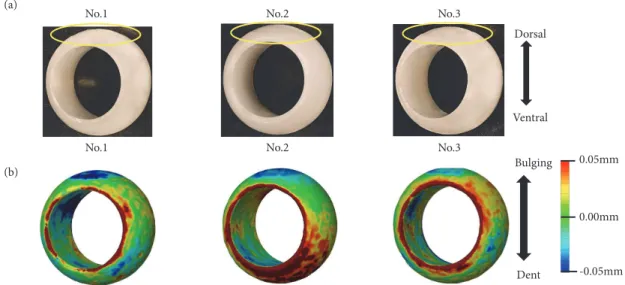
(a) No.1
No.2 No.3
Dorsal
Ventral
0.05mm
-0.05mm 0.00mm Bulging
Dent
No.1 No.2 No.3
(b)
Figure 4: (a) Appearance of polyethylene ball: in all units, a depression was found at the contact points with the set screw. (b) Shape change of polyethylene ball: the depression seen in all units was less than 0.1 mm.
After breeding for 6 months, autopsies were performed and vertebral bodies, screw, and rod were removed in one piece.
CT imaging was used to evaluate bone union of the facet joint. In addition, abrasive specimens were prepared. After Villanueva bone staining and Toluidine blue staining, the presence or absence of polyethylene and metal abrasion powder and bone soft tissue surrounding the screw were evaluated histologically under a microscope. This study has been approved by the Ethics Committee of Nihon University School of Medicine.
3. Results
3.1. Fatigue Wear Test. The loss of polyethylene was 0.02 mg for Unit No. 1, 0.01 mg for Unit No. 2, and -0.06 mg for
Table 1: Abrasion amount of polyethylene.
No. 1 No. 2 No. 3 Average
0.02 mg 0.01 mg -0.06 mg -0.01 mg
Unit No. 3. The average loss of polyethylene due to abrasion was -0.01 mg (Table 1). All units showed a polyethylene decrease of 0.1 mm or less at the contact point with the set screw (Figure 4). No obvious damage was observed on the sliding surface of the pedicle screw, the rod, and the set screw.
3.2. Biological Response in the Facet Joint. The facet joints between the conventional screws exhibited bone fusion, but
4 BioMed Research International
L2
L3
L4
L2/3
L3/4
Figure 5: CT image of the spine removed from a pig. The joint space disappears as a result of rigid fixation and bone fusion (L2/3). The joint space is retained as a result of Dynamic Stabilization (L3/4).
PE
METAL
500 m
Figure 6: Microscopic image (Villanueva bone stain: photograph magnification 20 times). Polyethylene (PE) and metal abrasion powder were not found in the soft tissue around the screw head.
Abnormal osteolysis was not observed in bone tissue around the screw, and inflammatory cell infiltration was only minimally observed.
the facet joint between the conventional and the new screw retained mobility with no bony fusion (Figure 5).
3.3. Biological Response in Bone Soft Tissue Surrounding the Pedicle Screw. No polyethylene or metal abrasion powder was found in the tissue.
Abnormal osteolysis was not observed and inflammatory cell infiltration was only minimally observed in the bone and soft tissue surrounding the screw.
Aseptic lymphocyte-dominant vasculitis-associated le- sion (ALVAL), a reactivity change caused by abrasion powder, was not observed (Figure 6).
4. Discussion
In order to avoid the complication of adjacent segmental disease after spinal fusion surgery, various systems that
preserve intervertebral disc mobility without rigid inter- vertebral fusion have been developed [2]. However, none of these have proved to be ideal. Existing systems have a sliding surface of metal on metal, but metal on metal generates metallic abrasive powder. Such systems have been reported to cause an adverse reaction due to metal debris (ARMD) mainly based on lymphocyte accumulation and tissue necrosis [15, 16]. Osteolysis, a result of the biological reaction caused by polyethylene abrasive powder, has also been reported in a system using a sliding surface of metal on polyethylene [17]. Therefore, it is important to use polyethy- lene which has excellent durability against abrasion on the sliding surface. Although artificial joints using polyethylene on sliding surfaces have been developed for various joints, Posterior Dynamic Stabilization using polyethylene on the sliding surface in the field of the spine has not been reported.
We have developed a new Posterior Dynamic Stabilization system using vitamin E-containing high molecular weight polyethylene, which has been reported to have excellent durability in impingement and friction under oxidative stress [10, 12]. In the durability test of the present study, the decrease in polyethylene due to abrasion was very small, and deformation in the polyethylene was also mild. However, in Unit No. 3, polyethylene weight exhibited an increase after the test. This may have been caused by the absorption of moisture and oil texts in the lubricating liquid. We believe that polyethylene wear in Unit No. 3 was as minimal as it was in the other two units, which would explain the increase seen in Unit No. 3 as being the result of absorption. In addition, since the polyethylene sample used to calculate the loss of polyethylene was not loaded, the conditions for absorption were different from that of the unit undergoing the wear test. Although this was a limitation in our research, it seems to be a measurement error indicating a very small degree of wear. In addition, no damage was observed on the pedicle screw, the rod, or the set screw including the sliding surface. It was possible to reduce the occurrence of abrasion powder to a very low level while confirming the durability of the new system. Furthermore, in the animal experiment using two mini pigs, no polyethylene or metal wear powder was found in the tissue. Inflammatory cell infiltration was only minimally observed and osteolysis and ARMD were not observed. These results indicate the safety of the new pedicle screw system in vivo. The facet joints between the conventional screws exhibited bone fusion, but the facet joint between the conventional and the new screw retained mobility with no bony fusion. These results indicate that new Posterior Dynamic Stabilization system functions in vivo and supports the possibility of its clinical application.
Artificial joints using polyethylene for sliding surfaces in the knee, elbow, and hip joints are widely used, and the ability to apply such mechanisms to the sliding surface of the spine is a subject that will continue to see further development in the future. The most important findings of this study are that polyethylene was used for the first time on the sliding surface in Posterior Dynamic Stabilization and that a high degree of durability and biological safety were attained. However, it is recognized that this system is the first prototype and that there is still much room for
research and development. The limitation of this study is the difference in the vertebral body characteristics in mini pigs and humans. This system is limited in the braking force of flexion-extension although there is a braking force in the anterior-posterior direction, and the long-term performance is unknown. Further development of instruments and long- term observation are desired.
5. Conclusion
A new Posterior Dynamic Stabilization system using polyethylene on the sliding surface exhibited a high level of durability and biological safety.
Data Availability
The data used to support the findings of this study are available from the corresponding author upon request.
Disclosure
This manuscript had been presented as a conference paper under Joint Meeting of Istanbul Spine Masters & ISMISS Turkey 2018.
Conflicts of Interest
We received assistance from Teijin Nakashima Medical for the production of screws and materials. The research was funded by Teijin Nakashima Medical. Neither the funding agency nor any outside organization has participated in study design or have any conflicts of interest. Teijin Nakashima Medical had final approval of the manuscript.
Acknowledgments
Department of Therapeutics for Aging Locomotive Disorders and donation to Orthopaedic Department were used for this study. Department of Therapeutics for Aging Locomotive Disorders was paid by Teijin Nakashima Medical. Donation to Orthopaedic Department was paid by Takeda Pharma- ceutical Company Limited, Zimmer Biomet Holdings Inc., Stryker Japan KK, Kaken Pharmaceutical Co.
References
[1] X.-P. Xia, H.-L. Chen, and H.-B. Cheng, “Prevalence of adjacent segment degeneration after spine surgery: a systematic review and meta-analysis,”The Spine Journal, vol. 38, no. 7, pp. 597–608, 2013.
[2] C. Gomleksiz, M. Sasani, T. Oktenoglu, and A. Ozer, “A Short History of Posterior Dynamic Stabilization,”Advances in Orthopedics, vol. 2012, Article ID 629698, 12 pages, 2012.
[3] T. Kaner, M. Sasani, T. Oktenoglu, and A. F. Ozer, “Dynamic stabilization of the spine: A new classification system,”Turkish Neurosurgery, vol. 20, no. 2, pp. 205–215, 2010.
[4] R. Sasso, D. M. Foulk, and M. Hahn, “Prospective, randomized trial of metal-on-metal artificial lumbar disc replacement: ini- tial results for treatment of discogenic pain,”The Spine Journal, vol. 33, no. 2, pp. 123–131, 2008.
[5] R. D. Guyer, P. C. McAfee, R. J. Banco et al., “Prospec- tive, randomized, multicenter Food and Drug Administration investigational device exemption study of lumbar total disc replacement with the CHARIT´E artificial disc versus lumbar fusion: Five-year follow-up,”The Spine Journal, vol. 9, no. 5, pp.
374–386, 2009.
[6] C. Ren, Y. Song, L. Liu, and Y. Xue, “Adjacent segment degener- ation and disease after lumbar fusion compared with motion- preserving procedures: a meta-analysis,”European Journal of Orthopaedic Surgery and Traumatology, vol. 24, pp. 245–253, 2014.
[7] J. Siewe, C. Otto, P. Knoell et al., “Comparison of standard fusion with a “topping off” system in lumbar spine surgery: a protocol for a randomized controlled trial,”BMC Musculoskeletal Disor- ders, vol. 12, article 239, 2011.
[8] R. E. McCarthy, D. Sucato, J. L. Turner, H. Zhang, M. A. W.
Henson, and K. McCarthy, “Shilla growing rods in a caprine animal model: A pilot study,”Clinical Orthopaedics and Related Research, vol. 468, no. 3, pp. 705–710, 2010.
[9] J. L. Lee, F. Billi, S. N. Sangiorgio et al., “Wear of an experimental metal-on-metal artificial disc for the lumbar spine,”The Spine Journal, vol. 33, no. 6, pp. 597–606, 2008.
[10] S. Teramura, H. Sakoda, T. Terao, K. Fujiwara, K. Kawai, and N. Tomita, “Reduction of wear volume from accelerated aged UHMWPE knee components by the addition of vitamin E,”
Journal of Biomechanical Science and Engineering, vol. 4, no. 4, pp. 589–596, 2009.
[11] M. P. Grevitt, A. D. H. Gardner, and J. Spilsbury, “The Graf stabilization system: early results in 50 patients,”European Spine Journal, vol. 4, no. 3, pp. 169–175, 1995.
[12] Y. Takahashi, T. Tateiwa, G. Pezzotti, T. Shishido, T. Masaoka, and K. Yamamoto, “Improved Resistance to Neck-Liner Impingement in Second-Generation Highly Crosslinked Polyethylene—The Role of Vitamin E and Crosslinks,” The Journal of Arthroplasty, vol. 31, no. 12, pp. 2926–2932, 2016.
[13] Y. Tokuhashi, M. Oshima, and Y. Ajiro, “A Novel Pedicle Screw with Mobile Connection: A Pilot Study,” BioMed Research International, vol. 2014, Article ID 841958, 6 pages, 2014.
[14] INTERNATIONAL STANDARD ISO 18192-1 Implants for surgery-Wear of total intervertebral spinal disc prostheses 2011.
[15] H. Willert, G. H. Buchhorn, A. Fayyazi et al., “Metal-on-metal bearings and hypersensitivity in patients with artificial hip joints,”The Journal of Bone and Joint Surgery-American Volume, vol. 87, no. 1, pp. 28–36, 2005.
[16] B. J. R. F. Bolland, D. J. Culliford, D. J. Langton, J. P. S. Millington, N. K. Arden, and J. M. Latham, “High failure rates with a large- diameter hybrid metal-on-metal total hip replacement: clinical, radiological and retrieval analysis,”The Journal of Bone & Joint Surgery—British Volume, vol. 93, no. 5, pp. 608–615, 2011.
[17] J. M. Wilkinson, A. J. Hamer, I. Stockley, and R. Eastell,
“Polyethylene wear rate and osteolysis: Critical threshold versus continuous dose-response relationship,”Journal of Orthopaedic Research, vol. 23, no. 3, pp. 520–525, 2005.
【題名】
ビタミンE入りポリエチレンを用いた新しい後方制動システムの耐久性と生物学的反応
【目的】
摺動面にビタミンE入りポリエチレンを用いた新しい後方制動システムの耐久性と生物学 的反応を評価すること。
【背景】
現在、腰部脊柱管狭窄症や腰椎すべり症に対して広く脊椎固定術が行われている。しかし、
脊椎固定術は隣接椎間の負荷を増加させることで、固定後の隣接椎間障害を発生させる。下 肢痛、間欠性跛行、麻痺が再燃し、再手術を必要とすることもある。隣接椎間障害の発生頻 度はreview paperにおいては隣接椎間障害の評価方法や観察期間の差により4.8-92.2%と 幅広く報告がなされている。自験例においては平均37.6カ月の観察期間でレントゲン上の 隣接椎間障害が43.3%であり、再手術を必要としたのは16.0%あった。この隣接椎間障害を 解決するために強固な椎体間固定ではなく椎間の可動性を残した様々なシステムが開発さ れ実際に続発する隣接椎間障害を防ぐことが報告されている。しかしながらこれらのシス テムは耐久性や摩耗紛による生物学的反応が問題となり、いまだに理想的なシステムはな い。一方、膝や股関節では摺動面にポリエチレンを用いた人工関節が多く使用され良好な成 績を収めている。さらに酸化安定性や耐摩耗性に優れたビタミンE入りポリエチレンが開 発され、より良好な臨床成績を収めている。ビタミンEはポリエチレン内にフリーラジカル を取り込み、酸素とフリーラジカルの反応を防ぐことで酸化安定性と耐摩耗性を得ること が知られている。当教室では以前に摺動面がmetal on metalの後方制動システムを開発し 研究を行った。しかしながら、金属摩耗粉の発生が問題となり、臨床応用には至らなかった。
そこで我々は摺動面に酸化安定性や耐摩耗性に優れたビタミン E 入りのポリエチレンを用 いた新しい後方制動システムを帝人ナカシマメディカルと共同開発した。本システムは隣 接椎間障害予防目的に固定隣接椎間に設置することを目的としている。現在まで、後方制動 システムにおいて摺動面にビタミン E 入りポリエチレンを用いたシステムは報告されてい ない。この新しいシステムの耐久性と生物学的反応を評価した。
【方法】
新しい後方制動システムはペディクルスクリューとボールとセットスクリューより構成さ れる。ボールはビタミンE入りのポリエチレン製であり中心にはロッドの貫通孔がありセ ットスクリューでボールを挟み込む構造になっている。
1. 新しい後方制動システムの疲労摩耗試験:
新しいペディクルスクリューシステムはロッドと本体スクリューが垂直になるように試験 機に固定した。器具で把持したロッドを50Nの荷重で引っ張りながら、±5°、1Hzのスピ ードで100万サイクルまで繰り返し動作させた。評価項目はポリエチレンボールの摩耗量、
ボールの外観と形状変化、摺動面の外観とした。3検体に試験を行った。
2. 豚に対する生物学的反応
の椎弓根スクリューを挿入した。つまり、第2.3腰椎間は制動術とし、第3.4腰椎間は完全 固定とした。もう一頭は第 4 腰椎に新しい椎弓根スクリューを挿入し、第2.3 腰椎に従来 の椎弓根スクリューを挿入しロッドで固定した。6カ月間飼育後に剖検し、6椎体とスクリ ューとロッドを一塊に切除した。CT撮影を行い椎間関節部の骨癒合の評価を行った。また、
顕微鏡下に摩耗紛の有無、スクリュー周囲の骨軟部組織の評価を行った。
本研究は当教室がすでに英語論文として発表したmetal on metalの後方制動システムに行 った試験に倣い、3検体の疲労摩耗試験と、2頭の動物実験を行った。また、本研究におい て著者、共著者個人に対する資金提供はない。
【結果】
・耐久性試験
3つの検体の平均摩耗量は-0.01mgであった。すべての検体でセットスクリューとの接触箇 所に圧痕を認めた、圧痕が認められた部位では、0.1mm以下の凹みを認めた。摺動面には 明らかな損傷を認めなかった。
・設置椎間の変化と生物学的反応
従来のスクリューで完全固定を行った椎間では椎間関節の関節裂隙が消失しており、骨癒 合していることをCTで確認した。一方、新しいスクリューを用いて可動性を残した椎間で は椎間関節の関節裂隙が保たれており椎間関節に骨癒合を認めないことを 2 頭の検体で確 認した。
・ペディクルスクリュー周囲の骨軟部組織の生物学的反応
組織内にポリエチレンや金属の摩耗紛は認めなかった。ペディクルスクリューと椎体の境 界部において異常な骨吸収は認めなかった。摩耗紛の反応性変化でみられるようなリンパ 球集積や組織壊死は認めなかった。
【考察】
ビタミンE入りのポリエチレンを用いる欠点としてはCTやMRI撮影でのアーチファクトの 発生があげられる。本研究において椎間関節の骨癒合を評価するうえではアーチファクト は問題とならなかった。しかし、ポリエチレンのより近傍の骨軟部組織の術後評価を詳細に 行う上では弊害となりうる。本耐久テストは 1Hzで 100万サイクル行った。人工膝関節の 摩耗試験方法に関する国際規格では 1Hzで 500万サイクル行うことになっており、本耐久 試験は初期摩耗の評価として十分であると考えるが、臨床応用に向けては追加の耐久テス トが必要である。本システムは、後方制動において摺動面にビタミンE入りポリエチレンを 用いた初めてのシステムである。膝や股関節ではビタミン E 入りポリエチレンが広く臨床 応用されていることを考慮に入れると今後の臨床応用が期待できる。
【結語】
摺動面にビタミンE入りポリエチレンを用いた新しい後方制動システムは高い耐久性と生 物学的安全性を認めた。