Clinical validity of biochemical and molecular analysis in diagnosing Leigh syndrome: a study of 106 Japanese patients
日本大学医学部小児科学系小児科学分野
小川えりか 2017 年
指導教員 高橋 昌里
ORIGINAL ARTICLE
Clinical validity of biochemical and molecular analysis
in diagnosing Leigh syndrome: a study of 106 Japanese patients
Erika Ogawa1,2&Masaru Shimura1&Takuya Fushimi1&Makiko Tajika1&
Keiko Ichimoto1&Ayako Matsunaga1&Tomoko Tsuruoka1&Mika Ishige2&
Tatsuo Fuchigami2&Taro Yamazaki3&Masato Mori4&Masakazu Kohda5&
Yoshihito Kishita6&Yasushi Okazaki5,6&Shori Takahashi2&Akira Ohtake3&
Kei Murayama1
Received: 6 September 2016 / Revised: 1 March 2017 / Accepted: 24 March 2017 / Published online: 20 April 2017
#The Author(s) 2017. This article is an open access publication
Abstract Leigh syndrome (LS) is a progressive neurodegen- erative disorder of infancy and early childhood. It is clinically diagnosed by typical manifestations and characteristic com- puted tomography (CT) or magnetic resonance imaging (MRI) studies. Unravelling mitochondrial respiratory chain (MRC) dysfunction behind LS is essential for deeper under- standing of the disease, which may lead to the development of new therapies and cure. The aim of this study was to evaluate the clinical validity of various diagnostic tools in confirming MRC disorder in LS and Leigh-like syndrome (LL). The re- sults of enzyme assays, molecular analysis, and cellular oxy- gen consumption rate (OCR) measurements were examined.
Of 106 patients, 41 were biochemically and genetically veri- fied, and 34 had reduced MRC activity but no causative mu- tations. Seven patients with normal MRC complex activities had mutations in theMT-ATP6gene. Five further patients with normal activity in MRC were identified with causative
mutations. Conversely, 12 out of 60 enzyme assays performed for genetically verified patients returned normal results. No biochemical or genetic background was confirmed for 19 pa- tients. OCR was reduced in ten out of 19 patients with nega- tive enzyme assay results. Inconsistent enzyme assay results between fibroblast and skeletal muscle biopsy samples were observed in 33% of 37 simultaneously analyzed cases. These data suggest that highest diagnostic rate is reached using a combined enzymatic and genetic approach, analyzing more than one type of biological materials where suitable.
Microscale oxygraphy detected MRC impairment in 50%
cases with no defect in MRC complex activities.
Keywords Mitochondrial respiratory chain disorder . Leigh syndrome . Enzyme assay . Genetic analysis . Oxygen consumption rate
Communicated by: Shamima Rahman
Shori Takahashi, Akira Ohtake and Kei Murayama contributed equally to this work.
Electronic supplementary materialThe online version of this article (doi:10.1007/s10545-017-0042-6) contains supplementary material, which is available to authorized users.
* Akira Ohtake
* Kei Murayama
1 Department of Metabolism, Chiba Children’s Hospital, 579-1 Heta-cho, Midori-ku, Chiba 266-0007, Japan
2 Department of Pediatrics and Child Health, Nihon University School of Medicine, 30-1 Ohyaguchikami-cho, Itabashi-ku,
Tokyo 173-8610, Japan
3 Department of Pediatrics, Saitama Medical University, 38 Morohongo, Moroyama, Saitama 350-0495, Japan
4 Department of Pediatrics, Matsudo City Hospital, Matsudo, 4005 Kamihongo, Matsudo, Chiba 271-8511, Japan
5 Division of Translational Research, Research Center for Genomic Medicine, Saitama Medical University, 1397-1 Yamane, Hidaka, Saitama 350-1241, Japan
6 Division of Functional Genomics and Systems Medicine, Research Center for Genomic Medicine, Saitama Medical University, 1397-1 Yamane, Hidaka, Saitama 350-1241, Japan
Introduction
Leigh syndrome (LS) (OMIM 256000), also known as sub- acute necrotizing encephalopathy, is a progressive neurode- generative disorder associated with primary or secondary dys- function of mitochondrial oxidative phosphorylation. Clinical manifestations include psychomotor regression or retardation and signs of brainstem dysfunction, such as respiratory distur- bance, nystagmus, ophthalmoplegia, or dysphagia (Thorburn and Rahman 1993). Symptoms often start in infancy, and many patients do not survive into childhood (Sofou et al.
2014). LS was originally defined neuropathologically by bi- lateral necrotic lesions in the basal ganglia and/or brainstem that were found at autopsy (Leigh1951). Such lesions can now be observed in vivo with brain magnetic resonance im- aging (MRI) or computed tomography (CT) (Gropman2013).
LS is clinically diagnosed based on typical manifestations and neuroimaging, accompanied by an elevated lactate or lactate- to-pyruvate (L/P) ratio in the blood or cerebrospinal fluid (CSF). The clinical diagnosis is followed by enzyme assays and genetic analysis to confirm the biochemical and molecular background (Baertling et al.2014).
With advances in biochemical techniques and genomic medicine, enzyme assays and genetic analyses are now stan- dard procedures for confirming mitochondrial respiratory chain (MRC) disorders. Numerous reports on the biochemical and molecular profiles of LS have been published, but there are limited studies on clinically diagnosed LS with negative biochemical or molecular findings (Sofou et al.2014), and the clinical validity of these diagnostic methods remains un- known. In this report, we present the results of 106 Japanese patients with LS and Leigh-like syndrome (LL) to evaluate the clinical validity of various diagnostic methods. We also assessed the detection rate of each type of biological material for the enzyme assays to determine which was optimal for diagnosing LS/LL patients. We also assessed the usefulness of microscale oxygraphy.
Patients and methods Patients
A total of 106 patients were included in this study. Patients were referred to either Chiba Children’s Hospital or Saitama Medical University for enzyme assay and genetic analysis of MRC disorders from February 2007 to February 2015 by pediatricians and neurologists across Japan. Written informed consent was obtained from the parents of each patient. Both institutions received approval for comprehensive MRC anal- ysis and genetic analysis from their appropriate ethics review boards. Data on the present illness, laboratory results, and
neuroimaging findings were extracted from case summaries that accompanied the samples.
We used the stringent criteria defined by Rahman as the inclusion criteria for LS (Rahman et al.1996). Those with atypical or normal neuroimaging results, or those with typical neuroimaging but with normal lactate levels in serum and CSF were classified as LL patients (Rahman et al.1996). Patients were excluded from the study when they were diagnosed with pyruvate dehydrogenase complex deficiency or eventually di- agnosed as having other metabolic diseases.
Measurements
Activities of MRC complexes I, II, III, and IV were assayed in mitochondria isolated from skin fibroblasts or in the crude supernatant following centrifugation at 600 g from tissues, as previously described (Kirby et al.1999; Murayama et al.
2009). Enzyme activities of each complex were presented as the percentage of normal control mean relative to appropriate reference enzyme activities, such as citrate synthase or MRC complex II. Enzyme activity was defined as being decreased at <40% in a cell line or <30% in a tissue, as reported (Bernier et al.2002).
The cellular oxygen consumption rate (OCR) of fibroblast- derived cell lines was measured using microscale oxygraphy (Seahorse XF96 system; Seahorse Bioscience, Billerica, MA, USA) in cases with negative enzyme assay results. Material was prepared as reported (Kopajtich et al.2014). After measure- ment of the basal OCR, oligomycin, carbonyl cyanide phenylhydrazone, and rotenone were added sequentially, and OCR was recorded after each addition. Maximum respiration rate (MRR) corresponds to the OCR after the addition of car- bonyl cyanide phenylhydrazone minus rotenone-insensitive OCR (Invernizzi et al.2012). Samples were measured in a 96- well plate, using 16 wells for each sample. Each sample’s data were normalized as 20,000 cells per well. We analyzed five control samples, each one being measured at least five times.
Cells in passages five through nine were used for controls and patient samples. In each run, we measured one or two controls with patient samples. OCR was expressed as percentage relative to the average of control(s).
Patients with MRC defects by enzyme assay were analyzed for mitochondrial DNA (mtDNA) mutations by whole mtDNA sequencing. Where no causative mtDNA mutations were found, we proceeded to whole-exome sequencing with next-generation sequencing for nuclear DNA (nDNA) muta- tions. Detailed information on this procedure was previously reported (Kohda et al. 2016). Those with negative enzyme assay results were screened for mutations using targeted gene panel of 251 nuclear genes known to cause mitochondrial diseases as well as the whole mitochondrial genome. In a few cases where referring clinicians had screened for and identified common mtDNA mutations before referring
patients to our institutions, findings were negative in our en- zyme assay. There was also one case in whom an outside laboratory identified an nDNA mutation, although it was bio- chemically negative in our assay. The results of these cases were incorporated into the study to estimate the detection rate of each diagnostic method.
Statistical analysis
Statistical analysis was performed using Microsoft Excel 2010 (Microsoft, Redmond, WA, USA). The Kruskal–WallisHtest was used to evaluate differences in continuous variables be- tween groups, chi-squared and Fisher’s tests were used to evaluate differences between categorical variables, and Wilcoxon test was used to evaluate differences between con- trol and patient samples. All statistical tests were two sided, andpvalues <0.05 were considered statistically significant.
Results Overview
All 106 analyzed patients were from different families, and no consanguinity was reported. Seventy-five patients showed MRC defects that satisfied Bernier’s criteria (Table 1).
Forty-one of those patients received a molecular diagnosis:
nDNA mutations in 19 and mtDNA mutations in 22. In 34 patients, the underlying genetic mutation was not identified.
Of the 31 patients with no apparent reduction in MRC activ- ities, seven had mutations inMT-ATP6, one inMT-ND6, three inECHS1, and one inSLC19A3. The remaining 19 patients had no biochemical defect in MRC and no confirmed genetic diagnosis, including two patients whose gene analysis was not performed due to lack of material. Microscale oxygraphy was performed in 19 available fibroblast cell lines, with no reduc- tion in enzyme activities and a significant reduction in OCR observed in ten.
Clinical presentation
Patient clinical features and metabolic status are summarized in Table 2 according to their biochemical and genetic backgrounds:
1. Positive assay and mutation identified (41 patients) 2. Mutation only (12 patients)
3. Positive assay only (34 patients)
4. Negative assay and no confirmed genetic diagnosis (19 patients).
There was no apparent clinical difference between groups.
Patient status, age of living patients, LS/LL ratio, and median age at onset were similar. Besides regression and developmen- tal delay, seizure and respiratory distress were the two major clinical symptoms observed in each group. There were no differences in serum or CSF lactate levels between groups.
The mean serum and CSF L/P ratios for the whole cohort were 23.0 ± 13.2 and 24.1 ± 18.9, respectively, which were higher than the L/P ratios in normal individuals (Saudubray and Charpentier 2001), with no significant difference between groups.
Enzyme assay
A total of 154 samples (92 fibroblast, 56 skeletal muscle, four liver, one cardiac muscle, and one lymph node) were submit- ted for enzyme assay, and a total of 151 assays (91 fibroblasts, 55 skeletal muscle, four liver, and one cardiac muscle sample) were completed. Of these, 89 assays (59%) exhibited de- creased activity: fibroblasts, 54/91 (59%); skeletal muscle, 31/55 (56%); liver, 4/4 (100%); and cardiac muscle, 0/1 (0%), confirming MRC disorder in 75 (71%) of the 106 pa- tients analyzed. No significant difference was found between the detection rate of fibroblasts and skeletal muscle biopsy samples. Isolated complex I defect was most frequently ob- served (37 patients), followed by isolated complex IV (17).
Combined complex defects were observed in 20 patients, and the most frequently observed combination was defects of complexes I and IV (13).
In 42 patients, more than one type of tissue material was assayed; results were inconsistent in 17. Excluding those with mutations in theMT-ATP6gene, 37 patients had both skeletal muscle biopsy samples and fibroblasts assayed; results were inconsistent in 13 (Supplementary Table 1). Inconsistency was observed in four patients with nDNA mutations, in one with mtDNA mutation, and in eight with no genetic back- ground confirmed. For genetically verified patients excluding those with mutations in theMT-ATP6gene, 60 samples were analyzed by enzyme assay; 12 returned normal or nonsignif- icant results, the majority of which were from patients with nDNA mutations (Supplementary Table2).
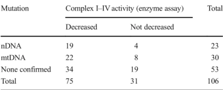
Table 1 Mitochondrial respiratory chain (MRC) complex activities and associated genetic mutations
Mutation Complex I–IV activity (enzyme assay) Total Decreased Not decreased
nDNA 19 4 23
mtDNA 22 8 30
None confirmed 34 19 53
Total 75 31 106
nDNAnuclear DNA,mtDNAmitochondrial DNA
Oxygen consumption rate
The OCR was measured in 19 of the 31 LS/LL patients who presented normal enzyme assay results. Seven cases with mtDNA mutations were omitted. Analysis was precluded in three cases from whom fibroblast cell lines were not available.
In an additional two patients, cell lines did not react properly to the experiment, and results were not obtained. Based on MRR distribution in our five controls, a reduction to
<71.6% was considered a significant decline (p < 0.05). In 19 patients, it ranged from 36% to 136%, with a median of 69% of normal control(s). Ten patients showed a significant decline, suggesting mitochondrial respiratory dysfunction (Table3).
mtDNA analysis
Analysis of mtDNA mutation was performed for 103 patients and were identified in 30 patients across seven different genes (Table4), resulting in a yield of 29%.MT-ATP6was the gene most frequent (ten patients). We also identified 19 patients with 11 different mutations in mtDNA genes related to com- plex I.
Previously unreported variants were considered as poten- tial novel causative mutations of LS/LL when they coincided with positive enzyme assay results. Mutation m.14439G>A was shown to be pathogenic using cybrid analysis (Uehara
et al. 2014). One of two cases with a mutation in m.14487T>C showed a reduction in enzyme activity of com- plex I. Mutations m.3946G>A and m.14687A>G had been reported to cause other mitochondrial diseases (Kirby et al.
2004; Spruijt et al.2007; Bruno et al.2003) and were consid- ered as causative in our patients who showed defects in re- spective MRC complexes. Enzyme analysis of patients with confirmed pathogenic mutations m.3697G>A, m.10158T>C, m.10191T>C, m.13513G>A, and m.14459G>A all showed defects in complex I (Kohda et al.2016).
nDNA analysis
Seventy-six patients proceeded to nDNA analysis, and 17 pa- tients were identified with mutations in nine genes related to MRC complexes (SURF1, NDUFA1, NDUFAF6,NDUFS4, NDUFS6, NDUFV2, BOLA3, SCO2, and GTPBP3, see Table4). Mutations inNDUFAF6andSURF1were most fre- quent (five patients each), with all patients showing reduced activity in complex I(NDUFAF6) or IV (SURF1). Mutations in genes related to complex I constituted more than half of the nDNA mutations. The genetic defects were all in agreement with the biochemical defects.
Four cases were identified with a mutation in ECHS1, a gene involved in valine degradation. An outside laboratory identified one more patient with a mutation in the same gene (Yamada et al. 2015). Accumulation of toxic intermediates Table 2 Clinical presentations of patients with Leigh syndrome
Defect and mut Mut only Defect only No defect, no
validated mut
Total
Number of patients 41 12 34 19 106
Leigh-like 6 4 10 4 24
Livinga 71% (20/28) 78% (7/9) 62% (16/26) 83% (10/12) 71% (53/75)
Age of living patientsa[median (range)]
9 (3–17) years 8 (3–15) years 9.5 (3–38) years 8.5 (6–20) years 8 (3–38) years Age at onset [median (range)] 10.5 months
(0 months–8 years)
9 months (0 months–5 - years)
5.5 months (0 months–6 - years)
10 months (0 months–2 - years)
9 months (0 months–8 - years)
Neonatal onset 2 (5%) 2 (17%) 7 (21%) 1 (5%) 12 (11%)
Seizure 20% 33% 41% 42% 32%
Involuntary movement 10% 25% 18% 16% 15%
Hypotonia 24% 42% 9% 32% 23%
Nystagmus/ ophthalmoplegia 17% 33% 26% 11% 21%
Dysphagia 10% 17% 29% 32% 21%
Respiratory distress 24% 17% 41% 37% 31%
Serum L/P (mean ± SD) (number of data available)
26.4 ± 16.4 (36) 22.9 ± 14.9 (10) 21.4 ± 9.9 (27) 18.2 ± 5.7 (16) 23.0 ± 13.2 (89) CSF L/P (mean ± SD) (number of
data available)
27.2 ± 28.0 (30) 20.7 ± 4.5 (9) 25.0 ± 9.6 (20) 18.7 ± 7.1 (14) 24.1 ± 18.9 (73)
Mutmutations in mitochondrial and nuclear DNA,L/Plactate-to-pyruvate ratio,SDstandard deviation,CSFcerebrospinal fluid
aAs of November 2016
caused by impairment in this pathway is suspected to cause MRC complex defect (Peters et al.2014). Three of our five patients showed no decline in enzyme activities, one patient showed a defect in complex IV and another in complex I.
Lastly, one patient was identified with a mutation in SLC19A3, a gene encoding a thiamine transporter, which is essential for cerebral thiamine metabolism.
Mutations in all these genes exceptBOLA3had been re- ported to cause LS (Tiranti et al. 1998; Budde et al.2000;
Fernandez-Moreira et al. 2007; McKenzie et al. 2011;
Kopajtich et al. 2014; Peters et al. 2014; Gerards et al.
2013).BOLA3had been identified in patients with other mi- tochondrial diseases (Cameron et al.2011; Haack et al.2013), and our case was previously reported as the first evidence of this mutation in an LS patient (Kohda et al.2016).
Discussion
We demonstrated the importance of combining multiple methods of diagnosing LS/LL patients. Genetic analysis iden- tified a causative mutation in 51% (53/104) of analyzed cases.
Enzyme assay recognized MRC complex defects in 71% (75/
106) of patients. With those approaches combined, MRC de- fects were confirmed in 82% (87/106) of cases. The highest diagnostic rate was reached by a combined enzymatic and genetic approach. Seven patients with normal enzyme activi- ties had mutations in theMT-ATP6gene, which encodes for complex V, which is measured in few laboratories. Screening forMT-ATP6mutations should be performed in such settings at an early stage of diagnosis, as they comprise a significant proportion of LS/LL etiology, and screening is readily available.
Detection rates in our study of various biopsy samples were
<60% individually, which confirms previous results. Most importantly, the rate in muscle biopsies was no higher than in fibroblast cell lines, a finding not reported previously. For the diagnosis of mitochondrial diseases, skeletal muscle is often considered the tissue of choice (Thorburn and Rahman 1993), and fibroblasts have been considered less sensitive than skeletal muscle biopsy samples, detecting MRC defects in only half of cases with positive skeletal muscle assay results (Thorburn et al.2004; Heuvel et al.2004). A similar sensitiv- ity was observed in our study, although skeletal muscle biopsy samples returned negative results in six out of 19 cases with reduced MRC activity in fibroblasts, resulting in similar over- all detection rates. Tissue specificity of mitochondrial diseases was attributed to heteroplasmy of mtDNA, but inconsistencies between materials were frequently observed in nDNA- mutated cases. These findings suggest that, when possible, more than one type of patient biological sample should be analyzed, regardless of genetic background, to improve the detection rate of mitochondrial disorder.
In pediatric practice, it can be difficult to obtain multiple biological samples, and physicians must choose selectively.
Although tissues used for analysis should be taken from the most affected organ (Munnich and Rustin2001), this is diffi- cult to apply in principle to LS/LL, a neurodegenerative dis- order of the central nervous system. So the choice would be between skeletal muscle biopsy samples and cultured fibro- blast cell lines in most cases. Skeletal muscle biopsy is inva- sive and requires general anesthesia, which poses a risk to pediatric patients (Baertling et al.2014). Fibroblasts, on the other hand can be obtained in office settings with local anes- thesia. If only one type of material can be obtained, fibroblasts should be prioritized, as cell lines from cultured fibroblasts can be used in future studies such as those involving cybrid analysis and rescue experiments to verify the pathogenicity of novel variations (Haas et al.2008). Should no defect be ob- served in fibroblasts, or if the clinical status calls for a rapid result, skeletal muscle biopsy should also be considered.
Relatively high numbers of enzyme assays return negative results in genetically verified cases of LS/LL (Sofou et al.
2014). In our study, the rate of negative assay results in genet- ically verified cases was 20%, excluding MT-ATP6mutated cases. This observation implies that a normal MRC result in Table 3 Oxygen consumption rate (OCR) measured with a Seahorse
analyzer
Patient Enzyme analysis MRR (%)
Pt139 ns (Fb) 136
Pt156 ns (Fb) 69
Pt161 ns (Fb) 36
Pt207 ns (M, Fb) 90
Pt216 ns (M, Fb) 94
Pt394 ns (Fb) 94
Pt430 ns (M, Fb) 62
Pt536 ns (M, Fb) 96
Pt545 ns (M, Fb) 53
Pt668 ns (M, Fb) 62
Pt696 ns (Fb) 127
Pt701 ns (Fb) 61
Pt703 ns (M, Fb) 81
Pt794 ns (Fb) 48
Pt822 ns (M, Fb) 108
Pt840 ns (Fb) 43
Pt1038 ns (Fb) 51
Pt1065 ns (M, Fb) 78
Pt1120 ns (Fb) 51
MRR reduction to <71.6% of normal control value was considered to indicate mitochondrial impairment and is shown in bold
OCRoxygen consumption rate,MRRmaximum respiration rate,nsnot significant,Mskeletal muscle,Fbcultured fibroblast,CIVcomplex IV,P partial decline
Table 4 Mutations in mitochondrial DNA (mtDNA) and nuclear DNA (nDNA)
Patient Gene Mutation LS/LL Enzyme assay Heteroplasmy rate (%) Tissue
Pt27 SURF1(NM_003172.2) c.743C>A:p.A248D LS CIV c.743C>A:p.A248D
Pt756 SURF1(NM_003172.2) c.367_368del:p.R123Gfs LS CIV
c.54+1G>T
Pt981 SURF1(NM_003172.2) c.743C>A:p.A248D LL CIV
c.54+1G>T
Pt1066 SURF1(NM_003172.2) c.367_368del:p.R123Gfs LS CIV
c.867G>A:p.W289X
Pt1143 SURF1(NM_003172.2) c.743C>A:p.A248D LS CIV
c.826_827ins18:p.V276_T277ins6
Pt312a NDUFA1(NM_004541) c.55C>T:p.P19S LS CI
Pt286 BOLA3(NM_212552) c.287A>G:p.H96R LS CC (I, II)
c.287A>G:p.H96R
Pt376 ECHS1(NM_004092) c.98T>C:p.F33S LS CIV
c.176A>G:p.N59S
Pt536 ECHS1(ENST00000368547) c.5C>T:p.A2V LS ns
c.1A>G:p.M1V
Pt1038 ECHS1(NM_004092) c.5C>T:p.A2V LS ns
c.176A>G:p.N59S
Pt1135 ECHS1(NM_004092) c.5C>T:p.A2V LS CI
c.176A>G:p.N59S
Pt101 NDUFAF6(NM_152416) c.371T>C:p.I124T LS CI
c.805C>G:p.H269D
Pt330 NDUFAF6(NM_152416) c.820A>G:p.R274G LS CI
c.820A>G:p.R274G
Pt512 NDUFAF6(NM_152416) c.226T>C:p.S76P LS CI
c.805C>G:p.H269D
Pt598 NDUFAF6(NM_152416) c.206A>T:p.D69V LL CI
c.371T>C:p.I124T
Pt866 NDUFAF6(NM_152416) c.371T>C:p.1124T LS CI
c.805C>G:p.H269D
Pt711 NDUFS4(NM_002495) c.340T>C:p.W114R LS CI
c.340T>C:p.W114R
Pt1087 NDUFS6(NM_004553) c.309+5G>A LS CC (I, IV)
c.343T>C:p.C115R
Pt1177 NDUFV2(NM_021074) c.427C>T:p.R143X LS CI
c.580G>A:p.E194K
Pt628 SCO2(NM_001169109) c.577G>A:p.G193S LS CC (I, IV)
c.773T>C:p.M258T
Pt751 GTPBP3(NM_032620) c.8G>T:p.R3L LS CC (I, IV)
c.923_947del:p.E309Rfs
Pt156 SLC19A3(NM_025243) c.372C>G:p.Y124X LS ns
c.265A>C:p.S89R
Pt416 MT-ND1 m.3697G>A:p.G131S LS CI 100 F
Pt619 MT-ND1 m.3946G>A:p.E214K LS CC (I, IV) 66 M
Pt179 MT-ATP6 m.8993T>G:pL156R LL ns nearly 100 B
Pt274 MT-ATP6 m.8993T>C:p.L156P LS CC (I, III) 100 F
Pt453 MT-ATP6 m.8993T>G:p.L156R LS CC (I, IV) 100 F
Pt341 MT-ATP6 m.8993T>C:p.L156R LS ns 100 M
Pt720 MT-ATP6 m.8993T>G:p.L156R LS ns nearly 100 B
Pt772 MT-ATP6 m.8993T>G:p.L156R LS ns nearly 100 M
Pt968 MT-ATP6 m.8993T>G:p.L156R LS ns nearly 100 B
Pt400 MT-ATP6 m.9176T>C:p.L217P LS ns 100 B
muscle and/or fibroblast cell line does not exclude the possi- bility of a mitochondrial disorder. A reasonable proportion of MRC defects may remain undetected if negative enzyme assay results prevent us from proceeding to genetic analysis.
Interestingly, negative assay results were more frequently ob- served in cases with nDNA than mtDNA mutations. In addi- tion, genetic causes such asECHS1mutations, which are not directly related to components of the MRC complexes, have been associated with LS/LL. In such cases, each separate MRC complex may not show reduced activity and thus remain un- detected by enzyme assay. If marker substances detected by basic metabolic analysis leads directly to diagnosis, as is the case with urinary organic acids inECHS1mutation, the next step is to proceed directly to analyzing the candidate gene.
In addition to genetic screening and spectrophotometric assays that measure the activity of individual respiratory com- plexes, we used microscale oxygraphy to help analyze mito- chondrial activity. Microscale oxygraphy has a high efficiency for detecting mitochondrial respiratory defects in genetically proven mitochondrial disease patients, an observation by
Invernizzi but not adopted by many diagnostic laboratories (Invernizzi et al.2012). Half the cases in our cohort with no apparent defect in activities of MRC complexes showed a significant decline in OCR. Moreover, two nDNA mutations were identified in this group. Although evidence needs to be accumulated, this finding suggests the promising value of mi- croscale oxygraphy as a screening tool to detect MRC defect, especially in cases in whom each complex remains intact. If cellular OCR shows a significant reduction, genetic screening should be considered, even if MRC defects were not detected by enzyme assays of fibroblasts or peripheral organs.
With advances in molecular technologies, genetic screening is becoming increasingly utilized over enzyme analysis and invasive biopsies (Lake et al. 2016; Taylor et al 2014).
Enzyme assays are considered a confirmatory method for di- agnosis of LS/LL in cases with ambiguous genetic results or where genetic analysis fails to detect causative mutations (Morava and Brown2015). However, in our study, gene anal- ysis could not identify underlying mutations in 45% of cases with reduced MRC complex activities. The genetic spectrum Table 4 (continued)
Patient Gene Mutation LS/LL Enzyme assay Heteroplasmy rate (%) Tissue
Pt698 MT-ATP6 m.9176T>C:p.L217P LS CIV 100 B
Pt127 MT-ATP6 m.9185T>C:p.L220P LL ns 80 B
Pt728 MT-ND3 m.10158T>C:p.S34P LS CI 80 B
Pt994 MT-ND3 m.10158T>C:p.S34P LS CI 100 B
Pt43 MT-ND3 m.10191T>C:pS45P LS CI 100 F
Pt44 MT-ND3 m.10191T>C:pS45P LS CI 69 F
Pt58 MT-ND3 m.10191T>C:pS45P LS CI na
Pt83 MT-ND3 m.10191T>C:pS45P LS CI 100 F
Pt108 MT-ND3 m.10191T>C:p.S45P LS CI 95 B
Pt965 MT-ND3 m.10197G>C:pA47P (VUS)b LL CC(I,III,IV) na
Pt190 MT-ND4 m.11246G>A:pA163T (VUS) LS CC (I, IV) 73 F
Pt153 MT-ND5 m.13094T>C:pV253A LS CC (I, IV) na B,M
Pt467 MT-ND5 m.13513G>A:p.D393N LL CI 59 B
Pt744 MT-ND5 m.13513G>A:p.D393N LL CC (I, IV) 50 B
Pt377 MT-ND6 m.14439G>A:pP79S LS CI 100 F
Pt28 MT-ND6 m.14459G>A:pA72V LS CI 54 F
Pt593 MT-ND6 m.14459G>A:p.A72V LS CI 96 F
Pt224 MT-ND6 m.14487T>C:p.M63V LS CI 99 B
Pt1063 MT-ND6 m.14487T>C:p.M63V LS ns Nearly 100 B
Pt396 tRNAGlu m.14687A>G LS CI 85 M
Pt255, identified with a mutation inECHS1gene, is not listed here, and therefore the number of patients does not add up to the total number of patients with nDNA mutations on Table1. The patient was omitted from this table because the gene analysis was processed in an outside laboratory Segregation analyses have been completed for all autosomal recessive mutation cases
mtDNAmitochondrial DNA,nDNAnuclear DNA,LSLeigh syndrome,LLLeigh-like syndrome,CIisolated complex I deficiency,CIVisolated complex IV deficiency,CCcombined complex deficiency,VUSvariant of unknown significance,nsnot significant,nanot available,Ffibroblasts,Mskeletal muscle,Bblood
aPt312 is a male patient
bm.10197G>C is designated as VUS because the mutation confirmed in MITOMAP is m.10197G>A
of LS/LL is still expanding, and biochemical data obtained via enzyme assays enable the efficient selection of candidate genes (Thorburn et al.2004) and provide essential information in the pathogenicity of identified gene variants. Thus, enzyme anal- ysis remains an important part of the diagnostic process of mitochondrial disorders.
Based on our increasing understanding of the biologi- cal and molecular background of the disease, new thera- peutic methods are being proposed (Martinelli et al.2012;
Morava and Brown2015). Precise biochemical and genet- ic diagnosis is imperative in considering the possible gene-specific therapeutic options. It is also essential to provide appropriate genetic counseling. All available bio- chemical and molecular methods should be combined to not only diagnose the disease but also to provide optimal care to the LS/LL patients.
Acknowledgements We thank all the patients and their doctors in charge for supplying us with data. We also thank Dr Holger Prokisch and Mr Vicente Yepez for their professional advice.
Compliance with ethical standards Conflict of interest None.
Details of funding This work was supported in part by the Practical Research Project for Rare/Intractable Diseases from the Japan Agency for Medical Research and Development, AMED to Kei Murayama (http://
www.amed.go.jp/en/) and the Project Promoting Clinical Trials for Development of New Drugs and Medical Devices, Japan Medical Association, from the Japan Agency for Medical Research and Development, AMED to Akira Ohtake (http://www.jmacct.med.or.jp/
en/what-we-do/investigator.html).
This work was also supported by an Innovative Cell Biology by Innovative Technology grant (Cell Innovation Program) from the Ministry of Education, Culture, Sports, Science and Technology (MEXT), Japan to Yasushi Okazaki (http://cell-innovation.nig.ac.jp/
mext-life/english/index.html), the Support Project, and a Strategic Research Center in Private Universities grant from MEXT, Japan to Saitama Medical University Research Center for Genomic Medicine (http://www.mext.go.jp/a_menu/koutou/shinkou/07021403/002/002/
1218299.htm). Further, this work was supported by Grants-in-Aid of the Research on Intractable Diseases (Mitochondrial Disorder) from the Ministry of Health, Labor and Welfare of Japan, and a special research grant from Takeda Science Foundation (http://www.takeda-sci.or.jp/) to YO. The authors confirm independence from the sponsors.
Informed consent All procedures followed were in accordance with the ethical standards of the responsible committee on human experimen- tation (institutional and national) and with the Helsinki Declaration of 1975, as revised in 2000. Informed consent was obtained from parents of all patients for being included in the study.
Open AccessThis article is distributed under the terms of the Creative C o m m o n s A t t r i b u t i o n 4 . 0 I n t e r n a t i o n a l L i c e n s e ( h t t p : / / creativecommons.org/licenses/by/4.0/), which permits unrestricted use, distribution, and reproduction in any medium, provided you give appropriate credit to the original author(s) and the source, provide a link to the Creative Commons license, and indicate if changes were made.
References
Baertling F, Rodenburg RJ, Schaper J et al (2014) A guide to diagnosis and treatment of Leigh syndrome. J Neurol Neurosurg Psychiatry 85(3):257–265
Bernier FP, Boneh A, Dennett X, Chow CW, Cleary MA, Thorburn DR (2002) Diagnostic criteria for respiratory chain disorders in adults and children. Neurology 59(9):1406–1411
Bruno C, Sacco O, Santorelli FM et al (2003) Mitochondrial myopathy and respiratory failure associated with a new mutation in the mito- chondrial transfer ribonucleic acid glutamic acid gene. J Child Neurol 18(4):300–303
Budde SM, van den Heuvel LP, Janssen AJ et al (2000) Combined enzy- matic complex I and III deficiency associated with mutations in the nuclear encoded NDUFS4 gene. Biochem Biophys Res Commun 275(1):63–68
Cameron JM, Janer A, Levandovskiy V et al (2011) Mutations in iron- sulfur cluster scaffold genes NFU1 and BOLA3 cause a fatal defi- ciency of multiple respiratory chain and 2-oxoacid dehydrogenase enzymes. Am J Hum Genet 89(4):486–495
Fernandez-Moreira D, Ugalde C, Smeets R et al (2007) X-linked NDUFA1 gene mutations associated with mitochondrial encephalo- myopathy. Ann Neurol 61(1):73–83
Gerards M, Kamps R, van Oevelen J et al (2013) Exome sequencing reveals a novel Moroccan founder mutation in SLC19A3 as a new cause of early-childhood fatal Leigh syndrome. Brain 136(3):882– 890
Gropman AL (2013) Neuroimaging in mitochondrial disorders.
Neurotherapeutics 10(2):273–285
Haack TB, Rolinski B, Haberberger B et al (2013) Homozygous missense mutation in BOLA3 causes multiple mitochondrial dysfunctions syndrome in two siblings. J Inherit Metab Dis 36(1):55–62 Haas RH, Parikh S, Falk MJ et al (2008) The in-depth evaluation of
suspected mitochondrial disease. Mol Genet Metab 94(1):16–37 Heuvel LP, Smeitink JA, Rodenburg RJT (2004) Biochemical examina-
tion of fibroblasts in the diagnosis and research of oxidative phos- phorylation (OXPHOS) defects. Mitochondrion 4(5-6):395–401 Invernizzi F, D’Amato I, Jensen PB, Ravaqlia S, Zeviani M, Tiranti V
(2012) Microscale oxygraphy reveals OXPHOS impairment in MRC mutant cells. Mitochondrion 12(2):328–335
Kirby DM, Crawford M, Cleary MA, Dahl HH, Dennett X, Thorburn DR (1999) Respiratory chain complex I deficiency: an underdiagnosed energy generation disorder. Neurology 52(6):1255–1264
Kirby DM, McFarland R, Ohtake A et al (2004) Mutations of the mito- chondrial ND1 gene as a cause of MELAS. J Med Genet 41(10):
784–789
Kohda M, Tokuzawa Y, Kishita Y et al (2016) A comprehensive genomic analysis reveals the genetic landscape of mitochondrial respiratory chain complex deficiencies. PLoS Genet 12(1):e1005679. doi:10.
1371/journal.pgen.1005679
Kopajtich R, Nicholls TJ, Rorbach J et al (2014) Mutations in GTPBP3 cause a mitochondrial translation defect associated with hypertro- phic cardiomyopathy, lactic acidosis, and encephalopathy. Am J Hum Genet 95(6):708–720
Lake NJ, Compton AG, Rahman S, Thorburn DR (2016) Leigh syn- drome: one disorder, more than 75 monogenic causes. Ann Neurol 79(2):190–203
Leigh D (1951) Subacute necrotizing encephalomyelopathy in an infant. J Neurol Neurosurg Psychiatry 1(14):216–221
Martinelli D, Catteruccia M, Piemonte F et al (2012) EPI-743 reverses the progression of the pediatric mitochondrial disease–genetically de- fined Leigh syndrome. Mol Genet Metab 107(3):383–388 McKenzie M, Tucker EJ, Compton AG et al (2011) Mutations in the gene
encoding C8orf38 block complex I assembly by inhibiting
production of the mitochondria-encoded subunit ND1. J Mol Biol 414(3):413–426
Morava E, Brown GK (2015) Next generation mitochondrial disease:
change in diagnostics with eyes on therapy. J Inherit Metab Dis 38(3):387–388
Munnich A, Rustin P (2001) Clinical spectrum and diagnosis of mito- chondrial disorders. Am J Med Genet 106(1):4–17
Murayama K, Nagasaka H, Tsuruoka T et al (2009) Intractable secretory diarrhea in a Japanese boy with mitochondrial respiratory chain complex I deficiency. Eur J Pediatr 168(3):297–302
Peters H, Buck N, Wanders R et al (2014) ECHS1 mutations in Leigh disease: a new inborn error of metabolism affecting valine metabo- lism. Brain 137(Pt 11):2903–2908
Rahman S, Blok RB, Dahl H-HM et al (1996) Leigh syndrome: clinical features and biochemical and DNA abnormalities. Ann Neurol 39(3):343–351
Saudubray J-M, Charpentier C (2001) Clinical phenotypes: diagnosis/
algorithms. In: Scriver CR, Sly WS (eds) The metabolic and molec- ular bases of inherited diseases, 8th edn. McGraw-Hills, New York, pp 1327–1403
Sofou K, De Coo IF, Isohanni P et al (2014) A multicenter study on Leigh syndrome: disease course and predictors of survival. Orphanet J Rare Dis 9:52
Spruijt L, Smeets HJ, Hendrickx A et al (2007) A MELAS-associated ND1 mutation causing leber hereditary optic neuropathy and spastic dystonia. Arch Neurol 64(6):890–893
Taylor RW, Pyle A, Griffin H et al (2014) Use of whole-exome sequenc- ing to determine the genetic basis of multiple mitochondrial respi- ratory chain complex deficiencies. JAMA 312(1):68–77
Thorburn DR, Rahman S (1993) Mitochondrial DNA-associated Leigh syndrome and NARP. In: Pagon RA, Adam MP, Ardinger HH et al (eds) GeneReviews. University of Washington, Seattle
Thorburn DR, Chow CW, Kirby DM (2004) Respiratory chain enzyme analysis in muscle and liver. Mitochondrion 4(5–6):363–375 Tiranti V, Hoertnagel K, Carrozzo R et al (1998) Mutations ofSURF-1in
Leigh disease associated with cytochrome c oxidase deficiency. Am J Hum Genet 63(6):1609–1621
Uehara N, Mori M, Tokuzawa Y et al (2014) NewMT-ND6andNDUFA1 mutations in mitochondrial respiratory chain disorders. Ann Clin Transl Neurol 1(5):361–369
Yamada K, Aiba K, Kitaura Y et al (2015) Clinical, biochemical and metabolic characterization of a mild form of human short-chain enoyl-CoA hydratase deficiency: significance of increased N-ace- tyl-S-(2-carboxypropyl)cysteine excretion. J Med Genet 52(10):
691–698
1
論文タイトル:Clinical validity of biochemical and molecular analysis in diagnosing
Leigh syndrome; a study of 106 Japanese patients
邦題:Leigh 脳症の診断における生化学的・分子生物学的分析の臨床的妥当性に関する研究
【はじめに】
ミトコンドリアはエネルギー産生を担う細胞内小器官で、赤血球を除くほとんどすべて の細胞に存在する。特にエネルギー需要の大きい細胞には多数存在し、独自の
DNA(ミト コンドリア
DNA)を持つと同時に、核
DNAによる支配も受けている。構造的には、脂質 二重膜である外膜と内膜からなり、内膜の内側をマトリックス、内膜と外膜の間のスペース を膜間腔という。エネルギー産生は、内膜に埋め込まれている呼吸鎖が担っており、呼吸鎖 は複合体
Iから
Vまである。複合体
I~
IVが電子伝達系を、複合体
Vが
ATP合成を担って おり、それぞれ多数のサブユニットから構成されている。複合体
IIを除き、各複合体はミ トコンドリア
DNAと核
DNAの二重支配を受けているが、核遺伝子にコードされている蛋 白のほうが多い(複合体
IIは、核遺伝子にのみコードされている。 ) 。
ミトコンドリア病は、ミトコンドリアによる
ATP産生が低下することにより各種臓器に 障害がでる疾患または症候群の総称であり、特にエネルギー需要の大きい臓器に障害が出 やすい。ミトコンドリアはさまざまな臓器においてエネルギー産生を担っていることから、
あらゆる臓器であらゆる症状を呈し、あらゆる年代において発症しうる。また、その遺伝的 二重支配から、母系遺伝だけでなく、常染色体遺伝、性染色体遺伝のすべての遺伝形式をと る。
臨床病型としては、代表的な
MELASや
Leigh脳症、ミトコンドリア糖尿病や難聴のほ か、小児分野においては、ミトコンドリア肝症、ミトコンドリア心筋症、新生児ミトコンド リア病といった病型も認識されるようになっている。
病因分類としては、
各呼吸鎖複合体(
I~
V)欠損症
複数の複合体の活性が低下する複合型欠損症(ミトコンドリア
DNAの翻訳、呼吸鎖 の生合成、ミトコンドリア蛋白の輸送、ミトコンドリア膜の動力学にかかわる遺伝子 の異常等による。 )
ミトコンドリア
DNA枯渇症候群
がある。例えば
Leigh脳症は、すべての病因から発症しうるが、呼吸鎖複合体
I欠損症がも
っとも多い。逆に、呼吸鎖複合体
I欠損症は、
Leigh脳症のほかに、
MELASや致死型乳児
ミトコンドリア病など様々な臨床病型をとりうる。生化学的な理論に依拠した治療法は、病
因によりその有効性が異なると考えられるため(例えば、高脂肪・低炭水化物栄養療法は、
2
が期待される等) 、臨床病型診断にとどまらず、病因を明らかにすることが重要になってく る。
各病因の背後にはその原因となる遺伝子異常がある。ミトコンドリア呼吸鎖に関連する 遺伝子は、現在
250以上あるといわれており、ミトコンドリア病を起こすことが確認され ている遺伝子として
200程度が知られている。
現在、ミトコンドリア遺伝子解析 のプロジェクトが各国で進められて おり、日本においては、特に小児の ミトコンドリア呼吸鎖異常症につい て、埼玉医科大学と千葉県こども病 院の共同研究として「ミトコンドリ ア呼吸鎖異常症の酵素診断および責 任遺伝子解析に関する研究」が進め られている。この研究のスキームは 右図に示すとおり、全国から提供さ れる細胞や組織検体の酵素活性を測
定し、活性低下を示すものについてその原因遺伝子を同定し、創薬や治療法の開発につなげ るものである。診断の付いていない小児症例が多いため、この研究で解析している症例は、
Leigh
脳症を初めとする神経変性疾患、ミトコンドリア呼吸鎖異常が関与していると思われ
る肝症や心筋症、非常に重篤な致死型乳児ミトコンドリア病、そして乳幼児突然死症候群と して法医学教室から依頼されてくるものなどが多い。
【本研究の目的】
生化学的な分析技術や遺伝子解析技術の進歩により、ミトコンドリア病の診断において、
酵素活性測定や遺伝子解析はいまやスタンダードとなっている。小児ミトコンドリア病の 代表的な病型である
Leigh脳症についても、
Leigh脳症における呼吸鎖複合体欠損症につ いて論じた文献や、
Leigh脳症を呈する新たな遺伝子異常の報告は枚挙にいとまがない。そ の一方で、呼吸鎖酵素活性の低下や原因遺伝子が特定されていない
Leigh脳症について論 じた文献はほとんどない。そのため、これらの診断技術の有用性、すなわち、臨床診断され
た
Leigh脳症患者の何割において現在の生化学的、分子生物学的手法がミトコンドリア呼
吸鎖異常を確定できるのかは、明らかになっていない。
そこで、本研究では、
Leigh脳症における生化学的、分子生物学的解析の現時点での有用
性を明らかにすることを目的に、
106例の
Leigh脳症及び
Leigh様症候群(以下
Leigh脳
症等)患者の酵素活性測定結果と遺伝子解析結果を分析した。また、従来からミトコンドリ
ア病では筋生検がスタンダードとされてきたが、
Leigh脳症等において材料間の診断率を比
3
ない
Leigh脳症等におけるマイクロスケールオキシグラフィーの有用性を明らかにした報
告はないため、酵素活性低下を認めない症例について酸素消費量の測定と遺伝子パネルで の解析を行い、酸素消費量測定の有用性について検討した。
Leigh
脳症は、小児期ミトコンドリア病として
MELASとともに多く見られる病型であ
り、中枢神経系のエネルギー産生障害により、精神運動発達遅滞や退行を示す、乳幼児期に 発症する難治性の慢性進行性疾患である。
1951年に
Leighが剖検例の脳病理において特徴 的な左右対称性の壊死性病変を指摘したことに端を発しており、当初は死後において剖検 でのみ診断される病理学的診断名であった。現在は、
CTや
MRIにより、病理学的変化に 対応する画像変化をとらえることができるようになったため、生存中に診断がされるよう になり、典型的な臨床経過、血液・髄液検査所見、および画像所見から臨床的に診断されて いる。有病率は、
40000人に一人程度と考えられている。
【対象と方法】
<対象>
2007
年から
2015年の間に「ミトコンドリア呼吸鎖異常症の酵素診断と責任遺伝子解析 に関する研究」に解析依頼の
あった症例のうち、
Leigh脳 症等と考えられる症例を抽 出し、そのうち、
Rahmanの 診断基準(表
1)に照らして
Leigh
脳症等と診断できる
ものを対象とした。
<方法>
各症例について、発症年 齢、現況、主な症状、血中・
髄液中の乳酸/ピルビン酸比、酵素活性測定の結果、遺伝子解析の結果を収集・分析した。
酵素活性低下が認められない症例については、酸素消費量を測定するとともに、遺伝子パネ ルで遺伝子変異のスクリーニングを行った。
① 精神運動発達の退行を伴った進行性疾患
② 不随意運動、哺乳嚥下障害、呼吸障害、眼球運動障害、運動失 調などの脳幹・大脳基底核症状を伴う
③ 血中・髄液中の乳酸値の上昇
④ 次のうちの一つ以上
画像上の対称性基底核・脳幹病変
典型的神経病理学的変性(海綿状壊死)
同様症状の同胞の存在
以上のすべてを満たすものをLeigh脳症とする。
臨床所見は Leigh 脳症を強く示唆するものの、非典型的な病理所 見、画像所見が不存在、画像所見が正常または非典型的、典型的な 画像所見だが乳酸値が正常のものを ”Leigh様”とする。
表
1 Rahmanの診断基準
1)4
皮膚由来の線維芽細胞または
組織検体を用いて、各呼吸鎖複 合体の酵素反応の基質または生 成物の増減を分光光度計で測定 した。
Bernierらの診断基準
(表
2)に照らして、培養細胞で
40%未満、組織検体で
30%未満の場 合活性低下とした。
<酸素消費量の測定>
皮膚線維芽細胞を用いて、細
胞外フラックスアナライザーで測定した。基礎呼吸を測定後、オリゴマイシン、脱共役薬、
ロテノン(ミトコンドリア呼吸鎖複合体阻害薬)を順次添加し、その都度酸素消費量を測定 した。脱共役薬添加後の最大酸素消費量からロテノン添加後の酸素消費量を引いた差分を
MRR(最大酸素消費量)とした。酸素消費量の測定にあたっては、毎回1ないし2の正常 検体を同一プレートで測定した。すべての正常検体について
5回測定を行い、正常検体の 測定値の分布から
5%ile未満となる値
(71.6%)を下回るものを低下とした。
<遺伝子解析>
酵素活性低下が認められたものは、ミトコンドリア
DNAの全周囲解析を行った。ミトコ ンドリア
DNAで変異が確認されないものは次世代シークエンサーで核遺伝子の変異を検 索した。酵素活性が正常のものは、
Mayr(2015)に記載する
251のミトコンドリア関連遺伝 子を搭載した遺伝子パネルでスクリーニングを行った
3)。
本研究外で主治医により他の研究施設、検査機関等において遺伝子解析が行われ、高頻度 変異等が同定された症例については、遺伝子解析の診断率を明らかにするためその結果を 本研究に盛り込んだ。
【結果】
酵素活性測定の診断率は
71%(75/106)、遺伝子解析の診断率は
51%(53/104)(内訳:ミ トコンドリア遺伝子
29%(30/103)、核遺伝子
30%(23/76))だった。生化学的手法と遺伝 子解析を組み合わせると、診断率は
82%(87/106)に向上した。 (別表
1)
一般的な呼吸鎖酵素活性では測定しない複合体
Vの欠損による症例が少なからず存在 し、それらではミトコンドリア
DNAの高頻度変異を多く認めた。 (別表
2)
複合体
V欠損以外にも、
Leigh脳症関連の遺伝子に変異があるにもかかわらず、呼吸
大基準(抜粋)In vitro呼吸鎖酵素活性
1つの臓器で20%以下または2つ以上の臓器にまたがっ て30%以下
1つの培養細胞で30%以下 小基準(抜粋)
In vitro呼吸鎖酵素活性
1つの臓器で20-30%または2つ以上の臓器にまたがって 30-40%
1つの培養細胞で30-40%
Definite:大基準2つ 又は 大基準1つ+小基準2つ Probable:大基準1つ+小基準1つ 又は 小基準3つ
Possible:大基準1つ 又は 小基準のI(臨床症状:略)+他の
小基準1つ
表2 Bernierらのミトコンドリア呼吸鎖異常症の診断基準2)
5
酵素学的にミトコンドリア呼吸鎖異常が確かめられているが、エクソーム解析をもっ てしても遺伝子変異が確認できていない症例が
34例存在した。
酵素活性と遺伝子解析の結果により、
106例の
Leigh脳症等を、次の四群に分類し、予 後、発症年齢、新生児発症の頻度、患者年齢、主な症状や乳酸/ピルビン酸比等を比較 したが、四群間に有意な差は認めなかった。
(別表
4)① 酵素活性が低下し、遺伝子変異も同定されたもの
41例(核
DNA変異
19例、
mtDNA
変異
22② 酵素活性の低下はあるものの、遺伝子変異が同定されなかったもの
34例
③ 酵素活性は正常だったが、遺伝子変異が同定されたもの
12例
④ 酵素活性が正常で、遺伝子変異も同定されなかったもの
19例
検体種別酵素活性低下検出率は、皮膚線維芽細胞で
59%、骨格筋で
56%であり、この 二者間に有意差は認めなかった。一種類の検体(例:骨格筋生検検体)で酵素活性の低 下を認めない症例においても、異なる検体(例:皮膚培養細胞)で活性低下を認めるも のがあることが確認された。
酵素活性低下のない症例のうち、皮膚線維芽細胞が提出されていた
19例で酸素消費量 を測定したところ、
10例で有意な酸素消費量の低下を認めた。このうち
2例は遺伝子 パネル解析で核遺伝子の変異を認め、家系解析にて確定された。
(別表
5)
酵素活性測定、遺伝子解析、酸素消費量の測定すべてを合わせると、
Leigh脳症等にお けるミトコンドリア異常症の診断率は
90%(
95/106)となった。
【結論】
以上から、
1.
Leigh脳症等では、生化学的または分子生物学的手法の一方のみでは
Leigh脳症等の
ミトコンドリア機能異常をすべて確定することはできず、これらの手法を組み合わせ ることで診断率が向上すること
2. 原因遺伝子がミトコンドリア遺伝子か核遺伝子かにかかわらず、皮膚線維芽細胞と骨 格筋検体で酵素活性低下の検出率に有意差がないこと、また、一種類の材料で活性低下 がなくとも、ほかの臓器から採取した材料で活性低下を認めることがあり、複数の臓器 から検体を採取することで診断率が向上すること
が明らかになった。
【考察】
本研究にはいくつかの限界がある。
本研究は、対象患者の選定基準をあらかじめ定めた研究ではなく、酵素診断および遺伝子
解析目的に集められた母集団から
Leigh脳症等を取り出して検討している。
Leigh脳症ま
6
らの時間経過は様々である。酵素活性は、年齢や発症からの時間、また検体の保存状態によ っても結果に差が生じるため、酵素活性の測定結果がこれらの要素の影響を受けた可能性 は否定できない。また、
8年にわたり蓄積されたデータを対象としているため、この間の小 児分野におけるミトコンドリア病の認知度の高まりにより、集まってくる症例の性質(蓋然 性、重症度、発症からの期間等)が時とともに変化している可能性も否定できない。さらに、
同一時点でみても、きわめて重症の症例から、年長まで生存している比較的軽症といえる症 例まで、重症度が様々であり、酵素活性や遺伝子変異の種類に影響を与えている可能性があ る。
ミトコンドリア病では、同一の遺伝子異常が、異なる臨床病型を呈することが観察される。
ミトコンドリア遺伝子異常では、その背景に、ミトコンドリアの
fusion、
fission、変異の偏 り(
heteroplasmy) 、臓器・組織による
heteroplasmyの閾値の違いなどが発症に関与して いると考えられるが、詳しいメカニズムについては依然として不明なことが多く、今後の研 究課題である。核遺伝子変異例においても、病型や重症度の多様性が認められ、これは
heteroplasmy