介した大腸AQP3の発現増加により発症する
著者 今 理紗子
雑誌名 星薬科大学紀要
号 57
ページ 19‑24
発行年 2015‑12‑10
URL http://id.nii.ac.jp/1240/00000785/
モルヒネは、 がん性疼痛などに用いられている麻薬性 鎮痛薬である。 モルヒネは、 強力な鎮痛作用を有するこ とに加え、 様々な剤型があることから、 痛み治療の第一 選択薬となっている。 モルヒネの鎮痛作用は、 中枢のオ ピオイドμ受容体を介して発現する。 しかしながら、 オ ピオイド受容体は、 中枢だけではなく、 末梢にも発現し ているため、 モルヒネ服用患者には末梢のオピオイド受 容体を介した様々な副作用が現れる。 モルヒネの副作用 の中で、 臨床において最も深刻なものは便秘である1)。 モルヒネによる便秘は、 鎮痛用量以下で発生することか ら、 ほとんどすべてのモルヒネ服用患者に生じる。 また、
モルヒネによる便秘は耐性ができにくく、 放置しておく と重症化し、 腸閉塞などの重大な合併症を誘発する場合
もある2, 3)。 このように、 便秘は患者のQOLの低下を招
くばかりではなく、 モルヒネの継続投与が困難となる場 合もあるため、 モルヒネ服用と同時に十分な対策を行う 必要がある。
現在、 モルヒネによる便秘症患者には対症療法として、
浸透圧性下剤や大腸刺激性下剤などの瀉下剤が経験的に 使用されている。 しかしながら、 現存の瀉下剤を駆使し ても治療が困難である場合も多い4, 5)。 その理由は、 こ れまでモルヒネによる便秘は、 腸の蠕動運動が抑制され ることにより発症するものと考えられてきたが、 その他 のメカニズムについてはほとんどわかっていなかったた めである。 したがって、 モルヒネ誘発性便秘症の発症要 因を把握し、 その全容を解明することは、 単にモルヒネ の副作用を軽減するにとどまらず、 新規メカニズムを有 する下剤の開発や、 便秘に対するこれまでにない治療方 法や予防方法を提案する上で、 非常に重要となる。
一方、 近年、 腸管における水の移動に、 水チャネルで あるアクアポリン (aquaporins; AQPs) が関与してい
ることが明らかになってきた。 AQPsはヒトにおいて、
様々な臓器に発現しており、 現在AQP0からAQP12 までの13種類のAQPsが同定されている。 腸管にも多 数のAQPsが発現しており、 特に、 大腸粘膜上皮細胞 においては、 AQP3が優位に発現している。 我々はこれ までに、 大腸のAQP3の機能や発現が低下すると、 腸 管側から血管側への水の吸収が抑制され、 下痢が発症す ることを明らかにした6-9)。 この知見は、 大腸のAQP3 が便の水分調節に重要な役割を担っていることを示すも のである。
本研究では、 大腸AQP3に着目し、 モルヒネ誘発性 便秘症における大腸AQP3の役割およびその発現変動 メカニズムについて解明することにより、 便秘症の新規 治療法および薬物の適正使用の提案を試みた。
1.
AQP3
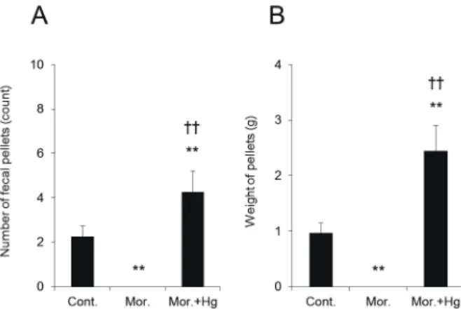
ラットにモルヒネを経口投与し、 12時間後までの排 便総重量、 排便回数および糞中水分量を調べることによ り、 便秘の程度を評価した。 モルヒネ投与後の排便回数 はコントロール群と比べて有意に低下し、 これに伴い、
排便総重量も著明に低下した (Fig. 1Aおよび1B)。 ま た、 モルヒネ投与後の糞中水分量は、 投与1時間後か ら低下し始め、 投与5時間後ではコントロール群 (0 h) の約60%であり、 最も低値を示した (Fig. 1C)。 これ らのことから、 ラットにモルヒネを投与することにより 便秘が発症することが確認できた。
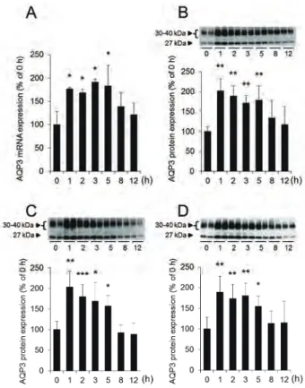
大腸においてはAQP3が粘膜上皮細胞に優位に発現 しており、 便の水分調節に重要な役割を担っている。 そ こで、 モルヒネを経口投与した際の大腸におけるAQP3 の発現量を解析した。 大腸AQP3のmRNA発現量は、
モルヒネ投与1時間後から有意に増加し、 投与5時間 後においても、 投与前と比べて約2倍高い値を示して
! "
#$%&'()*+,-./01 AQP3 234'56
今 理紗子
星薬科大学 薬動学教室
Morphine-induced constipation develops with increased aquaporin-3 expression in the colon via increased serotonin secretion
Risako KON
Department of Clinical Pharmacokinetics, Hoshi Unversity
いた (Fig. 2A)。 次に、 摘出した大腸から細胞膜を豊 富に含むPM (plasma membrane) 画分、 小胞体膜を 豊富に含むIV (intracellular vesicle) 画分およびこれ らを合わせた細胞全体の膜を含むCM (crude mem- brane) 画分を調製し、 それぞれの画分におけるAQP3 の発現量を解析した。 大腸CM画分におけるAQP3の タンパク質発現量は、 モルヒネ投与1時間後から約2 倍増加し、 この増加は投与5時間後まで持続していた (Fig. 2B)。 また、 大腸PM画分およびIV画分におけ るAQP3のタンパク質発現量についても、 CM画分と 同様の挙動を示した (Fig. 2Cおよび2D)。 加えて、 モ ルヒネによる大腸AQP3の発現増加は、 大腸粘膜上皮 細胞のapical側およびbasal側の両部位において見ら れた (Fig. 3)。
以上のことから、 モルヒネにより便秘が発症した際に は、 大腸AQP3の発現量が増加することがわかった。
2. AQP3
塩化水銀 (HgCl2) などの水銀化合物はin vitro試験 において、 AQP3の水の透過機能を阻害することが報告 されている10, 11)。 また、 我々はこれまでに、 AQP3の水 透過機能を約70%程度阻害する濃度のHgCl2をラット に直腸内投与すると、 腸管側から血管側への水の移動が 阻害されるため、 投与後1時間までに重度の下痢が発 症することを明らかにしている8)。 そこで、 モルヒネに よる便秘の発症に大腸のAQP3の発現増加が関与して いるかどうかをHgCl2を用いて調べた。 具体的には、
モルヒネ投与30分後のラットにHgCl2を直腸内投与し、
モルヒネ投与直後から1.5時間後までの便秘の程度を調 べた。 その結果、 モルヒネとHgCl2を併用投与した際 の排便回数および排便総重量は、 いずれもモルヒネ単独 投与群と比べて有意に増加していることがわかった (Fig. 4Aおよび4B)。
Fig. 1. Effects of morphine on the constipation score and fecal water content
Rat fecal samples were collected for up to 12 h after mor- phine administration. The number of fecal pellets (A), the weight of pellets (B), and the fecal water content (C) were measured. The mean fecal water content immediately after administration (0 h) was indicated as 100%. Data represent means ± SDs for six rats. Student’s t-test or Dunnett’s test: *p<0.05, **p<0.01, and ***p<0.001 vs.
Control or 0 h.
Fig. 2. Effect of morphine on AQP3 expression level in rat colon
Rat colons were harvested for up to 12 h, beginning im- mediately after morphine administration.
A : The mRNA expression levels of AQP3 were measured using real-time RT-PCR, and normalized to 18S rRNA.
B-D : The CM (B), PM (C), and IV (D) fractions were pre- pared, and the protein expression levels of AQP3 were analyzed using western blotting.
Mean levels of AQP3 mRNA and protein expression im- mediately after administration (0 h) were indicated as 100%. Data represent means ± SDs for six rats. Dunnet t’s test: *p<0.05, **p<0.01, and ***p<0.001 vs. 0 h.
Fig. 3. The distribution of AQP3 in rat colon after morphine administration
Rat colons were removed 5 h after water (control) or mor- phine administration.
A : AQP3 (green) and nuclei (blue) were immunostained.
B : Enlarged view of AQP3 in rat colon epithelial cells.
以上のことから、 モルヒネによる便秘の発症には大腸 AQP3の発現増加が関与していることがわかった。
3. AQP3
経口投与されたモルヒネは、 その大部分が肝臓の UGT (UDP-glucuronosyltransferase) によって、 鎮痛 作 用 を ほ と ん ど 持 た な い M-3-G (morphine-3- glucuronide) と 、 強 い 鎮 痛 作 用 を 持 つ M-6-G (morphine-6-glucuronide) に代謝される。 しかしなが ら、 ラットにおいては、 M-6-Gはごくわずかしか生成
されないことが知られている12, 13)。 そこで本研究では、
モルヒネあるいはM-3-Gの大腸AQP3の発現に及ぼす 影響をヒト結腸癌由来HT-29細胞を用いて調べた。
HT-29細胞は、 ヒトの正常な大腸の生理状態を示すこ とから、 AQP3に関する研究に広く用いられている。
モルヒネによる便秘が発症している際の血中および腸 管内の濃度に相当するモルヒネあるいはM-3-GをHT- 29細胞に添加し、 AQP3のmRNA発現量を調べたとこ ろ、 いずれの濃度においても添加3時間後まで変化は 認められなかった (Fig. 5Aおよび5B)。
4. AQP3
!"#$%&
モルヒネは、 腸管壁からのセロトニンの遊離を促進さ せ、 腸の蠕動運動を抑制することにより便秘を誘発させ ることが報告されている14, 15)。 そこで、 モルヒネ投与時 のAQP3の発現増加にもセロトニンが関与しているの ではないかとの仮説を立て、 その立証を試みた。
まず、 セロトニンが大腸のAQP3の発現を増加させ るかどうかをin vitro試験で調べた。 HT-29細胞にセ ロトニンを添加したところ、 1μM以上の濃度でAQP3 のmRNA発現量が増加することがわかった (Fig. 6A)。
また、 このセロトニンによるAQP3の発現増加は、 添 加1時間後が最も高く、 6時間後においては、 添加前と ほぼ同レベルまで戻っていた (Fig. 6B)。 このセロト ニンによるAQP3のmRNAの変化は、 モルヒネ投与後 のAQP3の発現変化 (Fig. 2A) と類似していた。
消化管には、 体内のセロトニンの約95%が存在して おり、 その合成や貯蔵、 分泌は腸内分泌細胞であるEC 細胞が主役を担っている。 一般にEC細胞は、 セロトニ
Fig. 5. Effects of morphine and M-3-G on AQP3 mRNA expression in HT-29 cells
HT-29 cells were treated with morphine (A) or M-3-G (B) for 3 h. AQP3 mRNA expression levels were measured using real-time RT-PCR, normalized to the GAPDH, and presented with the mean of the control cells set to 100%.
AQP3 mRNA expression levels of the control cells did not change 3 h post-treatment with original growth medium.
The data represent the means ± SDs for five experiments.
Fig. 6. Effect of 5-HT on AQP3 mRNA expression in HT-29 cells
HT-29 cells were treated with 5-HT (0-10μM) for 1 h (A).
HT-29 cells were treated with 5-HT (1μM) for up to 6 h (B). The expression level of AQP3 mRNA was measured using real-time PCR, normalized to the GAPDH, and then presented with the mean of the control cells set to 100%. AQP3 mRNA expression levels of the control cells did not change 6 h post-treatment with original growth medium.
The data represent the means ± SDs for five experiments.
Dunnett’s test: **p<0.01 and ***p<0.001 vs. 0μM or 0 h.
Fig. 4. Changes in the morphine-induced constipa- tion score by HgCl2 administration
HgCl2 or saline was administered intrarectally to rats 30 min after treatment with oral morphine. The number (A) and the weight (B) of fecal pellets were measured. The data represent the means ± SDs for five rats. Tukey’s test: **p<0.01 vs. Control. ††p<0.01 vs. Morphine.
ンを分泌する際にその形態を球状から突起状に変化させ ることが知られている16)。 そこで、 AQP3の著明な増加 がみられたモルヒネ投与1時間後のラットの大腸にお いて、 セロトニン陽性EC細胞の局在およびその形態が どのように変化するかを免疫染色により調べた。 コント ロール群においては、 セロトニン陽性EC細胞は大腸に 散在しており、 その多くは大腸粘膜固有層の陰窩に存在 していた。 それに対して、 モルヒネ投与群のセロトニン 陽性EC細胞は、 陰窩よりも上層の粘膜層に多く存在し ていた。 さらに、 モルヒネ投与群のEC細胞 は、 コン トロール群でみられた球状の形態とは異なり、 突起を伸 ばすような形態に変化していた (Fig. 7)。
5. AQP3
大腸にはセロトニン受容体やセロトニン選択的トラン スポーター (serotonin reuptake transporter; SERT) が存在する。 これらの阻害剤を用いて、 セロトニンがど のようなメカニズムでAQP3の発現を増加させたのか について調べた。 その結果、 HT-29細胞にセロトニン 受容体アンタゴニスト (5-HT2; ketanserin、 5-HT3 ;Y- 25130、 5-HT4; RS39604) を前処置しても、 セロトニ ンによるAQP3の発現増加には影響を及ぼさないこと がわかった (Fig. 8A)。 一方、 SERT阻害剤であるフ ルオキセチンを前処置した際には、 セロトニンによる AQP3の発現増加がほぼ完全に抑制されることがわかっ た (Fig. 8B)。
セロトニンはSERTにより細胞内に取り込まれると、
核内受容体ペルオキシソーム増殖因子活性化受容体γ (peroxisome proliferator-activated receptor γ; PPA Rγ) のリガンドとなることが報告されている17)。 一方、
PPARγは、 リガンドが結合すると核内に移行しAQP3 の転写を亢進させることが報告されている18)。 そこで、
HT-29細胞にセロトニンを添加した際のPPARγの核 内移行量を調べた。 その結果、 セロトニン添加1時間 後において、 PPARγの核内移行量は約2倍有意に増加 していることがわかった。 このセロトニンによるPPA Rγの核内移行は、 フルオキセチンで前処置することに よりほぼ完全に抑制された (Fig. 8C)。
以上のことから、 セロトニンは、 SERTにより細胞内 に取り込まれた後、 PPARγのリガンドとして機能し、
PPARγの核内移行を亢進することによりAQP3の発 現を増加させることがわかった。
6. SERT
SERT阻害剤であるフルオキセチンをラットに前処置 することにより、 モルヒネの便秘および大腸における AQP3の発現増加が抑制されるかどうかを調べた。 具体 的には、 ラットにフルオキセチンを腹腔内投与し、 投与 15分後にモルヒネを経口投与した際の排便状況および 大腸AQP3のタンパク質発現量を調べた。
モルヒネとフルオキセチンを併用投与した際の排便回 数および排便総重量は、 モルヒネ単独投与群よりも増加 Fig. 8. Effects of 5-HT receptor or SERT inhibitor on AQP3 mRNA and PPARγ expression in HT- 29 cells
A: HT-29 cells were treated with ketanserin (5-HT2 recep- tor antagonist), Y-25130 (5-HT3 receptor antagonist), or RS39604 (5-HT4 receptor antagonist) for 1 h and then with 5-HT (1μM) for an additional 1 h. AQP3 mRNA ex- pression levels were measured using real-time RT-PCR and normalized to GAPDH.
B : HT-29 cells were treated with fluoxetine (SERT inhibi- tor) for 1 h and 5-HT (1μM) for an additional 1 h. AQP3 mRNA expression levels were measured using real-time RT-PCR and normalized to GAPDH.
C : PPARγprotein expression levels in nuclei were meas- ured using western blotting and normalized to Lamin B levels.
AQP3 and PPARγ expression levels were presented with the means of the control cells set to 100%. The data rep- resent the means ± SDs for five experiments. Tukey’s test : *p<0.05 and ***p<0.001 vs. Control. †p<0.05 and
†††p<0.001 vs. 5-HT.
Fig. 7. The distribution of 5-HT-positive EC cells in rat colon after morphine administration
Rat colons were removed 1 h after water (control) or mor- phine administration. 5-HT-positive EC cells (green) and nuclei (blue) were immunostained.
することがわかった。 しかしながら、 この増加値はコン トロール群よりも低値であった (Fig. 9Aおよび9B)。
一方、 モルヒネ投与により2倍増加した大腸PM画分 のAQP3のタンパク質発現量は、 フルオキセチンの併 用により約30%低下した (Fig. 9C)。
以上のことから、 SERT阻害剤はモルヒネによる大腸 AQP3の発現増加を抑制し、 便秘を改善させ得ることが わかった。 なお、 フルオキセチンの阻害率とモルヒネ誘 発性便秘症の改善率との関係、 およびモルヒネ誘発性便 秘症の改善率とAQP3の発現抑制率の関係については、
一定の疑問が残り、 これらの点に関しては今後の課題と 考える。
本研究の結果から、 モルヒネは大腸におけるAQP3 の発現量を増加させることにより、 腸管側から血管側へ の水の吸収を亢進し、 便を硬化することによって便秘を 引き起こしている可能性が示唆された。 さらに、 モルヒ ネによるAQP3の発現増加には、 EC細胞から分泌され るセロトニンが関与していることがわかった。 すなわち、
EC細胞から過剰分泌されたセロトニンがSERTを介し て細胞内に取り込まれた後に、 PPARγのリガンドとし て機能し、 AQP3の転写を亢進させ、 発現量を増加させ たものと考えられた (Fig. 10)。
本研究の結果は、 モルヒネによる便秘が蠕動運動の抑
制作用ばかりではなく、 AQP3の発現増加も伴って発症 することを初めて示すものである。 さらに、 大腸の AQP3の機能や発現を制御する物質がモルヒネ誘発性便 秘症の新規治療薬になり得ることを示唆するものである。
加えて、 腸管でのセロトニンの作用を調節している SERTをターゲットにした薬物が、 モルヒネ誘発性便秘 症の治療や予防に有効である可能性を示している。 今回、
我々が見出した新しい知見は、 新しい便秘の治療薬開発 や治療法の提案においても重要なものとなるであろう。
本研究を遂行するにあたり、 平成26年度星薬科大学 大谷記念研究奨励金を賜りましたことに対し、 大谷卓男 理事長ならびに田中治学長に厚く御礼申し上げます。
また、 本研究の遂行のために、 多大なるご指導を賜りま した星薬科大学薬動学教室の杉山清教授ならびに落合和 准教授、 五十嵐信智助教に心より御礼申し上げます。 ま た、 本研究を進めるなかで、 ご支援、 ご助言いただきま した鈴木勉名誉教授に深く御礼申し上げます。 最後に、
本研究を進めるにあたり、 多大なるご協力をいただきま した薬動学教室の教室員の皆様に深謝いたします。
Fig. 10. The mechanism of morphine-induced consti- pation via AQP3 in rat colon
Fig. 9. Changes in the morphine-induced constipa- tion score and the colon AQP3 protein expres- sion by fluoxetine administration
Saline or fluoxetine was administered intraperitoneally to rats 15 min after oral water or morphine administration.
The number of fecal pallets (A) and the weight of pellets (B) were measured for up to 5 h after morphine admini- stration. The colon was removed 5 h after morphine ad- ministration. AQP3 protein expression levels in the PM fraction were measured using western blotting (C). AQP3 protein expression levels were presented with the means of the control group as 100%. The data represent the means ± SDs for five rats. Tukey’s test: *p<0.05,
**p<0.01, and ***p<0.001 vs. Control. ††p<0.01 and
†††p<0.001 vs. Morphine.
1) Swegle JM, & Logemann C (2006). Management of common opioid-induced adverse effects. Am Fam Physician 74:
1347-1354.
2) Mercadante S, Villari P, Ferrera P, & Casuccio A (2006). Opioid-induced or pain relief-reduced symptoms in ad- vanced cancer patients? Eur J Pain 10: 153-159.
3) Mancini I, & Bruera E (1998). Constipation in advanced cancer patients. Support Care Cancer 6: 356-364.
4) McNicol E, Horowicz-Mehler N, Fisk RA, Bennett K, Gialeli-Goudas M, Chew PW, et al. (2003). Management of opioid side effects in cancer-related and chronic noncancer pain: a systematic review. J Pain 4: 231-256.
5) Larkin PJ, Sykes NP, Centeno C, Ellershaw JE, Elsner F, Eugene B, et al. (2008). The management of constipation in palliative care: clinical practice recommendations. Palliat Med 22: 796-807.
6) Ikarashi N, Baba K, Ushiki T, Kon R, Mimura A, Toda T, et al. (2011a). The laxative effect of bisacodyl is attrib- utable to decreased aquaporin-3 expression in the colon induced by increased PGE2 secretion from macrophages. Am J Physiol Gastrointest Liver Physiol 301: G887-895.
7) Ikarashi N, Ushiki T, Mochizuki T, Toda T, Kudo T, Baba K, et al. (2011b). Effects of magnesium sulphate admini- stration on aquaporin 3 in rat gastrointestinal tract. Biol Pharm Bull 34: 238-242.
8) Ikarashi N, Kon R, Iizasa T, Suzuki N, Hiruma R, Suenaga K, et al. (2012). Inhibition of aquaporin-3 water channel in the colon induces diarrhea. Biol Pharm Bull 35: 957-962.
9) Kon R, Ikarashi N, Nagoya C, Takayama T, Kusunoki Y, Ishii M, et al. (2014). Rheinanthrone, a metabolite of sennoside A, triggers macrophage activation to decrease aquaporin-3 expression in the colon, causing the laxative ef- fect of rhubarb extract. J Ethnopharmacol 152: 190-200.
10) Ishibashi K, Sasaki S, Fushimi K, Uchida S, Kuwahara M, Saito H, et al. (1994). Molecular cloning and expression of a member of the aquaporin family with permeability to glycerol and urea in addition to water expressed at the basolateral membrane of kidney collecting duct cells. Proc Natl Acad Sci U S A 91: 6269-6273.
11) Kuwahara M, Gu Y, Ishibashi K, Marumo F, & Sasaki S (1997). Mercury-sensitive residues and pore site in AQP3 water channel. Biochemistry 36: 13973-13978.
12) Yue Q, von Bahr C, Odar-Cederl?f I, & S?we J (1990). Glucuronidation of codeine and morphine in human liver and kidney microsomes: effect of inhibitors. Pharmacol Toxicol 66: 221-226.
13) Kuo CK, Hanioka N, Hoshikawa Y, Oguri K, & Yoshimura H (1991). Species difference of site-selective glucuronidation of morphine. J Pharmacobiodyn 14: 187-193.
14) Burks TF, & Long JP (1967). Release of intestinal 5-hydroxytryptamine by morphine and related agents. J Pharmacol Exp Ther 156: 267-276.
15) Burks TF (1973). Mediation by 5-hydroxytryptamine of morphine stimulant actions in dog intestine. J Pharmacol Exp Ther 185: 530-539.
16) Kuramoto H, Kadowaki M, Sakamoto H, Yuasa K, Todo A, & Shirai R (2007). Distinct morphology of serotonin- containing enterochromaffin (EC) cells in the rat distal colon. Arch Histol Cytol 70: 235-241.
17) Waku T, Shiraki T, Oyama T, Maebara K, Nakamori R, & Morikawa K (2010). The nuclear receptor PPARγ indi- vidually responds to serotonin- and fatty acid-metabolites. EMBO J 29: 3395-3407.
18) Jiang YJ, Kim P, Lu YF, & Feingold KR (2011). PPARgamma activators stimulate aquaporin 3 expression in keratinocytes/epidermis. Exp Dermatol 20: 595-599.
Morphine-induced constipation develops with increased aquaporin-3 expression in the colon via increased serotonin secretion
Risako KON
Department of Clinical Pharmacokinetics, Hoshi Unversity
Aquaporin-3 (AQP3) is a water channel that is predominantly expressed in the colon, where it plays a critical role in the regulation of fecal water content. This study investigated the role of AQP3 in the colon in morphine-induced con- stipation. AQP3 expression levels in rat colon were increased during morphine-induced constipation. The combination of HgCl2 and morphine improved morphine-induced constipation. Treatment with 5-HT in HT-29 cells significantly increased the AQP3 expression level and the nuclear translocation of peroxisome proliferator-activated receptor gamma (PPARγ) 1 h after treatment. Pretreatment with fluoxetine significantly suppressed these increases. Fluoxetine pretreatment sup- pressed the development of morphine-induced constipation and the associated increase in AQP3 expression in the colon.
The results suggest that morphine increases the AQP3 expression level in the colon, which promotes water absorption from the luminal side to the vascular side and causes constipation. This study also showed that morphine-induced 5-HT secreted from the colon was taken into cells by SERT and activated PPARγ, which subsequently increased AQP3 expres- sion levels.