Japan6seJ・uma1・fTr・pica1MedicineandHygiene
第12巻 第4号 昭和59年12月15日
内 容
原 著
Diethylcarbamazine治療時におけるオンコセルカ症患者の臨床像と
治療検査成績の関係にっいて (英文)………坂本 信,三森 龍之,是永 正敬,
多田、 功,Zea G.F.,Castro J.C.
沖縄県在住ベトナム難民の寄生虫感染状況…………安里 龍二,長谷川英男,池城 毅 中国産ヒャッポダ毒よりホスホジエステラーゼの精製とその性質(英文)
一杉原 久義,二改 俊章,小森由美子,片田妃佐都,森 信博,岸田 充弘
学術記録
日本熱帯医学会九州支部第8回大会講演要旨………・・………・……・.…・……...…..一..
会 報
昭和59年度第2回幹事会記録…………一…・・………一………・…・….・.……・….…・..…曙…・.…・
国際熱帯医学・マラリア会議関係事項記録(1)…・………・…・………・…・…・.…・…・…・一.
投稿規定
231−238 239−246
247−254
255−273
275−276 276−317
日熱医会誌
JapanJ.T.M.H.
日 本熱帯医学会
ONCHOCERCIASIS =
A STUDY ON THE RELATION BETWEEN LABORATORY DATA AND REACTIONS ELICITED BY
DIETHYLCARBAMAZINE
MAKOTO SAKAMOTO1, TATsuvuKI MIMOR12, MASATAKA KORENAGA2 ISAO TADA2, GUILLERMO ZEA F.3 AND JULIO CASTRO C.3
Received May 2 1 984/Accepted September 28 1 984
Abstract : Parasitological, haematological, immunological and clinical examinations were carried out on 29 Guatemalan patients with onchocerciasis. The patients were given diethylcarbamazine (DEC) treatment (5 mg/kg/day for 7 days) and then examined clini‑
cally the adverse reactions at 3, 1 2, 24, 48 and 72 hours after the initial administration of DEC. The essential features of the adverse reactions elicited by DEC administration were pruritus, rash, pain, Iymphadenopathy and edema. The severity of those reactions was found to be roughly proportional to the eosinophil counts in blood and/or the serum
level of Onchocerca volvulus‑antigen specific lgG.
INTRODUCTION
Since Mazzotti reported that diethylcarbamazine (DEC) produced rather severe adverse reactions in the patients with onchocerciasis (Mazzotti, 1 948), difficulties have been experienced in the use of DEC in mass treatment. The reactions were especially dangerous if the patients were heavily infected or debilitated (Buck, 1 974;
Fuglsang and Anderson, 1974 ; Bryceson et al., 1 977). Hawking ( 1952) reported that these reactions were presumably due to the destruction of microfilariae with the liberation of antigens which excited allergic response in patients. Garner et al.
( 1 973) reported that the damaged microfilariae of O. volvulus or their products were not toxic when injected into normal animals, and therefore it was unlikely that these reactions were caused by themselves alone. There have been some haemato‑
logical and immunological studies on the pathogenesis and the mechanisms of inflammatory reactions in the patients with onchocerciasis following DEC admin‑
istration (Rodger, 1 962 ; Henson et al., 1979; Guerra‑Caceres et al., 1980) . However, little is known on the relation between laboratory data before drug administration and the adverse reactions elicited. The present study aimed at the clarification of this point.
1 Department of Parasitology, Institute for Tropical Medicine, Nagasaki University, Nagasaki, Japan 2 Department of Parasitic Diseases, Kumamoto University School of Medicine, Kumamoto, Japan 3 Departamento de Oncocercosis, Servicio Nacional de Erradicacion de la Malaria,
Guatemala
MATERIALS AND METHODS
Twenty‑nine male patients were from the plantation "San Pedro Corona" in Chicacao Suchitepequz located southwest of the endemic area of onchocerciasis in Guatemala. Their age varied from 1 6 to 70 years old (average ; 36.1 years old) . Two skin snips were taken from the left scapular and iliac regions by a Holth type corneoscleral punch. Microfilarial density (MFD) was expressed as a total number of microfilariae (mf) emerged from two 10 mm2 skin snips. Of these patients 24 were positive for microfilariae of O. volvulus by skin snip (mean MFD, 77.2/two lO mm2) , and 22 were positive for onchocercoma by palpation. The patients were classified into following four groups according to MFD ; negative, Iow ( I to 50 mf) ,
moderate (51 to 100 mf) and high density (lOl and more) groups. They were
treated with a single dose ofpyrantel pamoate (Combantrin) at the dosage of 10 mg/kg body weight in order to expel intestinal parasites three weeks before the administration of DEC. All patients were admitted to National Amatitlan Hospital in Guatemala during treatment and examination period. Just before DEC administration, a total number of white blood cells (WBC) per cubic millimeter of blood from cubital vein was counted by conventional method and differential cell count was assessed in the smear after Giemsa staining. The O. volvulus specific lgG was assayed by enzyme‑
linked immunosorbent assay (ELISA) using adult O. volvulus antigen before the initial dose of DEC. The amount of serum lgG was represented by optical density (OD) .
The procedure was described elsewhere (Korenaga et al., 1 983). The patients were then given the oral dose of DEC (Hetrazan) at 5 mg/kg/day for 7 consecutive days.
The adverse reactions were checked at 3, 1 2, 24, 48 and 72 hours after the initial administration of DEC. The reactions were classified into following four groups according to the severity.
No reaction (O) : No recognizable signs and symptoms.
Mild reaction ( +) : pruritus alone.
Moderate reaction (++) : More than two clinical manifestations among pruritus, rash, pain and lymphadenopathy.
Severe reaction (+ ) : Edema and at least one of the following clinical manifes‑
tations ; pruritus, rash, pain and lymphadenopathy.
The association between systemic complications and laboratory data was studied by using measures of association for I XJ Tables in "Discrete Multivariate Analysis : Theory and Practice" edited by Bishop et al. ( 1 975).
RESULTS
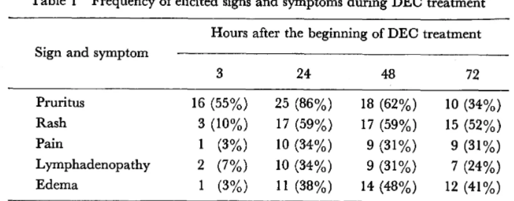
Table I summarized the chronological changes in the clinical manifestations during DEC treatment. The essential feature of the clinical manifestations during DEC treatment were shown as follows ; pruritus, rash, pain, Iymphadenopathy and edema. Twenty‑six of 29 patients experienced one or more of these manifestations.
Table I Frequency of elicited signs and symptoms during DEC treatment Hours after the beginning of DEC treatment Sign and symptom
3 24 48 72
Pruritus
Rash
Pai n
Lymphadenopathy Edema
16 (55・/・) 3 (10"/・)
1 (3・/・) 2 (7・1*) 1 (3・/*)
25 (86"/*) 17 (59"/・) lO (34"/・) lO (34・/ ) ll (38・/・)
18 (62"/*) 17 (59・/・)
9 (31'/・) 9 (31"/*) 14 (48"/・)
lO (34・/*) 15 (52・/・) 9 (31"/・) 7 (24・/・) 12 (41"/・)
Of 29 patients 25 experienced pruritus during this observation period, among them 1 6 complained the itching within three hours after the first dose of DEC (550/0)'
At 24 hours, this rate reached the maximum (860/0)' After 48 hours, the number of patients with pruritus gradually decreased. Only in a few patients rash, pain, lymphadenopathy and edema were noted at three hours, while at 24 hours, the frequency of these manifestations reached 59, 34, 34 and 38 per cent respectively.
Even after 48 hours, the frequency remained rather high. The pruritus appeared at various regions such as the face, neck, upper body and upper limbs. The rash was localized in the chest, scapular regions and upper limbs in most patients. The lymphadenopathy was noted more frequently in the inguinal region than the axillar one. The edema was localized in the face, chest and upper limbs, and when it appeared on the face, it was markedly severe than that in the other parts.
The relation between MFD and the severity of those adverse reactions is shown in Table 2 ( =0.391, 6=[ ] =0.154). Out of I I patients with high MFD, six
experienced the severe (+ ) , four experienced the moderate (++) and one patient showed mild (+) reaction. Of 1 3 patients with moderate and low MFD (1‑100), five showed severe reaction.
Table 2 Relation between the microfilarial density and adverse reactions seen in DEC treatment (29 patients)
Grade of reaction
MFD* No. of case
o + ++ +
O
1 ‑50
51‑lOO
1 OO <
5 10 3 11
1
2
3
1
l
4
1
4
3 2 6
* Number of microfilariae in two I O mm2 snips.
Among five cases of the microfilaria negative group, four patients showed no or mild reactions. The total number of WBC of the patients ranged from 7,000 to l 8,000lmm3 (mean ; 9,790 2,290/mm3) before DEC treatment. Of I I patients
whose WBC counts were 10,000lmm3 or more, 4 (36.40/0) showed severe reaction (+ ).
On the other hand, out of 1 8 patients with WBC counts less than l0,000lmm3 7 (38.90/0) showed severe reaction (Table 3; =0.374, a=[ ] =0.131). Eosinophil counts in the all patients were higher than the normal level, ranging from 660 to
Table 3 Relation between the WBC counts and reaction Grade of reaction
WBC counts/mm3 No. of case
o + ++ +
< 8,000 8,000‑ I 0,000 1 0,000 <
7 11 11
2
3
2
3 3 4
l
6 4
Table 4 Relation between the eosinophil reaction
counts of peripheral blood and
Grade of reaction Eosinophil countslmm3 No. of case
o + ++ +
< I ,OOO l ,OOC 2,000
2,000 <
4
12 13
1
2 3 2
3 5 2
2 9
Table 5 Relation between the number of neutrophil counts of peripheral blood and reaction
Grade of reaction Neutrophil countslmm3 No. of case
o + ++ +
< 4,000 4,000‑5,000 5,000 <
8 9 12
3
3
2
2 3 5
3 3 5
Table 6 Relation between the number of lymphocyte counts of peripheral blood nd reaction
Grade of reaction
Lymphocyte countslmm3
No. ofcase
o + ++ +
< 2,000 2,000‑3,000 3,000 <
lO 10 9
1
2
2 2
4 4 2
5 3 3
4,250lmm3 with the mean of 1,970 900/mm3. Nine (69.20/0) of 13 patients whose eosinophil counts were 2,000lmm3 or more showed severe reaction, while only 2
( 1 2.5 o/o) of 1 6 patients with eosinophils less than 2,000 showed severe reaction (Table 4;
=0.483, e=[ ] =0.120). A slight association with statistically significance between the severity of dverse reaction and the neutrophil counts was indicated (Table 5 ;
=0.428, 6=[V] =0.130). On the contrary, as seen in the Tables 6 ( =0.260 b=[V] =0.150), there were no apparent relation between the severity of adverse reactions a Id lymphocyte counts.
The relation between the serum lgG Ievels ofpatients and the severity of reactions after DEC administration , is shown in Figure I . The average OD values SD of
+++
' CQ
.5 co
ly a, ++
8
,::
o F:
O +
E:.
o
:Fl
o o
,o G, fo Urban ,
control
,e
O ,1
eap
" e e e
eoe e e 'e e e
o e e e
e
・・‑・e e
h‑'
0.2 1.0 1.2 1.4 1.6 1.8 2.0
O. 4 0.6 0.8 O D500Figure I Relation between O volvulus speclfic lgG and reactron
no, mild, moderate and severe reaction groups were 0.76 0.30,̲ 0.86 0.20, 1.03 0.20 and I .25 0.20, respectively. The mean OD of 9 urban control sera in Gua‑
temala was 0.18 0:09. Ten (90.90/0) of I I patients who experienced severe reaction (+ ) showed higher OD (over I .O). Only one of five patients with mild reaction (+) showed higher OD. In two of three patients without reactions, OD was near I .O.
As the OD for the onchocerciasis specific lgG increased the severity of adverse reactions after DEC administration tended to increase.
DISCUSSION
Although there were some reports which dealt with the change of blood eo‑
sinophil counts after DEC treatment in the patients with onchocerciasis (Guerra‑
Caceres et al., 1 980 ; Money, 1 969), none of them described a relation between the severity of reactions elicited by DEC and eosinophil counts before treatment. In this study, we clarified that the patients with high eosinophils showed severe adverse reactions. Of four patients with the eosinophils less than I ,OOO/mm3, nobody showed
severe reaction (+ ). The eosinophil counts in blood is thus likely to be used in predicting the severe reactions in the patients with onchocerciasis following DEC administration. With regard to the role of eosinophil in filarial infection, Spry ( 1 981) reported that the eosinophils released their granule products in response
to the presence of microfilariae in the patients with filariasis, and these products could induce local or systemic reactions. In the tissues, the accumulation and degranulation of eosinophils occured in patients with onchocerciasis around micro‑
filariae or adult worm which were damaged by DEC treatment, and this could give rise to some of the clinical features of the Mazzotti reaction (Guerra‑Caceres et al., 1980 ; Mimori, in press) . By DEC treatment, neutrophil counts increased and the relation between clinical manifestations and neutrophils has been discussed (Henson et al., 1 979; Sakamoto and Zea, 1 983). In this study, a slight association with statistically significance between systemic complications and neutrophil counts was observed. However, Iymphocyte counts in blood before DEC treatment were within normal level in most patients, and were not related to the severity of adverse reactions.
ELISA clarified the relation between the serum level of O. volvulus‑specific lgG and the severity of adverse reaction. The higher the OD of serum samples, the higher was the severity of reactions. However, three patients with higher level of lgG did not show severe reactions. Various factors would be involved in these cases, such as anti‑complement factors, poor antigenecity, acquisition of host 'antigen, immuno‑
tolerance and blocking antibodies (Henson et al., 1979). Greene et al. (1983) reported that very high pretreatment circulating immune complexes (CIC) Ievels were associated with an increase of ocular and systemic complications. The present study revealed that high eosinophil count in blood and/or high serum level of lgG were indicative laboratory data for severe adverse reactions caused by DEC treat‑
ment. A special precaution should be taken to treat such patients.
In the present study, the essential features of the clinical manifestations after
DEC treatment were pruritus, rash, pain, Iymphadenopathy and edema. The number of patients with pruritus, rash, pain, Iymphadenopathy and edema reached the maximum at 24, 24, 24, 24 and 48 hours, respectively. The number of patients who complained of itching was decreased at 72 hours, while the other clinical mani‑
festations remained stationary. Bryceson et al. ( 1977) reported that adverse reactions mentioned here appeared half an hour to 1 2 hours after DEC administration and continued for minutes, hours, or days.
Taylor et al. ( 1 980) reported that pruritus appeared within 1 2 to 36 hours, the rash started between days I and 3 in most cases, Iymphadenopathy and edema̲
occured between days 3 and 7. Our results observed are essentially similar to that of Taylor et al.
There are some reports which described that the severe reactions occured in the patients with heavy infection of O. volvulus (Buck, 1 974; Fuglsang and Anderson, 1974; Bryceson et al., 1 977; Martinez and Tello, 1 952). On the other hand, Greene et al. ( 1 983) reported that pretreatment skin microfilaria counts showed a weak correlation with the total number of ocular and systemic complications after DEC treatment. Our study showed that six of I I patients with high MFD experienced
severe reaction (+ ), and as many as five out 1 3 patients with moderate and low MFD showed severe reactions. The patients with high MFD tended to show severe reactions, although some of the patients with low or moderate MFD showed severe reaction. This finding suggests that the number of eosinophil counts, serum im‑
munoglobulin level such as lgG and lgE and the circulating immune complexes are more important factors than MFD, which predict severe adverse reactions by DEC administration.
ACKNOWLEDGMENT
This study was supported by the Overseas Scientific Research Grants No.
57041041 from the Ministry of Education of Japan. We wish to thank Dr.
H. A. Gody, Director de Servicio Nacional de Erradicacion de la Malaria, Ministerio de Salud Publica, Guatemala, and his colleagues for their kind cooperation. We also highly appreciate Dr. T. Suzuki, JICA, for his encouragement and Dra.
Mariantonieta of Amatitlan Nacional Hospital for the kind cooperation.
Appreciation is also expressed to Dr. T. Yoshimura, University of Occupational and Environmental Health, Kitakyushu, Japan, for his helpful criticism and sugges‑
tions in the preparation of the manuscript.
REFERENCES
l) Bishop, Y. M. M.. Fienberg, S. E. and Holland, P. W. ( 1975) : Discrete Multivariate Analysis : Theory and Practice. 385‑387, The MIT Press, Cambridge, Massachusetts and London‑
2) Bryceson, A. D. M.. Warrell, D. A, and Pope, H. M. (1977) : Dangerous reactions to treatment of onchocerciasis with diethylcarbamazine, Br. Med. J., 1, 742‑744
3) Buck. A. A. ( 1 974) : Onchocerciasis : Symptomatology. Pathology, Diagnosis, WHO Geneva.
4) Fuglsang, H. and Anderson, J. ( 1 974) : Collapse during treatment of onchocerciasis with diethylcarbamazine, Trans. Roy. Soc. Trop. Med. Hyg., 68, 72‑73
5) Garner, A., Duke, B. O. L. and Anderson, J. (1973) : A comparison of the lesions produced in the cornea of rabbit eyes by microfilaria of the forest and Sudan savanna strain of Onchocerca volvulus from Cameroon, II. The pathology, Z. Tropenmed. Parasit., 24, 385‑396
6) Greene, B. M., Taylor, H. R., Brown, E. J., Humphrey. R. L. and Lawley, T. J. (1983) : Ocular and systemic complications of diethylcabamazine therapy for onchocerciasis : Association with circulating immune complexes. J. Infect. Dis., 5, 890‑897
7) Guerra‑Caceres, J. G., Bryceson. A. D., Quakyi, I. and Spry, C.J. F. (1980) : Studies on the mechanisms of adverse reactions produced by diethylcarbamazine in patients with onchocerciasis‑
Mazzotti reaction, Parasit. Immunol., 2, 121‑131
8) Hawking, F. ( 1952): A histological study of onchocerciasis treated with Hetrazan. Br. Med.
J., l, 992‑99
9) Henson, P. M., Mackenzie, C. D. and Spector. W. G. (1979) : Inflammatory reactions in onchocerciasis : a report on current knowledge and recommendations for further study. Bull.
WHO., 27, 429‑i48
lO) Korenaga. M.. Tada. I., Lujan. T. A., Sakamoto, M., Zea. G. F., Castro, J. C. and Yarzabal, L.
( 1 983) : Enzyme‑Linked Immunosorbent Assay (ELISA) in the detection of lgG antibodies in onchocerciasis using blood collected on filter paper. Jap. J. Parasit., 32, 1 6‑24
1 l) Martinez, B. M. ahd Tello, P. R. (1952) : Modificacionnes en la eosinofilia de los oncocercosos
consecutivas乱1a del Hetrazan 、Medicina(Mexico),32,497
12)Mazzotti,L.(1948):Posibilidad de utilizar como medio diagnostico auxilar en la oncocercosis,
las alergicas consecutivas a la administracion del Hetrazan .R.ev.Inst、Salubr.En髭rm.Trop.,
9,235−237
13)Mimori,T.:A histopathological study of the skin and nodule during the course of diethy−
lcarbamazine treatment in onchocerciasis,Jap.」.Parasit.ン(in press)
14)Money,G.L.(1969): Hetrazan eosinopenia in onchocerciasis.A preliminary communication.
J.TroP.Med.Hyg.,63,238−241
15) Rodger,F.C、(1962):A review of recent advanccs in scientiHc knowlcdge of the symptoma−
tology,pathology and pathogenesis ofonchocercal in琵ctions.BulL Wld Hlth Org.27,429−448 16)Sakamoto,M.and Zea,G.F.(1983):The change of blood picture of patients with oncho−
cerciasis fbllowing administration ofdiethylcarbamazine.Trop.Med.,25,47−50
17) spry,cJ.F.(1981):Alterations in blood eosinophil morphology,binding capacity fbr com−
plexcd IgG and kinetics in patients with tropical(且larial)eosinophilia.Parasit.ImmunoL,3,
1−11
18)Taylor,H・R・,Greene,B・M・and Langham,M・E・(1980):Controlled clinical trial oforal and topical diethylcarbamazine in treatment ofonchocerciasis.Lancet,943−946
Diethylcarbamazine治療時におけるオンセルカ症患者の臨床像と 治療前検査成績の関係について
坂本 信1・三森 龍之2・是永 正敬2・多田 功2 Zea,G.F.・Castro,J.C.
中米グアテマラ国のオンコセルカ症患者29名についてDEC(5mg/kg/day7日間)で治療し,その 時におこる臨床像の観察と治療前における検査成績より,治療時における臨床像の重症度を予測した。
治療時の主な反応は掻痒感,疹痛,発疹,発赤,リンパ節腫脹,浮腫であった。これらはDEC投与 による炎症性反応であると考えられた。これら炎症性反応の重症度と治療前の検査成績の関係におい ては末梢血中の好酸球及び0,volvμ1μs−antigen speci且c IgGとの間に強い相関がみられ,治療上こ れらの成績の高い患者には充分な注意が必要と考えられた◎
1長崎大学熱帯医学研究所寄生虫学部門 2熊本大学医学部寄生虫病学教室 3グアテマラ共和国 マラリア防圧本部オンコセルカ部
本研究は文部省海外学術調査費によった。
沖縄県在住ベトナム難民の寄生虫感染状況
安里 龍二1・長谷川英男2・池城 毅3 昭和59年6月21日 受付/昭和59年10月16日 受理
緒 言
インドシナにおける長期の動乱は,霧しい難民 を産み出した。特にベトナムでは,戦争終了(1975 年)後,新政府の政策や生活に不満を持つ多数の 人々が,いわゆるボート・ピープルとして脱出し,
大きな国際問題となっている。漂流中に救助され たベトナム難民は主として先進諸国に収容され,
一部は定住が認められている。わが国では1975年 にベトナム難民の一時収容が開始されて以来,
1983年3月末までに収容難民数は7,477名に達し ている。沖縄県では1977年6月,八重山群島与那 国島に85名のベトナム難民が漂着したのが最初で あるが,1978年9月に沖縄本島本部町の日本赤十 字本部国際友好センターでベトナム難民の一時収 容が開始され,以来現在までに延べ1,033名が収 容されてきた。
ベトナムは熱帯に位置し,多くの感染症が蔓延 している地域であり,また低栄養状態,非衛生的 環境が加わって,難民の健康は著しく損われてい る。更にこれら難民と共に感染症が持ち込まれ,
収容地の住民に影響を与えることが考えられる。
沖縄県はわが国では唯一の亜熱帯に属する地域で あり,ベトナム難民が順化するには都合のよい風 土であるが,また一旦感染症が持ち込まれた場合 には,二次的感染,土着化が容易に起こりうる環 境でもある。本部国際友好センターでは難民の健 康を増進し,感染症の伝播を防ぐために,健康診 断,治療を積極的に行っているが,著者らはその うち寄生虫の検査を担当してきた。本論文はこれ
までの結果をまとめ,間題点について考察したも のである。
調 査 方 法
調査は1977年9月から1982年7月の間に行われ た。消化管寄生蠕虫および原虫の検査は原則とし て,入所後数カ月以内に行われる健康診断時に行 い,集めた糞便は翌日,沖縄県公害衛生研究所
(大里村)にて直接塗抹法,MGL法,試験管内 濾紙培養法を併用して検査した。原虫嚢子の検査 にはMGL法を用い,ヨード染色を施して検鏡 した。住血性寄生虫の検査は午後9時以降に耳朶 から採血し,厚層塗抹法ギムザ染色によって行っ た。自覚症状の調査は予め準備したアンケート用 紙を通訳を介して記入させた。また,駆虫薬の投 与は名護保健所長の指示により,面前にて服用さ せた。投薬後1カ月目に再検査を行って駆虫効果 を判定した。
結 果
1・消化管寄生蠕虫類
662名の検査を行い,その内441名(67.1%)に 1種以上の寄生蠕虫を認めた(表1)。内訳は線 虫類6種(回虫,鉤虫3種,鞭虫,糞線虫),条 虫類2種(小形条虫,無鉤条虫),吸虫類2種
(肥大吸虫,肝吸虫)の計10種であった。このう ち虫卵保有率が最も高いのは回虫で,次いで鞭虫,
鉤虫類の順であり,その他の寄生蠕虫の保有率は 著しく低く,条虫類,吸虫類の寄生は1彩以下で 1沖縄県公害衛生研究所 〒901−12沖縄県大里村大里高嶺原2085
2琉球大学医学部寄生虫学教室 〒903−01沖縄県西原町上原宮里207 3沖縄県予防医学協会 〒903沖縄県那覇市首里平良町1−23−3
Table l Prevalence of intestinal helminths in Vietnamese re蝕gees residing in Okinawa Pre驚cture
Year ofexamination l977 No.・fpersonsexamined 79
1979 329
1980 198
1981 56
Total 662 No.ofpersonsin偽cted
(Incidence,%)
∠5 σr露」π規6r∫60歪485 49
(62.9)
7}∫σh㍑r∫5∫rJ hゴz rα 17
(21.5)
Eookworms 2
(2.5)
8ケoη9メo肋55嬬orα」ゑ∫ 0
砺規8㎜妙雰舷紹
乃8痂躍g吻如
勲5σ乞o妙廊伽短
(】Zoηor:δhゑ5語η8η語∫
0 0 0 0
173
(52.6)
86
(26.1)
60
(18.2)
10
(3.0)
0
0 1
(0.3)
0
l l9
(60.1)
29
(14.6)
45
(22.7)
3
(L5)
3
(1.5)
1
(0.5)
0
0
32
(57。1)
12
(2L4)
15
(26.8)
2
(3.6)
0
0 0
811
373
(56、3)
144
(2L8)
122
(18.4)
15
(2.3)
3
(0、5)
1
(02)
1
(02)
1
(0.2)
Tota1 53
(67.1)
208 141 42 444
(63.2) (7L2) (75.0) (67、1)
あった。鉤虫類は濾紙培養で陽性が確認された 106名中,103名がアメリカ鉤虫,2名がズピニ鉤
虫と同定されたが,残る1名の培養便から得た フ イラリア型幼虫はズビニ,アメリカ両種のもの とは異なり,固有虫体の大きさ630〜650x22.5
〜25・04,被鞘を含む大きさ700〜750×27・5〜32・5 μ,食道長157.5〜162.5μ,尾長72.5〜775μで あらた。無鉤条虫は自然排泄された片節5個が得
られた・(歯1)。肥大吸虫卵(大きさ130〜147.5
×85.0〜92.5μ;図2) と肝吸虫卵(大きさ27.5
〜32.5×13.8〜175μ;図3)は各1名に検出され
た。
消化管寄生蠕虫類保有者の6L5彩は1種のみの 寄生であったが,29.7%は2種,7.%は3種寄 生しており,4種寄生も5名(1.1%)にみられ
た。
年齢別ではほとんどの年齢層で50%以上の高い
保有率を示し,特に10代,20代,40代では70%以 上であった(表2)。回虫,鞭虫保有率は10歳未 満の小児と20歳以上の成人の間で差はみられな かった。しかし鉤虫では小児の7.4%に対し成人 は約3倍の2L6%の保有率を示した。性別では回 虫卵保有率が男女ともほぼ同じであったのに対し,
鞭虫卵,鉤虫卵,糞線虫幼虫の保有率はいずれも 男性が高く,特に糞線虫幼虫保有者は全て男性で あった。
2.消化管寄生原虫類
消化管寄生原虫類の検査は274名について行い
(表3),その内50名 (18.2%)に原虫嚢子を認 めた。内訳は小形アメーバ31名(11.3%),大 腸アメーバ13名(4.7%),ランブル鞭毛虫10名
(3.6彩)で,赤痢アメーバは検出されなかった。
3.住血性寄生虫類
血液検査は311名について行ったが,マラリア
恥u鵬博
ロぎぽ の
灘灘灘、灘
講一、一盗灘欝 鱈
1騒灘難醐懸
馨灘
撚 鞭鑑.。
霧難漁
難
翼
腱薫
・置蟹
鑛
距農r麟薦飾㈱農in伽艶c鱈菖o£V蜘鋤鷹rε魚騨菖ほ、翫痂諮紳蜘脚篤10tt編
(x7)・2・勲繭瘍瞬伽就ゴ豊舘(×蔓60)・3,α醜磁才5蜘繍聡(×玉,7鋤.
丁寓b醜P欝&1eぬ。eofi騰呂t姻照面漁呂圭庶Vi硫鰍齪ere無騨鋤ya農騨寵灘
M乱1農 蝕漁a量む To捻董
A欝彦rou整 翼o,of 騨「隙齢 蟹am勧¢媛
愛o呂量ti鴨
置at倉
/%〉
No.of persons 轍乱m並磁
P{}富itive
聡憶
(%)
Nの.of P磁t量v農 Pε蕊o鵬 撒t鵬
燈翼蕊箪r践瓶きd (%)
o−9 10−ig
20−29 30−39
4049
⑪v¢r50
75 i47 i33
婆6
17
4
66,7
74.驚
73.7 65,2
70.蔭 50。⑪
471出5自V欝材ノ噛82董 51.4
68,1 磁,2
尋8.0 畠o,0 33、露
149 194
灘4 71 27
7
澱,1 72.7
70,三
59,2 猴.1
些2。9
Tota星 蔓2慧 71、3 羅o 59.6 662 67.1
原虫,ミク鷺フィ夢顎アは見出されなかゆた。
4.自覚症状
蟷虫保有者203名に行鴫た自覚症状調査では,
7ゑ6鄭が何らかの症状を訴え救。最も多かったの は腹痛(3L2%〉で,次いで頭痛(23,夢%),食 欲不振(22,4%L腹部膨満感(欝,6%),全身倦 怠感(164%),便秘(12.2%),鵬熱(m.7%),
めまい(10鴻%〉の順であった。
萎.好酸球増多
嬬虫保有審145名中, 零%以上の好酸球増多を 示したものは船,7%であり,一1方蠕虫が検出され なか噂た灘名中でも25%に好酸球増多が翻められ
た。
礁駆虫成綾
Table3 Prevalcnce ofintestinal protozoans in Vicmamese re蝕gees Sex ofrefUgees
No.ofpersons examined
Male
185
FemalC 89
Tota1 274 No.ofpersons in琵cted
(lncidence,9も)
Eη如η208加 o露
Eη40だη2αXπαπα
ααア漉α」伽めあα
6
(3.2)
18
(9.7)
8
(4、3)
7
(7.9)
13
(14.6)
2
(2.2)
13
(4.7)
31
(lL3)
10
(3.6)
Tota1 30
(16.2)
20
(22.5)
50
(182)
回虫,鉤虫,鞭虫保有者にはピランテル・パモ エート10mg/kgを投与し,1カ月後の検査で回 虫保有者274名中247名 (90.1%),鉤虫保有者 96名中57名(59.4%),鞭虫保有者104名中26名
(25.0%)に虫卵の陰転が認められた。肥大吸虫 保有者もピランテル・パモエート10mg/kg投与 で陰転化した。
考 察
本調査で得られた成績では,蠕虫類のうち土壌 伝播性線虫類の寄生が著しく,特に回虫が半数以 上にみられ,鞭虫,鉤虫は共に約1/5の難民に見 出された。これまで報告されたベトナム難民の寄 生虫調査(L6scherε α」.,1980;Picher and Asp6ck,1980;Lermanθ 認,1982)でも,検出 率は低いものの回虫が最も高率(24.8−41%)を 示し,鞭虫(6−14.2%)と鉤虫(8−12.2%)がほ ぼ等しい寄生率となっている。但し建野ら(1982)
め成績では鞭虫(6.8%)は鉤虫(27。3%)の約 1/4の保有率である。ベトナム難民に回虫が多く
鉤虫が少ないことは,ラオス・カンボジア難民 に鉤虫が多く回虫が少ないこと (Lindes,1979;
Wiesentha1ε 01.,1981;Tatenoθω1、,1981;
Lerman8 認,1982;Keittivuti8 鋤,1982;建 野ら,1982)と対照的であり,ベトナム難民に特 徴的な傾向と思われる。一方,ベトナム国内での
検査成績では,農村部で回虫より鉤虫が高率に見 出され,逆に都市とその近郊では鉤虫より回虫が 高率であることが知られている(Goodrich,1967;
Kimθ α1.,1970;Colwe11θ 01.,1971)。従って 難民の寄生虫感染状況は都市型であるといえるが,
これは難民の多くが都市とその近郊の出身者であ ることに関連しているものと思われる。ところで,
都市部と農村部でこのような差が生ずる原因につ いてKim8 αム(1970)とColwe11ε∫oム(1971)
は異なった見方をしているが,いずれも現象を十 分に説明していない。恐らく農村部と都市部にお ける食習慣,生活様式の違いが,寄生虫の種によ る虫卵の発育条件,抵抗性,感染様式の差と関連 しながら寄生率の違いをもたらすものと考えられ る。条虫類,吸虫類はベトナム難民には一般に低 率であることが知られているが,本調査でも同様 な傾向が認められた。肝吸虫類はラオス・カンボ ジア難民には高率に見出されるが,これは食習慣 の違いによるものであろう。
今回1例から検出されたズピニ,アメリカ両種 と異なったフィラリア型幼虫は,大きさがセイロ ン鉤虫についてのYoshida(1971)の値よりはや や小さいが,影井ら(1978)がベトナム難民から 得たセイロン鉤虫のフィラリア型幼虫の値にほぼ 一致していたことから,セイロン鉤虫と推定した。
また肝吸虫卵は大きさがタイ肝吸虫卵より肝吸虫 卵に近いので,肝吸虫としたが,両者の虫卵での
区別は困難であるため,今回見出されたものがタ ィ肝吸虫である可能性もある。Tatenoθ101,
(1981)はベトナム難民の1例をタイ肝吸虫であろ うとしている。肥大吸虫卵は肝蛭卵との区別が困 難であるが,ピランテル・パモエートによる駆虫 が奏効したので,今回のものを肥大吸虫と同定し た。なお,無鉤条虫,肥大吸虫が確認されたのは 本邦に収容されたベトナム難民では初めてと思わ
れる。
蠕虫保有者は75彩が何らかの自覚症状を訴えて いたが,そのうち腹痛,食欲不振,腹部膨満感等 の多くは蠕虫寄生と関連を有するものと考えられ,
寄生が難民の生活に大きく影響していることが推 測される。また好酸球増多を示した比率は,蠕虫 保有者群と蠕虫の証明されなかった群の問でほと んど差はなかった。このことは今回の検査で検出 されなかった蠕虫感染が存在する可能性を示唆し ていると考えられる。
沖縄県ではかつて各種の腸管寄生蠕虫類が広く 蔓延し,寄生率は著しく高かった(Hunter8 α1.,
1951;佐々ら,1958)が,集団検査,駆虫,生活 状態の改善,衛生知識の普及等によって,寄生虫 感染は激減し,現在では糞線虫等がみられるに過 ぎない(沖縄県予防医学協会,1982)。今日では 糞便の処理は衛生的に行われており,寄生虫の生 活環はほぽ完全に断ち切られているといえる。
従ってベトナム難民に腸管寄生蠕虫が高率に見ら れるとしても,現在の沖縄県民にとって大きな危 険性を有するものではないと考えられる。
病原性腸管寄生原虫類は難民での寄生率がベト ナム国内での寄生率より高い報告が多い(picher and Asp6ck,1980;L6scherθr o1.,1980;建野
ら,1982)。本調査でもランブル鞭毛虫は比較的 高値を示したが,赤痢アメーバは検出されなかっ た。しかし赤痢アメーバは重要な病原性アメーバ で,アメーバ性肝膿瘍を起こしたベトナム難民の 例もあり(天野ら,1983),また他者への感染も 比較的容易に起こりうるので警戒が必要である。
本調査では血液から寄生虫を検出することはで きなかった◎ベトナムはバンクロフト,マレー両 糸状虫が分布している地域である(Sasa,珍76)
が,難民の多くが都市部の出身者であるためか,
他の難民調査成績でもミクロフィラリアを検出し た例はみあたらない。一方,マラリアに関して は,難民に時々発症が報じられており(田村ら,
1981;大友ら,1981;荒木ら,1981;金ら,1983),
沖縄県に収容されたベトナム難民にもこれまで3 例の三日熱マラリアが発生している(安里,長谷 川,1981;祖堅ら,1983)。そのうち1例は入所 後1年2ヵ月経過してから発症し,再発例と考え
られた。また他の1例は収容後に出生した新生児 の症例で,ベトナム難民から輸血を受けた後に発 症したものである。この例では母体,供血者いず れからも事後の血液検査でマラリア原虫を証明す ることはできなかった。これらの事例はマラリア 原虫を保有した難民が長期にわたって滞在してい ることを示しており,この状態は既に土着に近い ものといえる。沖縄県はかつてフィラリアと共に マラリアの流行地であり,媒介蚊の繁殖に好適な 条件を有している。事実,現在難民が収容されて いる友好センターで捕獲された蚊の中にもマラリ ア媒介種であるシナハマダラカが含まれている
(宮城ら,私信)。従って現在の状態が長く続けば マラリアの土着化が起こる危険性が大きい。
以上のように,ベトナム難民には高率に種々の 蠕虫,原虫が寄生して難民の健康を害しており,
更にその一部は収容施設周辺の住民に影響を及ぼ す可能性を有している。従ってベトナム難民の受 け入れに際しては十分な寄生虫対策がとられなけ ればならない。即ち,入国時に速やかに糞便検査,
血液検査を行い,またその後も定期的に検査を反 復することが必要であり,特に赤痢アメーバ,マ ラリア原虫については血清学的検査によるスク リーニングも実施して,検出率を高めることが望 まれる。また寄生の認められた難民には十分に治 療を行い,完全な駆虫が確認されるまで検査,治 療を続けることが必要であろう。更にマラリア,
フィラリア症が発生した際に,二次的感染,土着 化の起こる可能性を予測するために,難民収容施 設周囲の媒介蚊相を把握しておくことも重要であ
る。
一要 約
沖縄県に収容されたベトナム難民662名にっい て消化管寄生蠕虫類の調査を行い,444名(67.1
%)に寄生を認めた。回虫が最も高率(56.3%)
で,次いで鞭虫(21.8%),鉤虫(18.4彩),糞線 虫(2,3%)の順であり,他に小形条虫(3名),無 鉤条虫,肥大吸虫,肝吸虫(各1名)がみられた。
年齢別ではほとんどの年齢層で50%以上の高い保 有率を示し,性別では男子の保有率が高かった。
これら蠕虫類の寄生状況はベトナム国内での都市 部のものに類似していた。消化管寄生原虫類の調 査は274名について行い,50名(18.2彩)に原虫 嚢子を認めた。内訳はランブル鞭毛虫(3.6%),
大腸アメーバ(4・7%),小形アメーバ(1L3彩)
で,赤痢アメーバは検出されなかった。住血性寄 生虫の検査は311名について行ったが,マラリア 原虫,ミクロフィラリアは証明されなかった。ベ
トナム難民の寄生虫が難民自身並びに収容施設周 辺の住民に与える影響,及び対策等について論じ
た。
謝 辞
本調査に御協力を頂いた沖縄県名護保健所比嘉 政昭所長,島袋憲氏並に検査室各位,日本赤十字 社沖縄県支部仲原英敏国際友好センター所長,兼 本幸三郎氏,照屋全豊氏,浦崎廣子氏,ファン・
デイン・コア・ロン氏,グエン・ギア・フン氏,
沖縄県予防医学協会泉川ツヤ子氏に謝意を表しま
す。
文 献
1) 天野博之,田畑隆文,森田 博,猪木正三,荒木恒治 (1983):ベトナム難民に見られたア メーバ性肝膿瘍例,日熱医会誌.,11(2),130−131
2) 荒木恒治,森田 博,瀬川武彦,猪木正三,天野博之,赤沢寛治(1981): ベトナム難民の検 診結果一1980年奈良県下グループと1977年滋賀県下のグループを比較して一, 日熱医会誌.,9
(2),118−119
3) 安里龍二,長谷川英男(1981):沖縄県における輸入マラリアの現状,寄生虫誌.,30(6),527−
531
4)Colwell,E.J.,Welsh,J.n,Bonne,S.C.and Legters,L.L.(1971):Intestinal parasitism in residents of the Mekong Delta of Vietnam,Southeast Asian J.Trop.Med.Publ.Hlth.,2 (1),25−28
5) Goodrich,1.(1967):Prevalence of intestinal nematodes in civilian,adult,some Vietna−
mese population,Am.J.Trop.Med.Hyg.,16(6),746−749
6)Hunter,G.W.皿,Ritchie,L.S.,Pan,C.and Lin,S、(1951):Parasitological studies in the Far East.XI.An epidemiological survey of Okinawa,Ryukyu Islands,406Medical General Laboratory,Japan Logistical Command Bulletin3,29pp.
7) 影井 昇,木畑美知江,浅野和人(1978):ベトナム難民に対する腸管寄生虫類の検査.特に セイロン鉤虫感染者にっいて,日熱医会誌.,6(1),43−49
8)Keittivuti,B.,D agnes,T.,Keittivuti,A.and Varavaidya,M.(1982):Prevalence of schis−
tosomiasis and other parasitic diseases among Cambodian refugees residing in Bang−
Kaeng Holding Center,Prachinburi Province,Thailand,Am.J.Trop.Med.Hyg.,31(5),
980−990
9) Kim,J.H.,Yoon,J.J.,Lee,S.H.and Seo,B.S.(1970):Parasitological studies of Korean Forces in South Vietnam.II.A comparative study on the incidence of intestinal para一
sites,Korean J.Parasit.,8(1),30−35
10)金光明,上野山裕己,吉村文秀,富次貞造,中林敏夫(1983):新生児期に発症したマラリ アの1例,日熱医会誌.,11(2),130
11)Lerman,n,Barett−Connor,E.and Norcross,W.(1982):Intestinal parasites in asympto・
matic adult Southeast Asian immigrants,」.Fam.Pracり15(3),443−446
12) Lindes,C.(1979):lntestinal parasites in Laotian refugees,」.Fam.Prac.,9(5),819−822 13) L6scher,T.,PrUfer,L and Sonnenburg,F.F.von(1980): Intestinal Parasiten bei vietna・
mesischen Flロchtlingen,Deu.Med.Woch.,105(14),488−489
14) 沖縄県予防医学協会(1982):鉤虫病予防特別対策,事業年報,11,184−185
15)大友弘士,日置敦巳,中林敏夫,海老沢功,石崎 達(1981):1977−1979年におけるマラリア 患者184人の感染状況に関する疫学的考察,日熱医会誌.,9(2),101
16) Picher,0.and Asp6ck,H.(1980):Haufigkeit und Bedeutung parasitarer Infektionen bei vietnamesischen Fmchtlingen,Wiener Med.Woch.,(5),190−193
17)Sasa,M.(1976):Human Filariasis:A Global Survey of Epidemiology and Control,
University of Tokyo Press,819pp.
18) 佐々 学,照屋寛善,池宮喜春,国吉真英,城間盛吉,金城 進(1958):沖縄農村の寄生虫 罹患状況について,日本医師会誌.,39(9),601−604
19)祖堅 尚,伊之幸信,小堂欣弥,照屋 彰,比嘉政昭,安次嶺馨,安里龍二(1983):交換輸 血後に発症した新生児マラリアの1例,52回日本感染症学会西日本地方会総会プログラム,24 20)田村遵一,脇誠治,五十嵐郁男,長谷川誠,石川晋介,鈴木守,田辺和桁,高田季久 (1981):難民集団内の潜在マラリア検索における間接螢光抗体法の有用性,日熱医会誌.,9
(2),100
21)Tateno,S.,Takeuchi,T、,Kobayashi,S.,Tanabe,M.,Miura,S.and Asami,K(1981):
HealthstatusofIndochinarefugeesinJapan:Prevalenceofintestinalparasitesandeo・
sinophilia, Jpn.J.Parasit.,30(5),485〜496
22)建野正毅,大塚英司,伊藤秀夫,亀谷志郎,上田利久,斉藤竜太,竹内 勤,小林正規,三浦 左千夫,浅見敬三(1982):インドシナ難民の健康調査,日本医事新報,(3047),47−51
23)Wiesentha1,AW.,Nickels,M.K,Hashimoto,G.,Endo,T.and Ehrhard,H.(1980):ln・
testinal parasites in Southeast−Asian refugees,JAMA,244(22),2543−2544
24) Yoshida,Yし(1971):Comparative studies onイηのノ10s o配σゐrαz 1㎞sなand44nのノ105 o襯αoεy−
」απ o躍η. 2.The infective larval stage, J.Parasit.,57(5),990−992
PREVALENCE OF PARASITES AMONG VIETNAMESE REFUGEES RESIDING IN OKINAWA PREFECTURE,
JAPAN
RYUJI ASATOl, HIDEO HASEGAWA2 AND TSUYOSHI IKESHIR03
Received June 2 1 1 984/Accepted October 1 6 1 984
Parasitological surveys were carried out on the fecal and blood samples from the Vietnamese refugees living in the Motobu House for the Vietnamese, Motobu, Okinawa, Japan. Intestinal helminths were detected in 444 (67. I o/o ) of 662 refugees examined. Ascaris infection was the most prevalent (56.3010), followed by Trichuris (21.80/0)' hookwonns (18.40/0) and Strongyloides (2.30/0) infec‑
tions. Hymenolepis nana infection was observed in three cases, and Taenia saginata, Fasciolopsis buski and Clonorchis sinensis infections in each one case. In almost all age groups, the prevalence was over 50 per cent, and incidence of infection was higher in males than in females. In general, the condition of helminthic infections in the refugees resembled those reported in the urban areas in South Vietnam.
Intestinal protozoans were observed in 50 (18.20/0) of274 persons examined. Endolimax nana (1 1 .3010)' Entamoeba coli (4.70/0) and Giardia lamblia (3.60/0) were found but E. histolytica was not detected. Malarial parasites and microfilariae were not demonstrated in the blood samples from 3 1 1 persons although three cases of benign tertian malaria have been recorded among refugees in the house. The significance of these parasitic infectiohs in the health of Vietnamese refugees and the infiuence to the public health status of the inhabitants living around the refugee camp were discussed.
1 Okinawa Prefectural Institute ofPublic Health 2 Department ofParasitology, School of Medicine, University of the Ryukuys 3 Okinawa Prefectural Cooperation of Preventive Medicine
PURIFICATION AND CHARACTERIZATION OF PHOSPHODIESTERASE FROM THE VENOM OF
AGKISTRODON ACUTUS (CHINA) L
HISAYOSHI SUGIHARA, TOSHIAKI NIKAI, YUMIKO KoMORI, HISATO NoBUHIRO MORI AND MITSUHIRO KISHIDA
Received May 25 1984/Accepted Nobember 1 1 1984
KATADA,
Abstract : Phosphodiesterase was isolated from the lyophilized venom of Agkistrodon acutus from China using gel filtration on a Sephadex G 75 column, followed by chroma‑
tography on diethylaminoethyl (DEAE) ‑Sephadex A‑50, carboxymethyl (CM) ‑Cellulose and Affi‑Gel Blue af nity. Homogeneity was established by the formation of a single band in acrylamide gel electrophoresis, isoelectric focusing, sodium dodecyl sulfate (SDS) acrylamide gel electrophoresis and immunodiffusion. Phosphodiesterase activity was inhibited by ethylenediamine tetraacetic acid (EDTA), ethyleneglycol (p‑aminoethyl) N, N, N ', N '‑tetraacetic acid (EGTA), o‑phenanthroline, cysteine, thioglycolic acid, glutathione or p‑chloromercuribenzoate (PCMB) but not by soylpean trypsin inhibitor (SBTI), Iimabean trypsin inhibitor (LBTI), egg white trypsin inhibitor (EWTI), benzamidine or trasylol. The molecular weight of this enzyme was determined to be approximately 1 40,000 and the isoelectric p6int was found to be pH 7.4 by isoelectric focusing with carrier ampholyte. The phosphodiesterase activity of the final preparation was 2,190 units/mg. This protein was unstable to heat treatment and stable between pH 7 and 12. Its michaelis constant (Km) and inhibition constant (Ki) values for p‑
nitrophenyl thymidine‑5 '‑phosphate or EDTA were found to be 1.6x I 0‑3M and 5.8x I 0‑6M, respectively. This protein did not contain any carbohydrates.
INTRODUCTION
Snake venoms contain many enzymes that hydrolyze phosphomonoester, phosphodiester bonds, arginine ester, casein and adenosine triphosphate. Among venom phosphodiesterases, exonuclease is one of the most extensively studied enzymes and is used commonly for the degradation of nucleic acid. Substrates for this enzyme are DNA, RNA or any polynucleotide chains. Boman and Kaletta ( 1 957) reported that the venom of Crotalus adamanteus has three different exonucleases whose activities are parallel to those of DNAase. Suzuki et al. (1960) showed that there were three different fractions of exonuclease from the venom of Aghistrodon blomhoffii blomhoffii. Philipps ( 1 975) isolated a phosphodiesterase from Crotalus spp. venom.
The molecular weight was 1 1 5,000. In this paper we describe the purification and characterization of the phosphodiesterase (Aa‑p) from the venom of Agkistrodon
acutus.
Department of Microbiology, Faculty of Pharmacy, Meijo University, Tenpaku‑ku, Nagoya, Japan