ISSN O304一2146
Japanese Joumal of Tropical Medicine and Hygiene
第17巻第1号 平成元年3月15日
内 容
原 著
ヌマカ属(アシマダラヌマカ)の幼虫および蝋の呼吸器の走査電子顕微鏡
による形態観察……・………・・…………一……・・……・…………・………岩城 操 1−7 丁吻αηoso㎜わ耀06プg脚6碗s召感染マウス血清中のIL−2産生抑制物質
…・陶 道,日笠 檎宮本彦四郎申田 和彦,小松 俊憲,新家 荘平・ 9−15 濾紙使用による微量血清中のHBsAgのELISAによる測定(英文)
・・松田 晋哉,華表宏有 17−25 喀疾液化剤dithiothreito1(SputasoI)処理によるP%8㈱o釧s漉oo吻露
の診断(英文)……一…・…・・…………一…・・一…………一…¶…一……・・塩田 恒三 27−31
第30回日本熱帯医学会総会講演英文抄録
目 次一… 33−36
特別講演・ 37−39
シンポジウム
1 熱帯性感染性と薬剤・…… 40−50 11熱帯性感染症とワクチン開発の現状とトピックスー…… 51−58 パネルディスカッション… 59−68
一般講演・ 69−103 会 報
会則一 105−108
会員名簿・役員……… 109−130
投稿規定
日熱医会誌
Japan.」.T.M.H. 日本熱帯医学会
日本熱帯医学会雑誌 第17巻 第1号 1989 1−7頁 1 ヌマカ属(アシマダラヌマカ)の幼虫および蜻の
呼吸器の走査電子顕微鏡による形態観察
岩城 操
昭和63年8月18日受付/昭和63年12月26日受理
緒 言
孤αnson彪属の蚊は,マレー糸状虫励嘱g伽 吻吻頭の媒介蚊として知られ,疫学的に非常に重 要な蚊である(Samarawickrema,19681Laur−
ence,1960)。この蚊の幼虫および蠣の水中におけ る特殊な呼吸法に関しては,他のCπ」砿属や
・4646s属などとは異なり,幼虫や蝿の生息する水 域に自生する水生植物の根や茎に呼吸管,または 呼吸角を挿入して直接植物組織内に含まれる酸素 を呼吸するものと考えられていた(Laurence,
1960)。しかし,著者は,以前〃伽son彪属の幼虫 および蝿を生体のままホールスライドグラス内に 遊泳させ,光学顕微鏡によって観察した結果を報 告した(Iwaki,1984)。即ち,水中に静止している 幼虫の呼吸管先端を静かに刺激すうと,呼吸管先 端内部から千鳥型の爪と非常に薄い膜状組織が突 出すること,また,この組織が水中の溶存酸素を 呼吸する呼吸鯉(larvalrespiratorygill=LRG)
であることを示した。さらに,蝿の呼吸角先端部 については,水生植物の根や茎に呼吸角先端を挿 し込み体を固定するために鋭く尖った角状を呈し ていること,また,植物組織から酸素を吸収する ための穴は開口していないばかりでなく,呼吸角 先端部を包む網目状組織の呼吸角鯉(pupal res−
piratorygill=PRG)を有し,これによって呼吸 すると考えられることを報告した(Iwaki,
1984)。今回は,走査電子顕微鏡を用いて,幼虫の 呼吸管および婦の呼吸角にみられる微細構造につ いて観察した。1令幼虫の呼吸管先端部の両側に
カギ状の爪と,先端内部から一対の千鳥型の爪お よび薄い花冠状の膜組織が突出していること,そ して2令から4令幼虫の呼吸管先端部にはさらに 多くの爪と花冠状の騰組織とそれに包まれて特殊 なスポンジ状の構造をもった組織がみられた。一 方,嫡の呼吸角の最先端は鋭く尖った角状を呈し,
先端部側面に毛細血管状の管によって作られた レース状組織がみられ,いずれも,水中の溶存酸 素を呼吸するための呼吸鯛であることが認められ た。また,呼吸管および呼吸角内部の微細構造に ついても観察した結果から,幼虫および蝿の体内 の血液循環に関する経路についての知見を得たの で考察を加えて報告する。
材料および方法
観察に供した〃伽so%勿(ル勉㎜拓燃)観面撚
(Theobald)アシマダラヌマカの幼虫,および蠕 は,タイ国・マヒドール大学熱帯医学部医用昆虫 学教室において飼育した個体(Supat6!α1.,
1982)であり,比較のために用いたC%1疏
渉7」如召初ozh翅6h%s s%郷郷ozos%s(Dyar)コガタ アカイエカおよび。4θ惚sノ ψo痂o郷(Theobald)
ヤマトヤブカの幼虫および蝋は,京都市内で採集 し飼育して得た個体である。試粍は,あらかじめ 70%アルコール液に保存しダー1騰成時に80%か
ら100%アルコールに置換した後,自然乾燥させ た(Matsuo召地!.,1972〉己この試料をミニポー ル濾紙上に少量の接着剤を付けて固着させ,更に これをアルミニウム試料台に両面テープによって
京都女子大学家政学部生物学教室
2
,
固着させた。
この試料を日立E10110N蒸着用スパッター により金蒸着し,日立S−530走査電子顕微鏡を 用い,加速電圧10〜15KVのもとに観察した。な お,幼虫および蝿の呼吸器の各部名称は,Har−
bach and Knight(1980)およびIwaki(1984)
を適用した。
結果および考察 1.幼虫の呼吸管の観察結果
〃伽son彪属のアシマダラヌマカ1令幼虫の 呼吸管先端は,図1のごとく,C祝傭属のコガタ アカイエカおよび・4ε48s属のヤマトヤブカの呼 吸管と類似し,円筒型を呈して細長いが,呼吸管 先端部の両側にカギ状の爪(outer spiracular teeth=OST)が一対みられる。そして,この1令
幼虫が水生植物の根や茎に固着した場合,図2に 示されるごとく,呼吸管先端内から一対の千鳥型 の爪(innerspiracularteeth=IST)と薄い花冠 状の膜組織(thin田my tissue=tft)が突出する。
アシマダラヌマカの幼虫の呼吸管は,2令から4 令へと令の進行にしたがって円錐型に変化するが,
呼吸管先端部には顕著な変化がみられないので,
本報告では,2令・3令幼虫については省き,4 令幼虫について記述する。図3は,4令幼虫の呼 吸管先端を上側面より観たところであるが,ノコ ギリ状歯(saw)とISTがみられ,図4に示すご
とく水生植物の根や茎に体を固着した場合に呼吸 管先端内から突出する多数のISTとtftが認め られた。さらに,先端中心部には,図5に示すご とくtftに包まれたスポンジ状組織(sponge−like tissue=slt)がみられる。また,円錐型の呼吸管の
中央部を横に切断した内部には,図6の矢印に示 すごとく,中心部に1本の太い管(cylindrical tube=ct)と両側面に2本の筋束(muscular
band=mb),そして呼吸管内壁に試料を作成した 際に少し扁平に変形しているが,気管状の管
(thick trachea=tt)が左右一対あることが,そ れぞれ観察された。
2.幼虫の呼吸管に関する考察
3
C%忽属や・46磁s属の幼虫の呼吸管は細長い 管状を呈し,その先端は図7および図8におのお の示されるごとく,水面に浮上して呼吸管先端を 水表面に突出したときはsapを開き空気中の酸 素を直接呼吸管内にとり込み,また,水中を遊泳 する場合はsapを閉じ気管内に水が侵入するこ
とがない仕組みになっている(Gillett,1971)。し かし,砿αnson毎属の幼虫の水中における呼吸法 は,Bates(1949)によれば,幼虫は水生植物の根 や茎の組織を呼吸管側面に付属するsawによっ てくだき,その中に呼吸管先端を挿し込み植物組 織内に含有する酸素を呼吸するので,幼虫は水面 にはほとんど現われることはないと述べている。
またGillett(1971)は,幼虫の行動について,普 通の蚊と同様に後方向に水中を遊泳し,強く水生 植物の根や茎に呼吸管を挿し込む。このとき体を リズミックに動かしsawを使って植物組織に穴 を開け細い呼吸管先端を挿し込み,同時に6対の 爪をもって体を固着させると述べている。さらに Kettle(1984)は,呼吸管は扁平で短かい円錐型 を呈し,堅い頑丈な弁をもち,水生植物の空気を 含有している組織にまで呼吸管を挿し込み呼吸す ることが可能な形をもっていると述べている。し かしながら,今回走査電子顕微鏡(SEM)を用い てアシマダラヌマカの幼虫の呼吸管各部の微細構 造について観察した結果,1令幼虫の呼吸管には 水生植物の根や茎に穴をあけるためのノコギリ状 歯は全くみられず,また呼吸管先端が開口した場 合には内部からISTおよびtftが突出すること が観察された。このことから,1令幼虫は呼吸管 先端部のOSTによって水生植物の根などに懸垂
して体を固着させ,同時に呼吸管先端内部より突 出するLRGによって水中の溶存酸素を呼吸する
と考えられる。さらに高令幼虫の呼吸法について は,水生植物の根や茎に呼吸管先端を強く触れ ISTおよびOSTを用いて体を固着させ,呼吸管
先端内部より突出したtftに包まれたLRGに よって呼吸することが示唆された。このようにし てLRGから水中の溶存酸素がとり込まれた血液 の循環については,すでにIwaki(1984)が光学 顕微鏡による生体観察の結果について述べている
ごとく,左右2本に分岐したttを通り幼虫体内に
4
運ばれ,体の各部に酸素を供給したあとは体内の 中心部にあるポンプ状の管内に集められ,それに つながるct内を通り再び呼吸管先端のLRGに
もどる循環経路をもっている。なお図6の側面に みられるmbは,ctを前後に移動させctの先端 にあるLRGを呼吸管先端内から出し入れさせる ためにあり,また,同時にISTを強く振動させて 水生植物の根や茎に押しあてISTを植物組織に 懸けるために使われる筋肉である。
3.蜻の呼吸角の観察結果
アシマダラヌマカの呼吸角は,バナナ状で図9 に示すごとく呼吸角最先端は鋭く尖った角状で,
図10のコガタアカイエカや図11のヤマトヤブカ の蝿にみられるようなトランペット型とは全く異 なった形状を呈している。そして,先端部は図12 のごとく毛細血管状の管(slender loop=sl)が
レース状に組み合された鯉状組織PRGによって 被われている。また,呼吸角の最先端は水生植物 の根や茎に直接挿入し体を固定するための形を呈 し,図13に示されるごとく植物組織から酸素を吸 収するような穴は全く開口していない。さらに,
呼吸管先端部を縦に切断した内部には,図14のご とく多数の小穴が内壁に点在していることが観察 され,同部分の横断面には図15の矢印に示すごと く,内壁の小穴から外壁のPRGに連絡している
ことが明らかとなった。また,呼吸角基部の横断 面には,図16のごとく,内壁に細い管の管束(bun・
dleofslenderloop;bsl)の部分と空洞の2つの 部分に分かれていることが認められた。
4.蝋の呼吸角に関する考察
C%忽属や・4848s属などの蝿の呼吸角はトラ ンペット型を呈し,C%伽属では水中に潜る場合,
図10のごとく呼吸角先端内に細毛があり,その部 分に気泡を保ち呼吸管内に水が侵入しないように 仕組まれている。そして,再び水表面に浮上し呼 吸する際には,先端部を水表面につき出し水の表 面張力がはたらき管内には水が流入しないように なっている。一方、46惚s属では,呼吸角先端部に 図11に示されるごとくバルブを有し,水中に潜る 場合はバルブを閉じ,水表面に浮上した場合には
5
開口することを,著者は蚊の飼育中に観察し,ま たGillett(1971)もそのことを述べている。しか し,孤α郷on彪属の蝋の特殊な呼吸法について は,Bates(1949)によれば蝿の呼吸角先端を水生 植物の根や茎に強く挿入することによって体を植 物体に固定すると同時に,植物組織に含まれる酸 素を呼吸角先端から吸収することによって生活す るため,羽化するまで静止状態を続け水面には浮 上しないと述べている。また,Gillett(1971)は,
蠣の呼吸角先端はドリル状を呈し水生植物の根や 茎に穴を開け,その部分に呼吸角先端を挿し入れ 呼吸するとともに体を固着させると述べている。
しかし,今回の著者による走査電子顕微鏡での観 察によれば,蠣の呼吸角先端には細かい毛があり,
鋭く尖っていてドリル状の構造はみられないばか りでなく,最先端には空気を吸収するための穴も 全く開口していない。即ち,呼吸角先端は,単に 水生植物の根や茎に体を固着させる為に尖ってい て,実際に呼吸に関与する部分は毛細管状のs1か らなるレース状組織であると考えられる。このこ とから,体内から呼吸角に送り出された血液は,
呼吸角の中心部を通り呼吸角先端内部に達し,多 数開口しているshからslのレース状のPRGに 送られ,そこで水中の溶存酸素を血液内にとり込 み,再び呼吸角内のbslを通り体内にもどる循環 経路をもつことが示唆された。
結 論
〃1σnsoπ勿属の蚊の幼虫および蝿は,C認飢属 や・48磁s属とは異なり,特殊な呼吸法を営むこと が知られている。Bates(1949),Gillett(1971),
そしてKettle(1984)らのそれぞれの著書には,
〃伽so窺α属の幼虫および蠕は,水生植物の根や 茎に呼吸器の先端を挿し込むことによって植物組 織中に含まれる酸素を呼吸すると述べられている。
しかしながら,これらの記述は,いずれも肉眼的 または光学顕微鏡的観察による知見にすぎない。
今回,著者は走査電子顕微鏡を用いて幼虫の呼吸 管および蠕の呼吸角の微細構造をくわしく観察し し
た結果,次のような結論を得るに至った。
1.幼虫の呼吸法については,1令幼虫の場合,
6
呼吸管外壁に付属する気管外爪(OST)に よって水生植物の根や茎に懸垂して体を固着 し,2令から4令幼虫の場合は,呼吸管先端 を水生植物に触れさせ気管内爪(IST)を用い て植物体に付着して体を固定させる。そして,
実際の呼吸は,すべての令を通して,呼吸管 先端内から突出する呼吸管鯉(LRG)によっ て水中の溶存酸素を取り込むことによって行
う。
2.蠣の呼吸角全体はバナナ状を呈し,キチン 化して鋭く尖った最先端を水生植物の組織に 強く挿し込み,体を固着させる。そして,呼
吸角先端部に附属する毛細血管状の管がレー ス状になった呼吸角鯉(PRG)によって,水 中の溶存酸素を呼吸すると考えられることが 構造上示唆された。
稿を終るに臨み,本研究の資料の提供に御協力 下された,タイ国・マヒドール大学熱帯医学部医 用昆虫学教室のDr.Supat Sucharit教授並びに 教室員各位に深謝する。
尚,本稿の内容は,第36回日本衛生動物学会大 会(1983年10月)において発表した。
文 献
1)Bates,M.(1949):The natural history of mosquitoes,New York141−142 2)Gillett,J.D.(1971):Mosquitoes,Weidenfeld and Nicolson,London,27−55
3)Harbach,R.E,and Knight,K.L.(1980):Taxonomists glossary of mosquito anatomy,Plexus Pub.,U.S.A.,130−347
4)Iwaki,M.(1984):A note of the siphon and trumpet in larval and pupal stages of〃碗so%宛 観〃b槻お(Theobald),Japan.」.Trop.Med.Hyg.,12,33−38
5)Kettle,D.S.(1984):Medical and Veterinary Entomology,Croom Helm.,London and Sydney,
99−136
6)Laurence,B.R.(1960):The biology of two species of mosquito,〃1αnson㍍励肱nα(Theobald)
and 〃4%soη砒 %%加ηη鉛 (Theobald)belonging to the subgenus〃lonson o痂召s (Diptera;
Culicidae),BulL Ent.Res.,51,491−517
7)Matsuo,K.,Yoshida,Y.and Kunou,1.(1972):Scanning electron microscopy of mosquitoes.
1.The egg surfaces of five species of/1848s and且7別㎏召名召s s幼 z必4劾ε」.Kyoto Pref.Univ.
Med.,81,358−363
8)Samarawickrema,W.A.(1968):Laboratory culture and life cycle oftwo species ofmosquito,
〃1αnso磁z(〃1伽so痂oゴ4εs)襯加 n商 (Theobald)andκαnson勿 (〃碗sonJo躍召s)召n%%」吻解 (Theobald)from Ceylon,Ceylon J.Med.Sci.,17,7−19
9)Supat,S.,Chamnam,A.,Rachada,R.andVanida,K.(1982):Improved oviposition medium for 〃伽so痂αcolonization,Mosq.News,42,357−359
LARVAE
Photo1−6ル正 znson凌z%n〃わプ解ゑs:
Phote l Side view of the closed tip of siphon in lst instar larva,×1,000 2 Dorsal view of the opened tip of siphon in lst instar larva,×2,000 3Anteralside view of the opened tip of siphon in4th instar larva,×300 4 Dorsal view of the opened tip of siphon in4th instar larva,×1,000 5Ventral view of the opened tip of siphon in4th instar larva,×3,000 6 1nner part of the siphon of4th instar larva,×1,000
7Anterior view of the siphon of4th instar larva(C廊」ε究 痂磁ηわ功翅oh%s sκ御挽o処 os%s),×400
8Anterior view of the siphon of4th instar larva(Zし召46s勉ρo初6%s),×400
PUPAE
7
Photo 9 Photo
and 12‑16 Mansonia umformis:
9 Side view of the tip of trumpet, x300
10 Anterior view of the trumpet of pupa (Culex tritaeniorhynchus 11 Anterior view of the trumpet of pupa (Aedes 'japonicus), x 400 12 Respiratory "gill" of the structure of a slender loop, x6,000 13 Ventral view of the tip of trumpet, x500
14 Cross section of the tip part of trumpet, x3,000 15 Vertical section of the tip part of trumpet, x3,000 16 Cross section of central part of trumpet, x 500
summorosus)
, x 400
OST:
LRG:
saw:
IST:
tft:
slt:
sap:
ABBREVIATIONS outer spiracular teeth ct:
larval respiratory gill tt:
siphon mb:
inner spiracular teeth sl:
thin filmy tissue sh:
sponge‑like tissue bsl:
spiracular apparatus fa:
cylindrical tube thick trachea muscular band pupal respiratory gill slender loop
small hole
bundle of slender loop filter apparatus
MORPHOLOGICAL OBSERVATION OF LARVAL AND PUPAL RESPIRATORY APPARATUS OF MANSONIA (MANSONIOIDES)
UNIFORMIS (THEOBALD) (DIPTERA, CULICIDAE) BY SCANNlNG ELECTRON MICROSCOPE
MISAO IWAKI
Received August 18 1988/Accepted December 26 1988
The respiratory organs of Mansonia umformis larvae and pupae were minutely observed by scanning electron microscope (SEM) . Crown‑shaped, gill‑like, thin filmy and sponge‑like tissues were observed at the tip of the siphon of the Ist and 4th instar larva. Similarly, characteristic structure consisting of fine, slender loop (like capillary blood vessels) was observed at the tip of the pupal trumpet. The auther suggested that immatures of Mansonia take in water soluble oxygen with the respiratory "gills" which exist at the tip of the siphon of the larva and at the tip of the trumpet of the pupa.
Biological Laboratory, Kyoto Women's University
日本熱帯医学会雑誌 第17巻 第1号 1989 9−15頁 9
Tη勿%oso卿α67%06i gπ7nδ」伽s6感染マウス 血清中のIL−2産生抑制物質
陶 道,日笠 穣,宮本彦四郎 山田 和彦,小松 俊憲,新家 荘平 昭和63年9月5日受付/平成元年1月24日受理
はじめに
トリパノソーマ原虫は,アフリカ睡眠病を引き 起こす病原体である。この原虫が感染すると,原 虫の表面抗原に対する特異抗体が産生されるが,
トリパノソーマは抗原変異により,この宿主の防 御反応より逃れることが知られている。また,ト
リパノソーマ感染時には,非特異的免疫抑制状態 になることが報告されている。この抑制の成因に ついて,免疫細胞の機能低下,抑制細胞の存在,
或はインターロイキンー2(IL−2)活性の低下等 が考えられている。最近になって,トリパノソー マと同様に免疫抑制を起こすマラリア原虫に感染 すると,マウスの血清中にIL−2活性を抑制する,
いわゆる1レ2インヒビターが上昇することが報 告された(Lelchuk8∫α1.,1985)。このIL−2イ ンヒビターの上昇は,マラリア感染時のIL−2産生 能の低下の原因の1つと考えられ,したがってマ
ラリア感染時の免疫抑制に関与している可能性が 示唆されている。
今回の実験において著者らは,トリパノソーマ 感染による免疫抑制には,IL−2インヒビターが 関与しているかどうかを明らかにするため,
Tη餌noso解αわ鰯6切80窺6歪6ns8の感染時にお ける血清のIL−2インヒビター活性,正常脾細胞 のIL−2産生に対する影響ならびに脾細胞のIL−
2産生能を調べた。
材料と方法 1)実験動物
BALB/cマウスとC57BL/6マウスは日本
チャールス・リバー株式会社より購入した。感染 実験には,6週齢の雌性マウスを用いた。
2)原虫および感染方法
丁肋σnoso惚扉躍θ1即励ガ6πsd以下,Tbg と略する)はマウスで長期継代されたWellcome 株を用いた。Tbg原虫の分離は,Lanhamの方法 に準じた(Lanham扉α1.,1970)。すなわち,感 染マウスより心採血し,DEAEセルロースカラム を通過させて血液成分より分離した後,リン酸緩 衝生理食塩水(pH7.0)で3,000rpm,10分間の 遠沈により3回洗浄したものを実験に用いた。マ
ウスヘの感染は,Tbg原虫2×10⁴個を腹腔内に
投与することで行った。
3)脾細胞浮遊液の作成
Tbg感染後1日,2日,3日目のマゥス,また は正常マウスより,脾細胞を無菌的に摘出し,単 細胞浮遊液を作成した。細胞浮遊液は,トリパン ブルーにて生細胞数を数えて,5×106個/mlに 調整した。培養液は5×10⁻⁵Mの2一メルカプト エタノール,10%牛胎児血清(FCS)を含むRPMI
−1640を用いた。
4)IL−2産生能の測定
感染マウス脾細胞によるIL−2産生能の測定は,
Lelchukらの方法に準じて,脾細胞浮遊液の5×
106/mJにConcanavalin A(Con A,Miles社)
を最終濃度2μg/mJになるように加え,37C,
兵庫医科大学免疫学・医動物学教室 〒663 西宮市武庫川町1−1
10
5%炭酸ガス下に24時間培養した後,その培養上 清を回収し,IL−2活性測定用のサンプルとした
(Lelchuk召∫ z!,,1984)。
IL−2の活性は,Gillisらの方法に準じてIL−2 依存性のCTLL−2細胞の増殖反応を測定するこ
とで求めた(Gillis召∫α1.,1978)。即ち,段階希 釈したサンプル100μ」を96穴マルチプレートに 入れ,5x104/mJのCTLL−2細胞を100μ1加
え,37。C,20時間培養後,3H−thymidine(アマシャ ム・ジャパン株式会社)0.5μCi/we11を加え,さ らに4時間培養した後,セルハーベスタ(Semi−
automatic multiple cell harvester,Labo Sci・
ence Co.,Ltd.)でグラスファイバーフィルター
(ラボマッシュ用フィルター)上に細胞をハーベ ストした後,3H−thymidineの取り込みを液体シ ンチレーションカウンター(Packard460CD,
USA)で測定した。
5)IL−2インヒビターの測定
1.血清内に含まれるIL−2インヒビター活性の 測定は,Lelchukらの方法に従った(Lelchuk8孟 認.,1985)。即ちTbg感染1,2,3日目のマウ スの血清を培養液で段階希釈し,この希釈した血 清50μ」を1×10⁴個のCTLL−2細胞浮遊液100 μ」および100unit/mlのrecombinantIL−2
(rIL−2,シオノギ製薬)50μ」を含む96穴のマイ
クロプレート中で20時間の培養後,3H−
thymidineを0.5μCi/well加え,さらに4時間 培養した後,3H−thymidineの取り込みの減少に
よりIL−2インヒビター活性を測定した。
2.IL−2産生に対する抑制は,Tbg感染3日目 のマウスの血清が正常脾細胞のIL−2産生を抑制 するか否かで判定した。すなわち正常脾細胞(5×
106/mZ)を最終濃度2μg/mJのConAで刺激 し,同時にその培養系に上記の血清を総容量の 1%,2%,4%および8%の濃度で加え,24時 間後の培養上清中のIL−2 活性を,CTLL−2細胞 の増殖度により測定することで判定した。
結 果
1)Tbg感染脾細胞におけるIL−2産生能
Tbg感染マウス脾細胞のConA刺激に対する
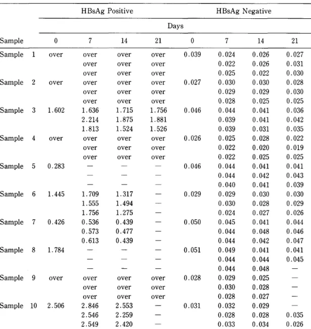
IL−2産生能を調べた。その結果,図1で示すよう
にTbg感染3日目のBALB/cおよびC57BL/
6マウス脾細胞のIL−2産生能は正常マウスに比 べて低下していることが明らかとなった。しかしな がら,感染1日目,2日目では,IL−2産生能はコン
トロールと較べて,明らかな低下を示さなかった。
2)Tbg感染マウス血清中のIL−2インヒビ
ター活性
Tbg感染3日目の血清中のIL−2インヒビ ター活性は,BALB/cおよびC57BL/6マウス の何れの系統においても正常血清とほぼ同様であ
り,感染によるIL−2インヒビター活性の増強は,
認められなかった(図2)。感染1日,2日目のIL−
2インヒビター活性も3日目と同様に正常血清と 比較して上昇していなかった。
3)正常脾細胞のIL−2産生能に対する影響 感染血清の正常脾細胞のIL−2産生への影響をみ
た。図3に示すように,感染3日目の血清は,8%
の濃度で正常血清より明らかな抑制作用を示した。
考 察
トリパノソーマ感染によって起こる免疫抑制に は,様々な機序が存在することが多くの研究者に よって報告されてきた(Ackerman召∫α」.,1976;
Person召∫α」.,1978;Romos6∫α1.,1979;Cm−
ningham6緬」.,1980;Maleckar召緬1、,1983)。
特に丁細胞の機能に関しては,細胞内寄生原虫で ある丁珈伽oso吻αo耀認あるいは細胞外寄生原 虫の感染において,マウスの脾細胞のConAに対 する増殖反応の低下や,IL−2産生能の低下が報 告されている(Bellan召∫認.,1983;Alcina8厩1.,
1985)。またIL−2をT6耀認感染マウスに投与 することによって,原虫に対する特異的な免疫反 応の増強,虫血症の減少,マウスの延命効果が報 告されている(Choromanski召∫認.,1987)。こ れらの事実は,丁細胞の機能低下が,T6剛4ま たはT6吻o召1感染時の免疫抑制現象の重要な要 因になっていることを示唆している。しかし,T わ.go励」伽s6感染については,いまだ報告されて いない。
近年,Lelchukらはトリパノソーマと同様に免
11
C O
:i:
O a
O C
(D C
.̲
E : b
C H
r‑1 I
LJ
e ‑X
E CL
O
50 40 30 20 10 O
(A)
r
, ,, ,
,,
‑/
BALB/C
, / , ,
../' ' IL .. ., L ,.
' 'L
¥
.¥
¥ ¥
¥..
¥ .
.¥ " ・・. . L' ¥,b' '
'¥ " t¥ !b
¥ " ' ' '.
' ¥ . "
21 D
Dilution
2‑
of culture
2*
supernatants
27
:p O eL
O O c O c
.̲
E )h J: H
‑I
‑
a'e
‑X
E c:
o
30
20
10
O
(B)
,
C57BL/6
̲. d""'!!., ,・ ‑
"I'F
‑ ¥
¥.
:‑
:・*
¥. ¥b,b :,b ,b "' ,b!b 'a'b tb
,,,
AD
Figure 1
Dilution of culture SUPernatantS
Titration curve of IL‑2 production. Spleen cells collected from
normal (e) and Tbg‑infected BALB/C (A) and C57B/6 (B) mice 1(O), 2(A) or 3([]) days after infection were cultured with Con A for 24 hr and IL‑2 activity of the culture super‑
natants were assayed using IL‑2 dependent CTLL‑2 cells.
Results indicate meaniS.D. of three experiments.
12
(A) BALB/C
C O :i
CO
O e
O O
C:
O =
l:: .
>
J= H
r‑l
‑
a'e
‑X
E a
o
80 60 40 20 o
l I Il 11 dS
+,: ‑‑ ;‑‑
..I ・・ I I ""I ..,1"' '
4%
Final serum
1010 O . 25%
concentration
c O : O
e O O C O C
1:,.‑
E )h J: H
r I
'‑, coe
‑X
E CL
O
60
40
20
o
(B) C57BL/6
‑‑
t:‑1' !‑ l I ・:・‑・JI:
4010
Final
1010 0.25010
serum concentration
Figure 2 Inhibition of the activity of recombinant IL‑2.
Fifty pl of serially diluted normal mouse serum (e) or sera sampled from mice I (O) , 2 (A) or 3 ([]) days after infection were added, together with 50 pl of 100 unit/ml rlL‑2 to I x 10' CTLL‑2 cells in 100 pl of culture medium. After 24‑hr culture, prolifera‑
tion of CTLL‑2 cells were measured by 3H‑TdR incorporation. In the control , culture medium was added instead of serum and c.p.m.
level were 64,600 6,578(A) or 49,758 4,127(B) . Results indicate meaniS.D. of three experiments.
13
qOgX∈OO
仁9結﹂OO﹂OOε〇二層E︾=﹂﹃﹇工出
50 40 30 20 10
serum1%
1 \︑ \\ 亟玉 \︑\︑ ︑
丞
/︑ /
F
21 23 20 27 serum4%
serum2%
./堅\.
/ \玄
享1一一一一・一一一{\、
\、㌔
50 40 30 20 10
/イド\、
/ 「臥、
蚤〆 \
}一一一一一
21 21 25 27
serum8%
㌔
寓\
\
奉4︑/一/一︸ひ
●、、r、亀隔
21 23 20 27 21 23 25 27
Dilution of culture suPernatants
Figure3 InhibitionofIL−2production.ConAactivatednormalspleencells
(5x10⁶/mJ)were incubated with serially diluted serum from either BALB/c normal mice(●)or mice infected for3days with Tbg(○)。After24−hr−culture,the remaining activity ofIレ2inthe c611−free supematants were assayed using CTLL−2cells.Results indicate mean±S.D、of three experiments.Maximum activity of IL−2released in the absence of additional serum(□)is shown as
a control of each condition.
疫抑制を起こすマラリア感染について,マウス血 清中のIL−2インヒビターの変動を報告した
(Lelchukゆσ1.,1985)。彼らによると,IL−2 インヒビターは無胸腺マウスや無菌条件下で飼育 されたマウス血清中には存在しないが,正常マウ ス血清中に存在し,マラリア(jp彪s窺04伽吻
ッ08」劾感染時に上昇するという。
今回の実験では,Tδ.gα郷6伽s召感染におけ る丁細胞の機能を検索するためにマウス脾細胞の IL−2産生能を調べたところ,感染3日目に著し
く低下していた。そこで,IL−2産生の低下にいか なる抑制機構が存在しているかを明らかにするた めに感染3日目のマウス血清中のIL−2インヒビ ターを測定した。その結果感染3日目のマウス血 清は,8%の濃度で正常脾細胞のIL−2産生を明
らかに抑制した。しかしながらCTLL−2細胞の IL−2依存性増殖に対しては,抑制反応を示さな
かった。このことからTわ.go吻6勉s召感染にお けるIL−2抑制因子は,IL−2と競合的に作用す るものではなく,IL−2産生を抑制するものと考 えられた。
IL−2インヒビターはHardtら(Hardt擁α」.,
1981)の報告に,Lyt−2,3+のsuppressor T細 胞より分泌されて丁細胞の機能調節の一環を担っ ているとされ,マウス血清中では,分子量5万の タンパク質とも言われているが,その詳細はいま だ不明である。ヒトでは,血清中にprostaglandin の様な1レ2活性を抑制する物質も含まれており
(Rappaport8∫oJ.,1982),かならずしもIL−2 インヒビターといわれる単一の物質が存在するか については異論もある(Lelchuk6勧1.,1987)。
さらにこれら,いわゆるIL−2インヒビター活性 は寄生虫の種や感染時期,寄生部位によって様々 な活性の変化をきたす可能性も考えられる。しか
14
Uf , v* ftL J 8 %o)J ,*. 7 7; l l IL‑ 2 m UUlt ; > in vivo e , c ) I ,*. f 7 7; mli , >ti ) & l Uf FFi c l
U v > I J , :D tL f*‑ .
h IJ / / ') ‑7' ,*. ec ; : UCl) i } e
:t c IL‑ 2 ) .. ' f F c, i #)lLJ : : l )
i ^ { :T b suppressor macrophage ) l : :tL v・ (Bagasra et al., 1981; Corsini et al., 1977; Eardley et al., 1977). C >1)jt
& t: )f ;T OA i c, interferon b NK ? f'
Ji LU :v>; v, I ) (Hatcher et
al., 1980). h IJ , / y ‑7 ec : : :,t: )
lH ) ,,* *,1 f 1 , h " ' ) ')‑7 { 5 l ) F > ) 1 1)ect tL .
x
1 ) Ackerman, S.B. and Seed, J.R. (1976): The effects of tryptophol on immune responses and its implications toward trypanosome‑induced immunosuppression, Experientia, 32, 645‑647 2 ) Alcina, A and Fresno, M. (1985): Suppressor factor of T‑cell activation and decreased
interleukin 2 activity in experimental African Trypanosomiasis, Infect. Immun., 50 (2) , 382‑387 3 ) Bagasra, O., Schell, R.F. and Le Frock, J.L. (1981): Evidence for depletion of la+ macro‑
phages and associated immunosuppression in African trypanosomiasis, Infect. Immun., 32 (1) , 188‑193
4 ) Bellan, A.H., Joskowicz, M., Fradelizi, D. and Eisen. H. (1983): Modification of T‑cell proliferation and interleukin 2 production in mice infected with Tr)panosoma cruzi, Proc. Natl.
Acad. Sci. USA, 80, 3466‑3469
5 ) Choromanski, L. and Kuhn, R.E. (1987): Use of parasite antigens and interleukin‑2 to enhance suppressed immune response during Tr)panosoma cruzi infection in mice, Infect. Immun., 55
(2) , 403‑408
6 ) Corsini, A.C., Clayton, C., Askonas. B.A. and Ogilvie, B.M. (1977): Suppressor cells and loss of B‑cell potential in mice infected with Tmpanosoma brucei, Clin. Exp. Immunol., 29, 122‑131 7 ) Cunningham, D.S. and Kuhn, R.E. (1980): Tr)panosoma cruzi‑induced suppressor substance. I.
Cellular involvement and partial characterization, J. Immunol., 124 (5), 2122‑2129
8 ) Eardley, D.D. and Jayawardena, A.N. (1977): Suppressor cells in mice infected with Tmpanosoma brucei, J. Immunol., 119 (3), 1029‑1033
9 ) Gillis, S., Ferm, M.M.. Ou. W. and Smith, K.S. (1978): T cell growth factor: parameters of production and a quantitative microassay for activity, J. Immunol., 120 (6) , 2027‑2032 lO) Hardt, C., Rollinghoff, M., Pfizenmaier, K., Mosmann, H. and Wagner, H. (1981): Lyt23+
cyclophosphamide sensitive cells regulate the activity of an interleukin 2 inhibitor in vivo, J.
Exp. Med, 154, 262‑274
11) Hatcher, F.M., Kuhn, R.E., Cerrone, M.C. and Burton, R.C. (1980): Increased natural killer cell activity in experimental American Trypanosomiasis, J. Immunol., 127 (3) , 1126‑1130
12) Lanham, S.M. and Godfrey, D.G. (1970): Isolation of salivarian Trypanosomes from man and other mammalian using DEAE‑cellulose, Exp. Parasitol., 28, 521‑534
13) Lelchuk, R., Rose, G. and Playfair, J.H.L. (1984) : Changes in the capacity of macrophages and T cells to produce interleukins during murine Malaria infection, Cell Immunol., 84, 253‑263 14) Lelchuk, R. and Playfair, J.H.L (1985): Serum IL‑2 inhibitor in mice. I. increase during
infection. Immunol., 56, 113‑118
15) Lehchuk, R., Schmidt, T.A., Hodson, K., Aston, R. and Liew, F.Y. (1987) : Is there an Interleu‑
kin 2 inhibitor in human serum?, Cell Immunol., 104, 126‑133
16) Maleckar, J.R. and Kierszenbaum, F. (1983) : Inhibition of mitogen‑induced proliferation of
15 mouse T and B Iymphocytes by bloodstream forms of Tr)panosoma cruzi, J. Immunol., 130 (2) , 908‑911
17) Person, T.W.. Roelants, G.E., Lundin, L.B. and Mayor‑Withey, K.S. (1978): Immune depres‑
sion in trypanosome‑infected mice. I. Depressed T Iymphocyte responses, Eur. J. Immunol., 8, 723‑727
18) Romos, C., Schadtler‑Siwon, I. and Ortiz‑Ortiz, L (1979): Suppressor cells present in the spleens of Tmpanosoma cruzi‑infected mice, J. Immunol., 122 (4), 1243‑1247
19) Rappaport, R.S. and Dodge, G.R. (1982): Prostaglandin E inhibits the production of human interleukin 2, J. Exp. Med, 155, 943‑948
IL‑2 PRODUCTION INHIBITORY ACTIVITY OF THE SERA IN TRYPANOSOMA BRUCEI GAMBIENSE INFECTED MICE
DAO TAO, MINORU HIGASA, HIKOSHIRO MIYAMOTO, KAZUHIKO YAMADA, TOSHlNORI KOMATSU AND SOHEI SHlNKA
Received September 5 1988/Accepted January 24 1989
African trypanosome infections result in severe immunosuppression. Previous studies have shown that this suppression is due, at least in part, to suppressor cells (T cells and macrophages) or impairment of IL‑2 production.
In the present study, the effects of Trypanosoma brucei gambiense infection on serum levels of IL‑2 inhibitor and Concanavalin A (Con A) induced IL‑2 production of spleen cells were investigated. The results showed the ability of infected spleen cells to produce IL‑2 was markedly reduced and IL‑2 production of normal spleen cells was inhibited by the infected sera. But the IL‑2 dependent CTLL‑2 cell proliferation was not inhibited by those sera.
These results suggest that depletion of IL‑2 production during T. b. gambiense infection may be due to the increase of an inhibitor of IL‑2 production.
Department of Immunology and Medical Zoology, Hyogo College of Medicine
Japan. J. Trop. Med. Hyg., Vol. 17, No. l, 1989, pp. 17‑25 17
ENZYME‑LlNKED IMMUNOSORBENT ASSAY
(ELISA) IN THE DETECTION OF HEPATITIS‑B SURFACE ANTIGEN IN BLOOD DRIED
ON FILTER PAPER
SHINYA MATSUDA AND HIROAKI KAHYO Received September 7 1988/Accepted December 26 1988
Abstract: In an attempt to find a convenient method for the seroepidemiological study of HBsAg in developing countries, ELISA using blood samples dried on filter paper was evaluated. The results of this method were in complete agreement with both those obtained on original sera by RIA and with those obtained on sera by ELISA. We did not experience any false positive or false negative results. In this study the sample dried on filter paper was eluted by I ml of PBS (pH 7.2). Dilution rates were calculated by the calibration line of each serum. Dilution rates were between 0.041 and 0.082. The coefficient of variation of absorbance value of 10 samples of each case was between 5.0%
and 14.8%, which indicated an acceptable intratest reproducibility of this method. In addition, storage at 20'C, 20'C with desiccants and 3'C for up to 21 days did not alter the sensitivity and specificity of this method. This method will be useful for the sero‑
epidemiological study of HBV in developing countries.
INTRODUCTION
While conducting a seroepidemiological study in developing countries, researchers often encounter the following difficulties:
1 . Collecting blood samples
It is often difiicult to take venous blood from newborns, children, anemic patients, pregnant women and other adults who are not accustomed to such a procedure 2 . Storage and transportation of blood samples
3 . Economical problems 4 . Technical problems
The planning method may not be available for local conditions. These difficulties are magnified when the population being studied is spread over a large area or living far away from research centers.
To overcome these di :culties, blood samples dried on filter paper have been used to detect markers of various infectious diseases like Onchocerciasis (Korenaga et al., 1983;
Tada et al., 1985) , Japanese encephalitis (Yamamoto et al., 1985) and Rubella (Sander et al., 1985). As far as HBsAg is concerned, the filter paper method has been applied to the detection using radioimmunoassay (RIA) and reversed passive hemagglutination (RPHA) (Farzadegan et al.. 1978; Villa et al., 1980; Kitazumi et al., 1981; Zhuang et al., 1982), and
Department of Human Ecology, School of Medicine, University of Occupational and Environmental Health, Kitakyusyu 807, Japan
18
some seroepidemiological studies have been carried out by this method (Prince et al., 1981;
Werner et al., 1985).
Enzyme‑linked immunosorbent assay (ELISA) which was invented by Engyall and Perlmann (1971) has been used to detect various pathogenic agents because of its high sensitivity, specificity and rapidity. Ukkonen et al., (1977) demonstrated the usefullness of ELISA to detect HBSAg. Compared to RPHA, ELISA is a more sensitive method. Also, ELISA does not have any regulations which RIA have about radioactive agents. In this study we intended to detect HBsAg in blood dried on filter paper using ELISA.
MATERIALS AND METHODS
1 . Collection of samples
Ten HBsAg positive cases were selected from out‑patients of the Ist Department of Internal Medicine, University of Occupational and Environmental Health (UOEH). Ten HBsAg negative cases were selected from student volunteers of UOEH. All cases were retested by a commercial RIA kit (Ausria, Abbott Laboratories) on HBSAg. Twenty blood samples dried on filter paper were collected from each HBsAg positive case and ten samples from HBsAg negative cases.
2 . Filter paper
In this study we used a commercial blood sampling paper (Type I , Toyo Roshi, Ltd., Japan). This paper, which was developed by Nobuto (1970), is composed of two areas, an absorbing area and a diffusion area (Fig. 1). The absorbing area is allowed to absorb as much blood as possible. An excess of blood is allowed to be diffused into the diffusion area.
In this way the amount of blood contained in the absorbing area is considered to be almost constant (0.1 ml which is equal to 0.04 ml of serum) . Whole blood was collected by this type of paper and air‑dried for 2 days at room temperature with the absorbing area up, avoiding direct sunlight. The blood samples dried on filter paper were stored in small polyethylene envelopes under 3 different conditions, 20'C, 20'C with desiccants and 3'C to be tested after 7, 14 and 21 days from collecting.
3 . Elution
The absorbing area was cut into 4 portions and placed in a test tube. Then I ml of PBS (pH 7.2) was added and the test tube was incubated at 37'C for 3 hr with vibrat ron for elution.
4 . ELISA procedure
In this study we used a commercial kit, IMMUNlS HBsAg EIA (Sanko Junyaku Co., Ltd.). The procedure was as follows: 1) 0.1 ml of eluate was added to each well. 2) The
18mm
30 m m
10
a b
Ca] blood absorbing area Cb) blood diffusion area Figure I Blood sainpling paper.
19
plate was incubated for 2 hr at 37'C. 3) After the reaction, the eluate in well was aspirated.
4) The wells were washed 5 times with wash fluid supplied by the manufacturer. 5) 0.1 ml of horse radish peroxidase conjugated antibody (mouse anti‑HBs*) was added to each well.
6) The plate was incubated for 2 hr at 37'C. 7) The wells were emptied by aspiration. 8) The wells were washed 5 times by wash fluid. 9) 0.1 ml of freshly prepared substrate solution (o
‑phenyl‑diamine, OPD) was added to each well. 10) The plate was incubated for 30 min at room temperature in the dark. 11) The enzymatic reaction was stopped by adding 0.05 ml of 4 N H2SO*. 12) The plate was read by an autoreader (EAR340, Australia S.L.T. Co., Ltd.) . In addition to the above tests the original sera were tested by ELISA to compare them with the results obtained from blood dried on filter papers. The results were interpreted according to the manufacturer's recommendation, that is, positive ELISA values were>2.5x (mean ELISA values of negative control sera) .
RESULTS
1 . Comparison of results obtained by RIA and our method
Table I shows the comparison of results obtained by RIA and this method. The results are in complete agreement with those obtained on original sera by RIA, indicating acceptable sensitivity and specificity of this method.
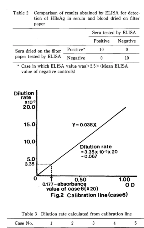
2 . Comparison of results obtaind by ELISA in serum and in whole blood dried on filter paper Table 2 shows the comparison of results obtained by ELISA in serum and in whole blood dried on filter paper. The results were in complete agreement.
3 . Dilution rate
Theoretically speaking, the dilution rate is 0.04 when the sample dried on the absorbing area of the filter paper was eluted by I ml of PBS (pH 7.2) . In this study we made calibration lines of the original sera to calculate the dilution rate from the absorbance value of each sample (Fig. 2). The results are shown in Table 3. The dilution rates were between 0.041 and 0.082.
4 . Reproducibility
The intratest reproducibility of this method was assessed by 10 blood samples dried on filter paper 10 times for each case within the same test. The results of 6 cases are shown in Table 4. Four cases were excluded from discussion because their absorbance values overfiowed. The coefficients of variation (C.V.) were between 5.0% and 14.8%. According
Table I Companson of results for detection of HBsAg obtained by RIA for sera and those obtained by ELISA for blood dried on filter paper
Sera tested by RIA Posrtrve Negative
Sera dried on the filter paper tested by ELISA
Positive* 200 o
Negative o 100
* Case in which ELISA value was>2 .5 x (Mean ELISA value of negative controls)
20
Table 2 Comparison of results obtained by ELISA tion of HBsAg in serum and blood dried paper
f or
on detec‑
filter
Sera tested by ELISA Positive Negative Sera dried on the filter
paper tested by ELISA
Positive* 10 O
Negative o 10
* Case in which ELISA value was>2.5x (Mean ELISA value of negative controls)
Diiution rate
xl0‑3
20.0 15.0
10.0
5.0
3.35
t l , ,
Y = O. 038X
Dilution rate
= 3.35x l0‑3x 20
‑ o.067
O 0.50
0.1 77 * absorbance
value of case6<x20) Fig.2 Calibration
1.00
O
line(case6)
D
Table 3 Dilution rate calculated f rom calibration line
Case No. l 2 3 4 5
Dilution rate O . 069 o . 082 O . 041 o . 044 o . 063
Case No. 6 7 8 9 10
Dilution rate o . 067 o . 069 O . 047 o . 044 O . 055
Mean: 0.058 SD: 0.014
to the explanatory note of IMMUNIS HBsAg EIA, the C.V. of the original ELISA procedure are 7.7% and 9.6%. The results indicated the acceptable intratest reproducibility of this method.
5 . Influence of storage
Samples dried on filter paper were tested after 7, 14 and 21 days from the first test. The