Japanese Joumal of Tropical Medicine and Hygiene
第12巻第1号 昭和59年3月15日
内 容 原 著
実験的狂犬病におけるearly deathに関する研究(英文)
…小原博 ケニア在住日本人およぴその家庭で働くケニア人の腸管寄生虫検査………井関 基弘 ナイジェリアにおける学童の寄生虫調査ならびにPyrantel pamoate
による細駆虫成績(英文)……、___……,_……金子酸N晶Ish。1⑩
ベネズエラで経験じたスナノミ症(英文)
・野田 伸一,高岡 宏行,鈴木、博,多田 功
アシマダラヌマカの幼虫期および蠕期の呼吸管と呼吸角についての考察(英文)
・岩城 操
会 報
昭和58年度第1回幹事会記録 …・…
幹事選挙の経過と結果……
会員名簿…………一……・
投稿規定
1−15 17−20
21−26
27−31
33−38
39−40 40−41 43−73
日熱医会誌
Japan」・エM耳 日 本熱帯医学会
STUDIES ON EARLY DEATH IN EXPERIMENTAL ANIMAL RABIES
HIROSHI OHARA
(Received July 1 8 1 983/Accepted February 1 3 1 984)
Abstract : It has been shown that inadequately immunized humans dying of rabies succumb after an incubation period shorter than that of any unvaccinated human rabies cases. This report is concerned with the reproduction and analysis of this phenomenon called "early death" in experimental animals. A significant reduction of the incubati6n period was recognized in hamsters (P<0.01), and rabbits (P<0.05) immunized with potent anti‑rabies vaccine after being challenged by rabies virus, while the reduction was not significant among animals that were treated with anti‑rabies virus serum. The viral antigen in the central nervous system examined at the onset of disease by immunofluores‑
cent staining rather decreased and inclusion bodies were less plentiful among animals that died earlier than in the controls, whereas no marked deposition of complement or immuno‑
globulin in nerve cells was proved in this study. The phenomenon could be reproduced in experimental animals vaccinated after exposure, although it could not be shown clearly that "early death" in experimental animals is mediated through anti‑rabies antibodies.
I NTRODUCTION
Rabies is a fatal disease which causes severe encephalomyelitis and death after a long incubation period. To prevent the onset of this disease, various kinds of rabies vaccines have been investigated and applied sipce the development of the first vaccine by Pasteur in 1885. Rabies is usually prevented successfully if people are vaccinated before being bitten by rabid animals and enough antibody titer is produced (Gerichter t al., 1978; Anderson et al., 1980; Ajjan et al., 1980). However the effect of post‑
exposure treatment with vaccine is not complete. Moreover, it has been observed that among humans (Ohtani, 1959; Held et al., 1 967) or laboratory animals (Sikes et al., 1971 ; Baer and Cleary, 1972; Blancou et al., 1980) if vaccine was inoculated after infection with rabies virus, subjects could not be prevented from catch・ing the diseases, and some vaccinated subjects died sooner than non‑vaccinated ones. This phenomenon has been termed "early death", and the immunopathological mech‑
anism, especially that mediated by antibodies rather than immune T cells, is suspected of causing this phenomenon (Prabhakar and Nathanson, 1981 ; Porterfield, 1981).
This phenomenon has been observed during some experiments examining the eflicacy of vaccine on humah cases, but few experimental trials reproducing or
Department of Infectious Diseases, Ja pan
present address : Department of Ishikawa, 920‑02, Japan
Institute of Medical Science, University of Tokyo, Medical Zoology, Kanazawa Medical University,
Tokyo, l 08,
Uchinada,
investigating the mechanism have been made. The present study was undertaken to reproduce this phenomenon using experimental animals with post‑exposure treat‑
ment by vaccine or antiserum, and thereafter the author tried to examine the mech‑
anism from a morphological point of view.
MATERIALS AND METHODS
Animals : Japanese white rabbits and Syrian goldeTl hamsters were used with initial weight of 2.0‑2.3 kg and 25‑32 g, respectively. Virus titrations were made in ddY suckling mice 3‑7 days of age.
Virus : A 10 per cent emulsion of monkey brain infected with rabies virus waS prepared in Eagle's minimal essential medium (MEM, pH 7.4) . The supernatant after centrifuged at 3,000 rpm for lOmin was stored at ‑ 70'C until use. The titer of the virus in the preparation by intracerebral inoculation of suckling mice was 104.8LD50/0.01 ml.
Vaccine : Rabies vaccine prepared in chick‑embryo cell cultures (TC vaccine, Chemo‑Sero Therapeutic Research Institute, Kumamoto, Japan) was used. This vaccine was prepared by the tissue culture adapted strain of the HEP‑Flury strain of rabies virus and inactivated with p‑propiolactone. The lyophilized vaccine in single‑
does vials was reconstituted with sterile water just before being used. The infective rabies virus contained in this vaccine before inactivation was over 107.0LD50/ml.
Sera for postexposure treatment: Four kinds of sera were prepared' to reproduce
"early death" phenomenon ; anti‑rabies hamster serum with neutralizing antibody titer (NT) of 102.6 (serum A), anti‑rabies rabbit serum with antibody titer of I 02. 2 (serum B), anti‑rabies rabbit serum with antibody titer of 104. 2 (serum C), and anti‑rabies monkey serum with antibody titer of 103. 5 (serum D). Neutralizing antibody titer of these sera were calculated as shown below. Serum A was obtained after daily injections of the vaccine for 14 days (0.2 ml in each) , serum B after daily injections for 6 days ( I .O ml in each), serum C after 7 injections at an interval of one month ( I .O ml in each), and serum D after 4 injections at an interval of one month ( I .O ml in each) .
Serum B was separated on a Sephadex G 200 (Pharmacia Co., Uppsala, Sweden) using phosphate buffered saline (PBS, pH 7.2) and absorbance of each elution fraction was read at 280 nm by a spectrophotometer. Three peaks of absorbance were obtained, and each peak was concentrated to the initial volume of the serum, then separated into two aliquots. The first aliquot was used to measure the serum neutralization antibody titer, and the second was mixed with 0.2 M 2‑mercaptoethanol in PBS (2ME). Both samples were kept at room temperature for 1 8 hours and then titrated for anti‑rabies antibody. The neutralizing antibody titers of peak I, peak II and peak 111 without 2ME treatment were 102.0, 101.8 and O respectively. Titers fell significantly in the sample at peak I treated with 2ME (from I 02.0 to less than lO0.2), but not in the sample at peak II. So it was thought that peak I contained primarily lgM, peak 11 primarily lgG, and peak 111 proteins such as albumin that are smaller than the immunoglobulins.
Serum neutralization tests for assaying each immune serum were performed as
follows : A tenfold dilution of heat‑inactivated serum was prepared with MEM.
Each dilution was mixed with an equal volume of 10 per cent suspension of rabbit brain infected with Nishigahara strain of rabies virus ( 102. OLD50) ' The above mixtures were incubated for one hour at 37'C, and then inoculated into the brains of 4‑5 suckling mice. Fifty per cent end‑point titer for each specimen was deter‑
mined by the Reed‑Muench method.
Serafor immunofluorescent staining : Sera used for immunofluorescent (IF) staining in this experiment were as follows : FITC‑conjugated anti‑rabies rabbit serum prepared in our laboratory, FITC‑conjugated goat anti‑rabbit lgG (Eiken Co., Tokyo, Japan), and FITC‑conjugated goat anti‑rabbit lgM (Cappel Laboratories, Downington, U. S. A.) were used for direct IF staining. Rabbit anti‑hamster lgG (MBL Co., Nagoya, Japan), rabbit anti‑hamster C3 (Cappel Laboratories, Down‑
ington, U. S. A.), goat anti‑rabbit C3 (Fujizoki Co., Tokyo. Japan), and FITC‑
conjugated anti‑rabbit lgG (Eiken Co., Tokyo, Japan) were used for indirect IF
staining.
Method of IF staining : IF staining was done as described previously (Burns et al., 1978; Swoveland et al., 1979; Johnson et al., 1980). Formalin fixed tissues, embedded in paraffin were sectioned to 4 p thickness. Those sections were de‑
paraffinized by immersion in xylene for 20 minutes, rehydrated through 100, 90, 80 and 70 per cent ethanol, and washed with phosphate buffered saline (PBS, pH 7.2).
Tissues were digested with 0.2 per cent trypsin solution (Merck Co., Darmstadt, Germany) with 0.01 per cent CaC12 for 90 minutes at 37'C, followed by washing with PBS for 1 5 minutes. Direct or indirect methods of IF staining were followed.
Each section was covered with specific antiserum, then incubated overnight at 4'C.
After incubation they were washed with PBS, mounted with glycerine phosphate buffer, and observed under a fluorescent microscope with substage illumination (Olympus, Tokyo, Japan) .
Treatment: The vaccinated animals consisted of five groups of hamsters and one group of rabbits. In the hamster groups, there were 5‑6 hamsters in each contiol group and l0‑1 1 in each vaccinated group, while there were 3 rabbits in the control group and 6 rabbits in the vaccinated one. Virus was inoculated into the following tissues : the cornea of group I, the neck muscle of group II, the back muscle of group III, the fore footpad ofgroup IV, the rear footpad ofgroup V, and the cornea of group VI.
Each animal was anesthetized by an intraperitoneal injection of pentobarbital.
The cornea of groups I and VI was scarified and 0.02 ml of virus suspension was administered. In groups II‑V, 0.02 ml of stock virus was injected into the site indicated above. Groups I‑V received 0.2 ml and group VI, I .O ml respectively, of TC vaccine intraperitoneally immediately after the inoculation of virus. TC vaccine was injected daily and totally 14 doses were given to each hamster and rabbit.
The serum‑treated hamsters and rabbits were divided into 10 groups according to the serum injected. Setum A w2 s used for group I, serum B for groups 11 and V, serum C for groups II'l and VI, rabbit lgM for group VII, rabbit lgG for group VIII, and serum D for group IX. Groups IV and X were used as controls. One ml and I O ml of serum were injected intraperitoneally into hamsters (groups 1‑III) and
rabbits (groups V‑IX) respectively, immediately after the inoculation of virus. The injections were given on three consecutive days, hence the total amount of serum for each hamster was 3 ml, and 30 ml for each rabbit.
Animals were observed for 50 days after challenge of the virus and killed when showing clear clinical signs of rabies. Specimens were collected from the inoculation
site, spinal cord, trigeminal ganglion, cerebellum, cerebrum and pons. The
specimens were fixed in 10 per cent buffered formalin solution (pH 7.2) for 14 days.
After the process of paraffin embedding, 4 p thick sections were cut and stained with immunofluorescent (IF), hematoxylin‑eosin (HE) and luxol fast blue (LFB) .
RESULTS
Clinical signs : All rabid hamsters showed clinical signs as follows : weakness, elevation of body temperature, ataxia, and paralysis. Convulsion and tonic spasm were also observed and appeared more frequently among the vaccinated groups than in the controls. A11 rabbits showed similar clinical signs, i.e., weakness, elevation of body temperature and ataxia followed by paralysis. A few animals showed sali‑
vation or urinary incontinence.
Mortality rate and incubation period: The mortality rate among the vaccinated hamsters was lower than that of the control ones. All the control groups suffered lOO per cent mortality, while the vaccinated groups showed a mortality rate in the
Table 1 Effect of vaccination on incubation period and mortality rate
Site of Mortality Incubation period
Group Animal inocu‑
lation Control Vacci‑ Control
nated M* SD** Vacci‑n a ted M* SD**
I Hamster Cornea 5/5 8/10 18, 19, 22
(100'/.) ( 80"/.) 24, 27 22.0
II Hamster Neck 5/5 8/11 1 7 20 24 muscle (100 ) ( 73 ) 29 32' 24.4
III Hamster Back 5/5 7/11 25 25 27 muscle (100 ) ( 64'A ) 28 29' 26.8
3.3
5.5
l .6
10, 11, 15 15, 15, 16 16, 17 13, 14, 14 15, 17, 17 17, 17 16, 16, 17 18, 19, 19 19
14.3
15.5
17.7 2.3
1.6
1 .3
IV Hamster Fore 5/5 7/11 25 27 29 footpad (lOO ) ( 64'A ) 33 34' 29.6 14, 16, 19 3.4 19, 19, 20
25
18.9 3.2
V Hamster Rear 6/6 7/1 1 26, 29, 29
footpad (100'/.) ( 64"/.) 39, 39, 41 33.8
VI Rabbit Cornea 3/3 6/6 , 14 16, 17
(100*/.) (100"/*) 15.7
17, 18, 18 6.0 18, 19, 19
20 12, 13, 13 l.2 13, 14, 15
18.4
13.3 0.9
0.9 0.02 ml of stock virus (104. 8LD50) was inoculated into each site.
daily and was killed after showing clear clinical signs of rabies.
*M: mean, **SD: standard deviation.
Each animal was observed
range of 64‑80 per cent. In rabbits, the mortality rate was 100 per cent in all groups including the control.
The range of incubation periods varied in each group. It was shorter when the inoculation site was closer to the head. Within the same group of animals inoculated at the same site the incubation periods also varied. In the vaccinated hamsters, the incubation periods were markedly shorter than those of the controls. The decrease in incubation period, compared with that of control, was 35.0 per cent in group I, 36.5 per cent in group II, 34.0 per cent in group 111, 36.1 per cent in group IV, and 45.6 per cent in group V ; all the values were statistically significant (P<0.01 ) . In rabbits, the decrease was 1 5.3 per cent (P<0.05). The standard deviation (SD) of the incubation periods in all vaccinated groups were smaller than those of the controls ; especially in group V, the SD of the vaccinated hamsters was 0.9, whereas that of controls was 6.0 (Table I ) .
In all groups injected with serum, the mean incubation period differed from that of the control groups, but the difference was not statistically significant (P>0.05).
When sera with low antibody titer (sera A and B) were used, the incubation periods were slightly shorter than those of the cdntrol groups. But when hamsters were treated with anti‑rabies hyperimmune rabbit serum (serum C) for three consecutive days immediately following virus inoculation, the incubation periods were prolonged as long as 5.8 days. When injected with whole serum, however, regardle s of its antibody titer, the incubation periods of rabbits were slightly shortened. The
Table 2 Effect of anti‑rabies serum on incubation period and mortality rate
Group Animal anti‑rabies
serum (antibody titer) Mortality Incubation
Period Mean
I II III
IV VI V
VII VIII
IX
X
Hamster Hamster Hamster Hamster
Rabbit Rabbit Rabbit Rabbit Rabbit Rabbit
Serum A Serum B Serum C
(NT= I 02. 6) (NT= 108. 2) (NT= I 04. 2) (control)
Serum B (NT= 102. 2) Serum C (NT= 104. a) Rabbit lgM (NT= 102. o) Rabbit lgG (NT= 101'8) Serum D (NT= 103. 5)
‑ (control)
315 315 2j5 5/5
( 60"/・) ( 60'/・) ( 40"/・) (100'/・)
3j3 (100"/・) 3/3 (lOO'/・) 3/3 (100"/・) 3/3 (100 /・) 3/3 (100"/・) 4/4 (100'/ )
17, 20, 22 19, 20, 22 24, 32 20, 21, 22 24, 24
13, 14, 14 14, 15, 15 15, 15, 15 14, 14, 14 12, 14, 19 15, 15, 16 16
19!7 20.3 28.0 22.2 13.7 14.7 15.0 14.0 15.0 15.5
Serum A : anti‑rabies hamster serum. Serum B and C : anti‑rabies rabbit serum, Serum D : anti‑rabie monkey serum, NT : neutralizing antibody titer.
Serum A was produced after I O‑ 14 consecutive injections of TC vaccine, serum B after 5‑7 consecutive injections, serum C : after 6‑7 injections at intervals of one month, and serum D after 4 injections at intervals of one month.
Group 1‑III, one ml and V‑IX, I O ml of serum was injected immediately after inoculation of virus. The injections were performed for three consecutive days.
decrease was largest when they were injected with serum B, although this decrease rate was only I .8 days. The results of treatment with serum components showed that both lgM and lgG slightly reduced the incubation periods and the decre se was slightly larger (one day) when using lgG than using lgM but the difference was not statistically significant (P>0.05) (Table 2).
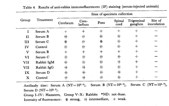
Results of IF staining : Direct IF staining for rabies virus in various parts of the CNS showed that in the vaccinated groups the intensity of fluorescence was generally weaker than in the controls, and some were almost negative. The fluorescence also differed in different parts of the nervous system in the same animal. Generally the fluorescence could be recognized in the cerebellum, cerebrum, pons, and cervical cord. However, no fluorescence was detected at the site of inoculation in all the groups. In the control of group I, the strongest fluorescence was shown in the cerebrum ; the same was in the cervical cord in groups 111‑V. In vaccinated groups, the distribution of viral antigen was almost the same as that in the control groups but the intensity of fluorescence was weaker. The viral antigen was located in the cytd‑
plasm of the nerve cells, and was completely negative in the nuclei. In the vaccinated groups, nerve cells did not show strongly positive reactions like those of the control
ones .
Intravascular lgG, IgM. C3, and surface lgG of lymphocytes infiltrating around blood vessels or in meninges were stained, but those deposited in nerve cells could not
Table 3 Results of anti‑rabies immunofluorescent (IF) staining (vaccinated animals) Sites of specimen collection
Group Treatment Spinal Trigeminal Site of Cerebrum Cere‑ pons
bellum cord ganglion inoculation
I Control
Vaccination
I I Control
Vaccination I I I Control
Vaccnation
IV Control
Vaccination
V Control
Vaccination
VI Control
Vaccination
+
+
+h
+h
+h
+h
+h
+h
+h
+ +
+h
+h
+h
+h
+h
+F
+h
+h
+h
+
+
+h
+h +h
+ +
+ + ND*
ND ND ND ND ND ND ND + +
Group ILV : Hamsters, Group VI : Rabbits.
In group I, rabies virus was inoculated into the cornea ; in group II, into the neck muscle ; in group 111, into the back muscle ; in group IV, into the fore footpad ; in group V, into the rear footpad ; and in group VI, into the cornea:
*ND : not done, Intensity of fluorescence : f strong, +h intermediate, + weak.
Table 4 Results of anti‑rabies immunofluorescent (IF) staining (serum‑injected animals) Sites of specimen collection
Group Treatment Spinal Trigeminal Site of Cerebrum bellum ganglion inoculation Cere‑ Pons cord
I II lll
IV VI V
VII VII IX
X
Serum A Serum B Serum C Control Serum B Serum C Rabbit lgM Rabbit lgG Serum D Control
+h +h +F +h +h H+
+h
+h +h +h +h +h +f +h
+h +h +h
rl+
+h + +h
rl+
H+
+h +h H+
H+
+h +h
+ + + + + + + + +
Antibody titer: Serum A (NT=102.6), Serum B (NT=102.2), Serum C (NT=10a.2),
Serum D (NT= 103.. 5).
Group 1‑IV: Hamsters, Group V‑X: Rabbits *ND: not done.
Intensity of fluorescence : strong, +h intermediate, + weak.
be stained by the IFmethod. Deposition of C3 was recognized around some blood . vessels ofvaccinated animals but this finding was also observed in some control animals
(Table 3).
The diffe ences in the intensity offluorescence among serum‑injected groups were small. Generally the shorter the incubation period, the weaker the fluorescence.
The fluorescence was the strongest in groups 111 in which the incubation period was the longest among the above groups. Like the vaccinated groups, no viral antigen was found in the cornea (Table 4) .
The chronological spread of rabies virus in CNS was studied. Rabbits were divided into three groups (control, vaccinated, and serum‑injected) and 0.02 ml of stock virus was inoculated into the cornea. One ml of TC vaccine or 10 ml of anti‑
rabies monkey serum (serum D) was injected intraperitoneally in each rabbit; the vaccine was injected every five days and the serum seven days after inoculation of virus. A rabbit of each group was killed each on day 6, I I , 1 2, 14, 1 8 and 1 9 after inoculation of virus, and the CNS of each hamster was examined by the IF and HE staining methods.
In all groups of rabbits, the viral antigen was recognized in the trigeminal ganglion six days after inoculation, and after 1 2 days in various parts of the brain in all the groups. The fluorescence in CNS increased day by day, the strongest fluo‑
rescence was observed in the brain of a control rabbit 19 days after inoculation. The intensit,y of fluorescence in the vaccinated group was slightly weaker than that of the control when these were examined at the same time after virus inoculation (Figure I ,
Results of HE and LFB staining : Examination of various iparts of the CNS with HE staining indicated that the inflammatory findings in the meninges or perivascular
Days after inoculation
2D
15
lO
5
o
+ + +
+ +
I
l +
l
l
l
l +
l
I + +
l
l
l
+
l
l
l
l
l
+ + +
+
+ + +
+
++ +++ +++ ++ ++ +++
++ +++
+ + +
+ + +
l ‑ ‑ I ‑
v S C
CORNEA
v s c
TRI GEMI NAL GANGLION
v s c
CEREBELLUM
v s c
CEREBRUM
V vacclnated group S: serum InJected group c: control group
Figure I Chronological spread ofrabies virus in the CNS ofrabbits after inoculation into the cornea.
region were stronger in the vaccinated groups than in the contols when both were compared at the same time after virus challenge and also at th time of onset of rabies.
The changes in nerve cells were rather weak in the vaccinated groups. Infiltration of mononuclear cells around blood vessels and nerve cells was observed in almost all sections, and in some of them vacuolation of cytoplasm was visible. Control ani‑
mals revealed a number of inclusion bodies which, however, were few in the animals that died earlier after vaccination. No demyelinated lesion was seen in either group (Photos 5, 6).
DISCUSSION
Many references have reported the eificacy of preexposure treatment against rabies with various types of vaccines '(Nicholson et al., 1 978; Ajjan et al., 1 980 ; Anderson et al., 1 980) , but the exact mechanism underlying the phenomenon is still poorly understood, although humoral immunity has been shown to play an important role in clearing rabies virus from the nervous tissues (Miller et al., 1 978 ; Moreno et al., l 979). Weinmann et al. ( 1 979) reported that administration of human leukocyte
interferon could reduce the mortality rate in cynomolgus monkeys. Smith ( 1 981) and other investigators (Wiktor et al., 1974; Lagrance et al., 1979; Nicholson et al., 1979)
reported that T Iymphocytes were critical for normal clearance of rabies virus in‑
fection and they stressed the importance of relationship between T Iymphocytes and antibody responses.
According to Sikes et al. ( 1 971) a single dose of TC vaccine gave effective pro‑
tection to monkeys but "early death" was also observed in animals that died after challenge, if they had been treated with vaccine before or after the challenge. Ac‑
cording to some other investigators (Baer et al., 1 972 ; Blancou et al., 1 980 ; Prabhaker and Nathanson, 1 98 1 ) , postexposure treatment with vaccine was often effective, but when the death could not be prevented, the incubation period was usually shortened.
But different results have been obtained as to the effect of postexposure treatment with immune serum ; in an experiment on mice, postexposure treatment with hyper‑
immune serum was unable to reduce the mortality rate, however the incubation period had been prolonged (Koprowski et al., 1 950).
In the present study, "early death" phenomenon was reproduced by postexposure treatment with TC vaccine, but the hamsters treated with the hyperimmune serum (serum C) succumbed after a longer incubation period. These results coincide with the previous reports described above, while treatment by sera with low antibody titers (sera A and B) caused animals to succumb slightly sooner than the control groups. The previous evidence that vaccination can produce "early death" but hyperimmune serum cannot, makes us suspect that this phenomenon is mediated by lgM rather than by lgG. But in the present study we could not confirm this antic‑
ipation, because no significant difference in the incubation period was obtained between lgM and lgG fractions.
Apart from rabies, there are some other infectious diseases in which antibodies appear to produce effects detrimental to the host. Dengue fever is usually a curable disease which produces fever and rash, but in some cases severe hemorrhagic syn‑
dromes are seen (Halstead, 1980) . As a mechanism of this phenomenon, antibody‑
mediated enhancement of viral replication is suspected ' (Halstesd and O'rourke, 1977; Halstead, 1 979; Peiris and Porterfield, 1 979). According to this hypothesis, antibody combined with antigen can be taken by macrophages easily through Fc receptors and then viral replication is enhanced. This phenomenon was confirmed by in vitro and in vivo studies and has been regarded as one of the mechanisms causing dengue hemorrhagic fever and West Nile fever (Peiris and Porterfield, 1979) .
The same mechanism may be applicable to explaining "early death" from rabies.
But in the present study the CNS of animals that died earlier than controls for all vaccinations or serum injections showed decreased fluorescence for rabies viral antigen although the clinical signs of both vaccinated and control groups were almost the same. These results coincide with our unpublished data which were obtained by direct IF staining of fresh frozen tissues in suckling mice. The decrease of fluo‑
rescence may be only an apparent phenomenon, because antigenic sites may be covered with antibodies. Ieaving fewer antigenic sites which will bind fluorescence conjugated antibodies. Furthe studies are necessary to confirm whether viral antigen is truly decreased or not.
Immunolysis mediated by the immune complex and complement is also suspected as it is regarded as a causative factor of atypical measles and some types of glom‑
erulonephritis. According to Wiktor et al. ( 1 968), cultivated nerve cells infected with rabies virus could be fell into lysis by being added with antibody and comple‑
ment. Data of electron microscopic studies suggested that virus clearance might require both neutralization of free virus and virus infected cells (Murphy et al., 1 973 ; lwasaki and Clark, 1 975), and the importance of participation of complement on neutralization of virus is suspected (Daniels et al., 1970) . These evidences, though still fragmentary, well suggest that "early death" might well be the result of tissue
injury mediated by complement which became bound to antigen and antibody complex.
In the present study, complement deposited in nerve cells was not proved, although there were some areas where deposition of complement was seen around blood vessels. This flnding may be one of the causative factors of "early death", though not yet affirmed by the present experiments. Detailed study using a more sensitive method is required to prove whether complement and immunoglobulins are deposited around blood vessels or nerve cells.
In the animals that died earlier than controls, inflammatory findings were mainly observed in the meninges and perivascular area rather than the gray matter without any demyelinated lesions. Considering the appearance of inclusion bodies in the cytoplasm of nerve cells and the decreased viral antigen in the nerve cells, not only direct injury by virus but also any immunopathological mechanism must be relevant to the phenomenon. It is true that this phenomenon is strongly suspected of being caused by inadequate immunization with subneutralizing antibody ; at the same time, the participation of some other factors is also suspected. Although it was not proved clearly in the present study that this phenomenon is antibody mediated, experirTlents using this model may lead to further understanding of the contribution of host immunological responses to rabies pathogenesis and the mechanism of the
"early death"
ACKNOWLEDGEMENT
The author expresses his cordial thanks to Prof. Sugishi Ohtani, the Medical Science, the University of Tokyo, for the instructive advice and
Institute of supervision.
REFERENCES
1) Ajan, N., Soulebot, J. P.. Triau, R. and Biron. G. (1980) : Intradermal immunization with rabies vaccine, JAMA, 244, 2528‑2531
2) Anderson, L.J., Winkler, W. C., Hafkin, B., Keenlyside, R. A.. D'Angeio, L. J, and Deitch, M. W. (1980) : Clinical experience with a human diploid cell rabies vaccine. JAMA, 244,
78 1 ‑784
3) Baer, .G. M. and Cleary. W. F. (1972) : A model in mice for the pathogenesis and treatment of rabies, J. Infect. Dis., 125, 520‑526
4) Blancou, J., Andral. B. and Andral, L. (1980) : A model in mice for the study of the early death phenomel on after vaccination ahd challenge with rabies virus, J. gen. Virol., 50, 433J 35 5) Burns, J., Hambridge, M, and Taylor, C. R. (1978) : Intracellular immunoglobulins. J. clin.
Pathol., 27, 548‑557
6) Daniels, C. A., Borsos, T., Rapp, H. J., Snyderman, R. and Notkins, A. L. ( 1970) : Neutraliza‑
tion ofsensitized virus by purified components ofcomplement, Proc. Natl. Acad. Sci., 65, 528‑533 7) Gerichter, C. B., Shtark, J, and Braunstein, I. ( 1978) : Clinical trial with an anti‑rabies human
diploid cell rabies vaccine (HDCV), Develop. biol. Standard, 41, 241‑244
8) Halstead, S. B. and O'rourke, (1977) : Dengue viruses and mononuclear phagocytes, I. Infec‑
tion enhancement by non‑neutralizing antibody, J. Exp. Med., 146, 201‑2 15
9) Halstead, S. B. (1979) : In vitro enhancement of Dengue virus infection in rhesus monkeys by passively transferred antibody, J. Infect. Dis., 140, 527‑532
10) Halstead, S. B. (1980) : Dengue haemorragic fever ‑ a public health problem and a field for research, Bull. Wld Hlth Org., 58 (1), 1‑21
1 1) Held, J. R., Tierkel, E. S, and Steele, J. H. ( 1967) : Rabies in man and animals in the United States, 1946‑65. Publ. Hlth. Rep., 82, 1009‑l018
12) Iwasaki, Y. and Clark, H. F. (1975) : Cell to cell transmission of virus in the central nervous system II. Experimental rabies in mouse, Lab. Invest., 33, 391‑399
13) Johnson, K. P., Swoveland. P. T. and Emmons, R. W. ( 198,0) : Diagnosis of rabies by immuno‑
fluorescence in trypsin‑treated histologic sections, JAMA, 244, 41‑43
14) Koprowski, H.. Van der Scher, J. and Black, J. (1950) : Use of hyperimmune antirabies serum concentrates in experimental rabies, Am. J. Med., 8, 412 20
15) Lagrance, P. H., Tsiang, H., Hurtrel, B. and Ravisse. P. ( 1979) : Delayed‑type hypersensitivity to rabies virus in mice : assay ofactive or passive sensitization by the footpad test, Infect. Immun., 2 1 , 931‑939
16) Miller, A., Morse, H. C.. Winkelstein, J. and Nathanson, N. (1978) : The role of antibody in recovery from experimental rabies, J. Immunol., 121, 32 1‑326
17) Moreno, J. A., Baughcum. S. D., Levy. H. B. and Baer. G. M. ( 1979) : Further study on rabies postexposure prophylaxis in mice : a comparison of vaccine with interferon and vaccine, J. gen.
Virol., 42, 219‑222
18) Murphy, F. A., Harrison, A. K., Winn, W. C. and Bauer, S. P. (1973) : Comparative patho:
genesis of rabies and rabies like viruses, Infection of the central nervous system and centrifugal spread of virus to peripheral tissues, Lab. Invest., 29, 1‑16
19) Nicholson, K. G., Turner, G. S. and Aoki, F. Y. (1978) : Immunization with a human diploid cell strain of rabies virus vaccine, J. Infect. Dis., 137, 783‑788
20) Nicholson, K. G., Cole, P. J., Turner, G. S. and Harrison, P. (1979) : Immune responses of humans to a human diploid cell strain of rabies virus vaccine : Iymphocyte transformation, pro‑
duction of virus‑neutralizing antibody and induction of interferon, J. Infect. Dis., 140, 1 76‑182 2 l) Ohtani, S. ( 1959) : Epidemiological studies on rabies in Japan. Symposium on polyomyelitis,
rabies and other viral infections, 1 5th General Assembly of the Japan Medical Congress 22) Peiris, J. S. M. and Porterfield J. S. (1979) : Antibody‑mediated enhancement of Flavivirus
replication in macrophage‑like cell lines, Nature, 282, 509‑5 1 1
23) Porterfield. J. S. ( 1 98 1 ) : Antibody‑mediated enhancement of rabies virus, Nature, 290, 542 24) Prabhakar. B. S. and Nathanson, N: (1981) : Acute rabies death mediated by antibody, Na‑
ture, 290, 590‑591
25) Sikes. R. K.. Cleary, W. F., Koprowski, H., Wiktor, T. J. and Kaplan, M. M. (1971) : Effec‑
tive protection of monkeys against death from street virus by post‑exposur administration of tissue culture rabies vaccine, Bull. Wld. Hlth. Org., 45, 1‑11
26) Smith, J. S. ( 1 98 1 ) : Mouse model for abortive rabies infection of the central nervous system, Infect. Immun., 31, 297‑308
27) Swoveland, P. T. and Johnson, K. P. (1979) : Enhancement of fluorescent an.tibody staining of viral antigens in formalinfixed tissues by trypsin digestion; J. Infect. Dis., 1 40, 758‑763
28) Weinmann, E., Major, M. and Hilfenhaus, J. (1979) : Intramuscular and/or intralumbar
treatment of rabies virus−in艶cted cynomolgus monkeys with human inter飴ron,In飴ct.Immun.,
24ン24−31
29)Wiktor,T・J・,Kuwert,E.and Koprowski,H.(1968):Immune lysis of rabies virus−in驚cted cells,J.ImmunoL,lo1,1271−1282
30)Wiktor,T.」.,Kamo,1.and Koprowski,H.(1974):In vitro stimulation ofrabbit lymphocytes a丘er immunization with live and inactivated rabies virus vaccines,J.Immunol.,112ン2013−2019
実験的狂犬病におけるearly deathに関する研究
小 原 博
葎犬病ワクチンの接種を受けてなお発症した例では,しばしば潜伏期が短いという現象が知られて おりearly deathといわれている。その機序解明のために,本研究では,実験動物を用いて感染後発 病阻止実験を行い,この現象を再現し,形態学的検討を行った。
街上毒狂犬病ウイルス(104・8LD5。)を実験動物(ハムスター,ウサギ)に感染後,ワクチンある いは各種免疫段階の血清を一定期間毎に接種し,明確な臨床症状を呈した時点で致死せしめ,ホルマ リン固定後,中枢神経各部位について,螢光抗体法,HE染色法,LFB染色法による検索を行った。
その結果,ワクチン接種群では,ハムスター(P〈0.01)ウサギ(P<0,05)ともに潜伏期の有意な短 縮が認められ,この現象を再現することができた。免疫初期血清(抗体価NT=102・2)接種群では 潜伏期の僅かな短縮が,免疫後期血清(抗体価NTニ104・2)接種群では延長の傾向が認められた。
発症時点で螢光抗体法により検索した脳内ウイルス抗原量は,early deathを呈した動物では,対照 群に比し著明に低下しており,ウイルスによる直接的細胞障害の他に,免疫学的機序による障害が関 与していると考えられた。 /
組織学的検索の結果,early death群では対照群に比し,灰白質の障害よりも,髄膜,血管周囲へ の炎症所見が強く認められたが,封入体は少なかった。脱髄巣は認められなかった。補体c3の血管 周囲への沈着が認められるものもあったが,対照群に比し有意な差ではなかった。IgG,IgM,補体 C3の細胞内沈着は証明されなかった。
東京大学医科学研究所感染症研究部 現住所:金沢医科大学医動物学教室
P紅oto.1 Rabies vir積鋸葺ntige籔in (きpithelium of cornea3 d翫ys a£ter in鋤acornea亙inoculatioぬ 纂s r〔妻vealed by immunof垂uorescence・ Contrd房roup・ ・4GO×・
蟄昼o悉o、2 Rab圭εs・▽irus&ntig⑳intrige細段1gang1圭・n7daysa丘er豊nt蹴・mea茎inQcul農ti。n乱s reve抗1edby三mmun・丑u。res灘ce・C。ntro1言r・up・200×・
Photo.3 Rabie騨irusa沿tigenincerebel量u皿1些days識¢r量良trac。mea1㎞。culat三・nasreveale曲y i㎜un・伽・rescence・Contrd呂roup.・200×.
Photo.4 Rabies vi罫雌鼠箆t壌ε聡麺 p{凱s 14 d訊y懸 a衰er intr翫corneal inoculation 訊s 三mmunoHuore畠cence・Contぎ01宮roup.200×、
revealed by
Photo . 5 Infiltration of mononuclear cells around a sn all blood vessel in ceirebrum of vaccinated group. 400 x .
Photo. 6 Inclusion b.odies are clearly seen in the cytoplasm of nerve cells m cerebrum of cOntrol group:. 200 x .
ケニア在住日本人およびその家庭で働く
ケニア人の腸管寄生虫検査
井 関 基 受付昭和58年9月16日/受理
弘
昭和59年2月6日
国際交流が進むなかで日本人の海外滞在者は 年々増加する傾向にあり,家族連れ,長期滞在の ケースが増えている。熱帯地をはじめ衛生状態の 悪い発展途上国に長期間滞在する人達にとって健 康管理には不安が多い。著者が医療協力プロジェ クトで1980年4月から1年間ケニアのナイロピに 滞在した際にも日本人学校や青年海外協力隊,そ の他長期滞在の人達から寄生虫疾患について種々 相談を受けた。その内容は,個人からはアタマジ ラミや条虫など肉眼的に見える寄生虫感染に自分 で気付いて,駆虫をどうすればよいかという相談 や,発熱した人からのマラリア検査の依頼,マラ リアの予防内服方法についての相談などであり,
団体としては日本人学校,日本人会婦人部や留学 生サークルなどからの,ケニアで注意すべき寄生 虫病やその感染予防方法に関する講演依頼などで あった。当時,ナイロビ日本人会には454人(成 人:男168人,女142人;15歳未満の子供:男80人,
女64人)が登録されていたが,これ以外に地方で 働く青年海外協力隊員や建設関係者も多く,短期 出張者や観光旅行者を除いて,ケニア全体に滞在 する日本人の数は相当なものであった。在ケニア 日本大使館には医務官(当時は大利昌久博士)が 駐在しておられ,これらの人達の健康相談にも応 じておられたが出張などで不在の際には私達に相 談がもたらされるのである。
長期滞在者はこの国の社会慣習上,掃除・洗 濯・皿洗いなどをするサーヴァントを家庭に雇わ ざるをえず,また,料理・子守りを手伝うメイド,
運転手,アスカリと称する守衛など数人を雇って いる日本人家庭も多い。この現地人使用人達は通 いのこともあるが,その家や同一敷地内に住み込
みで毎日働いている。従って,この人達からの感 染にも不安を感じている人が多かった。
1981年3月,日本人会からの要望で大使館を通 じて在ケニア日本人およびその家庭で働くケニァ 人の糞便検査の依頼があり,寄生虫検査と細菌学 的検査を行った。著者は寄生虫検査を担当したの で,その結果を報告する。
検 査 対 象
ナイロビに在住する日本人と,主としてその家 庭で働くケニア人。提出された検体数は日本人 205(成人:男79,女64;子供:男33,女29)お よびケニア人65(成人:男33,女28;子供4)で あった。日本人検体提出者のケニア滞在期間につ いては正確なデータは得られなかったが,3カ月 位から10年以上になる人もあり,平均すると1年 以上になると思われる。
検査 方 法
ホルマリン・エーテル法によ る集卵・集シスト を行い,ヨード・ヨードカリ染色をして鏡検した。
検査一結 果
表に示したように,日本人からは大腸アメーバ のシスト陽性者が1例(成人・女)と横川吸虫卵 陽性者が1例(成人・男)検出されたのみであっ
た。
一方,ケニア人においては赤痢アメーバと大腸 アメーバのシスト陽性者がそれぞれ11例(16.9%),
大阪市立大学医学部医動物学教室
The prcvalence ofintestinal parasites among205Japanese Iiving in Nairobi and
・65Kenyans working in theJapanese homes
Positive case mmber in Parasites
205Japanese
(%)
65Kenyans
(%)
Eη伽08加廊oひ伽
Eη如蹴086α60あ Eη吻」珈ακη伽α
1b吻伽6加6灘50hJ露
ααr 如」α解ゐ伽 σ窺Jo規σ3∫伽膨5痂あ
」50πrゑ5」%窺み吻0∫吻5
Hookworm
7ン露h撹rゑ5 r露hゴz ブロ
S roη40」伽謝 oγα」お(?)*
漉惚0η勉螂ク0乃09αωα∫
36h5認050η観㎜η50η∫
乃6π毎sp.
1(0.5)
1(0.5)
11 (16.9)
11(16・9)
17(26.2)
(1.5)
(1.5)
(4.6)
(3.1)
15(23.1)
7(10.8)
2(3.1)
3(4.6)
2(3.1)
*Suspected fめm the皿orphology ofthe rhabditifbrm larvae.
小形アメーバは17例(26.2%)など原虫感染も多 く認められ,また蠕虫類では鉤虫が15例(23.1%)
で最も多く,鞭虫7例(10.8彩)の他,マンソン 住血吸虫,回虫,条虫(無鉤条虫と思われる)な どの感染もみられた。2例からラブジチス型幼虫 が検出された。培養によるフィラリア型の観察は 行っていないが,食道の形態および生殖原基の大 きさなどからおそらく糞線虫であろうと思われた。
ケニア人65人のうち41人が何らかの腸管寄生虫に 感染しており,総陽性率(総陽性者数/総検査数)
は63.1彩であった。このうち,1種感染は19例で あり,2種混合感染が11例,3種混合が10例,4 種混合が1例であウた。
考 察
山浦ら(1976,1981)によると青年海外協力隊 で海外に長期滞在して帰国した日本人の消化器系 寄生虫感染率は25〜30彩という高い値を示してい るが,今回の調査では日本人の寄生虫感染者は予 想以上に少なかった。横川吸虫はおそらく日本国 内でアユを食べて感染したのであろうから,ケニ
アでの感染が疑われたのは205人中1例のみで あった。これはナイロビで日本人が住んでいる生 活環境は上水道 ・下水道・水洗便所が完備してお り,他の熱帯地に比べると街全体の衛生状態が優 れているし,2年以上も滞在している人の中には 自分で定期的に駆虫薬を服用している人も多いと 聴くので,その効果が出ているのかも知れない。
また,青年海外協力隊員の場合は地方で働くこと が多いので,その生活環境や生活様式とナイロビ 在住者のそれとは大きく異なる点もあげられる。
それではナイロビで生活している日本人にとっ て寄生虫感染の恐れは無いのであろうか。著者は 1年間のナイロピ滞在中にNaivasha,Machakos,
Kituiなどケニア各地の一般住民2,000人余の腸 管寄生原虫感染状況を調査したが,その平均感染 率は赤痢アメーバ31・8%,大腸アメーバ52.3彩,
原虫のみの総陽性率は75.1%にも達した(Iseki θ∫認,1983)。また,Pamba and Mulega(1981)
はナイロピのケニア人学童698人の糞便検査を行 い,回虫47.7彩,鞭虫12.2%,鉤虫7.9%,マ ンソン住血吸虫6.9%,小形条虫3.9%,テニア 3.0彩という陽性率を報告している。一見清潔そ
うにみえるナイロピも人口の急激な都市集中化で 地方からの人口流入が多く,郊外に住みついてい る彼等の生活レベルは低いし,衛生状態はあまり 良くないことがうかがえる。そして今回の調査で,
日本人家庭で働くケニア人もその60彩以上が何ら かの腸管寄生虫に感染しており,その半数以上が 2種混合感染あるいは3種混合感染であったこと は,彼等の生活する環境の衛生状態がいかに悪い ものであるかを如実に示しているといえる。ナイ ロビの日本人家庭で働いている間はまだ良いが,
彼等の多くは田舎の出身であり,クリスマスなど の休暇には里帰りすることが多いので,そのよう な時に感染するのであろう。特に,17彩近くが赤 痢アメーバのシストを排出していることは,彼等 が料理や,食器洗い,子守り,掃除,洗濯など家
庭内で直接密接に日本人の生活に係わっている点 で留意しなければならない。赤痢アメーバは,中 間宿主を要する住血吸虫などと違って,手指など についたそのシストが食物や食器,飲料水などを 汚染し,感染が容易に起こりうるからである。
これらのことから,ナイロビの都市部で生活し ている日本人にとっても寄生虫感染の機会は身近 に存在する。現に,私の滞在期間中に日本人で条 虫症3例,イソスポーラ症1例を経験した。また,
大利(1983)によると,在ケニア日本大使館医務 室では1980年に腸管寄生虫に関して,アメーバ症 6例,蠕虫症7例があったことを報告している。
今回Φ調査では,日本人における感染者は少な かったが,長期滞在者はその家庭内使用人を含め て日常の感染予防に十分注意する必要がある。
文 献
1)Iseki,M,,Hayashi,K,Gatika,S.M.and Siongok,T.A(1983):The prevalence ofintestinal protozoa in Naivasha,Kitui,Machakos,Taveta and Nandi Hills areas in Kenya,Japan.
J・Trop・Med.Hyg,,11(3/4),249−256
2) 大利昌久(1983):海外医療ハンドブック・東アフリカ,19−34,日本熱帯医学協会,東京 3)Pamba,H.0.and Mulega,P.G.(1981):Comparison of Kato thick smear technique and formol−ether sedimentation method for qualitative diagnosis of intestinal helminths,East Afr.Med.J.,58,95−100
4) 山浦 常,和田芳武,松本克彦,鈴木雅子,白坂龍暖(1976):海外駐在員の寄生虫感染一青年 海外協力隊員の感染状況からr熱帯,1α18−20
5) 山浦 常,松本克彦,和田芳武,小林和代,岡本雅子,白坂龍噴(1981):海外長期滞在者の消 化器系寄生虫検査,寄生虫誌,3q85−89
A 'SURVEY ON INTESTINAL PARASITE INFECTION AMONG JAPANESE LIVING IN NAIROBI AND KENYANS WORKING ' IN THE JAPANESE HOMES
MOTOHIRO ISEKI
(Received September 1 6 1 983/Accepted February 6 1 984)
The stool examinations on 205 Japanese living in Nairobi, Kenya, and 65 Kenyans working in the Japanese homes as an indoor‑ or outdoor servant, maid or driver were carried out in March, 1 98 1 . The Japanese group was composed of 1 43 adults ( 79 males and 64 females) and 62 children under 1 5 years old (33 males and 29 females). Most ofthem had been staying in Nairobi for 6 months or more.
The Kenyan group was composed of 6 1 adults (33 males and 28 females) and 4 children. The stool specimens were examined by formol‑ether sedimentation method followed by iodine‑staining.
Out of 205 specimens from the Japanese group, only one case of Entamoeba coli infection and one case of Metagonimusyokogawai infection were detected. The latter case was suspected that the infection had occurred by ingestion of undercooked Ayu, a fresh water fish Plecoglosus altivelis, during his stay in Japan. On the other hand, out of 65 Kenyan persons 41 (63.lo/o) were infected with one or more intestinal parasite species. The parasite species detected and their infection rates were 1 6.9 per cent for Entamoeba histolytica, 16.9 per cent for E. coli, 26.2 per cent for Endolimax nana, 1.5 per cent for Iodamoeba butschlii, I .5 per cent for Giardia lamblia, 4.6 per cent for Chilomastix mesuili, 3.1 per cent for Asearis lumbricoides, 23.1 per cent for hookworm, l0.8 per cent for Trichuris trichiura, 3.1 per cent for Strongyloides stercoralis, 4.6 per cent for Schistosoma mansoni and 3. I per cent for Taenia sp., respectively.
Department of Medical Zoology, Osaka City University Medical School, Abeno‑ku, Osaka 545, Japan
A PARASITIC SURVEY AND MASS‑TREATMENT USING PYRANTEL PAMOATE ON
NIGERIAN SCHOOL CHILDEN*
KlYOTOSHI KANEKO1, FRANCIS NG2 AND OSISAMI ISHOLA3
(Received August 22 1 983/Accepted February 1 5 1 984)
Abstract : The survey and mass‑teratment of helminthic infections with pyrantel pamoate was carried out on 1 40 school children in lle‑Ife, Western State of Nigeria. It was marked that 1 29 children were positive for Ascaris lumbricoides (As. Iumbricoides), T;ichuris trichiura ( Tr. trichiura) or Necator americanus (Ne. americanus) . Of the parasite‑
pdsitive children, 82 could be allocated for the treatment with pyrantel pamoate. They were divided on the basis of having single, double or triple infections of the following intestinal nematodes ; As. Iumbridoides, Tr. trichiura and Ne. americanus.
The results demonstrate that pyrantel pamoate is effective in curing As, Iumbricoides almost completely, but is less effective against Tr. trichiura and Ne. americanus.
I NTRODUCTION
Pyrantel pamoate has been reported to be highly effective with less side or toxic effects in the treatment of infections with As. Iumbricoides (Yokogawa et al., 1 970;
Kobayashi et al., 1970; Desowitz et al., 1970; Bell and Nassif, 1971 ; Kojima et al., l 978), Ancylostoma duodenale (Yokogawa et al., 1970 ; Hori, 1 971 ; Simwogerere, 1972;
Nassif and Bell, 1972), and Ne. americanus (Yokogawa et al., 1970; Ishizaki et al., 1971; Kobayashi et al., 1971; Simwogerere, 1972; Sato et al., 1973; Bortero and Castaso, 1973). The effect of pyrantel pamoate on single or multiple infections with As. Iumbricoides, An. duodenale and Trichostrongylus spp. in Arab Republic of Egypt has been reported by Nassif and Bell (1972).
The present investigation was to determine the effect of pyrantel pamoate on single and multiple infections with As. Iumbricoides, Ne. americanus and Tr. trichiura in Nigeria .
MATERIALS AND METHODS
The present study was carried out in lle‑Ife which lies about 250 km north=east of Lagos, the capital city of Nigeria, with a population of about 1 50.000. The 1 Department of Parasitology, Aichi Medical University, Yazako, Nagakute‑cho, Aichi‑gun, Aichi
480‑1 1 , Japan
2 Department of Pediatrics, S. D. Adventist Hospital, Ile‑Ife, Nigeria
3 Department of Morbid Anatomy, Faculty of Health Sciences, University of Ife, Ile‑Ife, Nigeria
* This study was supported by the Japan‑Nigeria Cooperative Medical Science Program conducted by Japan International Cooperation Agency