JAPANESE JOURNAL OF
TROPICAL MEDICINE AND HYGIENE
JAPANESE SOCIETY OF TROPICAL MEDICINE
日 本 熱 帯 医 学 会
第3 0巻 第2号 平成1 4年6月
内 容
原 著
抗体データ解析の新たなアプローチ:兵庫県におけるトキソプラズマ抗体陽性率の 数学的分析と小児における周期的感染流行の示唆(英文)
直井 幸二,小西 英二,松村 武男,矢野 明彦………99 ベトナムで分離したコレラ菌の性状(英文)
Nguyen Binh Minh,比嘉 直美,垣花 シゲ,岩永 正明 ………103
ラオスにおける薬剤耐性黄色ブドウ球菌の監視経過(英文)
Sithivong, N., Phantouamath, B., Chomlasak, K., Sisavath, L., Munnalath, K., Insisiengmay, S.
比嘉 直美,垣花 シゲ,岩永 正明………109 タイで採集されたアシマラダブユ亜属griseifrons種群の分類学的検討:
2新種の記載およびSimulium (S.) digrammicum Edwardsの雄成虫,蛹,幼虫の記載(英文)
高岡 宏行,Wej Choochote ………115 カトマンドゥ(ネパール王国)においてモンスーン初期にみられる成人下痢症の病因に
ついて(英文)
Basu Dev Pandey, Laxmi Bikram Thapa, Jeevan Bahadur Sherchand,
Nirmal Rimal, Achyut Bhattarai,森田 公一 ………133
会報・記録
理事会・評議員会・会務総会記録………139 2002(平成14年)年度日本熱帯医学会役員名簿(2002年6月1日現在)………143 日本熱帯医学会雑誌編集委員名簿………144 投稿規定………146 著作権複写に関する注意………148
日本熱帯医学会表紙 ! − " MK 稲 芦塚 中島 宮副
日熱医会誌
VOL. 30 No. 2 JUNE 2002
CONTENTS
Original article
Naoi, K., Konishi, E., Matsumura, T. and Yano, A.
A new approach for the analysis of seroprevalence data: a mathematical analysis of the seroprevalence of Toxoplasma gondii infection in Hyogo prefecture (Japan)
with an implication of periodic outbreaks among young children.………99 Nguyen, B. M., Higa, N., Kakinohana, S. and Iwanaga M.
Characterization of Vibrio cholerae O1 isolated in Vietnam ………103 Sithivong, N., Phantouamath, B., Chomlasak, K., Sisavath, L., Munnalath, K.,
Insisiengmay, S., Higa, N., Kakinohana, S. and Iwanaga, M.
Monitoring of drug resistant Staphylococcus aureus in People’s Democratic Republic of Lao…………109 Takaoka, H. and Choochote, W.
Taxonomic notes on the griseifrons species-group in Simulium (Simulium) (Diptera: Simuliidae) from Thailand: Descriptions of two new species and
description of the male, pupa and larva of S. (S.) digrammicum Edwards………115 Pandey, B. D., Thapa, L. B., Sherchand, J. B., Rimal, N., Bhattarai, A. and
Morita, K.
Etiology of Diarrhoea Among Adult Patients During the Early Monsoon Period
in Kathmandu, Nepal ………133
背は3mm (決)
Jpn. J. Trop.
Med. Hyg.
JapaneseJournalofTropicalMedicineandHygieneVol.30No.2JUNE2002
A new approach for the analysis of seroprevalence data:
a mathematical analysis of the seroprevalence of
Toxoplasma gondii infection in Hyogo prefecture (Japan) with an implication of periodic outbreaks among young children.
KOJINAOI1, EIJIKONISHI2, TAKEOMATSUMURA3, and AKIHIKOYANO1*
Accepted 26, April, 2002
Abstract: We propose a new approach for the analysis of seroprecvalence data. The seroprevalence date on Toxoplasma gondii infection published in 1997 at Kobe University Hospital were analyzed, with a simple mathe- matical model, Y = exp(-λt), where Y ,λand t represent the percentage of seronegative people, annual infection rate and age, respectively. After calculating the mean annual infection rate (MAIR) on the basis of the above data, we determined the relationship between MAIR and the year of birth of the female participants in the study. Our present study indicates that MAIR for women born between 1940 and 1960 decreased over the years, and that in- fection rates correlated with the year of birth. Moreover, assuming that the historically declining trends of MAIR mainly reflect infection rate changes in childhood, we created a simulation of MAIR for the age under 5 years.
This simulation demonstrated that MAIR for the age under 5 years decreased from around 4% for females born in 1940, to about 1% for those born in 1960. For women born after 1960, it implied that infection rates might have been cyclically fluctuating from 0% to 1%, with an approximate 10-year interval. Our analyses imply periodic out- breaks of T. gondii infection among young children in Hyogo prefecture. In Japan, it is difficult to make a sophis- ticated statistical analysis of seroprevalence of T. gondii mainly due to the lack of available data. Despite simplic- ity of our new approach, we believe the approach will be useful to grasp the current and the past situations of T.
gondii infection even without enough data.
Keywords: Toxoplasma gondii, congenital toxoplasmosis, mathematical analysis, mean annual infection rate (MAIR), cyclical outbreaks.
INTRODUCTION
Toxoplasma gondii, an obligate intracellular parasitic protozoan, induces commonly benign or asymptomatic toxoplasmosis in healthy persons (Dubey and Beattie, 1988).
However, it may have harmful effects on the fetus when pregnant women contract primary infection, although rare cases of congenital toxoplasmosis (CT) from patients with chronic infection have been reported (Dollfus et al., 1988).
As we reported previously (Naoi and Yano, 2002), the risk of CT is dependent on annual infection rates before 15 years of age, as well as during the gestational period (equal to or older than 15 and younger than 45 years old). Hence, the historical trends of annual infection rates are crucial for evaluating the risk of CT currently faced by pregnant women. In 1997, Win et al. (1997) compared the sero- prevalence data of T. gondii infection between 1981 and
1995. The study shows that less people under 60 acquired new T. gondii infections from 1981 to 1995, and that indi- viduals under 40 contracted few T. gondii infections throughout their life. Furthermore, the data suggest that the incidence of T. gondii infection is relatively higher in chil- dren than adult women. Although these implications are in- formative, the study does not reveal historical trends of in- fection, since only two sets of seroprevalence data (taken in 1981 and 1995) are available.
Using a simple mathematical model, we devised a simulation of the historical trends of annual infection rates for females, based on the sampled data. We initially calcu- lated the mean annual infection rates (MAIR) and examined the relationship between MAIR and the year of birth of the women taking part in the sampling study. Next, we calcu- lated MAIR of women for the age under 5 years from 1940 to 1990, with the assumption that MAIR changes mainly re-
Jpn. J. Trop. Med. Hyg., Vol.30, No.2,2002, pp.99-102
1 Department of Infection and Host Defense, Chiba University Graduate School of Medicine, Chiba, Japan 2 Department of Health Sciences, Kobe University School of Medicine, Kobe, Japan
3 Department of Medical Zoology, Kobe University School of Medicine, Kobe, Japan
99
Year of birth
Mean annual infection rate
0.008 0.006 0.004 0.002 0 0.012
0.01 1981
1995
1900 1920 1940 1960 1980 2000
flect infection rate trends at a relatively young age.
METHODE
In this study, we employed an exponential curve with a constant hazard, or Y = exp (-λt) (1), mainly used for sur- vival analysis. For survival analysis, Y , λ and t represent the percentage of people alive, annual mortality rate (which is constant) and years after interventions, respectively. By assuming that the annual infection rate is constant and the percentage of seropositivity is cumulative, Eq. (1) is appli- cable to the simulation of T. gondii infection, in which, Y,λ and t represent the percentage of seronegative people, an- nual infection rate and age, respectively.
By solving Eq. (1), we obtain the equation:λ= (-t)-11 n Y (2), in whichλis defined as the mean annual infection rate (MAIR). In this study, we calculated MAIR for the fe- male participants examined by Win et al., (1997), using Eq.
(2).
We simulated MAIR for the age under 5 years in the following manner: Consider a hypothetical case in which the percentage of seronegative women 30 years old in 1995 is Y *. With the assumption that MAIR for the age between 5 to 30 years isλ2, we obtain a hypothetical age, or t*, as t*
=(-λ2)-11n Y * (3), which may be different from the real age, or 30 years old. The percentage of seronegative women at 5 years of age, or Y ** is defined as Y ** = exp {-λ2[ t*-(30- 5)]} = exp [-λ2( t*-25)] (4). If we define MAIR for the age under 5 years asλ1, we acquire Y ** = exp [-λ2( t*-25)] = exp (-5λ1) (5).
By solving Eq. (5),λ1is calculated asλ1= (-5)-11n Y **
(6).
RESULTS
MAIR are presented in Table 1. The same data are il-
lustrated in Fig. 1, in which historical downward trends are clearly indicated from 1940 to 1960. We analyzed data from 1940 to 1960 with a regression model to determine whether there is a correlation between the year of birth and MAIR. In addition, we evaluated statistical differences be- tween the data of 1981 and 1995.
The regression model
MAIR =α+β(year of birth) +γD +ε D = 0 (for 1981), = 1 (for 1995).
We acquired the following results:
MAIR = 37.38-0.01904 (year of birth) + 0.02329D D = 0 (for 1981), = 1 (for 1995).
R2=0.9676, adjusted R2= 0.8724, s = 0.03942 95% confidence interval forβ: -0.02977<β<-0.08311
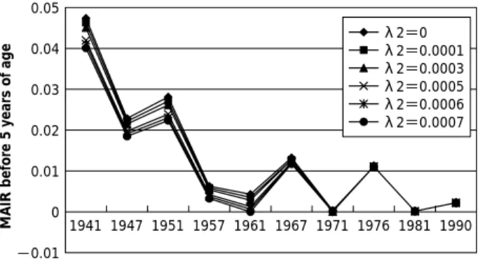
Figure 1 Longitudinal trends of MAIR (mean annual infec- tion rate) of female in Hyogo prefecture.
Table 1 Seroprevalence of T. gondii in Kobe University Hospital
1981 1995
age MA YOB (mean) MAIR (%) age MA YOB (mean) MAIR (%)
1to9 10to19 20to29 30to39 40to49 50to59 60to69 70to79
5 14.5 24.5 34.5 44.5 54.5 64.5 74.5
1976 1966.5 1956.5 1946.5 1936.5 1926.5 1916.5 1906.5
1.110 0.449 0.124 0.328 0.438 0.324 0.471 0.386
1to9 10to19 20to29 30to39 40to49 50to59 60to69 70to79
5 14.5 24.5 34.5 44.5 54.5 64.5 74.5
1990 1980.5 1970.5 1960.5 1950.5 1940.5 1930.5 1920.5
0.201 0.000 0.000 0.059 0.313 0.433 0.446 0.334 (Calculated with the simple mathematical model, Y = exp (-λt), Y = seronegativity,
λ= mean annual infection rate, t = age)
MA: mean age, YOB: Year of birth, MAIR: mean annual infection rate in % 100
Year of birth
MAIR(%)
0.25 0.2 0.15 0.1 0.05 0 0.5
0.35 0.4 0.45
0.3
1995 1981 Estimation
1935 1940 1945 1950 1955 1960 1965
Year of birth
MAIR before 5 years of age
0.02 0.01 0
−0.01 0.05 0.04 0.03
λ2=0 λ2=0.0001 λ2=0.0003 λ2=0.0005 λ2=0.0006 λ2=0.0007
1941 1947 1951 1957 1961 1967 1971 1976 1981 1990
(statistically significant forβ< 0, p < 0.05)
95% confidence interval forγ: -0.1319 <γ< 0.1785 n = 5, the birth year from 1940. 5 to 1960. 5 in Table 1.
The real value and the estimated value of MAIR based on the regression model are illustrated in Fig. 2. The results indicate that MAIR is negatively correlated with the year of birth. However, no statistically significant evidence of dif- ferences or a shift of curves, let alone historical decrease, were noted between the data of 1981 and those of 1995.
We presume that the historical downward trends in MAIR mainly reflect a decline in the incidence of T. gondii infection among younger children. As shown in Table 1 and Fig. 1, MAIR of children for the age under 5 years sig- nificantly decreased (from 1.11% in 1981 to 0.201% in 1995). However,our regression model analyses do not indi- cate a statistically valid reduction in MAIR from 1981 to
1995 for women born between 1940 and 1960 (age vari- ation from 21 to 50 years). These results imply that MAIR changes historically during childhood rather than adulthood.
We simulated MAIR of children for the age under 5 years, as explained in Methods, in which we varied λ2be- tween 0% and 0.07% (Table 2 and Fig. 3). Figure 3 implies that MAIR for the age under 5 years basically decreased from 1940 until 1960, and may heve been fluctuating cycli- cally within a range of 0% to 1%, with an approximate 10- year interval, since 1960.
DISCUSSION
A number of reports demonstrate historically declining trends in the incidence of T. gondii infection in Europe (Ades and Nokes, 1993; Nokes et al., 1997). In these re- ports, reasons for the decrease in incidence include increas- ing consumption of frozen meat, improvement in hygiene, and the increased use of processed and sterilized cat food, among others. In our present report, it is noteworthy that the term of MAIR reduction (from 1940 to 1960) includes 1945, the year of the end of World War II. Dramatic social changes related to the war as well as society reconstruction during the aftermath may have affected the decline in MAIR through this period.
An epidemiological study on Helicobacter pylori by Japanese scientists presents an interesting viewpoint on the correlation between the year of birth and incidence of infec- tious diseases in Japan (Asaka et al., 1992). This study re- ported that seropositivity for H. pylori is higher in individu- als born before 1950. They attributed this decline in sero- positivity to the Westernization of Japan. Although many differences are prevalent between H. pylori and T. gondii, including species and infection routes, epidemiological studies on the bacteria may provide new perspectives for further research on the causes of historical reduction in Figure 2 MAIR (mean annual infection rate): real vaues and
estimations.
Figure 3 Simulation of MAIR for female before 5 years of age.
λ2: Hypothetical value of MAIR after 5 years of age, varied between 0% and 0.07%.
Table 2 Simulation of MAIR before 5 years of age λ2 1940.5 1946.5 1950.5 1956.5 1960.5 0
0.0001 0.0003 0.0005 0.0006 0.0007
0.0471 0.0462 0.0452 0.0422 0.0412 0.0402
0.0226 0.0220 0.0215 0.0197 0.0190 0.0185
0.0279 0.0271 0.0263 0.0239 0.0231 0.0223
0.00609 0.00570 0.00531 0.00414 0.00375 0.00336
0.00404 0.00345 0.00286 0.00109 0.00050 (<0) λ2 1966.5 1970.5 1976 1980.5 1990 0
0.0001 0.0003 0.0005 0.0006 0.0007
0.0130 0.0128 0.0126 0.0121 0.0119 0.0117
0 0 0 0 0 0
0.0111 0.0111 0.0111 0.0111 0.0111 0.0111
0 0 0 0 0 0
0.00201 0.00201 0.00201 0.00201 0.00201 0.00201
*λ2: MAIR (mean annual infection rate) after 5 years of age.
101
MAIR by the protozoan.
Based on our simulation, possible periodical outbreaks of T. gondii infection among young people are speculated.
This speculation does not contradict our clinical experi- ences, which indicate periodic outbreaks of CT. Mothers are generally expected to be exposed to the same risk factors for T. gondii infection as their younger offspring as they care for them, and therefore T. gondii infection via contact with oocysts in cats’feces or contaminated soil is a signifi- cant threat for women as well as children. From the stand- point of risk management, public preventive measures, based on comprehensive research on infection routes for mothers and children, behavioral patterns and lifestyles are required.
From the viewpoint of strict statistical analysis, how- ever, our simulation may face some criticism, mainly on the original data (Takahashi et al., 1985; Win et al., 1997), as follows:
(1) The seroprevalence data of T. gondii used in this simu- lation are of outpatients in a certain hospital, not of randomly selected samples from the general people in Kobe prefecture.
(2) The data of 1981 and those of 1995 are not prospective but cross-sectional data.
(3) For obtaining the data, self-identification was not exe- cuted.
(4) The number of samples is 100 by each age category in the 1995 survey, while it varies from 7 to 149 in the 1981 survey. Hence, for devising the simulation, inter- val estimation rather than point estimation will be pre- ferred.
Currently in Japan, starkly different from the European nations, seroprevalence data of T. gondii infection are scarcely available, not to speak of those of randomly se- lected samples. While clinical cases of CT are reported in Japan, the recognition of the disease is still less prevalent even among medical practitioners here than in Europe.
Therefore, new analytical methods, which are useful for re- searchers of CT to enrich their understanding about the dis- ease even without enough data, are required. We believe that our new approach would be one of those methods. And we hope that our simulation, an example of analysis with MAIR, would be of use to find new perspectives for future epidemiological studies, albeit some controversies over original data and the way of estimation.
We believe that MAIR is an effective tool in mathe- matical analysis, which reflects the history of infection as a whole and presents an average of incidence in the past. Al-
though in the current analysis, it is simply presumed that MAIR alterations mainly feflect changes during childhood, other assumptions based on comprehensive epidemiological studies in the future are also possible. On the basis of these presumptions, conclusions distinct to those of the present study may be drawn.
Current model analyses are made in an age-and time- dependent manner by European statisticians, based on sev- eral decades of seroprevalence data (Ades and Nokes, 1993).
However, these kinds of data are not available in Japan. Al- though continuous sampling studies of seroprevalence are requested, it may take numerous more decades in Japan to obtain data extensive enough for similar statistical analyses as those in Europe. In the meantime, MAIR and related methods for mathematical speculations on T. gondii infec- tion are effective, and the current procedures of choice in Japan.
REFERENCES
1)Ades, A. E. and Nokes, D. J. (1993): Modeling age- and time-specific incidence from seroprevalence: Toxoplas- mosis. Am. J. Epidemiolo., 137, 1022-1034
2)Asaka, M., Kimura, T., Kudo, M., Takeda, H., Mitani, S., Miyazaki, T., Miki,K. and Graham, D. Y. (1992): Rela- tionship of Helicobacter pylori to serum pepsinogens in an asymptomatic Japanese population. Gastroenterology, 102, 760-766
3)Dollfus, H., Dureau, P., Hennequin, C., Uteza, Y., Bron, A. and Dufier, J. L. (1998): Congenital toxoplasmosis transmitted by preconceptionally immune women. Brit J Ophtalmol., 82, 1444
4)Dubey, J. P. and Beattie, C. P. (1988): Toxoplasmosis of Animals and Man. Florida: CRC Press. 41-60.
5)Naoi, K. and Yano, A. (2002): A theoretical analysis of the relations between the risk of congenital toxoplasmosis and the annual infection rates with a convincing argu- ment for better public intervention. Parasitol. Int., 51, 187-194
6)Nokes, D. J., Forsgren, M., Gille, E. and Ljungström,!. (1997): Modelling toxoplasma incidence from longitudi- nal seroprevalence in Stockholm, Sweden. Parasitology, 107, 33-40
7)Takahashi, J., Konishi, E. and Matumura, T. (1985): A sur- vey of antibodies to Toxoplasma gondii among patients of a hospital in Hyogo prefecture, Japan, by Enzyme- Linked Immunosorbent Assay. Jpn. J. Parasitol, 34, 47- 52
8)Win, K. S., Matsumura, T., Kumagai, S., Uga, S. and Kon- ishi, E. (1997): Prevalence of antibody to Toxoplasma gondii in Hyogo prefecture, Japan: Comparison at 10- year interval. Kobe J. Med. Sci., 43, 159-168
102
CHARACTERIZATION OF
VIBRIO CHOLERAE O1 ISOLATED IN VIETNAM
NGUYENBINHMINH1, NAOMIHIGA2, SHIGEKAKINOHANA3, and MASAAKIIWANAGA2 Accepted 9, March, 2002
Abstract: V. cholerae O1 isolated in Vietnam in 1995 and 2000 were characterized. Most of the isolates in 2000 showed moderate resistance to tetracycline and chloramphenicol, and strong resistance to sulfamethoxazol- trimethoprim. The susceptibilities to ampicillin, erythromycin, nalidixic acid and ofloxacin were not very different from those of the isolates in 1995. V. cholerae O1 strains showing this kind of drug susceptibility pattern are unique to the seventh cholera pandemic, and they have become widely distributed in Southeast Asian countries.
Genetic analysis of the strains from Vietnam by arbitrarily primed polymerase chain reaction suggested that they might be clonal derivatives. In prophage type, 6 of 20 strains (30%) in 1995 and 1 of 24 strains (4%) in 2000 be- longed to Ubol type. Considering the current spreading of V. cholerae O1 with such unique susceptibility to the drugs, we recommend careful monitoring of the drug susceptibility of V. cholerae O1 throughout the world.
Key words: Vibrio cholerae O1, drug susceptibility, Vietnam
INTRODUCTION
It has been more than 40 years since the seventh chol- era pandemic started in 1961. During that period, the re- sponsible pathogen, Vibrio cholerae O1 El Tor has under- gone some changes in its biological behavior. This pathogen was first distinguished from classical cholera vibrios based on its hemolytic property, and thereafter, some other unique properties of El Tor vibrios were described (Feeley, 1966).
These unique properties include the following: El Tor vi- brios are resistant to polymyxin B and cholera phage IV, produce acetoin from glucose (positive VP reaction), and agglutinate chicken erythrocytes when grown on nutrient agar plates. Soon after the beginning of this pandemic, the hemolytic property, as examined by Feeley and Pittman’s routine method of testing (Feeley and Pittman, 1963), dis- appeared in most isolates, but it reappeared in African iso- lates in the 1980s (Iwanaga et al., 1982). Cholera phage IV- sensitive El Tor vibrio appeared in Thailand in 1986 (Iwanaga et al. 1989). Drug resistant vibrios have appeared and disappeared (Ichinose et al., 1986; Glass et al., 1980;
Mhalu et al., 1979). Recently, the isolates in Laos were well characterized, and unique phenotypes such as polymyxin B sensitive and moderately resistant to tetracycline were found (Toma et al., 1997; Phantouamath et al., 2001). Poly-
myxin B sensitive strains were isolated in Southern Laos facing the border to the central area of Vietnam. However, general characterization of V. cholerae O1 in Vietnam has not been carried out yet. In this study, we characterized V.
cholerae O1 isolated in Vietnam in 1995 and 2000 in com- parison with the previously characterized strains in the neighboring country, Laos, in an attempt to understand the transmission of cholera through the border.
MATERIALSANDMETHODS 1. Bacterial strains:
Forty-four strains of V. cholerae O1 isolated from cholera patients in Vietnam were used. Twenty strains were isolated in 1995 and 24 were isolated in 2000. Several strains of Lao isolates in 2000 were used for the compara- tive studies. The strains H218 (biotype classic, serovar Ogawa) and UDT109 (non-O1 V. cholerae) were used as the indicator organisms for kappa phage and fs phages, re- spectively. Identification of V. cholerae O1 was made based on standard laboratory tests.
2. Hemolytic property:
The hemolytic property was examined using Feeley and Pittman’s method (Feeley and Pittman, 1963) and sheep blood agar plate cultures with one point stabbing inocula-
Jpn. J. Trop. Med. Hyg., Vol.30, No.2,2002, pp.103-107
1. National Institute of Epidemiology and Hygiene, Hanoi, Vietnam 2. Department of Bacteriology, 3. Department of Fundamental Nursing,
Faculty of Medicine, University of the Ryukyus, Okinawa 903-0215, Japan
103
tion. Strains that appeared to be non-hemolytic based on these methods were further examined by using heart infu- sion broth (HIB) supplemented with 1% and 3% glycerol following the same procedures as in Feeley and Pittman’s method.
3. Production of acetoin
The organisms were inoculated into VP-semisolid agar (Eiken) and cultured at 37°C for 24 hours. Production of acetoin was then examined by pouring a few drops of 5%
alpha-naphthol and 40% potassium hydroxide over the cul- tures, and assessing the development of red color.
4. Production of and sensitivity to phages:
To determine the prophage type of the isolates, kappa phage was examined as described by Takeya and Shimodori (1963). Production of the filamentous phages fs-1 and fs-2 was examined by using the indicator strain V. cholerae UDT109. V. cholerae O1 isolates were cultured in HIB (10 ml in 100-ml Erlenmeyer flasks) at 37°C overnight with re- ciprocal shaking, and the culture supernatants were used as the samples for detection of fs-1 and/or fs-2. The indicator strain was cultured overnight in broth, and 0.1 ml of the cul- ture fluid was mixed with 4 ml of soft agar medium (1%
peptone, 0.5% yeast extract, 0.5% NaCl, 0.6% agar, pH 7.2) and overlaid on a glass slide. Ten microlitters of this sample was dropped on to a layer of the indicator strain and incu- bated at 37°C overnight. Sensitivity to the filamentous phages (fs-1 and fs-2), cholera phage IV, and El Tor phage 5 was examined according to the method described by Ikema and Honma (1998).
5. Drug susceptibility:
The minimum inhibitory concentration (MIC) of each drug was examined by the plate dilution technique. The drugs examined were ampicillin (Meiji), tetracycline (Nacarai), chloramphenicol (Wako), erythromycin (Dainip- pon), ofloxacin (Daiichi), nalidixic acid (Wako), sulfamethoxazol-trimethoprim (sulfamethoxazol: Wako, trimethoprim: Sigma) and polymyxin B (Pfizer). A series of heart infusion agar plates containing serial 2-fold dilutions of the drugs at concentration ranging from 100µg/ml to 0.1 µg/ml were prepared. In the case of ofloxacin, the dilution series was extended to 0.0125µg/ml. Sulfamethoxazol and trimethoprim (19:1) were mixed, and the concentration of the drug was expressed as the total drug weight / ml of the mixture. A dilution series from 640µg/ml to 0.078µg/ml of the drug combination was made in agar plates. A10-fold di- lution of overnight broth culture was inoculated onto each plate using a Micro planter (Sakuma Co., model MITT # 00257), and incubated at 37°C for 24 hours. The susceptibil- ity was expressed as the MIC of each drug.
6. Arbitrarily primed polymerase chain reaction (AP-PCR) The clonal variation of the isolates was evaluated
based on the pattern of the products of AP-PCR obtained using a random primer AP42 (5’-CCGCAGCCAA-3’) and AP46 (5’-GAGGACAAAG-3’) as described by Akopyanz et al. (1992). The PCR conditions were 45 cycles of 94°C: 2 min, 38°C: 2 min, 72°C: 2 min, with 3 min of transitional time when raising the temperature from 38°C to 72°C, fol- lowed by a final incubation at 72°C for 10 min. The PCR products were electrophoresed on a 2% agarose gel for 2 hr at 50 V in an ice box. Then the gel was stained with ethid- ium bromide, and observed under a UV illuminator.
RESULTS
General properties of the isolates:
All isolates showed the following typical properties of V. cholerae O1: motile Gram-negative curved rods, yellow colonies on TCBS agar (sucrose fermentative), glucose fer- mentation without gas production, lactose non-fermentative, cytochrome oxidase positive, lysine decarboxylase positive and arginine hydrolase negative.
Hemolysis:
Although the hemolytic activity was generally very weak, 19 of 20 isolates (95%) in 1995 and 18 of 24 isolates (75%) in 2000 were hemolytic on sheep blood agar. When they were examined by Feeley and Pittman’s method, 18 of 20 (90%) in 1995 and 12 of 24 (50%) in 2000 were hemo- lytic. The strains which did not show hemolysis in either method were cultured in heart infusion broth supplemented with 1% and 3% glycerol, but the hemolysis did not appear under these conditions, either.
Production of acetoin:
Examination of the production of acetoin in semi-solid VP medium (Eiken) revealed that 35% of isolates in 1995 and 62.5% of isolates in 2000 were positive.
Production of and sensitivity to phages:
The isolates in 1995 included 12 strains that produced kappa phage (Celebes type), 2 that did not produce the phage but was sensitive to it (Cured Celebes type) and 6 that neither produced nor were sensitive to it (Ubol type).
The isolates in 2000 included 23 strains that produced kappa phage (Celebes type) and 1 that did not produce and was not sensitive to it (Ubol type). Most isolates (18 of 20) in 1995 were sensitive to filamentous phages fs-1 and/or fs- 2. Three isolates produced fs-2, but none produced fs-1.
None of the 24 isolates in 2000 produced fs phages, and 21 were not sensitive to them, while the other 3 isolates were sensitive only to fs-1 (Table 1).
Drug susceptibility:
The drug sensitivities of the isolates in 2000 were very different from those in 1995, as shown in Table 2 and Fig- ure 1. Although the sensitivities of the isolates in 1995 and 104
Table 1 Production of and sensitivity to phages
Phage 20 isolates in 1995 24 isolates in 2000
production sensitivity No. production sensitivity No.
kappa +
- -
- + -
12 2 6
+ - -
- + -
23 0 1
fs-1 +
- -
- + -
0 17
3
+ - -
- + -
0 3 21
fs-2 +
- -
- + -
3 13
4
+ - -
- - -
0 0 24
Table 2 (A) Frequency distribution of MIC for 20 isolates in 1995
µg/ml ABPC TC CP EM PLB NA OFLX µg/ml ST
0.0125≧ 8 0.08
0.025 7 0.156
0.05 3 0.313 14
0.1 1 1 0.625 1
0.2 5 17 1 1.25 4
0.39 14 2 2.5 1
0.78 18 5
1.56 4 2 10
3.13 13 1 1 20
6.25 3 7 40
12.5 12 1 80
25 5 160
50 4 320
100 2 640
100< 8 640<
(B) Frequency distribution of MIC for 24 isolates in 2000
µg/ml ABPC TC CP EM PLB NA OFLX µg/ml ST
0.0125≧ 4 0.08
0.025 14 0.156
0.05 6 0.313 1
0.1 0.625
0.2 20 1.25
0.39 1 2.5
0.78 1 4 5
1.56 10
3.13 23 21 20
6.25 1 2 20 40
12.5 3 24 80
25 160
50 320
100 3 640
100< 21 640< 23
105
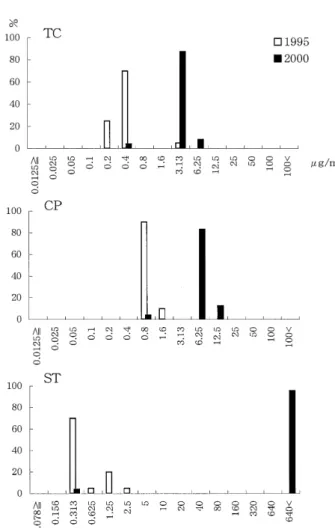
2000 to erythromycin, ampicillin, nalidixic acid and oflox- acin were almost the same, the isolates in 2000 were moder- ately resistant to tetracycline and chloramphenicol, and highly resistant to sulfamethoxazol-trimethoprim. The MIC of tetracycline against the isolates in 1995 was 0.4µg/ml or lower, except for 1 isolate against which it was 3.13µg/ml, whereas the MIC against the isolates in 2000 was 3.13 or 6.25µg/ml except for 1 isolate against which it was 0.4µg/
ml. Chloramphenicol showed almost the same pattern as that seen for tetracycline. The MIC of sulfamethoxazol- trimethoprim against the isolates in 1995 was in most case 0.313 µg/ml, but it was higher than 640µg/ml against the isolates in 2000.
AP-PCR:
Chromosome analysis by AP-PCR using primers AP42 and AP46 was performed to determine the genetic diversity among the isolates from different areas and years. The PCR products revealed almost the same pattern, regardless of dif- ferences of phenotype, the year of isolation and the place of
isolation, except in the case of 1 strain when the primer AP 46 was used. This strain was isolated in Vietnam in 1995.
The AP-PCR patterns of representative isolates are shown in Figure 2.
DISCUSSION
The general properties of V. cholerae O1 in Vietnam were clarified in this study. Although the isolates in 2000 were slightly different in hemolytic property, prophage type, and acetoin production from those in 1995, the changes of the drug- susceptibility patterns were notable. The charac- teristic drug susceptibility of the isolates in 2000 was that they were moderately resistant to tetracycline and chloram- phenicol, and highly resistant to sulfamethoxazol- trimethoprim, while the susceptibilities to ampicillin, eryth- romycin, nalidixic acid and ofloxacin were not very differ- ent from those of the isolates in 1995. These features were very similar to those of the isolates in Laos after 1998 (Phantouamath et al ., 2000), and also to those of the iso- lates in Thailand (unpublished data), except that polymyxin B sensitive strains were not found in Vietnam and Thailand.
Since no plasmid was detected in the Lao isolates (Iwanaga et al. 1999) as well as in the Vietnam isolates (data not shown) and the phenotype of the Vietnam isolates was very similar to that of the Lao isolates, this specific susceptibility pattern may have been mediated by a chromosomal gene.
Previously reported tetracycline-resistant V. cholerae O1 strains isolated in a cholera epidemic were highly resistant Fig. 1 Distribution of MIC of tetracycline (TC), chloram-
phenicol (CP) and sulfamethoxazol-trimethoprim (ST).
The frequency is expressed by per cent.
Fig. 2 AP-PCR pattern. Lane1:molecular marker (100-bp lad- der), 2: strain 00Vie27 (Vietnam isolate in 2000), 3:
strain 00LA4 (Lao isolate in 2000), 4: strain LA1 (Lao isolate in 2000, polymyxin B sensitive), 5: strain Vie47 (Vietnam isolate in 1995), 6: strain Vie156 (Vietnam isolate in1995). The left half of the panel shows the re- sult obtained using primer AP42, and the right using AP46.
106
to tetracycline as well as to other drugs; the resistance was mediated by a plasmid, and the resistant strains disappeared soon after the epidemic (Glass et al., 1980). However, the chromosomally mediated resistance in organisms detected in the present study could be persistent in future epidemics.
Indeed, such strains have spread in Southeast Asian coun- tries since 1998.
Their indistinguishable phenotype and genotype imply that it is likely that the distribution and spread of these drug resistant cholera vibrios are due to the same clone. It is also suggested that the resistant strains arose from the mutation of previously sensitive strains and were not a new clone coming from some other source. One strain isolated in Viet- nam in 1995 had a slightly different AP-PCR pattern (Fig. 2, lane 5 in AP46), and this strain was moderately sensitive to polymyxin B (MIC = 12.5 µg/ml), but its AP-PCR pattern was not identical to those of strains highly sensitive to poly- myxin B isolated in Laos (Fig. 2, lane 4 in AP46). There- fore, the origin of the polymyxin B sensitive El Tor vibrios in Laos has still not been clarified.
In the present study of cholera due to organisms mod- erately resistant to tetracycline, clinical data about the prog- nosis of the patients treated with ordinary doses of tetracy- cline was not available, but it is likely that the diarrhea lasted longer than the diarrhea caused by sensitive organ- isms. If the use of tetracycline continues, there is a possibil- ity that highly resistant clones will develop. Considering the current spreading of these organisms in Southeast Asian countries, careful monitoring of the drug susceptibility throughout the world is required.
This study was done under the Core University Program, Tropical Medicine, sponsored by Japan Society for the Promortion of Science.
REFERENCES
1)Akopyanz, N., Bukanov, N.O., Westblom, T.U., Kreso- vich, S., and Berg, D.E. (1992): DNA diversity among clinical isolates of Helicobacter pylori detected by PCR- based RAPD fingerprinting. Nucleic Acid Res. 20, 5137- 5142
2)Feeley, J. C. (1966): Minutes of IAMS committee on tax- onomy of Vibrios. Intern. J. Syst. Bacteriol. 16, 135-142 3)Feeley, J. C., Pittman, M. (1963): Studies on hemolytic
activity of El Tor vibrios. Bull. Wld. Hlth. Org. 28, 347- 356
4)Glass, R. I., Huq, I., Akim, ARMA., Yunus, M. (1980):
Emergence of multiply antibiotic-resistant Vibrio chol- erae in Bangladesh. J. Infect. Dis.142, 939-942
5)Ichinose, Y., Ehara, M., Watanabe, S., Shimodori, S., Waiyaki, P.G., Kibue, A.M., Sang, F.C., Ngugi, J., and Kaviti, J.N. (1986): The characterization of Vibrio chol- erae isolated in Kenya in 1983. J. Trop. Med. Hyg. 89, 269-276
6)Ikema, M. and Honma, Y. (1998): A novel filamentous phage, fs2, of Vibrio cholerae O139. Microbiology 144, 1901-1906
7)Iwanaga, M., Mori, K., Kaviti, J. N. (1982): Vibrio chol- erae O1 in Kenya. J. Clin. Microbiol. 16, 742-743 8)Iwanaga, M., Nakamura, S., Kuyyakanond, T. (1989):
Hemolytic Vibrio cholerae O1 that is sensitive to Muker- jee’s cholera phage IV and the phages produced by the hemolytic vibrio lysogenized with the infection of Muk- erjee’s cholera phage IV. Microbiol. Immunol. 33, 609- 618
9)Mhalu, F. S., Mmari, P. W., Ijumba, J. (1979): Rapid emergence of El Tor Vibrio cholerae resistant to antimi- crobial agents during the first six months of fourth chol- era epidemic in Tanzania. Lancet 1, 345-347
10)Phantouamath, B., Sithivong, N., Sisavath, L., Munnalath, K., Chomlasak, K., Insisiengmay, S., Higa, N., Kakino- hana, S., Iwanaga, M. (2001): Transition of drug suscep- tibilities of Vibrio cholerae O1 in Lao People’s Demo- cratic Republic.
Southeast Asian J. Trop. Med. Pub. Hlth. 32, 96-99 11)Takeya, K. and Shimodori, S. (1963): “Prophage typing”
of El Tor vibrios. J. Bacteriol. 99, 339-340
12)Toma, C., Sithavath, L., Higa, N., Iwanaga, M. (1997):
Characterization of Vibrio cholerae O1 isolated in Lao People’s Democratic Republic. Jpn. J. Trop. Med. Hyg.
25, 85-87
107
Monitoring of drug resistant Staphylococcus aureus in People’s
Democratic Republic of Lao
NOIKASEUMSYSITHIVONG1, BOUNNANHPHANTOUAMATH1, KHAMPHENGCHOMLASAK1, LAYSISAVATH1, KHAMPHYEUMUNNALATH1, SITHATINSISIENGMAY1
NAOMIHIGA2, SHIGEKAKINOHANA3, MASAAKIIWANAGA2 Accepted 31, May, 2002
Abstract: Drug susceptibility of Staphylococcus aureus has been examined in the past 9 years to monitor the ap- pearance of drug resistant strains especially methicillin resistant S. aureus (MRSA). The pattern of drug suscepti- bility has been relatively constant, but S. aureus is gradually becoming resistant to ampicillin. One MRSA was isolated in 1996 from the nasal vestibulum of a non-infected patient at Mahosot Hospital but not isolated thereafter.
In 2001, 2 strains of MRSA which belonged to different clones were first isolated from the infection foci of pa- tients. One was isolated from Setthathirath Hospital, where many Japanese staff have been dispatched since 2000.
Careful monitoring and epidemiological studies of MRSA are recommended.
Key words: Staphylococcus aureus, drug susceptibility, Laos
INTRODUCTION
Staphylococcus aureus is one of the most important pathogens throughout the world. Because S. aureus easily develop resistance to antibiotics when these drugs are used to treat infections, much attention has been focused on the appearance of drug-resistant strains. In the past 2 decades, the incidence of methicillin-resistant Staphylococcus aureus (MRSA) has been an especially hot issue in the clinical field. The proportion of MRSA among S. aureus isolates varies from country to country. One report showed that the proportion was 1.5% in the Netherlands but 25.1% in Bel- gium, a neighboring country, and 33.6% in France (Voss et al ., 1994). In Thailand, which borders the People’s Demo- cratic Republic of Lao (Lao PDR or Laos), a high propor- tion of MRSA was reported (Wongwanich et al ., 2000). It is a well-known fact that the proportion of MRSA among S.
aureus isolates from hospitalized patients in Japan is over 50%.
Antibiotic availability was very restricted in Laos until the end of the 1980s and there was almost no problem of drug-resistant pathogens until then. Once hospitals become heavily contaminated with MRSA, eradication is almost im- possible (Thompson et al ., 1982). Therefore, prevention of the development of MRSA in the hospital is a critical prob-
lem. There must currently be an increased risk of develop- ing drug-resistant pathogens in Laos, since the kinds of an- tibiotics available and their consumption were steadily in- creased from 1990 until the present. Because we antici- pated the development resistant strains, we have been moni- toring the drug-resistant S. aureus in Laos since 1993. In this communication, we described the results of 9 years of monitoring of drug-resistant S. aureus in Laos.
MATERIALSANDMETHODS
Bacterial strains
S. aureus used in this study was isolated from clinical specimens submitted to Center for Laboratory and Epidemi- ology, Vientiane, Laos. The specimens included swabbed mateials (lesion of skin, throat, ear, or vagina), sputum, urine, aspirated fluid, etc. The number of isolates was 54 in 1993, 32 in 1995, 47 in 1996, 56 in 1997, 72 in 1998, 59 in 1999, 35 in 2000, and 61 in 2001 (416 in total).
Drug susceptibility
The susceptibility was expressed as the minimum in- hibitory concentration (MIC) of the following 5 drugs as examined by the plate dilution method. The drugs exam- ined were methicillin (DMPPC, Banyu), oxacillin (MPIPC,
Jpn. J. Trop. Med. Hyg., Vol.30, No.2,2002, pp.109-113
1 Center for Laboratory and Epidemiology, Ministry of Health Vientiane, Laos 2 Department of Bacteriology,
3 Department of Fundamental Nursing Faculty of Medicine, University of the Ryukyus, Okinawa 903-0215, Japan
109
Wako) as a substitute for methicillin, ampicillin (ABPC, Meiji), tetracycline (TC, Nacarai), erythromycin (EM, Dainippon), and cefdinir (CFDN, Fujisawa). A series of heart infuion agar plates containing serial 2-fold dilutions of the drugs at concentrations ranging from 100µg/ml to 0.1 µg/ml or lower were prepared. A 10-fold dilution of an overnight broth culture of the S. aureus strains indicated in the text was inoculated onto each plate using a Microplanter (Sakuma Co. model MITP #00257) and incubated at 37°C for 24 hours. The MICs of methicillin and oxacillin were determined using agar plates containing 2% NaCl and cul- tured at 30°C.
Detection of mecA gene
The mecA gene was detected by polymerase chain re- action (PCR). The PCR templates samples were prepared from the colonies on a nutrient agar plate by emulsifying a few colonies in distilled water and boiling them for 20 min.
The PCR primers designed to amplify a 630 bp fragment derived from a portion of mecA, 5’-GAACCTCTGCTCAA CAAGTT-3’ and 5’-GGATTTGCCAATTAAGTTTG-3’, were used as described by Song et al . (1987). The PCR conditions consisted of 30 cycles of 94°C for 1 minute, 52°C for 1 minute, and 72°C for 1 minute, and a final incubation at 72°C for 10 minutes.
Detection of PBP2’
The expression of mecA was examined by detecting the gene product, PBP2’. A kit for the detection of PBP2’, MRSA Screen (Denka Seiken Co., Tokyo) was used accord- ing to the instructions in the manual supplied with the kit.
Coagulase typing
Coagulase type was determined by neutralization test with the type-specific antiserum. A Coagulase Typing Im- mune Sera Kit (Denka Seiken Co. Tokyo) was used to clas- sify the antigenic type of coagulase produced by the isolates.
Coagulation inhibition with the type-specific anti-serum was examined using plastic microdilution plates as de- scribed by Tajima et al . (1992).
Detection of toxins
Production of Staphylococcal enterotoxins (SE) in- cluding the antigenic types A, B, C, D, and E was examined by using a SET-RPLA kit (Denka Seiken Co., Tokyo) for the detection and typing of staphylococcal enterotoxins.
Toxic shock syndrome toxin-I (TSST-I) production was ex- amined by using a TST-RPLA kit (Denka Seiken Co., To- kyo). The detection was performed according to the in- structions given in the manuals supplied with each kit.
RESULTS
Drug susceptibilities
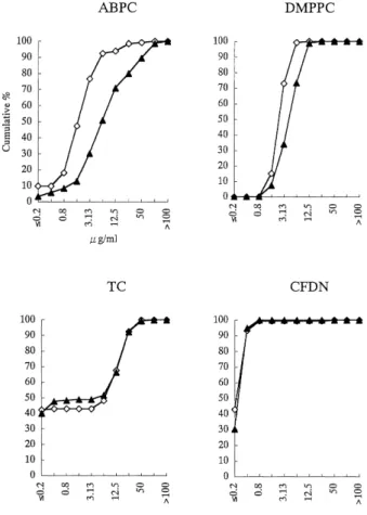
The MICs of ampicillin and methicillin gradually be- came higher during the past 8 years. This gradual change of drug susceptibility was shown by determining the cumu- lative distribution of MIC (Fig.1). The susceptibility of S.
aureus to erythromycin varied from year to year. All iso- lates were highly sensitive to cefdinir throughout the period of monitoring (Fig. 1). The MIC of tetracycline became slightly lower in the past few years. Taken altogether, the MICs values reveal that the drug susceptibility pattern has not much varied so far. The raw data regarding MIC are presented in Table 1.
Among the isolates obtained in 2001, there were 3 strains against which the MIC of oxacillin was 12.5 or 100 µg/ml, while the MIC against the other isolates was lower than 3.13 µg/ml. The MICs of oxacillin, ampicillin, tetra- cycline, erythromycin and cefdinir against these 3 strains were: 12.5, 6.25, 12.5, 0.2, and 0.8µg/ml, respectively, for strain 01LP32; 200, 200, 100, 6.25, 200µg/ml, respectively, for strain 01LP40; and 12.5, 200, 25, 0.4, 0.8µg/ml, respec-
Fig. 1: Cumulative frequency distribution (%) of MIC.
―◇―: total of the isolates in 1993, 1995, and 1996
―▲―: total of the isolates in 1999, 2000, and 2001 Data for DMPPC do not include 2001
110
Table 1 MIC of each of the indicated drugs in the past 9 years
The frequency distribution of MIC is expressed as the percentage of total isolates in each year (The number of total isolates for each year is shown in the bottom line).
DMPPC
93 95 96 97 98 99 0
!0.2 0 0 0 0 0 0 0
0 0 0 0 0 0 0 0
1 0 0 0 0 0 0 0
2 28 3 9 0 4 10 3
3 65 59 49 2 52 41 3
6 7 38 40 64 44 46 29
13 0 0 2 32 0 3 62
25 0 0 0 0 0 0 3
50 0 0 0 0 0 0 0
100 0 0 0 0 0 0 0
>100 0 0 0 2 0 0 0
number 54 32 47 56 72 59 35
TC
93 95 96 97 98 99 0 1
!0.2 42 28 51 0 3 24 45 51
0 0 0 2 30 31 12 3 7
1 0 0 0 0 30 2 0 0
2 0 0 0 0 0 2 0 0
3 0 0 0 0 3 0 0 0
6 0 22 0 0 1 3 6 0
13 24 25 11 4 0 17 20 10
25 28 25 21 25 19 26 17 30
50 6 0 15 36 13 14 9 0
100 0 0 0 5 0 0 0 2
>100 0 0 0 0 0 0 0 0
number 54 32 47 56 72 59 35 61
CFDN
93 95 96 97 98 99 0 1
!0.2 26 34 68 16 14 42 9 31
0 68 50 30 61 79 58 74 66
1 6 13 2 21 7 0 17 3
2 0 0 0 0 0 0 0 0
3 0 0 0 0 0 0 0 0
6 0 0 0 0 0 0 0 0
13 0 0 0 0 0 0 0 0
25 0 0 0 0 0 0 0 0
50 0 3 0 0 0 0 0 0
100 0 0 0 0 0 0 0 0
>100 0 0 0 2 0 0 0 0
number 54 32 47 56 72 59 35 61
ABPC
93 95 96 97 98 99 0 1
!0.2 11 19 2 4 4 5 0 3
0 0 0 0 2 5 2 3 3
1 6 13 9 7 3 5 3 0
2 11 6 66 11 5 10 3 0
3 35 37 17 28 5 25 14 11
6 28 19 0 27 14 14 34 20
13 0 6 0 4 36 5 26 32
25 7 0 4 5 17 7 6 13
50 2 0 0 7 4 12 0 13
100 0 0 2 5 4 15 11 2
>100 0 0 0 0 3 0 0 3
number 54 32 47 56 72 59 35 61
EM
93 95 96 97 98 99 0 1
!0.2 87 85 34 4 82 19 17 7
0 7 6 2 85 3 52 72 83
1 0 0 49 2 0 0 0 0
2 0 0 13 0 0 0 0 0
3 2 0 0 0 0 0 0 0
6 0 0 0 0 0 0 0 2
13 2 0 0 0 0 0 0 0
25 0 0 2 0 0 0 0 0
50 0 0 0 0 0 0 0 0
100 0 0 0 2 0 0 0 0
>100 2 9 0 7 15 29 11 8
number 54 32 47 56 72 59 35 61
111
tively, for strain 01LP63. The production of PBP2’ and the presence of the mecA gene were examined in these 3 strains.
Strain 01LP40 was isolated from the discharge from the middle ear of an 8-year-old female patient with otitis media (Friendship Hospital), and 01LP63 was isolated from the sputum of a 13-year-old male patient with a respiratory in- fection (Setthathirath Hospital). These 2 strains had the mecA gene, as demonstrated by the PCR (Fig. 2), and they expressed PBP2’ as examined by the agglutination test.
Strain 01LP32 was negative for both the gene and its prod- uct.
Characterization of MRSA strains 01LP40 and 01LP63 The biochemical characteristics of these MRSA strains were typical of S. aureus, and included fermentation of lac- tose, mannitol and maltose, and production of acetoin. The coagulase of 01LP40 did not belong to any of the 8 known types (I to VIII), and the strain did not produce enterotoxin or toxic shock syndrome toxin. However, 01LP63 pro- duced enterotoxin type B and type D but did not produce toxic shock syndrome toxin. The coagulase of 01LP63 was classified as type II.
DISCUSSION
This is the first report of MRSA isolated from the in- fection foci of patients in Laos. Although we reported an isolate of MRSA from the nasal vestibulum of a nonin- fected patient in Laos in 1996 (Iwanaga et al ., 1997), MRSA has not been found in the infection foci until this study. Considering the pattern of use of antimicrobial agents in Laos in the past, the incidence of MRSA is ex- pected to be very low. However, recently, many Laotians
have received medical care in neighboring Thailand, where MRSA is routinely isolated (Wongwanich et al., 2000), and have then returned to Laos. Moreover, numerous antibiotics are imported and their use has increased. As a result, there is a high risk of the appearance of MRSA or other highly drug-resistant pathogens. The fact that 2 strains of MRSA appeared in 2 different hospitals warns us of the increase of MRSA in Laos.
Ampicillin and tetracycline have been the major antibi- otics used in Laos, and this pattern of antibiotic use may ex- plain why the susceptibility of S. aureus to ampicillin is gradually getting resistant. However, the susceptibility to tetracycline has not changed much, and may even have got sensitive in the past few years. Although it seems that the use of tetracycline decreased in Laos after 1998, when tetra- cycline resistant Vibrio cholerae O1 appeared, there is no reliable data on the rate of use of tetracycline. Therefore, it remains unknown why S. aureus has become more sensitive to tetracycline in the past few years.
The 2 strains of MRSA isolated here showed different phenotypes, implying that they belonged to different clones.
One of the strains (01LP63) was isolated at a hospital which was donated by the Japanese government in 2000, and many Japanese staff members from a university hospital are working there under the name of International Cooperation.
Since coagulase type!is the major type of coagulase pro- duced by MRSA in Japan (Matsumoto et al., 1989; Kimura et al., 1993), it is possible that 01LP63 was carried from Ja- pan to Laos by the hospital staff. It should be clarified whether this MRSA strain was carried to Laos by Japanese staff or not. Regardless of whether it was or not, hospital staff members have to be careful to avoid the improper use of antibiotics in order to prevent the development of MRSA in Laos, and to avoid carrying MRSA from Japan to Laos.
Careful monitoring of MRSA in Laos should be continued.
REFERENCES Fig.2: Detection of mecA gene by PCR
lane 1: Molecular size marker (100 bp ladder), lane 2: strain 01LP32, lane 3: strain 01LP40, lane 4: strain 01LP63, lane 5:
mecA-positive S. aureus as control
1)Iwanaga, M., Sithavath, L., Higa, N., Honma, Y., and Kakinohana, S. (1997): Emergence of methicillin resis- tant Staphylococcus aureus in Laos. Jpn J. Trop. Med.
Hyg., 25, 103-106
2)Kimura, A., Igarashi, H., Ushioda, H., Okuzumi, K., Ko- bayashi, H., Otsuka, T., (1993): Relationship between co- agulase toxin-type and drug susceptibility in Staphylo- coccus aureus strains isolated in all the Japanese Na- tional University Hospitals. Jpn. J. Infect. Dis. 67, 223- 230
3)Matsumoto, K., Takahashi, A., Shishido, H., Watanabe, K., Rikitomi, N., Suzuki, H. et al. (1989): A comparison of the changes in Staphylococcus aureus during two peri- 112
ods. Chemotherapy 37, 549-562
4)Song, M.D., Wachu, M., Doi, M., Ishino, F., and Matsu- hashi, M. (1987): Evolution of an inducible penicillin tar- get protein in methicillin-resistant Staphylococcus aureus by gene function. FEBS Lett. 221, 167-171
5)Tajima, Y., Nagasawa, Z., Tanabe, I., Yamada, H., Kusaba, K., and Tadano, J. (1992): An Improved method for the serotyping of free coagulase from Staphylococcus aureus.
Microbiol. Immunol. 36, 1233-1237
6)Thompson, R.L., and R.P. Wanzel. (1982): Editorial. In- ternational recognition of methicillin-resistant Staphylo-
coccus aureus. Ann. Intern. Med. 97, 925-926
7)Voss, A., Milatovic, D., Wallrauch-Schwarz, C., Rosdahl.
VT., Braveny, I., (1994): Methicillin-resistant Staphylo- coccus aureus in Europe. Eur. J. Clin. Microbiol. Infect.
Dis. 13, 50-55
8)Wongwanich, S., Tishyadhigama, P., Paisomboon, S., Ohta, T., Hayashi, H., (2000): Epidemiological analysis of methicillin resistant Staphylococcus aureus in Thai- land. Southeast Asian J. Trop. Med. Public Health 31, 72- 76
113
TAXONOMIC NOTES ON THE GRISEIFRONS SPECIES-GROUP IN SIMULIUM (SIMULIUM ) (DIPTERA: SIMULIIDAE) FROM
THAILAND: DESCRIPTIONS OF TWO NEW SPECIES AND DESCRIPTION OF THE MALE, PUPA AND LARVA OF
S . (S .) DIGRAMMICUM EDWARDS
HIROYUKITAKAOKA1and WEJCHOOCHOTE2 Accepted 28, May, 2002
Abstract: Two new black-fly species, Simulium maenoi sp. nov. and S. choochotei sp. nov. are described, based on the reared females, reared males, pupae and mature larvae collected in Chiang Mai Province, northern Thailand. In addition, the male, pupa and mature larva of S. digrammicum Edwards, 1928, originally described from Peninsular Malaysia and Thailand, are described for the first time. All these three species are assigned to the griseifrons species-group within Simulium (Simulium) Latreille s. str.
Key words: black fly, Simuliidae, Simulium, Thailand, new species, griseifrons species-group
The griseifrons species-group within the subgenus Simulium (Simulium) Latreille s. str. is characterized by the female scutum with five vittae, simple claws, the male style with a basal protuberance furnished with spinules, the ven- tral plate untoothed posteriorly, the pupal gill with six fila- ments per side, and the larval postgenal cleft deep, subtrian- gular or bullet-shaped (Takaoka and Davies, 1996). In the world list of black flies by Crosskey and Howard (1997), this species-group includes 10 species, all but one recorded from the Oriental Region. Apparently five other species placed in the multistriatum species-group by Crosskey and Howard (1997), and two more species left unplaced by Takaoka and Davies (1995; 1997) belong to the griseifrons species-group: i.e., S. horokaense Ono, 1980, S. kawamurae Matsumura, 1915, S. konakovi Rubtsov, 1956, S. kurilense Rubtsov, 1956, S. subornatoides Rubtsov, 1947 (all from Palaearctic Region), and S. rudnicki Takaoka and Davies, 1995, and S. yongi Takaoka and Davies, 1997 (both from Peninsular Malaysia and Thailand). In Thailand, the grisei- frons species-group is represented by four species, S. di- grammicum Edwards, 1928 (Edwards, 1928), S. grossifilum Takaoka and Davies, 1995, S. rudnicki and S. yongi (Ku- vangkadilok and Takaoka, 2000), all originally described from Peninsular Malaysia.
During our recent surveys on aquatic stages of black flies in northern Thailand, we discoverd the pupae and lar-
vae of S. digrammicum, which was so far known only from female specimens, and found two new species assignable to the griseifrons species-group.
These new species are here described on the basis of the reared females, reared males, pupae and mature larvae;
in addition, the male, pupa and mature larva of S. digram- micum are described for the first time.
Collecting and rearing methods, as well as dissection of anatomical parts for description, were already mentioned in Takaoka (1983). The morphological features and terms used herein follow those of Crosskey (1969).
Holotype and most paratype specimens will be depos- ited at the Natural History Museum (BMNH), London, U.K.
Simulium (Simulium) maenoi Takaoka and Choochote, sp. nov.
DESCRIPTION. Female. Body length ca. 2.5 mm. Head.
Narrower than width of thorax. Frons black, white pruinose, shiny, widely bare except several dark stout hairs along each lateral margin; frontal ratio 1.4:1.0:1.2; frons-head ratio 1.0:
3.8. Fronto-ocular area (Fig. 1) moderately developed, sub- triangular in shape. Clypeus black, white pruinose, shiny, moderately covered with dark stout hairs except upper por- tion bare medially. Proboscis ca. 0.8×as long as clypeus.
Antenna composed of 2+9 segments, light to medium
Jpn. J. Trop. Med. Hyg., Vol.30, No.2,2002, pp.115‐132
1. Department of Infectious Disease Control, and Research Center for Asian and Caribbean Diseases, Oita Medical University, Hasama, Oita, 879 -5593, Japan
2. Department of Parasitology, Faculty of Medicine, Chiang Mai University, Chiang Mai 50200, Thailand
115
brown except scape, pedicel and most of 1st flagellar seg- ment yellow; 1st flagellar segment ca. 1.5×as long as 2nd one. Maxillary palp medium brown, composed of 5 seg- ments with proportional lengths from 3rd to 5th segments 1.0:1.1:2.3; 3rd segment (Fig. 2) of moderate size; sensory vesicle medium in size, ellipsoidal, with rugged surface, 0.37× length of 3rd segment, with large round opening apically. Maxillary lacinia with 13 or 14 inner and 14 or 15 outer teeth. Mandible with ca. 28 inner and 13 or 14 outer teeth. Cibarium (Fig. 4), at its posterior end, produced into pharynx as narrow projection covered with numerous indis- tinct minute tubercles. Thorax. Scutum dark brown, grey- pruinose, with 5 dark longitudinal unpruinose vittae (i.e., 1 narrow median, 2 rather broad submedian, and 2 rather broad lateral vittae), all united on prescutellar area when il- luminated from dorsally and viewed from anterodorsally (this color pattern reversed when viewed from behind);
scutum densely covered with yellow recumbent fine hairs, interspersed with long upstanding dark hairs on prescutellar area. Scutellum unpigmented, with long dark and short yel- low hairs. Postscutellum light brown, shiny, without hairs.
Pleural membrane bare. Ketepisternum longer than deep, and bare. Legs (coloration still incomplete, especially on femora and tibiae). Foreleg: coxa and trochanter whitish yellow; femur whitish yellow on basal 1/2, dark yellow on apical 1/2, with apical cap light brown; tibia whitish yellow except apical 1/5 medium brown; tarsus entirely medium brown; basitarsus, 2nd and 3rd tarsal segments moderately dilated; basitarsus ca. 5.5× as long as its greatest width.
Midleg: coxa medium brown; trochanter light brown except base yellow; femur yellow somewhat darkened apically, and with apical cap light brown; tibia whitish yellow except api- cal 1/4 dark brown; tarsus dark brown except basal 1/2 of basitarsus whitish yellow. Hind leg: coxa dark brown; tro- chanter whitish yellow; femur and tibia whitish yellow with apical cap dark brown; tarsus medium brown except a little more of basal 1/2 of basitarsus and basal 1/2 of 2nd tarsal segment whitish yellow; basitarsus (Fig. 5) nearly parallel- sided, ca. 6.7×as long as wide, ca. 0.7×and ca. 0.6×as wide as hind tibia and femur, respectively; calcipala distinct but shorter than wide; pedisulcus distinct. All tarsal claws simple, without subbasal or basal tooth. Wing. Length 2.2 mm. Costa with spinules and hairs; subcosta haired except apical 1/5 bare; basal section of radial vein bare; hair tuft at base of stem vein dark brown; basal cell absent. Abdomen.
Basal scale light brown with a fringe of long hairs; dorsal surface of 2nd segment pale yellow on anterior 3/4 with a pair of large dorsolateral whitish spots broadly connected to each other in middle; dorsal surface of posterior 1/4 of 2nd segment and those of other segments dark brown and with short hairs; tergites 3, 4 and 5 small, tergites 6-8 large and
shiny. Ventral surface of abdominal segment 7 without any sternal plate or any hair clusters. Genitalia (Figs. 7 and 8).
Sternite 8 well sclerotized, bare medially but with 18 or 19 long stout hairs as well as a few short slender hairs laterally on each side; anterior gonapophysis triangular in shape, membranous, covered with 8 or 9 short hairs as well as nu- merous microsetae; inner border slightly concave; narrow area along inner and posterior margins transparent, bare.
Genital fork of inverted-Y form, with well sclerotized stem;
arms slender, each with sclerotized apical bulge having a distinct projection directed anteriorly. Paraproct in ventral view nearly as long as wide, with distinct concavity on ven- tral surface along anterior margin; anteromedial surface of paraproct moderately sclerotized, and with a few short sen- silla; paraproct much produced ventrally, coverd with 33-35 short stout hairs on ventral and lateral surfaces. Cercus in lateral view rounded posteriorly, ca. 0.5×as long as wide, covered with many short hairs. Spermatheca nearly ovoid, well sclerotized with no definite reticulate pattern, with minute internal setae; tube and large area around tubal base unsclerotized; accessary tubes subequal in diameter to each other, and somewhat larger than major one.
Male. Body length 2.5-2.8 mm. Head. Width slightly wider than thorax. Upper eye consisting of large facets in 19 or 20 horizontal rows and in 18-20 vertical columns.
Clypeus brownish black, white pruinose, moderately cov- ered with dark stout hairs. Antenna composed of 2+9 seg- ments, dark brown except base of 1st flagellar segment somewhat pale; 1st flagellar segment elongated, ca. 2.2×
as long as 2nd flagellomere. Maxillary palp composed of 5 segments, with proportional lengths from 3rd to 5th seg- ments 1.0:1.2:2.5; 3rd segment (Fig. 3) of normal size; sen- sory vesicle small, globular or ellipsoidal, ca. 0.2×as long as 3rd segment, with small opening. Thorax. Scutum black, dull, uniformly and densely covered with golden- yellow recumbent fine hairs, interspersed with long upright hairs on prescutellar area; in certain angles of light, scutum with white-pruinose subquadrate spot on each shoulder, ex- tending posteriorly forming a white-pruinose broad band along each lateral margin up to base of wing. Scutellum brownish black, with several upright dark hairs as well as golden-yellow fine hairs. Postscutellum brownish black, shiny, without hairs. Pleural membrane and katepisternum as in female. Legs. Foreleg: coxa whitish yellow; tro- chanter light to medium brown; femur light brown except apical cap medium brown; tibia brownish black except me- dian large area of outer surface white and sheeny; tarsus en- tirely brownish black; basitarsus, 2nd and 3rd tarsal seg- ments somewhat dilated (basitarsus ca. 6.8×as long as its greatest width). Midleg: coxa dark brown to brownish 116