Prognostic value of vascular endothelial
growth factors‑A and ‑C in oral squamous cell carcinoma
著者 柳瀬 瑞希
著者別表示 Yanase Mizuki journal or
publication title
博士論文本文Full 学位授与番号 13301甲第4081号
学位名 博士(医学)
学位授与年月日 2014‑06‑30
URL http://hdl.handle.net/2297/42066
doi: 10.1111/jop.12167
Creative Commons : 表示 ‑ 非営利 ‑ 改変禁止 http://creativecommons.org/licenses/by‑nc‑nd/3.0/deed.ja
Prognostic value of vascular endothelial growth factors A and C in oral squamous cell carcinoma
Mizuki Yanase, Koroku Kato, Kunio Yoshizawa, Natsuyo Noguchi, Hiroko Kitahara, Hiroyuki Nakamura
Division of Cancer Medicine, Department of Oral and Maxillofacial Surgery, Kanazawa University Graduate School of Medical Science, Ishikawa, Japan
BACKGROUND: Vascular endothelial growth factor (VEGF) family members play a major role in angiogenesis and vascularization. VEGF-A promotes tumor angiogen- esis by stimulating the growth of tumor vascular endo- thelial cells. In addition, VEGF-C has been identified as a potent inducer of lymphangiogenesis in tumor and lymph node metastasis. Previous studies have investigated the association between clinicopathological factors and the expression of VEGF-A and VEGF-C in oral squamous cell carcinoma cancer (OSCC), but the results are contradic- tory. In this study, we investigated the relationship between VEGF-A and VEGF-C expression and OSCC clinicopathological factors and prognosis.
METHODS: Expression of VEGF-A and VEGF-C was evaluated in surgical specimens from 61 patients with OSCC and three human oral cancer cell lines (OSC-19, OSC-20 and HOC313) by immunohistochemical staining and enzyme-linked immunosorbent assay, respectively.
We also determined the relationship between the 5-year survival rate and clinicopathological factors, such as TNM classification (Union for International Cancer Control, UICC), lymph node metastasis, recurrence, histological differentiation, location, and mode of invasion.
RESULTS: VEGF-A expression correlated significantly with lymph node metastasis. VEGF-C expression was associated with lymph node metastasis, recurrence, and a poorer 5-year survival rate. A multivariate analysis demonstrated that VEGF-C is an independent prognostic factor for patients with OSCC. VEGF-C expression was significantly up-regulated in HOC313 cells compared to OSC-19 and OSC-20 cells.
CONCLUSIONS: These results indicate that VEGF-C may be a predictive factor for OSCC outcome, lymph node metastasis, and recurrence. Moreover, VEGF-C may be an important factor in the development of new therapies for OSCC patients.
J Oral Pathol Med(2014)
Keywords:
immunohistochemistry; oral squamous cell carci- noma; prognosis; VEGF-A; VEGF-CIntroduction
Oral squamous cell carcinoma (OSCC) is a malignant tumor found most often in the head and neck region (1, 2). The presence or absence of lymph node metastasis and the mode of invasion in fl uence the outcomes of OSCC patients.
Current treatment regimens are guided by traditional clinicopathologic factors such as TNM stage, histological grade, and patient age. Despite efforts over the past two decades, treatment approaches such as surgery, radiother- apy, chemotherapy, or combinations of these have had little effect on the improvement in the survival rate (3); for example, a reported 5-year survival rate of OSCC patients was poor (approximately 56%) (2). To improve the prognoses of OSCC patients, the mechanisms underlying this cancer ’ s pathogenesis and progression must be identi fi ed.
Tumor angiogenesis and lymphangiogenesis play an essential role in the growth, invasion, and metastatic spread of solid neoplasms (4). Various angiogenesis factors and the group of their related factors have signi fi cant effects on angiogenesis and lymphangiogenesis. Vascular endothelial growth factor (VEGF), platelet-derived endothelial cell growth factor (PD-ECGF), and basic fi broblast growth factor (bFGF) are the main factors involved in angiogenesis.
The VEGF family members that have been identi fi ed include VEGF-A, -B, -C, -D, and -E, and the receptors VEGFR-1, -2, and -3. Among them, VEGF-A promotes tumor angiogenesis by strongly stimulating the growth of tumor vascular endothelial cells and chemotactic factor (5), and it is reported that expression of VEGF-A is related to tumor progression or poor prognosis in several human malignancies, including breast cancer (6), lung cancer (7), gastric cancer (8), pancreatic cancer (9), and prostate cancer (10). Overexpression of VEGF-A has been reported to be signi fi cantly associated with lymph node metastasis, histo-
Correspondence: Koroku Kato, Department of Oral and Maxillofacial Surgery, Kanazawa University Graduate School of Medical Science, 13-1 Takara-machi, Kanazawa City 920-8640, Ishikawa, Japan. Tel:
+81 76 265 2444, Fax: +81 76 234 4268, E-mail: [email protected].
kanazawa-u.ac.jp
Accepted for publication January 7, 2014
wileyonlinelibrary.com/journal/jop
logical differentiation, recurrence, and poor prognosis in head and neck cancer (11). In addition, VEGF-C has been identi fi ed as a potent inducer of the lymphangiogenesis of tumor and lymph node metastasis (2, 12, 13). There are previous studies of the association between clinicopatho- logical factors and the expressions of VEGF-A and VEGF- C in oral SCC, but the outcome is controversial. In this study, we evaluated the expressions of VEGF-A and VEGF- C in oral SCC and investigated their relationship to clinicopathological factors and prognosis.
Materials and methods Tissue samples
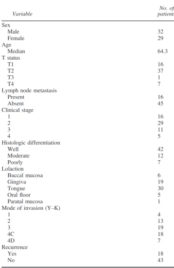
The subjects were 61 patients with primary OSCC who underwent surgical resection at the Department of Oral and Maxillofacial Surgery at Kanazawa University Hospital between 1989 and 2007. The tumors ’ TNM categories were classi fi ed according to the Union for International Cancer Control (UICC) system. The grade of tumor differentiation was determined according to the criteria proposed by the World Health Organization. The mode of tumor invasion was assessed according to the classi fi cation by Yamamoto et al. (14). Details of the patient and tumor characteristics are given in Table 1.
Cell lines
Three human oral squamous cell carcinoma cell lines with different invasive activities were used: OSC-20, OSC-19 (lower invasive type), and HOC313 (higher invasive type).
OSC-20 is a cell line derived a 58-year-old female with metastatic tongue cancer to the cervical lymph nodes (15).
OSC-19 was derived from a 61-year-old male with meta- static tongue cancer to the cervical lymph node (16).
HOC313 was derived from a 51-year-old female with metastatic squamous cell carcinoma (involving the mandib- ular gingival and oral fl oor) to the cervical lymph nodes (17). In addition, the normal human dermal fi broblast line (NHDF) was used as control.
Immunohistochemical staining
Tissue specimens were fi xed in 10% neutral buffered formalin and embedded in paraf fi n; then, 4-mm-thick sections were cut. Immunohistochemical detection of VEGF-A and VEGF- C was performed using an anti-VEGF-A rabbit polyclonal antibody at 1:200 dilution (LifeSpan BioScience, Seattle, WA, USA) and anti-VEGF-C rabbit polyclonal antibody at 1:100 dilution (Life Technologies, Carlsbad, CA, USA), respectively. Tissue sections were deparaf fi nized with xylene and rehydrated in graded alcohol. Endogenous peroxidase was blocked by treatment with 0.3% hydrogen peroxide in methanol for 30 min and incubated with the primary antibodies at 4 ° C overnight. Bound antibody was detected using the Envision system (Dako, Carpinteria, CA, USA).
Diaminobenzidine (1 mg/ml) in the presence of 0.03%
hydrogen peroxidase was used to visualize any bound peroxidase, and sections were counterstained with hematox- ylin. The speci fi cities of the staining were con fi rmed by using non-immune serum instead of the primary antibody as a negative control. The expressions of VEGF-A and VEGF-C were evaluated with immunohistochemically stained prepa-
rations using anti-VEGF-A antibody and anti-VEGF-C antibody. Immunohistochemical evaluation was carried out in accord with the report of Takanashi (18). The staining intensity of tumor cells in the leading invasion front regions was classi fi ed into four groups (0: none, 1: mild, 2: moderate, 3: strong), and cases were considered positive if their staining intensity was 2 or 3.
Enzyme-linked immunosorbent assay
For enzyme-linked immunosorbent assay analysis of VEGF-A and VEGF-C, supernatant of cultured several human oral cancer cell lines (OSC-19, OSC-20 and HOC313) and human normal dermal fi broblast cell lines (NHDF) were used. The supernatant was collected from cultured each cell line for 24 h. The concentrations of VEGF-A and VEGF-C were determined using ELISA kits (R&D Systems, Minneapolis, MN, USA) according to the manufacturer ’ s protocols.
Statistical analysis
JMP
â9 (SAS Institute Inc., Cary, NC, USA) was used for data analysis. The relationships between the expression of these proteins and clinicopathological parameters were examined by the chi-square test. We calculated the 5-year
Table 1 Clinicopathologic characteristics
Variable
No. of patients Sex
Male 32
Female 29
Age
Median 64.3
T status
T1 16
T2 37
T3 1
T4 7
Lymph node metastasis
Present 16
Absent 45
Clinical stage
1 16
2 29
3 11
4 5
Histologic differentiation
Well 42
Moderate 12
Poorly 7
Lolaction
Buccal mucosa 6
Gingiva 19
Tongue 30
Oralfloor 5
Paratal mucosa 1
Mode of invasion (Y–K)
1 4
2 13
3 19
4C 18
4D 7
Recurrence
Yes 18
No 43
J Oral Pathol Med
The expressions of VEGF-A and VEGF-C in OSCC Yanase et al
2
survival rates by the Kaplan – Meier method and compared them using the log-rank test. Factors found to be signi fi cant were then chosen for Cox ’ s multivariate proportional hazard model in order to ascertain their prognostic values. The signi fi cance level was set at 5% for each analysis. The data of ELISA are presented as the mean values SEM. The differences between groups were tested for statistical signi fi cance using the two-tailed Mann – Whitney U test.
P-values < 0.05 were considered statistically signi fi cant.
Results
Expression of VEGF-A and VEGF-C in oral SCC
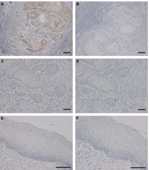
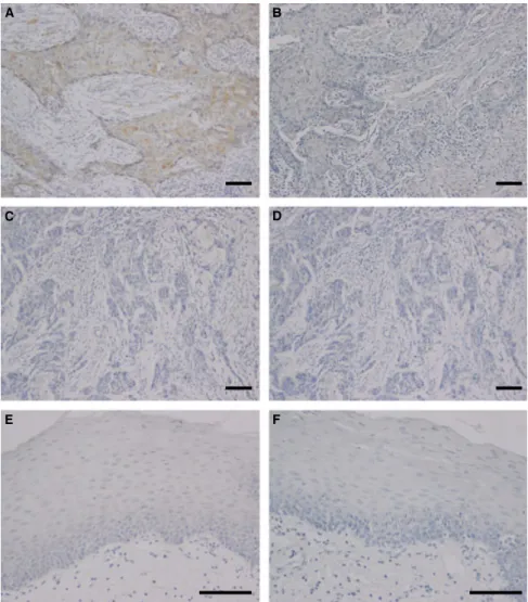
VEGF-A and VEGF-C were detected in the cytoplasm of tumor cells (Figs 1 and 2). According to the criteria for VEGF immunohistochemical evaluation, 46 cases (75.4%) were positive for VEGF-A, and 24 cases (39.3%) were positive for VEGF-C.
Association between VEGF-A or VEGF-C expression and clinicopathological features
Table 2 displays the correlations between VEGF-A and VEGF-C expression and the patients ’ clinicopathological
factors. VEGF-A expression was signi fi cantly correlated with lymph node metastasis (P < 0.05) but was not corre- lated with tumor size, local recurrence, histological differen- tiation, or mode of invasion. VEGF-C expression was signi fi cantly correlated with lymph node metastasis and local recurrence (P < 0.05) but was not correlated with tumor size, histological differentiation, location, or mode of invasion.
Correlations between VEGF-A or VEGF-C expression and survival time
Figure 3 shows the Kaplan – Meier survival curves of the VEGF-A-positive and VEGF-C-positive groups compared to their negative group counterparts. The 5-year survival rate was 57.6% for the VEGF-A-positive group and 72.7%
for the VEGF-A-negative group (Fig. 3A). The VEGF-A- positive group showed low survival rates, but there was no signi fi cant difference in survival compared with the negative group (Fig. 3A). The 5-year survival rate was 41.2% for the VEGF-C-positive group and 81.2% for the VEGF-C- negative group (Fig. 3B). The VEGF-C-positive group showed signi fi cantly poorer prognoses compared to the VEGF-C-negative group (Fig. 3B) (P < 0.05).
A B
E
D C
F
Figure 1 Immunohistochemical stain of Vascular endothelial growth factor-A (VEGF-A) in squamous cell carcinoma (A–D) and normal oral tissue (E, F).
According to the expression intensity of VEGF-A, the samples are differentiated as positive (A) and negative (C) groups. The specificities of the staining were confirmed by using non-immune serum instead of the primary antibody as a negative control (B, D and F). Scale bar, 100lm.
3
Cox proportional hazards regression analysis of the predictive factors for prognosis
We performed a Cox proportional hazards regression analysis to examine the signi fi cance of the predictive factors. A univariate analysis revealed that the expression of VEGF-C and the mode of invasion were signi fi cant prognostic indicators, but not VEGF-A. In addition, a multivariate analysis showed that VEGF-C expression and the mode of invasion were independent prognostic factors (Table 3).
Expression of VEGF-A or VEGF-C in human oral cancer cell lines
As demonstrated by the immunohistochemical analysis, the number of cancer cells expressed VEGF-A and VEGF-C.
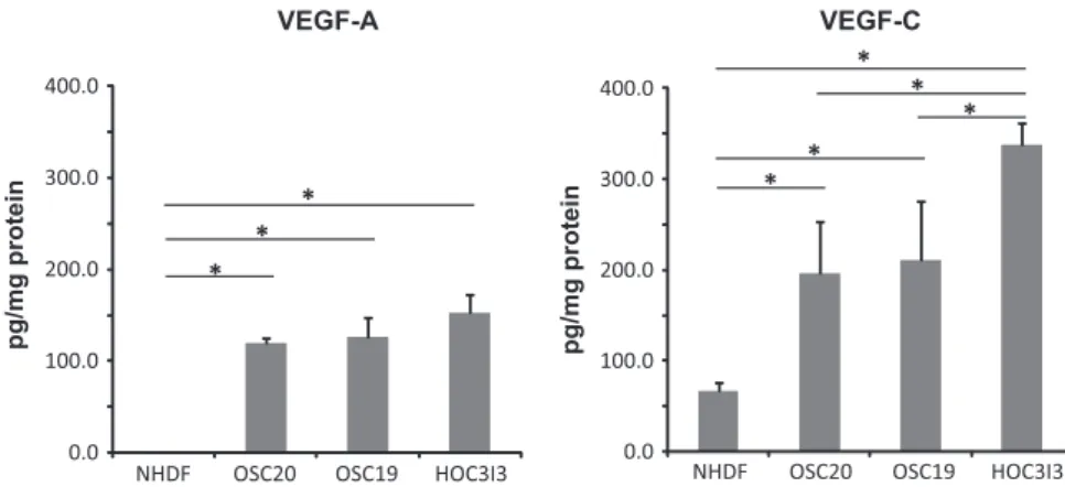
Therefore, we further examined several human oral SCC lines (OSC-19, OSC-20, and HOC313) with different invasive activities by measuring the concentration of the expressed VEGF-A and VEGF-C. The VEGF-A expression of OSC-19 (126.1 20.92 pg/mg protein), OSC-20 (121.5 4.66 pg/mg protein), and HOC313 (153.1 19.7 pg/mg protein) was signi fi cantly up-regulated compare
to NHDF (0.2 0.68 pg/mg protein) (mean SEM, Fig. 4). However, there was no signi fi cant difference between OSC-19, OSC-20, and NHDF. On the other hand, the VEGF-C expression of HOC313 (337.8 23.8 pg/mg protein) was signi fi cantly up-regulated compare to OSC-19 and OSC-20 (210.8 64.2, 196.6 56.7 pg/mg protein) (mean SEM, Fig. 4). There was no signi fi cant difference between OSC-19 and OSC-20. The VEGF-C expression levels of three oral cancer cells were signi fi cantly higher compared with NHDF (66.7 8.84 pg/mg protein) (mean SEM, Fig. 4).
Discussion
VEGF-A is a key tumor-derived growth factor that promotes the switch to an angiogenic phenotype in many tumors and other tissues. Interestingly, our study shows that the expression of VEGF-A is correlated with lymph node metastasis. This is most likely because VEGF-A can bind to VEGFR-1 and VEGFR-2, but not to VEGFR-3, which is expressed in lymphatic endothelial cells. Several studies have shown that VEGF-A can induce lymphangiogenesis
AE C
B
D
F
Figure 2 Immunohistochemical stain of Vascular endothelial growth factor-C (VEGF-C) in squamous cell carcinoma (A–D) and normal oral tissue (E, F).
According to the expression intensity of VEGF-C, the samples are differentiated as positive (A) and negative (C) groups. The specificities of the staining were confirmed by using non-immune serum instead of the primary antibody as a negative control (B, D and F). Scale bar, 100lm.
J Oral Pathol Med
The expressions of VEGF-A and VEGF-C in OSCC Yanase et al
4
(19, 20). Other groups have demonstrated that VEGF-A expression is associated with lymph node metastasis (11, 21, 22). A recent study reported that the VEGFR-2 receptor is occasionally expressed on lymphatic endothelial cells (23).
Unlike VEGFR-2, the af fi nity of VEGFR-1 for its ligand, VEGF-A, is approximately one order of magnitude higher than that of VEGFR-2. However, the kinase activity of VEGFR-1 is lower than VEGFR-2 (about one-tenth that of VEGFR-2), so VEGF-A is thought to act mainly through the activation of VEGFR-2 (3, 23 – 25). These data suggest that VEGF-A may have a direct stimulatory effect on lymphatic vessels in OSCC.
VEGF-C expression has been detected in a variety of tumors (26 – 29). VEGF-C, which binds to VEGFR-3, is known to be a major factor in lymphangiogenesis (30, 31).
VEGF-C binds to VEGFR-3 on the surface of lymphatic endothelial cells, inducing the proliferation and growth of new lymphatic vessels and leading to tumor cell dissemi- nation and lymph node metastasis (2, 11, 32). VEGF-C expression is associated with lymphatic vessel density (31, 33, 34) and lymph node metastases (30, 31, 35) in oral cancer. Pentheroudakis et al. (36) also reported an associ- ation between local recurrence and VEGF-C expression in head and neck cancer. In this study, we show that VEGF-C expression is associated with lymph node metastasis in OSCC. We also observed an association between VEGF-C
expression and local recurrence. The data in this study are consistent with previous reports. However, further studies are needed to understand the precise role of VEGF-C in local recurrence.
There was no signi fi cant difference in the 5-year survival rate between patients from the VEGF-A-positive group and the VEGF-A-negative group. In contrast, the survival rate of the VEGF-C-positive group was signi fi cantly decreased compared to the VEGF-C-negative group. The results of our analysis using a Cox proportional hazards model show that the mode of invasion and VEGF-C expression are indepen- dent prognostic factors. Our ELISA data show that VEGF-C expression is signi fi cantly higher in a highly invasive cell line (HOC313) than in cell lines with lower invasiveness (OSC-19 and OSC-20). Highly invasive OSCC is closely related to the prognosis (14). These results suggest that VEGF-C may affect OSCC prognosis. The clinicopatho- logical factors currently used to predict metastasis and prognosis include TNM classi fi cation, degree of differenti- ation, and mode of invasion. We propose including a molecular biological factor for comprehensive diagnoses in the future. More complete diagnostic methods not only improve the accuracy of diagnosing metastases, but could also help determine the prognosis and facilitate decision-
Table 2 Correlations between the expressions of vascaular endothelial growth factor-A, -C (VEGF-A, VEGF-C), and the oral squamous cell carcinoma (OSCC) patients’clinicopathologic factors
Factors
VEGF-A VEGF-C
Positive (n=46)
Negative (n=15)
P- value
Positive (n=24)
Negative (n=37)
P- value T status
T1 12 4 5 11
T2 29 8 15 22
T3 1 0 0.623 1 0 0.564
T4 4 3 3 4
Lymph node metastasis
Present 9 7 0.038 10 6 0.027
Absent 37 8 14 31
Recurrence
Yes 16 2 0.113 13 5 0.0006
No 30 13 11 32
Histologic differentiation
Well 33 9 0.222 16 26 0.997
Moderate 8 4 5 7
Poorly 5 2 3 4
Lolaction Buccal mucosa
6 0 0.221 2 4 0.627
Gingiva 14 5 8 11
Tongue 23 7 13 17
Oralfloor
3 2 1 4
Paratal mucosa
0 1 0 1
Mode of invasion (Y-K)
1 2 2 1 3
2 12 1 6 7
3 15 4 0.206 6 13 0.734
4C 11 7 7 11
4D 6 1 4 3
0.0 0.2 0.4 0.6 0.8 1.0 1.2
The 5-year survival rate
0 10 20 30 40 50 60
Months after surgery
VEGF-A-negative group
VEGF-A-positive group A
0.0 0.2 0.4 0.6 0.8 1.0 1.2
The 5-year survival rate
0 10 20 30 40 50 60
Months after surgery
VEGF-C-negative group
VEGF-C-positive group B
Figure 3 (A) The Kaplan–Meier survival rate of the VEGF-A-positive group shows low survival, but there was no significant difference in survival compared with the VEGF-A-negative group (P=0.6918). (B) The VEGF-C-positive group showed significantly poorer prognoses compared to the VEGF-C-negative group (P=0.029).
5
making regarding the necessity of prophylactic neck dissection. Our results suggest that VEGF-C may be a predictive factor for OSCC outcome.
Recently, new therapies have been developed to target VEGF-A. However, resistance to anti-angiogenic therapies limits the clinical bene fi t of these agents in cancer patients.
The single-agent response rate to anti-angiogenic drugs such as bevacizumab (a monoclonal antibody to VEGF-A) is
< 10%, and even in patients who do respond, the duration of the response is typically < 3 months (37 – 39). Similar response rates are observed in head and neck squamous cell carcinomas (40), for which bevacizumab is being evaluated in phase III clinical trials. Our results suggest that therapies targeting VEGF-C may be expected to play a major role in the treatment of bevacizumab-resistant OSCC in the future.
References
1. Oliveira-Neto HH, Gleber-Netto FO, De Sousa SF, et al. A comparative study of microvessel density in squamous cell carcinoma of the oral cavity and lip. Oral Surg Oral Med Oral Pathol Oral Radiol 2012;
113: 391–8.2. Liang X, Zhou H, Liu X, et al. Effect of local hyperthermia on lymphangiogenic factors VEGF-C and -D in a nude mouse xenograft model of tongue squamous cell carcinoma. Oral Oncol 2010;
46: 111–5.3. Johnstone S, Logan RM. The role of vascular endothelial growth factor (VEGF) in oral dysplasia and oral squamous cell carcinoma. Oral Oncol 2006;
42: 337–42.4. Villarejo-Campos P, Padilla-Valverde D, Martin RM, et al.
Serum VEGF and VEGF-C values before surgery and after postoperative treatment in gastric cancer. Clin Transl Oncol 2013;
15: 265–70.5. Kishimoto K, Yoshihama Y, Mandai T. Expression of VEGF, PD-ECGF, and VEGF-C and its clinicopathologic significance in oral squamous cell carcinoma. Nihon Koku Geka Gakkai Zasshi 2005;
51: 218–25.6. Berns EM, Klijn JG, Look MP, et al. Combined vascular endothelial growth factor and TP53 status predicts poor response to tamoxifen therapy in estrogen receptor-positive advanced breast cancer. Clin Cancer Res 2003;
9: 1253–8.
7. Fontanini G, Lucchi M, Vignati S, et al. Angiogenesis as a prognostic indicator of survival in non-small-cell lung carcinoma: a prospective study. J Natl Cancer Inst 1997;
89:881
–6.
8. Takahashi Y, Cleary KR, Mai M, et al. Significance of vessel count and vascular endothelial growth factor and its receptor (KDR) in intestinal-type gastric cancer. Clin Cancer Res 1996;
2: 1679–84.
9. Ikeda N, Adachi M, Taki T, et al. Prognostic significance of angiogenesis in human pancreatic cancer. Br J Cancer 1999;
79: 1553–63.
10. George DJ, Halabi S, Shepard TF, et al. Prognostic signifi- cance of plasma vascular endothelial growth factor levels in patients with hormone-refractory prostate cancer treated on Cancer and Leukemia Group B 9480. Clin Cancer Res 2001;
7: 1932–
6.
11. Chen SC, Shih CM, Tseng GC, et al. Vascular endothelial growth factor C as a predictor of early recurrence and poor prognosis of resected stage I non-small cell lung cancer. Ann Acad Med Singapore 2011;
40: 319–24.
0.0 100.0 200.0 300.0 400.0
NHDF OSC20 OSC19 HOC3I3
pg/mg protein pg/mg protein
VEGF-A VEGF-C
* *
* *
*
0.0 100.0 200.0 300.0 400.0
NHDF OSC20 OSC19 HOC3I3
* *
*
Figure 4 Expression of (vascular endothelial growth factor-A) VEGF-A or vascular endothelial growth factor-C (VEGF-C) in supernatant of cultured human oral cancer cell lines (OSC-19, OSC-20 and HOC313) and human normal dermalfibroblast cell lines (NHDF). The average concentration (pg/mg protein) of VEGF-A (left panel) and VEGF-C (right panel) is indicated. Error bars,SEM,*P<0.05.
Table 3 Results of the univariate and multivariate analyses with Cox’s proportional hazards mode
Factors
Univariate Multivariate
Hazard ratio
95% Confidence
interval P-value Hazard ratio
95% Confidence
interval P-value
VEGF-A (positive/negative) 1.541 0.508–6.657 0.4739
VEGF-C (positive/negative) 3.122 1.176–9.734 0.0214 2.771 1.041–8.661 0.041
Lymph node metastasis (present/absent) 1.616 0.560–4.181 0.3531 Histologic differentiation (moderate-poorly/well) 2.114 0.729–5.516 0.158
Mode of invasion (Y-K) (4C, 4D/1, 2, 3) 4.435 1.716–12.77 0.002 4.066 1.569–11.73 0.0037
VEGF-A, vascular endothelial growth factor-A; VEGF-C, vascular endothelial growth factor-C.
J Oral Pathol Med
The expressions of VEGF-A and VEGF-C in OSCC Yanase et al
6
12. Kondo K, Kaneko T, Baba M, et al. VEGF-C and VEGF-A synergistically enhance lymph node metastasis of gastric cancer. Biol Pharm Bull 2007;
30: 633–7.
13. Noda E, Maeda K, Inoue T, et al. Predictive value of vascular endothelial growth factor-C expression for local recurrence of rectal carcinoma. Oncol Rep 2007;
17: 1327–31.
14. Yamamoto E, Kohama G, Sunakawa H, et al. Mode of invasion, bleomycin sensitivity, and clinical course in squa- mous cell carcinoma of the oral cavity. Cancer 1983;
51:2175–80.
15. Yokoi T, Hirata S, Nishimura F, et al. Some properties of a newly established human cell line derived from an oral squamous carcinoma. Tumor Res 1990;
25: 93–103.16. Yokoi T, Homma H, Odajima T. Establishment and charac- terization of OSC-19 cell line in serum and protein free culture. Tumor Res 1988;
24: 1–17.17. Ishisaki A, Oida S, Momose F, et al. Identification and characterization of autocrine-motility-factor-like activity in oral squamous-cell-carcinoma cells. Int J Cancer 1994;
59:783
–8.
18. Takanashi S, Takimoto M, Ota H. Correlation with cycloox- ygenase-2, vasculogenetic factor and lymph node metastasis in esophageal squamous cell carcinoma. Showa Igakkai Zasshi 2006;
66: 331–9.
19. Nagy JA, Vasile E, Feng D, et al. Vascular permeability factor/
vascular endothelial growth factor induces lymphangiogenesis as well as angiogenesis. J Exp Med 2002;
196: 1497–506.
20. Hirakawa S, Kodama S, Kunstfeld R, et al. VEGF-A induces tumor and sentinel lymph node lymphangiogenesis and promotes lymphatic metastasis. J Exp Med 2005;
201:1089–99.
21. Sasahira T, Kirita T, Kurihara M, et al. MIA-dependent angiogenesis and lymphangiogenesis are closely associated with progression, nodal metastasis and poor prognosis in tongue squamous cell carcinoma. Eur J Cancer 2010;
46:2285–94.
22. Mineta H, Miura K, Ogino T, et al. Prognostic value of vascular endothelial growth factor (VEGF) in head and neck squamous cell carcinomas. Br J Cancer 2000;
83: 775–81.
23. Bj€ orndahl MA, Cao R, Burton JB, et al. Vascular endothelial growth factor-a promotes peritumoral lymphangiogenesis and lymphatic metastasis. Cancer Res 2005;
65: 9261–8.
24. Takahashi S. Vascular endothelial growth factor (VEGF), VEGF receptors and their inhibitors for antiangiogenic tumor therapy. Biol Pharm Bull 2011;
34: 1785–8.25. Shibuya M. Vascular endothelial growth factor and its receptor system: physiological functions in angiogenesis and patholog- ical roles in various diseases. J Biochem 2013;
153: 13–9.26. Niki T, Iba S, Tokunou M, et al. Expression of vascular endothelial growth factors A, B, C, and D and their relationships to lymph node status in lung adenocarcinoma.
Clin Cancer Res 2000;
6: 2431–9.27. Gunningham SP, Currie MJ, Han C, et al. The short form of the alternatively spliced
flt-4 but not its ligand vascularendothelial growth factor C is related to lymph node metastasis in human breast cancers. Clin Cancer Res 2000;
6: 4278–86.
28. Akagi K, Ikeda Y, Miyazaki M, et al. Vascular endothelial growth factor-C (VEGF-C) expression in human colorectal cancer tissues. Br J Cancer 2000;
83: 887–91.
29. Hashimoto I, Kodama J, Seki N, et al. Vascular endothelial growth factor-C expression and its relationship to pelvic lymph node status in invasive cervical cancer. Br J Cancer 2001;
85: 93–7.
30. Kishimoto K, Sasaki A, Yoshihama Y, et al. Expression of vascular endothelial growth factor-C predicts regional lymph node metastasis in early oral squamous cell carcinoma. Oral Oncol 2003;
39: 391–6.31. Sugiura T, Inoue Y, Matsuki R, et al. VEGF-C and VEGF-D expression is correlated with lymphatic vessel density and lymph node metastasis in oral squamous cell carcinoma:
Implications for use as a prognostic marker. Int J Oncol 2009;
34: 673–80.
32. Ishii H, Chikamatsu K, Sakakura K, et al. Primary tumor induces sentinel lymph node lymphangiogenesis in oral squamous cell carcinoma. Oral Oncol 2010;
46: 373–8.33. Liang X, Yang D, Hu J, et al. Hypoxia inducible factor- alpha expression correlates with vascular endothelial growth factor-C expression and lymphangiogenesis/angiogenesis in oral squamous cell carcinoma. Anticancer Res 2008;
28:1659
–66.
34. Watanabe S, Kato M, Kotani I, et al. Lymphatic vessel density and vascular endothelial growth factor expression in squamous cell carcinomas of lip and oral cavity: a clinicopathological analysis with immunohistochemistry using antibodies to D2- 40, VEGF-C and VEGF-D. Yonago Acta Med 2013;
56: 29–37.
35. Nakazato T, Shingaki S, Kitamura N, et al. Expression level of vascular endothelial growth factor-C and -A in cultured human oral squamous cell carcinoma correlates respectively with lymphatic metastasis and angiogenesis when transplanted into nude mouse oral cavity. Oncol Rep 2006;
15: 825–30.36. Pentheroudakis G, Angouridakis N, Wirtz R, et al. Transcrip- tional activity of human epidermal growth factor receptor family and angiogenesis effectors in locoregionally recurrent head and neck squamous cell carcinoma and correlation with patient outcome. J Oncol 2009;
2009: 854127.37. Yang JC, Haworth L, Sherry RM, et al. A randomized trial of bevacizumab, an anti-vascular endothelial growth factor antibody, for metastatic renal cancer. N Engl J Med 2003;
349: 427–
34.
38. Giantonio BJ, Catalano PJ, Meropol NJ, et al. Bevacizumab in combination with oxaliplatin,
fluorouracil, and leucovorin(FOLFOX4) for previously treated metastatic colorectal can- cer: results from the Eastern Cooperative Oncology Group Study E3200. J Clin Oncol 2007;
25: 1539–44.39. Sandler A, Gray R, Perry MC, et al. Paclitaxel-carboplatin alone or with bevacizumab for non-small-cell lung cancer. N Engl J Med 2006;
355: 2542–50.40. Cohen EE, Davis DW, Karrison TG, et al. Erlotinib and bevacizumab in patients with recurrent or metastatic squa- mous-cell carcinoma of the head and neck: a phase I/II study.
Lancet Oncol 2009;
10: 247–57.
7