INTRODUCTION
Hepatocellular carcinoma (HCC) is one of the com-monest malignant tumors in the world. Surgical resec-tion is the mainstay of treatment, but the prognosis after resection remains unsatisfactory because of the high incidence of recurrence (1-3). Most recurrences occur in the remnant liver rather than extrahepatic
ORIGINAL
Correlation of vascular endothelial cell proliferation with
mi-crovessel density and expression of vascular endothelial
growth factor and basic fibroblast growth factor in
hepa-tocellular carcinoma
Satoru Imura, Hidenori Miyake, Keisuke Izumi
1, Seiki Tashiro, and Hisanori Uehara
1Department of Digestive and Pediatric Surgery, and1
Department of Molecular and Environmental Pathology, The University of Tokushima School of Medicine, Tokushima, Japan
Abstract : Tumor-associated angiogenesis is essential for tumor growth or metastasis, and consists of multiple and sequential steps regulated by proangiogenic and antiangiogenic factors. Vascular endothelial cell proliferation is involved in this process. We investigated the correlation of vascular endothelial cell proliferation with microvessel density (MVD) and expression of major proangiogenic molecules, vascular endothelial growth factor (VEGF) and basic fibroblast growth factor (bFGF) in hepatocellular carcinoma (HCC).
Formalin-fixed paraffin-embedded specimens of surgically resected HCC from 67 patients were used. Proliferating endothelial cells were detected by immunofluorescence double staining for CD34 and proliferating cell nuclear antigen (PCNA). The proliferation activity of endothelial cells was determined by the rate of PCNA-positive endothelial cells, and evaluated at the periphery and center of the tumors and adjacent non-neoplastic livers. MVD and the expression of VEGF and bFGF in the tumors were also examined immunohistochemically.
The proliferation activity of endothelial cells at the periphery of the tumors was significantly higher than that at the center of the tumors (35.8% vs.12.7%, P<0.0001). The rate of PCNA-positive endothelial cells in the tumors with higher bFGF expression was significantly higher than that in the tumors with lower bFGF expression (44.8% vs. 32.5%, P<0.005) at the periphery of the tumors. There was no significant correlation between the rate of PCNA-positive endothelial cells and clinicopathological findings or MVD.
In HCC, the proliferation activity of vascular endothelial cells is suggested to be heterogeneous in the tumor and higher at the periphery of the tumor, and bFGF may play an important role in the positive regulation of tumor-associated vascular endothelial cell proliferation. J. Med. Invest. 51 : 202-209, August, 2004
Keywords : hepatocellular carcinoma, immunohistochemistry, proliferating cell nuclear antigen, angiogenesis,
endothelial cell proliferation activity
Received for publication February 23, 2004 ; accepted February 27, 2004.
Address correspondence and reprint requests to Hisanori Uehara, M.D., Ph.D., Department of Molecular and Environmental Pathol-ogy, Institute of Health Biosciences, The University of Tokushima Graduate School, Kuramoto-cho, Tokushima 770-8503, Japan and Fax : +81-88-633-7066.
The Journal of Medical Investigation Vol. 51 2004
sites. Intrahepatic metastasis resulting from portal ve-nous invasion is considered an important mechanism of intrahepatic recurrence (2, 3).
HCC is a hypervascular tumor characterized by neovascularization. The formation of neovasculature toward and within the tumor, i.e., tumor-associated angiogenesis, is essential for tumor growth or metas-tasis, and increasing intratumoral microvessel density (MVD), major of tumor-associated angiogenesis, cor-relates with greater tumor aggressiveness, such as a higher frequency of metastasis and/or poor prognosis in HCC and other solid tumors (4 -6). Tumor-associated angiogenesis is dependent on the local balance of proan-giogenic and antianproan-giogenic factors. Vascular endothelial growth factor (VEGF) and basic fibroblast growth factor (bFGF) are important positive regulators of tumor-associated angiogenesis, stimulate endothelial cell proliferation and enhance vascular permeability. Recent studies report that expression of the angiogenic factors such as VEGF, bFGF is closely related to growth, me-tastasis and prognosis of HCC (7-10).
Tumor-associated angiogenesis consists of multiple and sequential steps. In addition to vascular remodeling, sprouting and endothelial cell migration, endothelial cell proliferation is involved in this process (11). Tumor-associated endothelial cell proliferation has been in-vestigated in breast carcinoma and prostate carcinoma. Proliferation activities of vascular endothelial cells in these tumors were significantly higher than that in adjacent non-neoplastic tissue (12, 13), and the size and frequency of vessels with proliferating endothelial cells in breast carcinoma were associated with lymph node status (14). In this study, we evaluated the pro-liferation activity of vascular endothelial cells in 67 sur-gical specimens of HCC at the periphery and center of tumors and adjacent non-neoplastic liver using im-munofluorescence double staining technique (15, 16) for CD34, the most reproducible endothelial cell high-lighter (17), and proliferating cell nuclear antigen (PCNA), which is related to cell proliferative activity (18). We also investigated the correlation of proliferation activities of vascular endothelial cells with clinicopa-thological findings, MVD, and VEGF and bFGF ex-pression in tumors.
MATERIALS AND METHODS
PatientsSixty-seven patients who underwent initially curative surgery for HCC at Tokushima University Hospital between 1990 and 2000 were randomly enrolled in
this study. The mean age of the patients was 61.2 years (range, 38 -78 years). Fifty-two patients were male and fifteen were female. Patients whose resected specimens were inadequate or insufficient for this study were ex-cluded. None of these patients had previous chemo-therapy, including transarterial chemoembolization. Their formalin-fixed paraffin-embedded specimens were studied with informed consent.
Clinicopathological Findings
The clinicopathological findings used are listed in the General Rules for the Clinical and Pathological Study of Primary Liver Cancer (19).
Immunofluorescence Double Staining for CD34 and PCNA
Paraffin sections (4µm) were deparaffinized with xylene, rinsed thoroughly with graded ethanol solution, then soaked in 0.3% hydrogen peroxide in phosphate-buffered saline (PBS) for 15 min at room temperature in order to inactivate endogenous peroxidase activity. To retrieve antigen, slides were boiled with 10mM citrate buffer for 15min. All sections were incubated with a 1 : 50 dilution of monoclonal mouse anti-human CD34 antibody (DakoCytomation, Denmark) contain-ing 5% normal goat serum for1hour at room tempera-ture. After the samples were rinsed twice for 5 min each with PBS, they were incubated with a 1 : 400 dilution of secondary Alexa 594-conjugated goat anti-mouse IgG antibody (Molecular Probes, Inc., Eugene, OR) for 1 hour at room temperature in the dark. Samples were then rinsed twice with PBS, incubated at 4℃ for 18 hours with a 1 : 50 dilution of polyclonal rabbit anti-human PCNA antibody (Santa Cruz Biotechnology, Santa Cruz, CA). After the samples were rinsed twice for 5 min each with PBS, they were incubated with a 1:400 dilution of secondary Alexa 488-conjugated goat anti-rabbit IgG (Molecular Probes, Inc., Eugene, OR) for 1 hour at room temperature in the dark. The samples were then rinsed twice for 5 min each with PBS and mounted with Vectashield with DAPI (Vector Laboratories, Inc., Burlingame, CA). The images were captured using a CoolSNAP camera, Epi fluorescence upright microscopy system (Olympus Optical Co., Ltd., Tokyo, Japan), and MetaCam software (Universal Imaging Corporation, Downingtown, PA).
Immunohistochemical staining for CD34, VEGF and bFGF
Paraffin sections (4µm) were deparaffinized with xylene, rinsed thoroughly with graded ethanol solution, then soaked in 0.3% hydrogen peroxide in PBS for
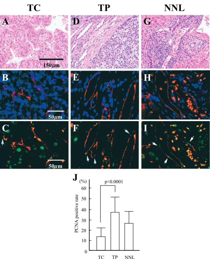
Figure 1. Analysis of vascular endothelial cell proliferation in HCC by immunofluorescence double staining for CD34 and PCNA at tumor center (TC) (A-C), tumor periphery (TP) (D-F) and non-neoplastic liver (NNL) (G-I). A, D and G, representative histological findings of three areas stained by hematoxylin and eosin (H & E). B, E and H, combined images of CD34-immunostaining (red) and nuclear staining with DAPI (blue) for the counting of endothelial cells. C, F and I, combined images of CD34-immunostaining (red) and PCNA-immunostaining (green) for the counting of PCNA-positive endothelial cells (arrows in C, F and I). The rate of PCNA-positive endothelial cells was defined as the ratio of PCNA-positive endothelial cells to the total endothelial cell count, and significantly higher at the periphery of tumors (35.8%) than the center (12.7%) at P<0.0001 (J).
S. Imura et al. Vascular endothelial cell proliferation in HCC
15 min at room temperature in order to inactivate en-dogenous peroxidase activity. To retrieve antigen, slides were boiled with 10mM citrate buffer for 15min. Sec-tions were incubated with a primary antibody containing 5% normal goat serum for1hour at room temperature. The primary antibodies used were as follows : mon-oclonal mouse anti-human CD34 (DakoCytomation, Denmark) at 1 : 50 dilution, polyclonal rabbit anti-human VEGF (Santa Cruz Biotechnology, Santa Cruz, CA) at 1 : 50 dilution, and polyclonal rabbit anti-human bFGF (Santa Cruz Biotechnology, Santa Cruz, CA) at 1 : 50 dilution. After washing they were incubated and visualized by the labeling streptavidin-biotin-peroxidase-complex method using DAKO LSAB!2 System (DAKO Co. Carpinteria, CA) with 3, 3’-diaminobenzidine tet-rachloride (DAB) as a chromogen.
Determination of the rate of PCNA-positive vascular endothelial cells
The rate of PCNA-positive vascular endothelial cells was defined as the ratio of positively stained cells to
the total cell count and evaluated at the center and pe-riphery of tumors and adjacent non-neoplastic livers. Ten fields were randomly selected from each area under
×400 magnification, and three images ; nuclear staining
with DAPI (blue fluorescence), immunohistochemical staining for PCNA (green fluorescence) and CD34 (red fluorescence), were independently captured at every field and combined using MetaCam software (Figure 1). Combined images of immunostaining for CD34 with nuclear staining were used for total endothelial cells counting, and combined images of immunostaining for CD34 with that for PCNA were used for PCNA-positive endothelial cells counting. More than 100 individual endothelial cell nuclei were counted.
Determination of MVD
Sections performed immunohistochemical staining for CD34 were assessed in five fields using a×200
mag-nifying lens to count the number of microvessels at the periphery of the tumor. MVD was expressed as the number of microvessels per square millimeter.
Figure 2. Immunohistochemical expression of bFGF and VEGF in HCC. The cytoplasmic intensity of epithelial cells of bile ducts in non-neoplastic liver was considered the control of immunohistochemical staining for bFGF and VEGF. If the tumor cells’ cytoplasmic intensity was similar in degree with the biliary epithelial cells’, the intensity was evaluated as 1+, and if the intensity was higher in degree than the biliary epithelial cells’, it was evaluated as 2+.
Determination of the degree of VEGF, bFGF expression The cytoplasmic intensity of epithelial cells of bile ducts in non-neoplastic liver was considered the control of immunohistochemical staining for VEGF and bFGF. If tumor cells’ cytoplasmic intensity was similar in de-gree with the biliary epithelial cells’, the intensity was evaluated as 1+, and if the intensity was higher in
de-gree than the biliary epithelial cells’, it was evaluated as 2+(Figure 2).
Histopathologic examination
Detailed histopathologic examination of all specimens in this study was performed by a senior pathologist specialized in HCC pathology, who was blind to the patients’ clinical findings.
Statistical analysis
The results were reported as the mean±standard
de-viation. Unpaired t-test was used for all statistical analy-sis. The differences were considered significant when P<0.05 in all examinations.
RESULTS
Correlation between the rate of PCNA-positive
vascu-lar endothelial cells and clinicopathological features
The rate of PCNA-positive vascular endothelial cells was evaluated at the center and periphery of tumors and adjacent non-neoplastic livers. In non-neoplastic livers, vascular endothelial cells were examined at portal tracts and fibrous septa surrounding cirrhotic nodules
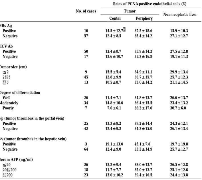
Table 1. Relationship between rates of PCNA-positive endothelial cells and clinicopathological findings in patients with HCCs
Rates of PCNA-positive endothelial cells (%)
No. of cases Tumor
Non-neoplastic liver Center Periphery HBs Ag Positive Negative HCV Ab Positive Negative Tumor size (cm) ≦2 2−5 >5 Degree of differentiation Well Moderately Poorly
Vp (tumor thrombus in the portal vein) Positive
Negative
Vv (tumor thrombus in the hepatic vein) Positive Negative Serum AFP (ng/ml) ≦20 20−200 >200 10 57 50 17 9 45 13 26 34 7 25 42 3 64 26 18 23 14.5±12.7* 12.4±8.5 12.4±8.7 13.6±10.7 15.5±5.4 12.8±9.9 10.5±8.7 11.4±7.1 14.8±10.6 7.6±6.1 13.3±9.2 12.4±9.2 19.1±13.0 12.4±9.0 13.2±9.4 11.7±7.7 13.0±10.2 37.5±18.6 35.4±14.2 35.9±14.2 35.3±16.8 34.9±11.1 36.7±15.7 33.0±14.2 34.8±13.7 36.4±15.5 36.2±17.0 38.2±14.4 34.3±15.0 45.1±7.8 35.3±14.9 33.0±13.7 35.0±13.7 39.4±16.5 15.9±10.3 27.1±12.7 27.5±12.8 19.1±11.3 29.9±13.4 25.7±12.3 21.1±14.5 26.6±13.7 23.4±13.2 30.7±6.0 24.3±12.1 26.1±13.4 19.7±19.8 25.7±12.7 26.5±12.8 25.1±12.6 24.4±13.8 * Mean±SD
No significant relation was found.
S. Imura et al. Vascular endothelial cell proliferation in HCC
because most of sinusoidal endothelial cells were nega-tive for CD34. PCNA-posinega-tive endothelial cells were ob-served in all sizes of vessels. PCNA positive rates at the center and periphery of tumors and non-neoplastic livers were 12.7% (0−43.8, median 10.5, SD 9.2), 35.8% (4.0−64.5, median 36.6, SD14.8) and 25.4% (1.3−59.2, median 24.0, SD12.9), respectively. PCNA positive rate was significantly higher at the periphery of tumors than the center (P<0.0001)(Figure 1). There was no
signifi-cant relation between PCNA positive rate and patients’ background, clinicopathological findings (Table 1). In addition, PCNA positive rates at non-neoplastic liver were not related to the proliferation activity of endo-thelial cells at the center and periphery of tumors and tended to be higher in cases with severe inflammation. MVD
MVD was counted at the periphery of tumors. The mean MVD was 409.8/mm2
with a range of 217.0 to 643.0 (median 404.0, SD 97.8).
Correlation between the rate of PCNA-positive vascular endothelial cells, MVD and the expression of VEGF, bFGF
Expression of bFGF and VEGF was observed in all tumors and the expression levels of them tend to be lower at the center of tumors. As shown in Table 2, the rate of PCNA-positive vascular endothelial cells of 1+
and 2+groups in bFGF expression, were 32.5±15.0
and 44.8±9.9. This difference was statistically
signifi-cant (P=0.0019). There was no signifisignifi-cant difference
in PCNA positive rates between 1+and 2+groups in
VEGF expression. When the cases were divided into three groups ; bFGF 1+/VEGF 1+group, bFGF 1+/
VEGF 2+and bFGF 2+/VEGF 1+group, and VEGF 2+/
bFGF 2+group, PCNA positive rates were 33.5±15.0,
34.8±14.7, and 45.8±10.2, respectively. There was
sta-tistically significant difference between bFGF 1+/VEGF
1+group and bFGF2+/VEGF2+group (P=0.0134).
MVD was not significantly related to the expression level of bFGF and VEGF, although MVD of 2+group
in VEGF expression tend to be higher than that of 1+
group. In addition, there was no significant relation be-tween PCNA positive rate of vascular endothelial cells and MVD.
DISCUSSION
Compelling data implicate tumor-associated angi-ogenesis as the central pathologic step in the process of tumor growth, invasion, and metastasis (11, 20). Numerous experimental models and several studies in human tumors have shown that tumor-associated angiogenesis involves endothelial cell proliferation (12-14, 21). Studies of endothelial cell proliferation using human cancer tissue have been conducted on breast (12, 14) and prostate (13), but endothelial cell proliferation in HCC has not been fully investigated. In this study, we evaluated the proliferation activities of vascular endothelial cells in 67 surgical specimens of HCC and investigated the correlation of proliferation Table 2. Expression of bFGF and VEGF in relation to rates of PCNA-positive endothelial cells and MVD at the periphery of tumors
No. of cases (%) Rates of PCNA-positive endothelial cells (%) MVD (/mm2 ) bFGF 1+ 49(73) 32.5±15. ** 0* 409.1±103.8 2+ 18(27) 44.8±9.9 411.7±81.7 VEGF 1+ 52(78) 34.8±14.7 397.8±96.0 2+ 15(22) 38.9±15.1 451.5±95.5 bFGF(1+)/VEGF(1+) 45(68) 33.5±15. *** 0 400.4±101.8 bFGF(1+)/VEGF(2+) and 11(16) 34.8±14.7 426.5±84.4 bFGF(2+)/VEGF(1+) bFGF(2+)/VEGF(2+) 11(16) 45.8±10.2 431.4±95.6 ***mean±SD ***P=0.0019 ***P=0.0134
activities of vascular endothelial cells with clinicopa-thological data, MVD and the expression of VEGF and bFGF in tumors.
Immunofluorescence double staining technique for CD34 and PCNA was used for the observation of pro-liferating endothelial cells. In this method, CD34 ex-pression, PCNA exex-pression, and nuclear staining were observed as red, green, and blue fluorescence, respec-tively. These images were captured independently in every field and two or all of them were combined. This process made the counting of endothelial cells easy and reliable.
In this study, we revealed that the mean of the rate of PCNA-positive vascular endothelial cells at the periph-ery of tumors was 35.8%, which is significantly higher than that at the center of tumors (12.7%) and is higher than that at non-neoplastic liver (25.4%). A highly ne-ovascular area can occur anywhere within tumors but most frequently appears at the margin of tumors (17). The higher proliferation activity of endothelial cells at the periphery of tumors may partly reflect high ne-ovascularization. There was no significant relation be-tween the rate of PCNA-positive endothelial cells and clinicopathological findings.
We simultaneously evaluated MVD at the periphery of tumors but there was no significant correlation be-tween MVD and the rate of PCNA-positive vascular endothelial cells. In the studies of prostate and breast carcinoma, vascular endothelial cell proliferation did not correlate with MVD, either (12, 13). MVD may not be reflected only by the degree of proliferation activity of endothelial cells but is also influenced by other an-giogenic factors, including positive and negative regu-lators.
Additionally we evaluated the expression of proan-giogenic factors such as VEGF and bFGF in HCC by immunohistochemistry. Poon et al. reported that there was a positive correlation between serum VEGF levels and tumor VEGF expression as assessed by immuno-histochemical study and there was a significant corre-lation between serum VEGF levels and tumor invasive-ness (8).They also reported regarding serum bFGF levels that a high preoperative serum bFGF level ap-peared to be predictive of invasive tumor and early post-operative recurrence (22). In our study, there was no significant correlation between the degree of VEGF expression and the rate of PCNA-positive vascular en-dothelial cells at the periphery of tumors. On the other hand, the rate of PCNA-positive vascular endothelial cells in the group with higher expression (2+) of bFGF
was significantly higher than that in the group with lower expression (1+) of bFGF. The rate of
PCNA-positive vascular endothelial cells in the group with higher expression of both VEGF and bFGF (VEGF 2+/bFGF 2+group) was also significantly higher than
that in the group with lower expression of both VEGF and bFGF (VEGF 1+/bFGF 1+group). In the process
of tumor-associated angiogenesis, proliferation activity of endothelial cells may be activated by positive regu-lation of angiogenic factors such as VEGF, bFGF. On the contrary, no significant correlation was found be-tween the expression of VEGF and bFGF and MVD at the periphery of tumors.
The findings of our study indicate that the prolifera-tion activity of vascular endothelial cells in HCC is higher at the periphery of tumors and the expression level of bFGF in tumors, at least in cases with HCC, signifi-cantly correlates with the proliferation activity of en-dothelial cells at the periphery of tumors. The lack of correlation between MVD and the proliferation activity of endothelial cells is consistent with the concept that angiogenesis consists of a complex process that in-volves endothelial cell migration in response to che-motactic factors, neovascular remodeling and shedding of tumor cells by capillary budding, besides endothelial cell proliferation (23-26).
In conclusion, the proliferation activity of vascular endothelial cells is suggested to be heterogeneous in the tumor and higher at the periphery of the tumor, and bFGF may play an important role in the positive regulation of tumor-associated vascular endothelial cell proliferation in HCC.
REFERENCE
1. Nagasue N, Uchida M, Makino Y, Takemoto Y, Yamanoi A, Hayashi T, Chang YC, Kohno H, Nakamura T, Yukaya H : Incidence and factors associated with intrahepatic recurrence following resection of hepatocellular carcinoma. Gastro-enterology 105 : 488-494, 1993
2. Yamamoto J, Kosuge T, Takayama T, Shimada K, Yamasaki S, Ozaki H, Yamaguchi N, Makuuchi M : Recurrence of hepatocellular carcinoma after surgery. Br J Surg 83 : 1219-1222, 1996 3. Poon RT, Fan ST, Lo CM, Liu CL, Wong J :
In-trahepatic recurrence after curative resection of hepatocellular carcinoma. Long-term results of treatment and prognostic factors. Ann Surg 229 : 216 -222, 1999
4. El-Assal ON, Yamanoi A, Soda Y, Yamaguchi M, Igarashi M, Yamamoto A, Nabika T, Nagasue N : Clinical significance of microvessel density and
S. Imura et al. Vascular endothelial cell proliferation in HCC
vascular endothelial growth factor expression in hepatocellular carcinoma and surrounding liver : possible involvement of vascular endothelial growth factor in the angiogenesis of cirrhotic liver. Hepa-tology 27 : 1554-1562, 1998
5. Poon RT, Ng IO, Lau C, Yu WC, Yang ZF, Fan ST, Wong J : Tumor microvessel density as a pre-dictor of recurrence after resection of hepatocel-lular carcinoma : a prospective study. J Clin Oncol 20 : 1775 -1785, 2002
6. Weidner N : Current pathologic methods for meas-uring intratumoral microvessel density within breast carcinoma and other solid tumors. Breast Cancer Res Treat 36 : 169 -180, 1995
7. Kin M, Sata M, Ueno T, Torimura T, Inuzuka S, Tsuji R, Sujaku K, Sakamoto M, Sugawara H, Tamaki S, Tanikawa K : Basic fibroblast growth factor regulates proliferation and motility of human hepatoma cells by an autocrine mechanism. J Hepatol 27 : 677-687, 1997
8. Poon RT, Ng IO, Lau C, Zhu LX, Yu WC, Lo CM, Fan ST, Wong J : Serum vascular endothelial growth factor predicts venous invasion in hepatocellular carcinoma : a prospective study. Ann Surg 233 : 227-235, 2001
9. An FQ, Matsuda M, Fujii H, Matsumoto Y : Ex-pression of vascular endothelial growth factor in surgical specimens of hepatocellular carcinoma. J Cancer Res Clin Oncol 126 : 153-160, 2000 10. Chow NH, Hsu PI, Lin XZ, Yang HB, Chan SH,
Cheng KS, Huang SM, Su IJ : Expression of vas-cular endothelial growth factor in normal liver and hepatocellular carcinoma : an immunohis-tochemical study. Hum Pathol 28 : 698-703, 1997 11. Folkman J : Tumor angiogenesis. Adv Cancer
Res 43 : 175 -203, 1985
12. Vartanian RK, Weidner N : Correlation of intra-tumoral endothelial cell proliferation with mi-crovessel density (tumor angiogenesis) and tumor cell proliferation in breast carcinoma. Am J Pathol 144 : 1188 -1194, 1994
13. Vartanian RK, Weidner N : Endothelial cell pro-liferation in prostatic carcinoma and prostatic hyperplasia : correlation with Gleason’s score, microvessel density, and epithelial cell prolifera-tion. Lab Invest 73 : 844-850, 1995
14. Edel M, Robbins P, D’Antuono M, Harvey J, Papadimitrion J, Mitchell C, Dawkins H : Assess-ment of endothelial cell proliferation in primary breast carcinoma and its association with axillary lymph node status. Breast 9 : 28 -34, 2000 15. Uehara H, Kim SJ, Karashima T, Shepherd DL,
Fan D, Tsan R, Killion JJ, Logothetis C, Mathew P, Fidler IJ : Effects of blocking platelet-derived growth factor-receptor signaling in a mouse model of experimental prostate cancer bone metastases. J Natl Cancer Inst 95 : 458-470, 2003
16. Baker CH, Kedar D, McCarty MF, Tsan R, Weber KL, Bucana CD, Fidler IJ : Blockade of epidermal growth factor receptor signaling on tumor cells and tumor-associated endothelial cells for therapy of human carcinomas. Am J Pathol 161 : 929-938, 2002
17. Weidner N : Intratumor microvessel density as a prognostic factor in cancer. Am J Pathol 147: 9 -19, 1995
18. Hall PA, Levison DA, Woods AL, Yu CC, Kellock DB, Watkins JA, Barnes DM, Gillett CE, Camplejohn R, Dover R, Waseem NH, Lane DP : Proliferating cell nuclear antigen (PCNA) immunolocalization in paraffin sections : an index of cell proliferation with evidence of deregulated expression in some neoplasms. J Pathol 162 : 285 -294, 1990 19. Liver Cancer Study Group of Japan : The General
Rules for the Clinical and Pathological Study of Primary Liver Cancer, 4th Edition. Japan, 2000 20. Folkman J : Endothelial cells and angiogenic growth factors in cancer growth and metastasis. Intro-duction. Cancer Metastasis Rev 9 : 171-174, 1990 21. Hobson B, Denekamp J : Endothelial prolifera-tion in tumours and normal tissues : continuous labelling studies. Br J Cancer 49 : 405-413, 1984 22. Poon RT, Ng IO, Lau C, Yu WC, Fan ST, Wong J : Correlation of serum basic fibroblast growth factor levels with clinicopathologic features and post-operative recurrence in hepatocellular carcinoma. Am J Surg 182 : 298 -304, 2001
23. Furcht LT : Critical factors controlling angiogene-sis : cell products, cell matrix, and growth factors. Lab Invest 55 : 505 -509, 1986
24. Denekamp J : Review article : angiogenesis, ne-ovascular proliferation and vascular pathophysi-ology as targets for cancer therapy. Br J Radiol 66 : 181-196, 1993
25. Folkman J, Klagsbrun M : Angiogenic factors. Science 235 : 442-447, 1987
26. Mahadevan V, Hart IR : Metastasis and angio-genesis. Acta Oncol 29 : 97-103, 1990