INTRODUCTION
Human esophageal carcinosarcomas are uncom-mon malignant tumors that have a characteristic gross polypoid configuration and contain carcinoma-tous and sarcomacarcinoma-tous component. Spindle cell car-cinoma (so-called carcinosarcoma) is composed of squamous cell carcinoma and sarcomatous spindle cells that are derived from metaplasia of the carci-noma cells (1).
The vascular endothelial growth factor (VEGF) is a highly specific mitogen for vascular endothelial cells, its expression is induced by hypoxia, oncogene
activation, and various cytokines. VEGF is impor-tant in angiogenesis and the neovascularization of solid tumor growth (2). Expression of VEGFs and VEGFRs in the esophageal so-called carcinosarcoma has not been fully undestood, although Kato et. al. reported that VEGF expression in the esophageal cancers is correlated with a poor prognosis (3). It is very important to investigate the expression of VEGFs and their receptors in clinically. The VEGF family members VEGF-A,-B,-C, and -D have been identified (4) of which VEGF-A is the most potent promoter of angiogenesis. Six VEGF-A isoforms, consisting of 121, 145, 165, 183, 189, or 206 amino acids, are generated as a result of alternative splic-ing from a ssplic-ingle gene (5). The 121 and 165 iso-forms are usually the predominant iso-forms. VEGF121 is more angiogenic and tumorigenic than the other isoforms (5). VEGFs bind to three receptor pro-tein tyrosine kinases, VEGF-R1 (Flt-1), VEGF-R2
ORIGINAL
Vascular endothelial growth factors and their receptors
in the novel human cell line, HN-Eso-1, established
from esophageal spindle cell carcinoma
Hajime Nakatani, Toyokazu Akimori*, Yuka Takezaki**, and Kazuhiro Hanazaki**
Department of Surgery, Kubokawa Hospital, Shimanto-cho, Kochi, Japan ; *Department of General Surgery, Hatakenmin Hospital, Sukumo, Kochi, Japan ; and **Department of Surgery, Kochi Medical School, Kochi University, Nankoku, Kochi, JapanAbstract : Human carcinosarcomas of esophagus are uncommon malignant neoplasms that are composed both carcinomatous and sarcomatous components. We established a novel cell line, HN-Eso-1, from the metastatic esophageal spindle cell carcinoma (so-called carci-nosarcoma). In this study, we estimated the vascular endothelial growth factors (VEGFs) and VEGF receptors (VEGFRs). Reverse transcription polymerase chain reaction (RT-PCR) studies revealed that VEGF-A,-C,-D and VEGFR-1,-2 were upregulated. Cisplatin reduced the cell viability of HN-Eso-1 cells and VEGF attenuated its effect. These results suggest that expression of VEGF-A, VEGF-C, VEGF-D, VEGFR-1, and VEGFR-2 are in-volved in the cell’s autocrine system and that VEGF protected these cells from the anti-tumor agent. J. Med. Invest. 57 : 232-236, August, 2010
Keywords : esophagus, so-called carcinosarcoma, vascular endothelial growth factor, vascular endothelial growth
factor receptor
Received for publication February 18, 2010 ; accepted March 20, 2010.
Address correspondence and reprint requests to Hajime Nakatani, Department of Surgery, Kubokawa Hospital, Shimanto - cho, Kochi 786 - 0002, Japan and Fax : + 81 - 880 - 22 - 1166.
(KDR/Flk-1), and VEGF-R3 (Flt-4) (4). VEGF-R2 which is a VEGF-A receptor plays an important role in pathological neovascularization via its tyrosine kinase activity and growth promoting signals to blood vessels (4). VEGF-R3 interacts with VEGF-C and is involved in lymphangiogenesis (4).
We have established a novel human cell line from a metastatic esophageal so-called carcinosarcoma. HN-Eso-1 is, to our knowledge, the only such cell line in existence. It is very important to estimate the biological aspects in the HN-Eso-1. We have dem-onstrated that VEGF plays an important role in pro-tecting HN-Eso-1 cells from the anti-tumor agent cisplatin.
MATERIALS AND METHODS
Establishment of the HN-Eso-1 cell lineTumor tissue was obtained from the metastatic adrenal gland of the esophageal so-called carcino-sarcoma in 2002, and the informed consent was ob-tained from the patient. Tumor tissue was cut thor-oughly by the scalpel and cultured in Dulbecco’s Modified Eagles Medium (DMEM) containing 8% fetal bovine serum (FBS) with penisilin and strep-tomycin on the dish. After 4 weeks, each colony was moved to a 96-well plate. HN-Eso-1 cells were es-tablished from one of the colonies and have been cultured for 2 years.
Cell culture
DLD-1 (colon adenocarcinoma), SW480 (colon adenocarcinoma), and Lovo (colon adenocarcinoma) cells were a kind gift from Dr. Ryoko Suzuki (Kochi Medical School). They were cultured in DMEM containing 8% FBS as described above.
Reagents
Cisplatin was purchased from Kyowa Hakko (Tokyo Japan). VEGF165 was purchased from PeproTech EC (London UK). Anti-VEGF antibody (A-20) was obtained from Santa Cruz Biotechnology (Los Angeles, CA, USA).
Isolation and detection of VEGF and VEGFR mRNA 1
!
106cells were seeded on 6-cm dishes and in-cubated for 24 hours. Cells were washed twice in ice-cold phosphate-buffered saline (PBS) and the total cellular RNA isolated using the TRIZOL re-agent (Gibco-BRL) according to the manufacturer’s protocol. Specific mRNA was assayed using reversetranscription polymerase chain reaction (RT-PCR). PCR conditions : VEGF-A : denaturing, 94!!for 1 min, annealing, 60!!for 1 min and elongation, 72!! for 1 min, for 30 cycles ; VEGF-B : denaturing, 94!! for 1 min, annealing, 60!!for 1 min and elongation, 72!!for 1 min, for 30 cycles ; VEGF-C : denatur-ing, 94!!for 1 min, annealing, 60!!for 1 min and elongation, 72!!for 1 min, for 30 cycles ; VEGF-D denaturing, 94!!for 1 min, annealing, 60!!for 1 min and elongation, 72!!for 1 min, for 30 cycles ; VEGFR-1 : denaturing, 94!!for 1 min, annealing, 60!!for 1 min and elongation, 72!!for 1 min, for 30 cycles ; VEGFR-2 : denaturing, 94!!for 1 min, annealing, 60!!for 1 min and elongation, 72!!for 1 min, for 30 cycles ; VEGFR-3 : denaturing, 94!! for 1 min, annealing, 60!!for 1 min and elongation, 72!!for 1 min, for 30 cycles ; Beta-actin : denatur-ing, 94!!for 30 s, annealing, 50!!for 40 s and elon-gation, 72!!for 1 min, for 30 cycles ; and final elon-gation at 72!!for 10 min-1 cycle. PCR products were subjected electrophoresis in 2% agarose gel. The primer sequences were VEGF-A sense : 5’-CGAAGTGGTGAAGTTCATGGATG-3’ ; VEGF-A antisense : 5’-TTCTGTATCAGTCTTTCCTGGTGA-3’ ; VEGF-B sense : ACATCACCCATCCCACTCC-AGGCTCCTTTGTTCCCCCACT-3’, VEGF-B an-tisense : 5’-GCTCCTTTGTTCCCCCACT-3’, VEGF-C sense : 5’-TGTTTTVEGF-CVEGF-CTVEGF-CGGATGVEGF-CTGGAG-3’, VEGF-C antisense : 5’-TGGGGCAGGTTCTTTTA-CATACAC-3’, VEGF-D sense : CCACTTGCTG-GAACAGAAGACCAC-3’, VEGF-D antisense : 5’-ATGACAGGGATGGGGAACTTGG-3’, VEGFR-1 sense : 5’-GCACCTTGGTTGCTG-3’, VEGFR-1 an-tisense : CGTGCTGCTTCCTGGTCC-3’, VEGFR-2 sense : 5’-CTGGCATGGTCTTCTGTGAAGCA-3’, VEGFR-2 antisense : 5’-AATACCAGTGGATGTG-ATGCGG-3’, VEGFR-3 sense : 5’-CAGGTGCCT-TCCCAGACACTGGCGTTACT-3’, VEGFR-3 an-tisense : 5’-ACTCATATTACCAAGGAATAACTGG-CGGGCA-3’, beta-actin sense : 5’-ATTGGCAATG-AGCGGTTCCGC-3’ ; beta-actin antisense : 5’-CT-CCTGCTTGCTGATCCACATC-3’, as previously de-scribed (4, 6).
Western blotting
We performed Western blotting as previously de-scribed (6-9). 4
!
106cells were seeded on a 10-cm dish for 24 hours. Cells were washed with PBS and lysed in RIPA buffer (Upstate Biotechnology Inc., NY, USA) containing 20 mM sodiumpyrophosphate, 20 mM NaF, 1 mM orthovanadate, 2 mM pyrophos-phate, 1 mM PMSF, 10μg/ml aprotinin, and 10 μg/ml leupeptin. Cell lysates containing comparable amounts of proteins, estimated by a Bradford assay (Bio-Rad, Munchen, Germany) were separated by SDS-PAGE and subjected to Western blotting. 3,-[4,5-dimethyl-thiazol-2-yl]-2,5-diphenyl tetra-zolium bromide (MTT) assay to determine cell vi-ability
The MTT assay was performed as previously de-scribed (6, 9). In a 96-well plate, 1
!
104cells/100μl of cell suspension were used to seed each well. After 24 h, reagents (cisplatin and/or VEGF) were added to each well and the cells were incubated for 24 h. After 24 h incubation with/without reagents, 10μl of a 2.5 mg/ml solution of MTT in PBS was added to each well and incubated for 2 hours at 37!!. The resulting violet formazan precipitate was solubilized by the addition of 100μl of a 50% N,N,-dimethyl formamide/10% SDS solution, and incu-bated for 6 hours at room temperature. The plate was then analyzed on a plate reader at 570 nm to measure sample absorbances.Microscopic analysis
Cells were seeded and cultured on the covered glass in the dish for 24 h and then covered glass were washed twice with PBS and fixed with 100% ethanol. Hematoxylin and eosine (H&E) staining was done and electron microscopic analysis was performed as previously described.
RESULT
RT-PCRRT-PCR was performed as described in the ma-terials and methods section and the results are shown in Fig. 1. VEGF-A was expressed more in
HN-Eso-1 than in the SW480, DLD-1, or Lovo cell lines. We previously revealed that VEGF in DLD-1 was detected in 35 cycles of PCR (6) . VEGF - C was detected only in HN - Eso - 1, while VEGF - D was detected in SW480, Lovo, and HN-Eso-1 cells. 1 was detected in all four cell lines. VEGFR-2 was detected in SW480 and HN-Eso-1 only. We could not detect the transcription of VEGF-B or VEGFR-3 in any cell line.
Expression of VEGF in the HN-Eso-1
Expression of VEGF was estimated by Western blotting (Fig. 2). VEGF was expressed in all cell lines, but most strongly in HN-Eso-1 and Lovo cells.
Cell viability
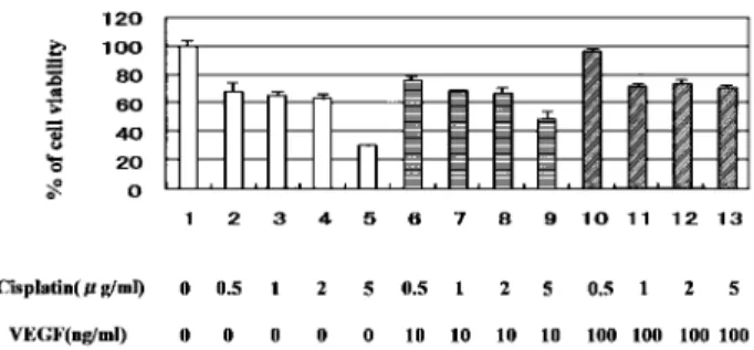
We tested whether VEGF could protect HN-Eso-1 cells from the anti-tumor agent, cisplatin. Cisplatin reduced the viability of HN-Eso-1 cells in a dose-dependent fashion as shown in Fig. 3 (lane2 - 5). The percent of cell viability"standard deviation (S.D.) was described as follows ; 68"6.44% (0.5μg/ ml), 65.2"2.86% (1μg/ml), 63.2"3.45% (2μg/ml),
Figure 2. Expression of VEGF
30μg protein was loaded in each lane. Lane 1, SW480 ; 2, DLD-1 ; 3, HN- Eso - DLD-1 ; 4, Lovo cells
Figure 1. RT- PCR
VEGF - A (VEGF121, VEGF165), VEGF - C, VEGF - D, VEGFR- 1, and VEGFR- 2 were detected by RT- PCR. Lane 1, SW480 ; 2, Lovo ; 3, DLD- 1 ; 4, HN- Eso - 1 cells
Figure 3. Assessment of cell viability
Cell viability was determined by MTT assay. Results were indu-cated as % of cell viability"standard deviation (S.D.). Triplicate experiments were performed. Treatment : 1 ; none, 2 ; cisplatin 0.5μg/ml, 3 ; Cisplatin 1 μg/ml, 4 ; Cisplatin 2 μg/ml, 5 ; Cis-platin 5μg/ml, 6 ; VEGF (10 ng/ml)+Cisplatin 0.5 μg/ml, 7 ; VEGF (10 ng/ml) + Cisplatin 1μg/ml, 8 ; VEGF (10 ng/ml)+ Cisplatin 2μg/ml, 9 ; VEGF (10 ng/ml)+Cisplatin 5 μg/ml, 10 ; VEGF (100 ng/ml) + Cisplatin 0.5μg/ml, 11 ; VEGF (100 ng/ ml) + Cisplatin 1μg/ml, 12 ; VEGF (100 ng/ml)+Cisplatin 2 μg/ ml, 13 ; VEGF (100 ng/ml) + Cisplatin 5μg/ml
30!1.23% (5 μg/ml). In the case of combination with VEGF (10 or 100 ng/ml), effect of cisplatin was attenuated. Results were described as follows ; 76.4! 2.91% (cisplatin 0.5 μg/ml and VEGF 10 ng/ml), 68.8!0.61% (cisplatin 1μg/ml and VEGF 10 ng/ ml), 67.8!2.88% (cisplatin 2 μg/ml and VEGF 10 ng/ml), 49.1!5.57% (cisplatin 5μg/ml and VEGF 10 ng/ml), 96.6!2.2% (cisplatin 0.5 μg/ml and VEGF 100 ng/ml), 72.2!1.28% (cisplatin 1μg/ml and VEGF 100 ng/ml), 73.7!2.56% (cisplatin 2μg/ ml and VEGF 100 ng/ml), 71!1.69% (cisplatin 5 μg/ml and VEGF 100 ng/ml).
Microscopic analysis
HN-Eso-1 cells are epithelial cell shape and they have a large nuclei and were developed mitochon-dria (Fig. 4). The mitotic cells were shown in Fig. 4 A.
DISCUSSION
We have established the novel HN-Eso-1 cell line from the esophageal so-called carcinosarcoma and used RT-PCR analysis to examine the VEGFs and VEGFRs in cells.
VEGF is a highly specific mitogen for vascular en-dothelial cells that is induced by hypoxia, oncogene activation, and variety of cytokines. VEGF is impor-tant in angiogenesis and neovascularization of solid tumor growth. Expression of VEGF and VEGFRs in the esophageal so-called carcinosarcoma has not been fully understood. It is very important to investi-gate the role of expression of VEGF and VEGFRs in the esophageal so-called carcinosarcoma. In these cells, transcription of VEGF-A, C, D, VEGFR-1, and VEGFR-2 were detected (Fig. 1). Expression of VEGF (VEGF-A) was detected in HN-Eso-1 cells (Fig. 2) and the other cell lines, Lovo, SW480, and DLD-1 also expressed VEGF (Fig. 2). VEGF-R2, which is a VEGF-A receptor plays an important role in pathological neovascularization via its tyrosine kinase activity and growth promoting signals to blood vessels (4). These results suggest that ex-pression of VEGFs and VEGFRs might be impli-cated not only in the HN-Eso-1 autocrine system but also in promotion of tumor environment. Indeed, the pathological analysis in a sample tissue suggests that both angiogenesis and lymphangiogenesis was well developed (data not shown).
Next we evaluated the effect of VEGF on HN-Eso-1 cells, although its direct effect on cancer cells has not been fully understood. Recombinant VEGF attenuated the cytotoxic effect of cisplatin on HN-Eso-1 cells (Fig. 3). However VEGF could not res-cue the cell death of SW480, DLD-1, or Lovo treated with cisplatin. (data not shown) VEGF appears to play an important role in the cytoprotective effect on HN-Eso-1 cells against anti-cancer agents.
SU11248, a VEGFR-2 inhibitor, has been ap-proved for cancer therapy, and undergone clinical setting (10). Our results suggest that SU11248 could be useful of esophageal so-called carcinosar-coma, in combination with other anti-cancer agents. Further studies are required to investigate in detail the cell survival signals of HN-Eso-1.
REFERENCE
1 Hemmi A, Terada N, Mizutani G, Fujii Y, Ohno S, Nemoto N : Squamous cell carcinoma with sarcomatous feature (so-called carcinosarcoma) of stomach probably metastasized from esopha-geal tumor : a case report with quick-freezing and deep-etching method. Med Electron mi-crosc 37 : 119-29, 2004
2 Stimpfl M, Tong D, Fasching B, Schuster E,
Figure 4. Microscopic analysis of HN- Eso - 1 cells
A ; Hematoxylin and eosin staining. B ; Electron microscopic analysis. Arrow : mitochondria, N : nucleus.
bermair A, Leodolter S, Robert Zeillinger R : Vascular endothelial growth factor splice vari-ants and their prognostic value in breast and ovarian cancer. Clin Caner Res 8 : 2253-9, 2002 3 Kato H, Yoshikawa M, Miyazaki T, Nakajima M, fukai Y, Masuda N, Fukuchi M, Manda R, Tsukada K, Kuwano H : Expression of vas-cular endothelial growth factor (VEGF) and its receptors (Flt-1 and Flk-1) in esophageal squamous cell carcinoma. Anticancer Res 6C : 3977-84, 2002
4 Aparicio S, Sawant S, Lara N, Barnstable CJ, Tombran-Tink J : Expression of angiogenesis factors in human umbilical vein endothelial cells and their regulation by PEDF. Biochem Bio-phis Res Com 326 : 387-94, 2005
5 Margit Stimpfl, Dan Tong, Barbara Fasching, Eva Schuster, Andreas Obermair, Sepp Leodolter, and Robert Zeillinger : Vascular En-dothelial Growth Factor Splice Variants and Their Prognostic Value in Breast and Ovarian Cancer. Clin Cancer Res 8 : 2253-2259, 2002 6 Jin T, Nakatani H, Taguchi T, Nakano T,
Okabayashi T, Sugimoto T, Kobayashi M, Araki K : STI571 (Glivec) suppresses the ex-pression of vascular endothelial growth factor in the gastrointestinal stromal tumor cell line, GIST-T1 World. J Gastroenterol 12 : 703-8, 2006
7 Nakatani H, Kobayashi M, Jin T, Taguchi T, Sugimoto T, Nakano T, Hamada S, Araki K : STI571 (Glivec) inhibits the interaction between c-KIT and Heat shock protein90 of the gastro-intestinal stromal tumor, GIST-T1 Cancer Sci 96 : 116-9, 2005
8 Nakatani H, Araki K, Jin T, Kobayashi M, Sugimoto T, Akimori T, Namikawa T, Okamoto K, Nakano T, Okabayashi T, Hokimoto N, Kitagawa H, Taguchi T : STI571 (Glivec) in-duces cell death in the gastrointestinal stro-mal tumor cell line, GIST-T1, via endoplasmic reticulum stress response. Int J Mol Med 17 : 893-7, 2006
9 Jin T, Nakatani H, Taguchi T, Sonobe H, Morimoto N, Sugimoto T, Akimori T, Nakano T, Namikawa T, Okabayashi T, Kobayashi M, Araki K : Thapsigargin enhances cell death in the gastrointestinal stromal tumor cell line, GIST-T1, by treatment with Imatinib (Glivec). J Health Sci 52 : 110-7, 2006
10 Faivre S, Delbaldo C, Vera K, Robert C, Lozahic S, Lassau N, Bello C, Deprimo S, Brega N, Massimini G, Armand JP, Scigalla P, Raymond E : Safety, Pharmacokinetic, and Anti-tumor Activity of SU11248, a novel Oral Multi-target Tyrosine Kinase Inhibitor, in Patients with Cancer. J Clin Oncol 24 : 25-35, 2006