IRUCAA@TDC : Age-related differences in expression of vascular endothelial growth factor by periodontal ligament cells in vitro
5
0
0
全文
(2) 143. Bull Tokyo Dent Coll (2007) 48(3): 143–146. Short Communication. Age-related Differences in Expression of Vascular Endothelial Growth Factor by Periodontal Ligament Cells In Vitro Kenichi Matsuzaka, Morito Tsuruoka, Eitoyo Kokubu, Akira Katakura, Takayuki Endo, Yoshihiro Shibukawa, Masuro Shintani, Masakazu Tazaki, Kazuyuki Ishihara, Sadamitsu Hashimoto, Masao Yoshinari and Takashi Inoue Oral Health Science Center HRC7, Tokyo Dental College, 1-2-2 Masago, Mihama-ku, Chiba 261-8502, Japan. Received 28 May, 2007/Accepted for publication 20 July, 2007. Abstract The purpose of this study was to evaluate age-related differences in expression of vascular endothelial growth factor (VEGF) by periodontal ligament (PDL) cells. PDL cells were obtained from Wistar male rats weighing approximately 150g each in the young group and 350g each in the old group. PDL cells derived from upper and lower incisors were seeded in 35-mm culture dishes after primary culture. For cell proliferation assays, cells were detached and counted at 1, 3, 5, 7, 11 and 14 days after culture. VEGF mRNA expression was analyzed with TaqMan®. The number of cells in both groups increased day by day, but the rate of increase in the young group was higher than that in the old group. VEGF mRNA expression in the young group increased from 3 to 14 days, but in the old group increased only slightly over the same time period. Expression ratios in the young group were higher than those in the old group, and there were significant differences between the young and old groups at 7 and 14 days of culture. In conclusion, the data revealed that PDL cells varied with age, and suggest that in view of such changes in cell proliferation and VEGF mRNA expression, age should be taken into consideration in periodontal treatment. Key words:. Aging— VEGF—Periodontal ligament—In vitro. Introduction Periodontal ligament (PDL) cells are heterogeneous cell populations containing fibroblasts and progenitor cells3). Furthermore, PDL cells are multifunctional cells that can differentiate into osteoblasts, cementoblasts or fibroblasts during wound healing in PDL. Vascular. endothelial growth factor (VEGF) has been demonstrated to have a remarkable potency to induce specific proliferation of endothelial cells4). Periodontal regeneration requires a coordinated series of events that includes not only the recruitment of PDL-specific cells, but vascular cells as well 9). VEGF is an angiogenic growth factor that elicits cellular responses to. 143.
(3) 144. Matsuzaka K et al.. injury1). Cell proliferation and VEGF expression play extremely important roles in the early stage of wound healing. Cellular activity is down-regulated with age, and the capacity for healing of wounds decreases with age. Angiogenesis plays an important role in homeostasis, wound healing and resistance to disease in PDL. Few studies have investigated age-related differences in characteristics of periodontal ligament cells5,8). The purpose of this study was to investigate age-related differences in cell proliferation and expression of VEGF mRNA in PDL cells.. Materials and Methods All animal studies were conducted in compliance with the Guidelines for the Treatment of Experimental Animals at Tokyo Dental College. PDL cells were obtained from Wistar male rats weighing approximately 150g each (5 weeks old) in the young group and 350g each (15 weeks old) in the old group. Five animals were sacrificed with an overdose of thiopental (RAVONAL®; Tanabe, Japan). Both the upper and lower incisors were extracted and washed in ␣-minimal essential medium (␣-MEM; GIBCO, Carlsbad, CA, USA) containing 10% gentamycin and 1.2% fungizone for 5 min. The pulp was then removed mechanically. Each tooth, with ameloblasts and tooth germ at the apical area removed, was placed with the PDL-side of the lingual aspect facing down in a 35-mm culture dish and cultured using ␣-MEM containing 10% fetal bovine serum (FBS, inactivated at 56°C for 35 min, GIBCO), ascorbic acid (50 l/ml, Wako, Japan) and antibiotic (gentamycin, SIGMA, St. Louis, MO, USA). The cells were cultured in a humidified atmosphere of 95% air and 5% CO2 at a temperature of 37°C. The culture medium was changed every 2 days. Fourteen days after primary culture, the cells were detached using trypsin/EDTA solution (0.1% trypsin, 0.02% EDTA, pH7.2) and subcultured. Cells from the 4th subculture were then used in the following experiments. For cell proliferation assays, approximately. 2⳯103 PDL cells were seeded in 24-well dishes and cultured. The culture medium was changed every 2 days. Cells were detached at days 1, 3, 5, 7, 11 and 14 using trypsin/EDTA solution, and counted with a Coulter counter (Coulter Z-1, Tokyo, Japan) at each time point. For quantitative RT-PCR, total RNA was extracted from each sample using the acid guanidium thiocyanate/phenol-chloroform method as follows. The cells were homogenized in Trisol® Reagent (Invitrogen, Carlsbad, CA, USA) after 3, 7 or 14 days of incubation after rinsing with PBS. Each solution was transferred to a tube containing chloroform and mixed. The solutions were centrifuged at 14,000 rpm at 4°C for 20 min, after which the supernatants were placed in tubes containing 70% isopropanol at ⳮ80°C for 1h. After centrifugation, the remaining mRNA pellets were washed with 70% cold ethanol. Finally, the mRNA pellets were dissolved in RNAasefree (DEPC-treated) water. Total RNAs were reverse transcribed and amplified using an RT-PCR kit (Takara Biomedicals, Shiga, Japan). RT-PCR products were analyzed by quantitative real-time RT-PCR in TaqMan® Gene Expression Assays for two target genes, VEGF (Rn 00582935-m1) and -actin (4352340E; as an endogenous control) (Applied Biosystems; Foster City, CA, USA), to determine variations in amounts of each RNA. All PCR reactions were performed using the real time PCR 7500 Fast System (Applied Biosystems). Gene expression quantitation using TaqMan® Gene Expression Assays was performed as the second step in a two-step RT-PCR. Assays were performed in singleplex reactions containing TaqMan® Fast Universal PCR Master Mix, TaqMan® Gene Expression Assays, distilled water and cDNA according to the manufacturer’s instructions (Applied Biosystems). Reaction conditions consisted of primary denaturation at 95°C for 20 sec, and cycling for 40 cycles at 95°C for 3 sec and 62°C for 30 sec. PCR data were measured in a relative manner as folds of the corresponding 3 days incubation in the young group with the threshold cycle. Three experimental runs were conducted,.
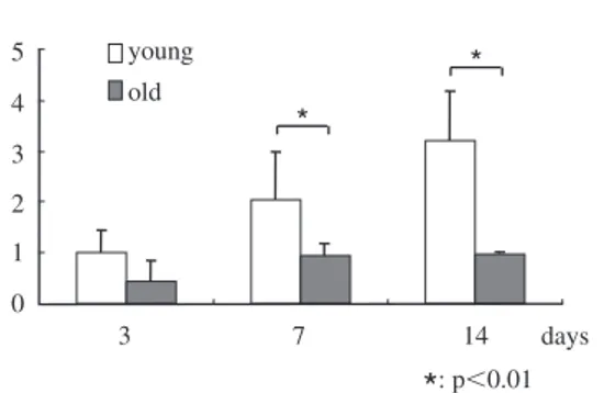
(4) 145. Age-related Differences in VEGF Expression by PDL. Number of cells. ⫻104 6. young. 5. old. 4 3 2 1. 5. young. 4. old. * *. 3 2. *. 0 0. 1. 3. *. *. *. 7. 11. 14. 1 0. 5. days. : p⬍0.01. *. 3. 7. 14. *. days. : p⬍0.01. Fig. 1 Cell proliferation assay The rate of increase in the young group is higher than in the old group.. Fig. 2 Expression of VEGF mRNA Although expression of VEGF by the old group increased slightly, expression of VEGF by the young group increased day by day. Data are shown as folds of the corresponding 3 days of incubation in the young group.. and the data were analyzed using the Student t -test (p⬍0.05).. stage6). This indicates that proliferation of PDL cells is absolutely imperative for wound healing in PDL. The results of this study indicate that a decline in cell proliferation in older people delays wound healing. Angiogenesis is essential in organ development and wound healing10). Booth et al. demonstrated that VEGF was involved in angiogenic processes in healthy as well as in diseased periodontal tissue2). Many studies have investigated angiogenesis in PDL, gradually clarifying its function been clarified. However, agerelated differences in angiogenesis are less well understood. VEGF is a crucial regulator of vascular development during embryogenesis (vasculogenesis), as well as in blood-vessel formation (angiogenesis) in adults7). Yoshino et al. reported that mechanical stress stimulated production of VEGF10). In this study, VEGF mRNA expression in the older rats was lower than that in the younger rats. The PDL in older animals is a disadvantage for orthodontic tooth moment. PDL cells play an important role in periodontal regeneration following treatment, including flap operation, guided tissue regeneration and emdogain application. PDL cells in older rats proliferate slowly compared to in younger rats, which suggests that some sort of growth factor is necessary during periodontal treatment in older patients. In conclusion, our data revealed that PDL cells varied with age and suggest that the age. Results The number of cells in both groups increased day by day, but the rate of increase in the young group was higher than that in the old group. There were significant differences between the young and old groups at 3, 11 and 14 days of culture (Fig. 1). VEGF mRNA expression in the young group increased from 3 to 14 days of incubation, but increased only slightly in the old group. Expression ratios in the young group were higher than those in the old group, and there were significant differences between the young and old groups at 7 and 14 days of culture (Fig. 2).. Discussion Angiogenesis plays a role in maintaining homeostasis in and restoring PDL after chronic inflammatory periodontal disease. Generally, wound healing commences with proliferation of blood vessels and cell proliferation. Ohta et al. concluded that stem cells were not involved in the regeneration of periodontium, although cells which migrated from residual PDL regenerated at an early.
(5) 146. Matsuzaka K et al.. of the patient needs to be taken into consideration during periodontal treatment.. Acknowledgements The authors would like to thank Ms. Saori Takano for her technical assistance, and associate professor Jeremy Williams for his assistance with the writing of the manuscript. This research was supported by Oral Health Science Center Grant HRC7 from the Tokyo Dental College by a “High-Tech Research Center” Project for Private Universities and matching fund subsidy from MEXT (Ministry of Education, Culture, Sports, Science and Technology) of Japan and 2007-2010 (No. 19592414) respectively.. References 1) Amemiya K, Kaneko Y, Muramatsu T, Shimono M, Inoue T (2003) Pulp cell responses during hypoxia and reoxygenation in vitro. Eur J Oral Sci 111:332–338. 2) Booth V, Young S, Cruchley A, Taichman NS, Paleolog E (1998) Vascular endothelial growth factor in human periodontal disease. J Periodont Res 33:491–499. 3) Fujita T, Iwata T, Shiba H, Igarashi A, Hirata R, Takeda K, Mizuno N, Tsuji K, Kawaguchi H, Kato Y, Kurihara H (2007) Identification of marker genes distinguishing human periodontal ligament cells from human mesenchymal stem cells and human gingival fibroblasts.. J Periodont Res 42:283–286. 4) Leung DW, Cachianes G, Kuang WJ, Goeddel DV, Ferrara N (1989) Vascular endothelial growth factor is a secreted angiogenic mitogen. Science 2465:1306–1309. 5) Oehmke MJ, Schramm CR, Knolle E, Frickey N, Bernhart T, Oehmke HJ (2004) Age-dependent changes of the periodontal ligament in rats. Microsc Res Tech 63:198–202. 6) Ohta S, Yamada S, Matuzaka K, Inoue T (2007) The behavior of stem cells and progenitor cells in the periodontal ligament during wound healing using immunohistochemical methods. J Periodont Res (in press). 7) Olsson AK, Dimberg A, Kreuger J, ClaessonWelsh L (2006) VEGF receptor signalling — in control of vascular function. Nat Rev Mol Cell Biol 7:359–371. 8) Ren Y, Maltha JC, Van’t Hof MA, KuijpersJagtman AM (2003) Age effect on orthodontic tooth movement in rats. J Dent Res 82:38–42. 9) Schlueter SR, Carnes DL, Cochran DL (2007) In vitro effects of enamel matrix derivative on microvascular cells. J Periodontol 78:141–151. 10) Yoshino H, Morita I, Murota SI, Ishikawa I (2003) Mechanical stress induces production of angiogenic regulators in cultured human gingival and periodontal ligament fibroblasts. J Periodont Res 38:405–410. Reprint requests to: Dr. Kenichi Matsuzaka Department of Clinical Pathophysiology, Tokyo Dental College, 1-2-2 Masago, Mihama-ku, Chiba 261-8502, Japan Tel: +81-43-270-3581 Fax: +81-43-270-3583 E-mail: [email protected].
(6)
図
関連したドキュメント
Effects of Cadmium Stress on Growth, Morphology, and Protein Expression in Rhodobacter capsulatus B10
In the present study, we identified six cadmium- induced proteins and five cadmium-binding proteins of R. Based on the elevated production of GroEL2, DnaK, and the ribosomal protein
mice, we found that S1P 2 was expressed in tumor vessels and normal blood vessels in many organs in both endothelial cells (ECs) and vascular smooth muscle cells, as well
In this study, we investigated whether tranilast inhibits the effects of TGF-b-induced EMT in HPMCs by inhibiting the TGF-b/Smad pathway, and whether fibrosis can be attenuated in
In our experiments, treatment of HUVECs with fluvastatin increased p38 phosphorylation; in addition, inhibition of p38 MAPK by SB203580 reversed the induction of TFPI by
TABLE 1: Primer sets, annealing temperatures (temp.) and expected amplified fragment sizes for reverse transcription–polymerase chain reaction analysis of genes encoding ephrin
Expression plasmids for JunB, c-Jun and JunD were each co-transfected with c-Fos expression plasmids as well as a luciferase reporter plasmid containing the wild-type LCR of
In the normal pancreas, moderate to marked basic FGF immuno- reactivity was present in a heterogeneous pattern at the basal aspect of acinar cells, and intense cytoplasmic FGF
PD-L1 expression was upregulated in macrophages and dendritic cells (DCs) in high-grade invasive human OSCC tissues or co-cultured with mesenchymal-phenotype OSCC cells in
