はじめに
心肺停止による急性脳障害はいまだ予後不良のことが多い
が,脳波上では多様なパターンの異常脳波が出現する1).な
かでも一側性周期性放電(lateralized periodic discharges; LPDs) は,脳卒中,頭部外傷など他の疾患でも多くみられるパターン であるが2)~4),低酸素脳症では予後不良の所見の一つとされ る5)6).また重積状態の一つである post-anoxic status epilepticus
(PSE)の場合も予後不良である7).我々は心停止後症候群
(post-cardiac arrest syndrome; PCAS)の男性例を経験した.そ の症例では昏睡状態における脳波で LPDs から electrographic seizureへの evolution(進展)を繰り返し,PSE と考えられな がら後遺症を残さずに回復した.脳低体温療法が一般化した 今日,PCAS において脳波上で LPDs や PSE のような予後不 良とされるパターンからも予測を上回る回復例もあることに 留意すべきである. 症 例 患者:53 歳,男性 主訴:心肺停止から蘇生後の意識障害 既往歴:高血圧症として 8 年前から近医で降圧剤を処方さ れていた. 家族歴:特記事項なし. 生活歴:喫煙 20 本 /30 年継続していた.飲酒は機会飲酒の みであった. 現病歴:2014 年 3 月下旬(発症第 1 日目)午後 3 時半ころ, 立ったままの軽作業中に呻いて倒れ意識消失した.Bystander cardiopulmonary resuscitation(CPR)は施行されず 8 分後に 救急隊到着時には心肺停止状態で,モニター上では心室細動 であった.電気的除細動,エピネフリン投与などの蘇生処置 が経過表に示す順序で施され,30 分後に自己心拍再開(return of spontaneous circulation; ROSC)した(Fig. 1).
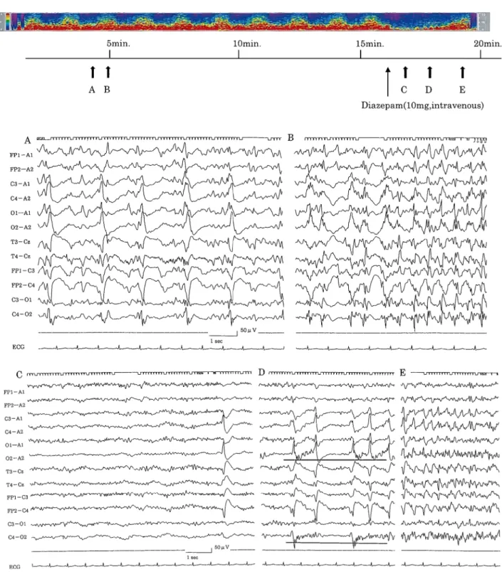
来院時現症および入院後経過:意識レベルは Japan Coma Scale-300の昏睡状態,血圧は 232/111 mmHg であった.自発 呼吸は保たれていたが不安定で気道確保のため気管内挿管 した.緊急で行われた心臓カテーテル検査では冠動脈には 狭窄なし.到着後もモニター上で心室頻拍(VT)が出現し, cardioversion(150J)で改善,アミオダロンを投与した.冠攣 縮予防にニコランジルを第 6 病日まで投与した.また来院当 日から四肢の痙攣も時折ありフェニトイン静注行いその後に バルプロ酸,レベチラセタムの経口投与へ変更した.また 34°C に維持した低体温療法(48 時間)を開始した.第 2 病 日の心エコー検査では駆出率 56%で心機能は良好と判断し た.第 3 病日には対光反射が出現し始めた.しかし第 4 病日 に 36°C まで復温した後も意識の回復はなかった.低体温療 法にあたりロクロニウム臭化物,プロポフォール(第 4 病日 まで),カルペリチド(第 5 病日まで)も投与した.血圧と自 発呼吸は安定してきたが,脳波記録を行った第 6 病日は昏睡 状態で右上下肢は時に自発的動きがあるのに対して,左上下 肢は筋トーヌス低下しており他動的な動きへの抵抗も無く弛 緩性麻痺と判断した.脳波記録中には不随意運動は無く,記 要旨: 53 歳の高血圧症の男性が心肺停止となり,蘇生処置で 30 分後に自己心拍が再開したが,昏睡は持続し 心停止後症候群と診断した.第 6 病日の脳波は,一側性周期性放電(lateralized periodic discharges; LPDs)から electrographic seizure への進展パターンを,約 15 分間に 10 回繰り返す cyclic seizure pattern であった.ジアゼ パム 10 mg の静注で背景活動のみとなった後に,再び LPDs へと変化した.抗てんかん薬を調整し,第 11 病日に 開眼し意識改善傾向となった以降は良好な回復で第 30 病日に退院した.
(臨床神経 2019;59:412-417)
Key words: 脳波,lateralized periodic discharges(LPDs),cyclic seizure pattern,心停止後症候群
*Corresponding author: 健和会大手町病院内科(〒 803-8543 福岡県北九州市小倉北区大手町 15-1) 1)健和会大手町病院内科
2)現:独立行政法人国立病院機構岡山医療センター小児外科
(Received May 8, 2018; Accepted April 23, 2019; Published online in J-STAGE on June 27, 2019) doi: 10.5692/clinicalneurol.cn-001183
脳波で cyclic seizure pattern がみられた心停止後症候群 59:413 録後の観察であるが体表に触れる刺激で頭部を右に向ける 動作と顔面にミオクローヌス様の微細な不随意運動が誘発さ れた.脳波所見は右後頭部(O2-A2)に最大電位を有する LPDsで振幅は 200~230 μV におよび,1 相性あるいは 2 相性 の鋭波で放電の間隔は 1.0~1.8 秒で,背景活動は抑制傾向で あったが棘波の重畳もみられた.周波数を増しながら分布も 右半球から対側半球にまで広がる spatiotemporal evolution の 過程がみられた(Fig. 2A, B).Color density spectral array 表 示(Fig. 2 upper panel)にあるように,この evolution の過程 は約 15 分間に 10 回繰り返した.ジアゼパム 10 mg を静脈内 投与したところこの高振幅の鋭波が一旦は消失し,α および β 帯域波の背景活動が約 16 秒間続いた.その後に再び鋭波が 出現し(Fig. 2C),さらに周波数を増しながら背景活動に低振 幅速波も重畳し(Fig. 2D 下線部),electro graphic seizure と なった時点までが記録された(Fig. 2E).頭部 CT では第 1 病 日には皮髄境界が不鮮明で全般的な脳浮腫と考えられた (Fig. 3A).第 9 病日には右頭頂部に局所性の低吸収域をみと めた(Fig. 3B).第 11 病日に開眼がみられ同時に抜管した.肺 炎の合併も経過中にあったが抗菌薬治療で改善した.第 15 病 日にはほぼ意識は清明となり,その後の経過は良好で第 30 病 日に独歩退院となり職場復帰した.退院後の頭部 CT では低吸 収域は消失していた(Fig. 3C).安静時の心電図では Brugada 症候群や QT 延長症候群の所見はなかった.回復後に他院で ホルター心電図にて冠攣縮性狭心症発作が確認され,植込み 型除細動器が装着された.装着後に 1 回作動していることか ら,今回の心肺停止は冠動脈攣縮からの致死的不整脈による ものであったと考えられた. 考 察
今回の例は持続脳波モニタリング(continuous EEG; cEEG) ではなく,低体温療法から復温後の,第 6 病日の約 20 分間の 単回の脳波記録である.Fig. 2A に示した周期性放電は,以前 は periodic lateralized epileptiform discharges(PLEDs)と呼ば
れてきたもので2),近年になり米国臨床神経生理学会は LPDs という用語への変更を提案した8)9).本稿では同学会の提案に したがい LPDs と記載しておきたい.背景に低振幅律動波や 速波が重畳している場合には LPDs+ と表記される.LPDs+ の脳波所見がみられる場合には,けいれん発作に移行しやすい ことが以前から知られている10).本例も波形から LPDs+ と 考えられ,昏睡状態であったが顔面の微細な痙攣,眼球偏倚が 挿間的にみられており comatose nonconvulsive status epilepticus (comatose NCSE)と考えられた11).脳波による NCSE の診断 基準として提唱された Salzburg consensus criteria に照らして spatiotemporal evolutionに該当する12).最近 Friedman らは持 続脳波モニタリングを施行した低酸素脳症も含む 13 例の critically ill patientsにおいて,短時間に起こる進展パターン (evolutionary pattern)の繰り返しを報告し,これを cyclic seizure
patternと名付けた13).彼らは cyclic pattern となる原因につい て詳細は不明であるとしながらもてんかん重積の一つのタイ プ(NCSE)と考えており,発作の停止状態を維持する機構 が破綻している可能性を考えている.本例ではジアゼパム投 与後に高振幅放電の出現はブロックされ,一時的に α および β 帯域の背景活動のみになった(Fig. 2C).しかし,その後す ぐに再度の LPDs からの進展がみられた(Fig. 2D, E).抗て Fig. 1 Clinical course of the patient.
On admission, the patient was deeply comatose but had multifocal intermittent activities involving the arms and legs. His pupils were dilated (5 mm) and the papillary reaction to light was initially absent on both sides. Therapeutic hypothermia was performed for two days. On day 3, light reflex emerged. An electroencephalogram was recorded on day 6. He opened his eyes on the 11th hospital day, after which his general conditions showed a good recovery. VF: ventricular fibrillation, ROSC: return of spontaneous circulation, EEG: electroencephalogram.
Fig. 2 The electroencephalogram of the patient.
Upper panel: Twenty minutes of color density spectral array (referenced as O1–A1, O2–A2) containing 10 evolving electrographic seizures recurring at relatively constant intervals. Time is plotted along the x-axis and the y-axis represents frequencies from 1–20 Hz. The frequency-specific power from 0 to 3.5 μV is visually represented using a color scale, with blue representing low power and light blue, green, yellow and red representing successively increasing power. The arrows (A through E) refer to the segments of raw EEG recording (see lower panel). About 15 minutes after the start of the recording (long arrow), 10 mg of diazepam was administered intravenously, which resulted in the attenuation of seizure activity. Lower panel: (A, B) Before the administration of diazepam. A representative example of raw EEG data showing lateralized periodic discharges (A) evolving to a seizure pattern (B) predominantly in the right scalp channels. (C–E) After the administration of diazepam. Following the loading of 10 mg of diazepam, high-amplitude discharges were blocked, and relatively stable background activity was observed for 16 seconds, followed by a single sharp wave emergence (C). Thereafter, LPDs accompanying low-amplitude fast rhythm on background activity (LPDs+: underlined) mainly in the occipital area were observed (D), finally evolving to electrographic seizure (E).
脳波で cyclic seizure pattern がみられた心停止後症候群 59:415 んかん薬が重積を完全に停止させるには不足あるいは適当で なかった可能性を考え,薬剤の調整を行った(Fig. 1).心肺 停止後では脳波は大きく変化していることが cEEG による知 見で推測されている1).今回は第 6 病日の単回の記録であり, 発症直後の脳波検査を行っていない.そのため回復過程にお ける脳波パターンの変動や推移に関する情報が今回は得られ ていない.知見の集積のためにも cEEG は施行困難であって も,発症早期からの脳波検査を繰り返すことは重要であると 思われた. 画像所見では初日の CT は皮髄境界が不鮮明で,低酸素に よる全般性の浮腫が示唆され(Fig. 3A),第 11 病日になると, 右の頭頂葉から後頭葉にかけて局在性の低吸収域が明瞭に なった(Fig. 3B).この低吸収域の所見は,退院後の評価では 改善しており(Fig. 3C)一過性にみられた左半身の麻痺の責 任病巣と考えられた.脳波で PLEDs が出現しながら運動麻痺 を含む神経脱落徴候が 7 日間持続した例も報告されており14),
本例も NCSE に伴う ictal paresis であった可能性がある.脳 全体に及ぶ虚血が想定される病態でありながら局在性の病変 となる機序としては,全般性浮腫からの回復過程が脳動脈硬 化などの要因により,部位によって差が出た可能性もある. しかし脳波所見と併せて考えると,焦点性の NCSE の結果とし
て頭部 CT で低吸収域が持続したと思われた15).浮腫には細胞
性と血管性が考えられるが,MRI での diffusion-weighted image (DWI)や apparent diffusion coefficient(ADC)の情報が無く,
区別は困難であった. 頭部偏倚の方向は,一般に焦点発作では,発作起始の時点で は病変の部位とは対側である場合が多いとする意見がある16). また発作初期には対側で,二次性全般化後には同側に変化す る場合や17),深部電極やビデオ脳波モニタリングによる検討 では,病巣と頭部の回旋の方向は一定せず同側の場合も多いと する報告もあるなど,側方性については一定していない18)19).
Epileptogenic zoneからsymptomatogenic zoneへの発作波の伝 播の様式によっても症状が異なってくると考えられた. 本例は Bystander CPR が行われず ROSC まで 30 分を要し た院外の心停止でありながら,後遺症無しに職場復帰できた 症例である.低酸素脳症後の昏睡からの回復予測は重要な テーマであり,臨床症状と並んで電気生理学検査による評価 が不可欠である.とりわけ短潜時体性感覚誘発電位(SSEP) で両側性の N20 消失は,高い精度で予後不良(1%以下の意 識回復)を予測できる20).本例のような脳波上の PSE の所見 も独立した予後不良因子と考えられているが,回復した例も 少数ながら含まれている7)21).Rossetti らは低体温療法を施さ れ,PSE の状態から覚醒に至った計 6 例について予測因子を 解析した.それによると①脳幹反射が保たれている,② SSEP で N20 が片側あるいは両側出現している,③脳波で背景活動 (10 μV 以上)がみられ,痛み刺激などへの反応性がみられる, の三つの全てを満たしていた場合には,重積状態の治療に よって比較的良好な結果が期待できるとしている22).すなわ ち脳幹-視床-皮質への投射系機能の保全が意識回復に重 要と考えられる.本例では①対光反射の第 3 病日での出現, ③脳波の背景活動の保持(刺激への反応性はみていない),の 二つの項目が該当していた.②の SSEP は施行しなかった. 脳低体温療法が広く行われるようになった今日では,鎮静剤 や筋弛緩剤はじめ神経系に影響する多くの薬剤が投与される ことが多い.薬剤の代謝系への影響も考えられるし,微細な ミオクローヌスなどの症状はマスクされる可能性もある.単 一の評価法や臨床所見のみでは完全な予後予測は未だ困難 で,早すぎる治療撤退の危険性がある.昏睡状態における脳 機能の評価のためには画像検査のみでは困難であり,持続脳 波モニタリングや SSEP,聴性脳幹反応などの神経生理学的 検査の重要性は,今後さらに増していくと思われる6)23)24). 本例の概要は第 314 回内科学会九州地方会(宮崎)で発表した. ※著者全員に本論文に関連し,開示すべき COI 状態にある企業,組 織,団体はいずれも有りません. 文 献
1) Bauer G, Trinka E, Kaplan P. EEG patterns in hypoxic Fig. 3 Brain CT findings of the patient.
A horizontal section of a CT image obtained at admission (A) revealed diffuse edematous changes of the brain. Ten days later, a low-density area became apparent on the right parietal zone (B), but had resolved completely on day 51 (C).
5) Pedersen GL, Rasmussen SB, Gyllenborg J, et al. Prognostic value of periodic electroencephalograpchic discharges for neurological patients with profound disturbances of conscious-ness. Clin Neurophysiol 2013;124:44-51.
6) Greer DM, Rosenthal ES, Wu O. Neuroprogonostication of hypoxic-ischemic coma in the therapeutic hypothermia era. Nat Rev Neurol 2014;10:190-203.
7) Rossetti AO, Logroscino G, Liaudet L, et al. Status epilepticus. An independent predictor after cerebral anoxia. Neurology 2007;69:255-260.
8) Hirsch LJ, Brenner RP, Drislane FW, et al. The ACNS subcommittee on research terminology for continuous EEG monitoring: proposed standardized terminology for rhythmic and periodic EEG patterns encountered in critically ill Patients. J Clin Neurophysiol 2005;22:128-135.
9) Hirsch LJ, LaRoche SM, Gaspard N, et al. American Clinical Neurophysiology Society’s Standardized Critical Care EEG Terminology: 2012 version. J Clin Neurophysiol 2013;30:1-27. 10) Reiher J, Rivest J, Grand’Maison F, et al. Periodic lateralize
epileptiform discharges with transitional rhythmic discharges: association with seizures. Electroencephalogr Clin Neurol 1991; 78:12-17.
11) Trinka E, Leitinger M. Which EEG patterns in coma are nonconvulsive status epilepticus? Epi Behav 2015;49:203-222. 12) Beniczky S, Hirsch LJ, Kaplan W, et al. Unified EEG terminology
2001;124:1683-1700.
17) Wyllie E, Luders H, Morris HH, et al. Ipsilateral forced head and eye turning at the end of the generalized tonic–clonic phase of versive seizures. Neurology 1986;36:1212-1217.
18) Robillard A, Saint-Hilaire JM, Mercier M, et al. The lateralizing and localizing value of adversion in epileptic seizures. Neurology 1983;33:1241-1242.
19) Ochs R, Gloor P, Quency F, et al. Does head-turning during a seizure have a laetralizing or localizing significance? Neurology 1984;34:884-890.
20) Robinson LR, Micklesen PJ, Tirschwell DL, et al. Predictive value of somatosensory evoked potentials for awakening from coma. Crit Care Med 2003;31:960-967.
21) Legriel S, Hilly-Ginoux J, Resche-Rigon M, et al. Prognostic value of electrographic status epilepticus in comatose cardiac-arrest survivors in the therapeutic hypothermia era. Resuscitation 2013;84:343-350.
22) Rossetti AO, Oddo M, Liaudet L, et al. Predictors of awakening from postanoxic status epilepticus after therapeutic hypothermia. Neurology 2009;72:744-749.
23) Taccone F, Cronberg T, Friberg H, et al. How to assess prognosis after cardiac arrest and therapeutic hypothermia. Crit Care 2014;18:202.
24) Fugate JE. Anoxic-ischemic brain injury. Neurol Clin 2017;35: 601-611.
脳波で cyclic seizure pattern がみられた心停止後症候群 59:417 Abstract
A case of post-cardiac arrest syndrome presenting with lateralized periodic
discharges evolving to a cyclic seizure pattern on electroencephalogram
Koji Shimozono, M.D., Ph.D.
1), Masanao Taba, M.D.
1)and Shojiro Hanaki, M.D.
1)2)1)Department of Internal Medicine, Otemachi Hospital 2)Present address: Department of Pediatric Surgery, Okayama Medical Center
A 53-year-old man with hypertension experienced sudden cardiopulmonary arrest. Ambulance crews detected
ventricular fibrillation that responded to defibrillation. Cardiopulmonary resuscitation resulted in return of spontaneous
circulation (ROSC) after 30 minutes. At admission to our hospital, he was in a comatose state. Therapeutic hypothermia
was performed for two days with other supportive care. However, despite these therapies, he remained comatose, and a
diagnosis of post-cardiac arrest syndrome (PCAS) was made. On the sixth hospital day, an electroencephalogram (EEG)
showed lateralized periodic discharges (LPDs) in the right occipital area evolving to electrographic seizures. Over
roughly 15 minutes, this evolution process repeated 10 times, demonstrating a cyclic seizure pattern. Intravenous
administration of 10 mg diazepam resulted in temporal attenuation of the high-amplitude discharges followed by LPDs
re-emergence accompanying a low-amplitude fast rhythm on the background activity (LPDs+). Antiepileptic drugs
administration was continued, along with supportive care. He opened his eyes on the 11th hospital day, after which his
general conditions showed a good recovery. He was discharged on the 30th
day without any sequelae.
(Rinsho Shinkeigaku (Clin Neurol) 2019;59:412-417)
Key words: electroencephalogram, lateralized periodic discharges (LPDs), cyclic seizure pattern,