第6巻 第1号 昭和53年6月15日
内 容
原 著
沖縄への夏季帰省が沖縄より本土に移住したヒトの耐熱性,耐寒性および局所耐 寒性に及ぼす影響について…………辻田 純三,田中 信雄,黛 誠,堀 九州産ヤマブユ亜属の1新種(英文)…………・・……・………一・………・………高岡 実験的犬バベシア感染に伴うビーグル犬の病態生理学的所見(英文)
・・石嶺 毅,牧村 進,北沢作治郎ジ田村 哲,鈴木 直腸生検を中心とした日本住血吸虫症の研究
3 皮内反応閾値・臨床的分析および臨床諸検査 「付 全篇の総括
・・加茂 悦爾,薬袋 藤,石崎 Vietnam難民に対する腸管寄生虫類の検査,特にセイロン鉤虫感染者について ・・影井 昇,木畑美知江,浅野
会則………
投稿規定・
会員名簿…一…一
記行清宏 1−8 9−14
直義15−26
達27−42
和仁43−49
51−54
−」 55−56
57−82
日熱医会誌
Japan.J.T.M.H. 日 本熱帯医学会
日本熱帯医学会雑誌 第6巻 第1号 19781−8頁 1
沖縄への夏季帰省が沖縄より本土に移住したヒトの耐熱性,
耐寒性および局所耐寒性に及ぽす影響について
辻田純三・田中信雄・黛 昭和53年3月4日
誠・堀 清記
受付
序 論
ヒトの体温調節機能は,季候の変化に伴って適 応的変化を生じることはよく知られている(Di11
8孟αム,1938;Robinson8孟α乙,1943)。
沖縄は亜熱帯に属し,夏は高温多湿の期間が長 く,冬は温暖である。沖縄住民は,温帯である日 本本土の住民と比較して,高温多湿の季候下に生 活する期間が長く,高温環境によく馴化している が,低温環境に曝露される機会が少なく,耐寒能 力が劣っていると推定されている。
最近沖縄住民で本土に移住する人の数は増加の 一途をたどっているが,その場合に沖縄住民がど のような過程で本土の季候に適応していくかは,
単に季候生理学的興味にとどまらず,本土での生 活活動の上からも重要な間題である。我々はすで に沖縄から本土に移住したヒトは本土住民と比較 して耐熱性は秀れているが,耐寒性が劣っており
(Horiθ孟謡,1975),耐寒性については,本土での 在住期間が長くなると,耐寒能を獲得して,本土 住民の耐寒能力に近づくことを観察している(中 村ら,1977)。
ところで,我が国の習慣によって夏(盆,大学 の夏休み)の一時期には出身地に帰る人が多い。
ことに大学生の場合は,夏休みが2ヵ月ほどあり,
沖縄より本土に来ている大学生が沖縄に帰る場合,
かなり長期間亜熱帯の季候下に生活することにな り,沖縄での生活がヒトの季候への馴化状態を変 化させるものと推定されているが,どの程度の変 化があるかを実測した報告がない。
そこで,沖縄より本土に移住した大学生が夏季 に沖縄で生活すると,高温環境曝露時および低温 環境曝露時の生理的反応,耐熱性,耐寒性,指の 局所寒冷血管反応にどのような影響を及ぼすかを 研究した。
研 究 方 法
沖縄より日本本土(関西)に移住して本土での 在住期間が2〜3年の男子大学生を被検者として,
夏休み前の7月上旬(平均気温26.O C,湿度80%)
と,夏休みを沖縄(平均気温28.1C,湿度79%)
で過した後,再び本土に来た9月上旬(平均気温 23.1C,湿度74%)に,暑熱曝露および寒冷曝露 時の生理的反応と局所寒冷血管反応を測定した。
実験は,体温の日内変動,運動,食事の影響を 避けるため,実験日には激運動を行わせず空腹状 態の被検者について,午後3時より測定を開始し た。暑熱曝露実験および局所寒冷血管反応は10人 の同一被検者について,寒冷曝露実験は5人の同 一被検者について行った。
1. 暑熱曝露実験
気温30C,湿度70%,風力17cm/secの人工 気候室内で被検者に30分安静をとらせたのち,水 泳パンツのみを着用させ42Cの下肢温浴を1時 間行わせ,その間の胸部および背部の局所発汗量 を15分毎に濾紙法で測定し,局所の汗のNa濃度 を求めた(Ohara,1966)。局所の汗の平均Na濃 度は4回の測定に含まれるNaの総量を総水分量 で除して求め,胸部と背部の平均値を汗の平均 Na濃度とみなした。実験終了時によく乾いたタ 兵庫医科大学生理学第一講座 663西宮市武庫川町1−1
オルで身体を充分ぬぐってから体重を測定し,体 重減少量にパンツの重量増加量を加えて真の体重 減少量を求め,この体重減少量を発汗量とみなし た。舌下温は15分毎に測定した。
耐熱性の判定を行うための指標は以下の式に よって算出した(Horiθ孟磁,1974a)。
相対的水分損失量ハー論灘叢辮。7
舌下温の上昇度(C)
相対的体温上昇度Bニ
40.6一初期舌下温(C)
相対的塩分損失量C=体重減少量(kg)×注璽 初期体重 平均Na濃度(mEq/1)×0、058 (kg)×0。75
耐熱性の指標∫ニぬ42+β2+(72
相対的体温上昇度と相対的水分塩分損失量の比 S=B/紐2+C2
1の値は,高温負荷によって体内に生じた水分塩 分代謝の平衡およぴ体温平衡に対して生じた生理 的歪みの大きさを示し,この値が少ない被検者ほ ど体温,水分塩分代謝の恒常性の維持能力が秀れ ており,耐熱性が秀れていると判定される。
2.寒冷曝露実験
1.8cloに相当する被服をつけた被検者を仰臥さ せ,寝具で充分保温した状態で20分間安静をとら せた後,寝具をとり去って,気温10C,湿度70%,
風力17cm/secの低温環境に1時間曝露した。
寒冷曝露前,寒冷曝露後30分および60分の代謝 量と全身6カ所の皮膚温を測定した。皮膚温の測 定部位および平均皮膚温算出のための加重平均係 数は次の通りである(緒方,1970)。
部 位 係数 前額中央眉上2cm
右側乳首部の下
左側前腕内面中央線,下から1/3 左側大腿前面中央線,上から1/2 左側下腿外側,上から1/3 左側足背中央線,上から1/2
0.07 0.35 0,17 0.19 0.13 0.09 耐寒性の判定のための指標は次のようにして算
出した(緒方,1970)。
曝露前の代謝量をMb(Kca1/m2/hr),曝露後30 分および60分の代謝量をそれぞれMi30(Kca1/m2/
hr)およびM6。(Kcal/m2/hr),曝露前の平均皮膚 温丁。(C),曝露後30分および60分の平均皮膚温を
T30(C),およびT60(C)として,曝露後30分およ び60分後の代謝量の増加量(」M)と平均皮膚温 の下降度(∠T)の比を次式によって求めた。
∠コ43。二沼43rM。 」ハ46。_ルf60一ルf。
」T30 To−T30 , 」T60 To−T60 寒冷環境に馴化すると,寒冷曝露時に皮膚温を 低下させ,放熱量を減少させる能力を獲得し,∠
Tが大きくなり,代謝量の増加を少なくして体温 が維持できるようになり」M が小さくなる。従っ て∠MμTの値が小さいほど耐寒性が秀れてい
ると判定できる(Carlson召∫磁,1965)。
3.局所寒冷血管反応の測定
室温20〜24Cの測定室に安静状態で30分間滞 在させたのち,被検者を静座させ,左中指末節背 面に銅コンスタンタン熱電対を絆創膏にてはりつ け,その上にワセリンを塗って防水し,室温での 指の皮膚温を測定したのち,指を第一節まで撹拝 されたO Cの氷水中に30分浸し,その間の指温の 変化を測定した(Yoshimura8孟磁,1950)。
局所耐寒性の判定は次のようにして行った。ま ず,浸水後5〜30分の平均皮膚温,mean skin
temperature M.S!r、(C),浸水後最初の皮膚温が 上昇するときの皮膚温,血管反応発現温度,tem−
perature of first rise after immersion T.F.R.(C),
および,浸水後最初の皮膚温が上昇するまでの時 間,血管反応発現時間,time for丘st temperature rise after immersionT.T.R.(分)を求めた。
以上の三つの測定値は,浸水前の指皮膚温に影 響されるので,浸水前指皮膚温丁(C)によって 補正した値を用いて指数を算出した (中村ら,
1g72)。M.S.T.にっいては0.4414T−10.0319未 満の場合を1点,この値と0.4414T−7。0509の間 の場合を2点,0.4414T−7.0509より高い場合を
3点とした。
T.F.R.については,logT,F.R.が0其)464T−
1.9141未満の場合には1点,0.0464T−0,8685よ り大きい場合は3点とし,これらの間の値であれ ば2点とした。
T.TR.にっいては,一〇4268T+22.7285より 大きい値の場合は1点,一〇.4268T+18.9765未満 の場合を3点とし,この間の値を2点として評価
した。
3
M.S.T.,TF.R.およびT.T.R.の評点の合計か ら抗凍傷指数(R.1.,Resistance index)を求め た。指標値が大きいほど局所耐寒能が秀れている
(Yoshimura8孟αム,1951)。
実 験 成 績
1. 暑熱曝露実験
被検者が沖縄へ帰省する前と後における暑熱曝 露時の発汗量,汗のNa濃度,口内温の上昇度,
相対的水分損失量,相対的体温上昇度,相対的塩 分損失量および耐熱性の指標の平均値と標準偏差 を表1に示した。
Table l Ef石ect of retum to Okinawa on sweating reaction,changes in body temperature during heat exposure and heat tolerance
Time Befbre After
体温の上昇度は0.08C減少している。しかし これらの差はいずれも統計的に有意ではなかった。
高温環境に馴化するときの発汗反応の変化は汗 量が増加し,汗の塩分濃度が減少するといわれて いる。また,体温の上昇度は馴化に伴って減少す ることが知られており,帰省中の沖縄での高温環 境下での生活が,高温馴化を招来せしめているこ
とが推定される。
耐熱性の指標とその成分については,帰省後は 帰省前に比べて,相対的水分損失量がかなり増加 し,相対的塩分損失量がわずかに増加している。
相対的体温上昇度はかなりの減少を示し,耐熱性 の指標1値およびS値の減少がみられる。しか
しこれらの差は全て有意差ではなかった。
2.寒冷曝露実験
沖縄への帰省前後における寒冷曝露時の代謝量 および平均皮膚温の変化と耐寒性の指標の平均値
と標準偏差を表2に示した。
W灘CT∬オBO13 59.3ゴニ6.9
0.41±0.18 60.9±21,8 36.9士0.30 0.60ニヒ0.30 0.101±0.046 0.158土0.046 0.034士0.020 0.201=ヒ0.056 1.72±0.87
59.3ニヒ6.4 0.48±0,14 59.5二L21.6 36.9士0.24 0,52±0.23 0.116=ヒ0.035 0.140ニヒ0.057 0.038±0.020 0.184ニヒ0.045 1.21ニヒ0.52
Table2 Ef琵ct of retum to Okinawa on changes in metabolismラmean skin temperature during cold exposure and cold tolerance indices
Time Befbre Af㌃er
W:Initialbodyweight(kg),∠W:Body weight loss(kg),C:Mean Na concen.
tration in sweat(mEqμ), T:Initial oral temperature(C), 」T:Rise in oral tem−
perature(C),且:Relativewaterl・ss,
B:Relative rise in oral temperature, 0:
Relativesaltloss, 1:Heattolerance index, S: Ef琵ctiveness of sweating,
Meanvalues aregivenwith theirstandard deviations.
ハ40 4M30
」ルf60 To 」T30
4T60
」M 30μT30
4M60μT60
36.7±3.4 37.0士4.4 1.2±0.87 11.9」二7.1*
9.0±9.9 9.1」二6.3 33.3ニヒ0.56 32.6ニヒ0.4r7
3.5士0.60 3.1士0.06 4.7±0.82 3.8±0.20
0.33ニヒ0.22 3.8士2.3*
2.2±2.5 2.4±1.8
M:Metabolic rate(Kcal/m2/hr), T:
Mean skin temperature(C),Su伍x in−
dicates time(min)a丘er cold exposure.
Mean values aregivenwith theirstandard deviations, * Signi且cant dif琵rence be−
tween two experiments at5%leveL
帰省後の測定値と帰省前の測定値とを平均値で 比較すると,汗量は約17%の増加をみたが,塩分 濃度はわずかではあるが低くなっている。
帰省後の値と帰省前の値を比較すると,代謝量 は安静状態ではわずかに増加しているが寒冷曝露 30分で著しく増加し,その差は有意であった。
帰省後の増加は,ふるえ shivering をおこす 被検者が増加したことによる。曝露60分値はわず かに増加している。
平均皮膚温の寒冷曝露前の値はわずかに低く なっているが,平均皮膚温の低下度は曝露後30分,
60分値ともにかなり減少している。
代謝量の増加度と平均皮膚温の下降度の比
∠ルf/∠Tは小さい値をもつ人が秀れた耐寒性を もつものとされているが,この値を30分値でみる と,帰省前の平均値3.8は帰省後の平均値0.33よ り有意に大きく,60分値でも大きな値を示した。
このことにより,沖縄への帰省によって耐寒性は 減弱したものと推定される。
3,局所寒冷血管反応
沖縄への帰省前と帰省後の指の寒冷血管反応の 測定値と抗凍傷指数の評点およびその三成分の評 点の平均値と標準偏差を表3に示した。
Table3 Ef琵ct of retum to Okinawa on skin temperature in hunting re−
action of Hnger dippeci in ice water and resistance index of frost bite
の皮膚温(T、F.R.)はともにわずかに高かったが 最初に皮膚温が上昇するまでの時間(T.T.R.)は 著しく遅延し,この差は有意差であった。
M.S.T.およびT.F.R.の評点は帰省前後でほと んど差がなかったが,T.T.R.の評点は,帰省前値 の平均値が2.0に対し,帰省後は1.1に減少してお りこの差は有意であった。
抗凍傷指数の評点の平均値は帰省前値が6.9で あったが帰省後の値は5.8で有意の減少を示した。
帰省中の沖縄での高温季候下での生活は,局所 の耐寒性を減弱させたものと思われる。
考 察
Time Befbre After
M.S』r.
T.F.R.
T.T.R.
9.2±3.9 4.9土2.1 8.6±4.1 2.4±0.49 2.5二LO.50 2.0±0.63 6.9土1.04
10.4二と3.3 5.3±1.4 17.2十7.4**
2.3十〇.46 2.4±0.50 1.1ニヒ0、30**
5.8士0.49*
M.S.T.:Mean skin temperature(C),
T.F.R.:Temperature of first rise afヒer immersion(C), T.TR.:Time fbr first temperature rise afヒer immersion(mln),
Index: Reslstancc index of fヤost bite,
Mean values are given with theirstandard deviations. * Significant diHもrence be−
tween two experiments*at5%level,
**at1%leveL
帰省後の平均値を帰省前の平均値と比較すると,
平均皮膚温(M.S.T.)および最初の皮膚温上昇時
ヒトが高温環境に馴化するときは,一定の暑熱 負荷に対して発汗が早くから発現し,汗量が増加 し汗の塩分濃度が減少して,発汗による放熱量が 増加する発汗反応の適応的変化が現れ,体温の上 昇度が減少することはよく知られている(Dillθ孟 α乙,1938;Robinsonααゐ,1943)。
表1より明らかなように,沖縄への帰省によっ て暑熱負荷時の汗量が増加し,体温上昇度は減少
している。
汗の塩分濃度はわずかしか減少していないが,
汗の塩分濃度は汗量の増加に伴って上昇するので あるから(Kittsteiner,1911),汗量の増加があっ て,汗の塩分濃度が一定あるいは減少する場合は,
一定発汗量に対する汗の塩分濃度が減少している ことを示す。
従って,沖縄での高温季候下での生活が,高温 環境への馴化を促進したものと推定される。
高温環境での曝露時にみられる障害には,原因 別にして高体温による熱射病,脱水による熱疲慧,
塩分損失による熱けいれんがあるといわれている。
しかし,通常はこれらの障害が混在した状態がみ
られる(Leithead8∫α1.,1964)。
これらの三つの障害が現れて,生命に危険が及 ぶ体温上昇度,水分損失量,塩分損失量と実験条 件下での体温上昇度,水分損失量,塩分損失量の 比を相対的体温上昇度,相対的水分損失量,相対 的塩分損失量として数量化し,この三つの変化量
5
の大きさを表したものが耐寒性の指標1である
(Horiε孟磁,1974a)。表1に示されたように沖 縄での生活は,相対的水分損失量を増加させてい るが,相対的体温上昇度および耐熱性の指標1を 減少させている。即ち,高温馴化によって,主と
して相対的水分損失量で表された水分代謝に関す るストレスを増加させる犠牲を払って相対的体温 上昇で表される体温平衡に関するストレスを減少 させ,全体として生体に生じるストレスの大きさ を減少させたものと考えられる。
相対的体温上昇度と相対的水分塩分損失量の比 Sは,暑熱曝露により生体内に生じたストレスの
パターンを示す指標になるが,S値は沖縄帰省前 の平均値1.72から帰省後は平均値1.21とかなり大 きく減少している。
S値と1値の関係が帰省前後でどのように変化 したかを図1に示した。図では測定時の5値と1 値をS値と1値の平均値を中心とし半径がそれ ぞれの値の標準誤差の楕円で表した。
∫値とS値の関係は,
1ニ4オ2+B2+02=躍2+c241+B2/(イ2+02)
ニzへ/1十(0/、4)2∀1十S玄となる。
ところでAの増減に伴ってCも増減し,本研究 で用いられた実験条件では表1より判るように C/Aは約1/3である。C2/A2は約1/9で,1に比べ
て小さく,Aれ+(C/A)2はAによってほとんど 決定されてしまう(Horiθ孟磁,1974a)。この値
をaとすれば1=a41+S2の関係がえられる。図 1中の4本の線はaが一定である場合の1と8
の関係を示している。
長期間の高温馴化を行わせたような場合は1の 減少とともにa値が大きく減少するのであるが,
短期間の高温馴化では1値の減少は少なく,a値 が同じiso−sweating lineにそってS−1関係が変 化するといわれている(Horiθ雄ム,1974b)。
図より明らかなように,沖縄帰省による5イ関 係の変化は大略aニ0.1〜0.12の間の変化であり,
短期間の高温馴化にみられる変化の特徴をよく示 している。
ヒトが寒冷環境に馴化すると,寒冷曝露時の皮
1
0.20
0.15
aニ0.14 a=0.12
a=0.10
B
a=0.08
2.0 s
Figure1
1.0 1.2 1,4 1.6 1.8
Relationship between heat tolerance indices l and乱
A:Af[er retuming to Okinawa,B:Befbre retuming to Okinawaラa:Parameter indicatingv翫ri・uslevels・fis・・sweatinglinewhicharedrawnbyc・nnectingthe
P・ints・fthesamevalue・fオ(1+象)百・Circles:Drawna−dthemeanswith
radiuses ofstandard errors,Arrow indicates direction of chan蓼e in r¢lationship of 1ndices・1andSa丘erretumin$t・Ok蜘w3↑
膚温の低下度(∠T)が増加し,産熱量 (∠ハ4)
の増加量が減少し,そのため耐寒性の指標∠ハ4/
∠丁比が減少するといわれている(Carlson8孟磁,
1965)。
表2に示されたように,沖縄での生活は寒冷曝 露時の皮膚温の低下度を減少させる。寒冷環境下 では皮膚血管が収縮して皮膚への循環血液量が減 少し,皮膚温を低下させ,体表面よりの熱の放散 を減少させる生理的反応が発現するのであるが,
沖縄の気候は日本本土に比べて平均気温が高いだ けでなく,気温の日内変動が少なく,沖縄での生 活では低温環境に曝露される機会がほとんどない ので皮膚血管の収縮力が低下し,低温曝露時の皮 膚温低下による体熱損失防衛能力が劣ってしまっ
たものと推定される。
低温環境曝露時には,産熱量が増加して体温の 下降を防ぐ生理的反応が生じるのであるが,本研 究に用いられた実験…条件はほとんど ふるえ
sh
ivering が現れず,寒冷曝露後徐々に代謝量 が増加する曝露条件を用いている(緒方,1970)。
しかし,表2より明らかなように帰省後は,曝露 後30分の代謝量増加量は11.9Kcal/m2/hrで曝露 後60分の値9・1Kca1/m2/hrより大きい。即ち,帰 省後は30分で ふるえ による代謝量増加をきた した被検者が多く含まれていることを示す。そし て,30分値では帰省後の代謝量の増加量は帰省前 の値と比較して有意の差をもって増加している。
寒冷曝露時の代謝量が多い被検者は耐寒能が 劣っているのであるが,その著しい場合には ふ るえ がみられて代謝量が増加するのである。
従って,帰省して沖縄での生活を終えて本土に移 住した時は,明らかに帰省前と比べて耐寒能力が 劣っていると結論できる。
指の局所寒冷反応は,手がきびしい寒冷に曝露 されたとき指に存在する動静脈吻合が開張して指 の温度を上昇させる生理的反応を観察しているの であるが,この反応は手が寒冷に曝露される機会 が多いと,動静脈吻合の開張度が増加して皮膚温 を高く維持する能力が充まり,凍傷にかかりにく くなるといわれている(Yoshimuraε孟磁,1950;
Yoshimuraθ孟αゐ,1951)。
抗凍傷指数(R.L)は,動静脈吻合の開張度がよ い場合に高い評点がつけられているので,この指 数が大きい程凍傷に対する抵抗性が強いことにな
る。
表3より,寒冷血管反応は沖縄への帰省後を帰 省前と比べると,平均皮膚温と血管反応発現温度 はほとんど変化がなく,従ってこの二つの成分に 関する評点は差がないのに,血管反応発現時間は,
帰省前の平均値8.6分が帰省後は17.2分と著しく 延長し有意差があった。これを評点で比較すると,
平均値で2.0点より1.1点まで有意差で減少したこ とになる。
RI.値でも帰省後の平均値5.8点は帰省前の6.9 点より有意差で小さい。
即ち,沖縄での生活は血管反応の発現時間を著 しく遅延させ,凍傷に対する抵抗性を減弱させた ことが判る。
以上の結果より,夏に沖縄へ帰省して沖縄での 平均気温が高く,気温の日内変動の少ない季候下 で生活すると,高温環境への馴化が促進され耐熱 性を獲得するが,その程度はわずかであるのに対 し,寒冷環境への適応能力がかなり失われて,全 身耐寒性および指の局所耐寒性を大きく減弱させ
ると結論できる。
要 約
沖縄より日本本土に移住した男子大学生にっい て,夏季沖縄に帰省する直前と直後に,暑熱曝露
(10名)およぴ寒冷曝露(5名)時の生理的反応 と局所寒冷血管反応(10名)を測定して次の結果
を得た。
1) 帰省後は帰省前に比べて暑熱曝露時の汗量 が増加し,汗量当たりの汗の塩分濃度が減少し,
体温の上昇度が低下しており,帰省中に高温環境 に対する馴化がすすみ耐熱性を獲得する傾向がみ
られた。
2) 寒冷曝露時の生理的反応は,帰省後は帰省 前と比較して,皮膚温の下降度が低く,代謝量の 増加と ふるえ の頻度が多く,代謝量の増加度 と皮膚温の下降度の比が増加しており,全身の耐
7
熱能力は減弱していた。
3) 指の局所寒冷血管反応は,帰省後は帰省前 と比べて,血管反応の発現が著しく遅延し,抗凍 傷指数が減少して凍傷に対する抵抗性が減弱して
いた。
4) 日本本土より沖縄に夏季に帰省すると,耐 熱性はわずかに増加するが,全身耐寒性および局 所耐寒性はかなり減弱することが示された。
文 献
1) Carlson,L n and Hsich,A.C.L.(1965):Cold15−51,The physiology of human surviva1,
Academic Press,London and New York
2) Dill,D.B.,Ha11,F.G.and Edwards,H.T.(1938):Changes in composition of sweat d皿ing acclimation to heat,Am.J.PhysioL,123,412−419
3) Hori,S.,Inoue,A.and Ihzuka,H.(1974a):Indices and sweating pattems for the assess−
ment of heat tolerance,Jap.J.PhysioL,24,263−275
4) Hori,S.,Inoue,A.,Ihzuka,H.and Yamada,T.(1974b):Study on seasonal variations of heat tolerance in young Japanese males and effects of physiological training thereon7Jap・
J.PhysioL,24,463−474
5)Hori,S.,Nakamura,M.,Sugawara,K.,Inoue,A.and Ihzuka,H・(1975):A comparative study on sweating pattem and heat tolerance,A丑eld study on residents of Okinawa in sum・
mer,Int.J.Biometeor.,19,184−193
6) Kittsteiner,C(1911):Sekretion,Kochsaltgehalt und Sekretion des Schweisses,Arch.
Hyg.Berl,73,275−306
7) Leithead,C.S.and Lind,A.R.(1964):Heat stress and heat disorders, Davis,Philadeレ
phia
8) 中村 正,渡辺 孟,菅原和夫,槌本六良,桑野絃一,山口洋一,深堀英彦(1972):寒冷血 管反応による局所耐寒性の評価法の新しい試み,長崎医学会雑誌,47(2),180−189
9) 中村正,堀 清記,戸田嘉秋,佐々木隆,赤松 隆(1977):沖縄台湾,本土各出身者の 耐熱耐寒能の比較追跡 第2報 発汗反応と寒冷曝露時の代謝量の比較,日生気誌,14,24 10) 緒方維弘(1970):日本人の耐寒性とその測定法,18−31,日本人の適応能,講談社,東京 11) Ohara,K、(1966):Chloride concentration in sweat; Its indivi{lual,regiona1・seasonal and
some other variations,and interrelations between them,Jap.J.PhysioL,16,274−290 12)Robinson,S.,Turrell,E,S.and Horvath,SM.(1943):Rapid acclimatization to work in hot
climates,Am.J.Physio1.,140,168−176
13) Yoshimura,H.and Iida,T.(1950):Studies on the reactivity of skin vessels to extreme cold・
partl.Apointtestontheresistanceagainstfrost−bite,Jap・J・PhysioL・1・147−159
14) Yoshimura,H.and Iida,T.(1951):Studies on the reactivity of skin vessels to extreme cold・
partII.Fact・rsg・verringtheindividualdi仔erence・fthereactivity・・rtheresistancea−
gainst frost−bite, Jap.」.Physiol.,2,177−185
THE EFFECT OF THE RETURN TO OKINAWA FOR SUMMER VACATION ON HEAT TOLERANCE, COLD TOLERANCE
AND PERIPHERAL COLD TOLERANCE OF THE SUBTROPICAL MIGRANTS TO THE
TEMPERATE ZONE
JUNZO TSUJITA, NOBUO TANAKA, MAKOTO MAYUZUMI AND SEIKI HORI
Received for publication 4 March 1978
Sweating test and peripheral cold tolerance test were performed on 10 young male university students born and raised in Okinawa (the subtropical zone) but moved to the Japanese Main Islands (the temperate zone) within last three years and cold tolerance test was made on five subjects. Two series of experiments were performed in Nishinomiya, using a climatic chamber. The first series of experiments was performed in July before the return of the subjects to Okinawa (the experiment 2).
The second series ofexperiments was performed in September after a month's stay in Okinawa in August (the experiment I ) . Sweating reaction was examined on the subjects dressed in shorts only 60 min by immersing both legs in stirring water of42 C in a room of 30 C with 70 R.H. Cold tolerance test was performed on the subjects clothed with I .8 clo by taking off a blanket after covering with a blanket in supine position for 20 min in a room of 10 C with 700/0 R.H. Physiological responses of the subjects to cold were observed for 60 min. Peripheral cold tolerance test was made by dipping the left middle finger with a thermocouple attached to the nail bed into ice water for 30 min and changes of the skin temperature were measured continuously. Greater sweat volume, Iower sodium concentration in sweat at the given sweat rate and less rise in oral temperature in the experiment I than in the experiment 2 were observed.
These results indicate acclimatization to heat ofthe subjects was induced by the return to Okinawa in summer and heat tolerance of the subjects was slightly improved when assessed by numetrical heat tolerance index after a month's stay in Okinawa in summer.
In cold tolerance test, signiflcantly greater increase in the metabolic rate, greater frequency of shivering and considerably lower drop of the mean skin temperature in the experiment I than in the experiment 2 were observed. These results indicate the ratio of increase in the metabolic rate to the drop in the mean skin temperature during cold exposure was greater in the experiment I than in the experiment 2. Thus it might be said that cold tolerance was reduced by the return to Okinawa in summer and living in a hot and humid climate. The mean value of time for first temperature rise of the vascular reaction offinger in the cold was significantly greater in the experiment I than in the experiment 2. The mean value of resistance index offrost bite caluculated by the Nakamura's method was signifi‑
cantly smaller in the experiment I than in the experiment 2. It may thus be concluded that the vascular reaction of finger in the cold is depressed and peripheral cold tolerance is reduced after the return to
Okinawa in summer.
T e Ist Department of Physiol0 y, Hyogo College of Medicine, Nishinomiya
Japan.J. Trop. Med. Hyg., Vol. 6, No. I 1978 pp 9 14 9
A NEW SPECIES OF BLACK‑FLY FROM KYUSHU, JAPAN
(SIMULIIDAE : DIPTERA)
HIROYUKI TAKAOKA
Received for publication 4 January 1 978
Abstract: The adult and immature stages ofanew species ofthe black‑fiy, Simulium (Gnus) kyushuense are described based on specimens collected from lowland rivers in Kumamoto, thc central part of Kyushu Island, Japan.
This new species is assigned to the subvariegatum group of the subgenus Gnus Rubzov and has a close affinity to S. (G.) nacojapi Smart, from which it differs by the deep postgenal cleft of larva and unbranched macrosetae on the 7th sternite of female.
In Kyushu area, the southern‑most main island in Japan, 25 species of Simuliidae have been reported (Shiraki, 1935; Bentinck, 1955; Ogata et al., 1956 and Takaoka et al., 1977). Among these, four species belong to the subgenus Gnus Rubzov, viz., Simulium (Gnus) nacojapi Smart, S. (G.) bidentatum (Shiraki), S. (G.) daisense (Takahasi) and S. (G.) malyshevi Dorogostajskij, Rubzov et Vlasenko.
The unknown black‑fly species of the subgenus Gnus which was collected from 10wland rivers in Kumamoto, the central part of Kyushu in September 1977 is apparently differentiated from these four known Gnus species.
In the present paper, this is described as a new species belonging to the sub‑
variegatum group defined by Rubzov (1959‑1964) .
DESCRIPTION
Simulium (Glws) kyushuense sp. nov.
Female
General body color shiny black. Body length about 2.5 mm. Wing length about I .8 mm.
Head: Narrower than thorax. Frons shiny black, with a few black hairs
along each lateral margin and on lower portion ; the greatest width of frons slightly over lj3 of the width of head; ratio of the greatest width, the narrowest one near antennal bases and the height of frons 4: 3 : 3. Clypeus black, grey‑pruinose and covered sparsely with black hairs. Antenna composed of 2+9 segments ; coloration somewhat variable, in some specimens almost black with scape, pedicel andjor Ist flagellar segment yellow, but in other specimens yellow on basal few segments, be‑
coming darker towards tip and black on distal segments. Maxillary palp black
Department of Medical Zoology. Faculty of Medicine, Kagoshima University, Kagoshima 890, Japan
10
J¥
1
c 1¥
̲
'== j]
/// )
8
I 6
7
i;s+i,.j
11
2 5
・
14
,l 17
', ¥
9 10
l9
12
18
a
'"
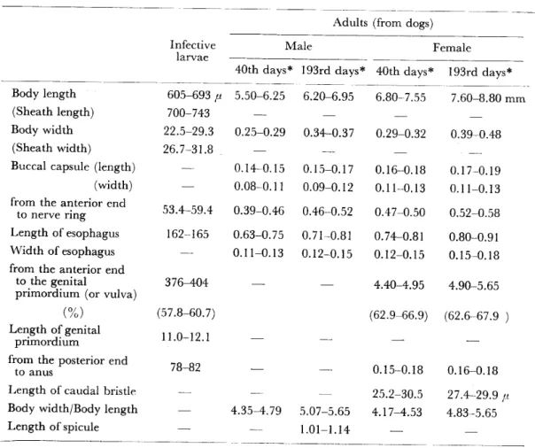
Figures of S. (G.) kyushuense sp, nov.
l , Dorsal view of head capsule of larva. 2, Ventral view of head capsule of larva.
3, Hypostomium of larva. 4, Tip of mandible of larva. 5, Gill filaments of pupa.
6, Dorsal view of cocoon. 7, Side view of cocoon. 8, Third segment of maxillary palp of female. 9, Fore leg of male. I O, Fore leg of female. I I , Mid leg of female. 1 2, Hind leg offemale. 13. Ventral view of female terminalia (eight sternite, anterior gonapophyses, genital fork and left paraproct and cercus) . 14, Spermatheca. 1 5, Lateral view of para‑
proct and cercus. 16. Ventral view of male genitalia (left coxite and style, ventral plate and parameres). 1 7, Styles viewed from different angles. 18, Side view of ventral plate.
19, End veiw of ventral plate. 20, Median sclerite.
wrth 5 segments In proportron of 6 6 8 1 1 21 3rd segment not enlarged ・ sensory vesicle small and oval in shape (Figure 8). Maxilla with l0‑14 strong teeth on each side. Mandible with about 26 inner teeth and about 14 outer ones. Cibarium with several weak denticles.
11
Thorax : Mesonotum shiny black, whitish gray‑pruinose and covered uniformly with fine recombent yellow pubescence and with upstanding black hairs on prescutel‑
lum region. Scutellum black with sparse long black hairs. Postscutellum black, gray‑pruinose and bare. Pleural membrane and katepisternum bare.
Legs (Figures l0‑12) : Blackish. Fore coxa and trochanter yellow. Mid and hind coxae and trochanters black. Fore femur yellowish brown to brown, gradually darkened distally. Mid and hind femora black with minute base of hind femur yellow. Fore tibia yellow on basal 4/5, brownish black on distal l/5 and with a conspicuous whitish silvery patch broadly on outer surface. Mid tibia black with basal I j3 of posterior surface yellow. Hind tibia black with basal I j2 of posterior surface yellow. Mid and hind tibiae each with a conspicuous whitish silvery patch broadly on posterior surface. Fore tarsi entirely black. Mid tarsi yellowish; distal lj6 of basitarsus and distal 1/2 of 2nd and 3rd segments somewhat darker; the rest of tarsal segments brownish black. Hind tarsi yellow with distal I !4 of basitarsus, distal I j3 of 2nd segment and the remaining segments brown to brownish black.
Fore basitarsus dilated, about 4.8 times as long as the greatest breadth. Hind basitarsus parallel‑sided. Calcipala short, not reaching pedisulcus. Pedisulcus marked. Each claw with a minute subbasal tooth.
Wing: Costa with 2 parallel rows of short spinules as well as hairs. Subcosta usually bare, at times very fewly haired. Basal section of radial vein bare. R1 with a single row of short spinules intermixed with hairs. Cross vein r‑m with a small pale brown spot. A taft of hairs on stem vein brown.
Halter: Whitish yellow with base black.
Abdomen: Basal scale black with a fringe of pale hairs. Dorsal surface of ab‑
domen dull brownish black on segments 2‑5, shiny black on the remaining posterior segments and with sparse brown hairs. The 2nd segment with a pair of conspicuous silvery patches dors0‑1aterally, which connected with each other broadly in the middle.
Terminalia : Macrosetae on ventral surface of 7th abdominal segment simple, not branched. Each arm of genital fork with a strong projection directed forwards.
Spermatheca (Figure 14) with minute internal hairs. Anterior gonapophyses, paraproct and cercus as shown in Figures 13 and 15.
Male
General body color black. Body length about 2.6 mm. Wing length about I .8 mm .
Head: Slightly wider than thorax. Holoptic, upper eye with about 13 trans‑
verse rows of large facets. Clypeus black, silvery‑pruinose and with sparse black hairs. Antenna consisting of 2+9 segments ; coloration usually brownish black, but at times basal few segments paler; Ist flagellar segment somewhat elongated, about I .5 times as long as the following segment. Maxillary palp black; ratio of each segment from base to apex 4: 4 : 8 : 10: 20.
Thorax : Mesonotum velvet black, uniformly covered with recumbent brown pubescence and with somewhat longer brown hairs on prescutellar region ; when viewed in certain angle of light, mesonotum with iridescent pattern ‑ a pair of
silvery spots on anterior region widely separated in the middle and a large silvery spot on posterior area, which connected with anterior ones through lateral silvery patches. Scutellum black and with long black hairs. Postscutellum black, gray‑
pruinose and bare. Pleural membrane and katepisternum bare.
Legs (Figure 9) : Blackish. Fore coxa yellow. Mid and hind coxae black.
Fore and hind trochanters brown. Mid trochanter black. Four femur dark brown.
Mid and hind femora black with minute base of hind one yellow. Fore tibia brown‑
ish black with middle large portion of outer surface yellow and with a conspicuous whitish silvery patch on pale portion. Mid and hind tibiae black with minute bases yellow and with a whitish silvery sheen basally on posterior surface. Coloration of all tarsi as in female. Fore basitarsus somewhat dilated, about 6.5 times as long as the greatest width. Hind basitarsus slender, parallel‑sided and much narrower than hind tibia. Calcipala and pedisulcus marked as in female.
Wing: Nearly same as in female. Rl with a few hairs intermixed with a
single row of short spinules.
Halter: Same as in female.
Abdomen: Basal scale black with a fringe of dark hairs. Dorsal surface of abdomen black with sparse brown hairs. A pair of silvery spots present dorso‑
laterally on segments 2 and 5‑7. Those on segment 2 connected with each other broadly in the middle.
Genitalia : As shown in Figures 1 6‑20. Style without apical spine. Paramere with numerous unequal hooks. Ventral plate setose on ventral surface and with dentate posterior margins.
Pupa
Body length (excluding gill filaments) about 2.6mm. Gill filaments about I .5 mm long.
Head: Integument yellowish brown, very sparsely covered with minute cone‑
like tubercles and with 3 pairs of short trichomes which are simple.
Thorax: Integument yellowish brown, covered moderately with minute cone‑
like tubercles with anterior I !2 almost bare, and with 4 pairs of simple trichomes antero‑dorsally. Gill organ (Figure 5) with 6 filaments in pairs, arising from short stalks of which ventral‑most one directed somewhat butwards. All filaments tapering towards tip, and with numerous transverse ridges and minute tubercles on their sur‑
face .
Abdomen: Terga 3 and 4 each with 4 hooked spines directed forwards on each side. Terga 7‑9 each with several stout spines in transverse row directed caudad on each side. Terminal hooks absent, if any, undeveloped. Sternum 5 with a pair of bifid or trifid hooks situated close together on each side. Sterna 6 and 7 each with a pair of bifid hooks widely separated from each other on each side.
Cocoon: As in Figures 6 and 7.
Mature larva
General body color reddish brown. Body length 4.2‑4.6 mm.
Head: Cephalic apotome with negative head spots as shown in Figure I .
13
Antenna with 4 segments in proportion of 10: 1 1 : 8 : I . Each cephalic fan with about 40 main rays. Tip of mandible as shown in Figure 4. Hypostomium (Figure 3) with a row of 9 apical teeth, of which corner ones rounded; Iateral serration developed on apical portion; hypostomial setae usually 4 in number lying subparallel to lateral margin on each side. Postgenal cleft deep and completely reaching the posterior margin of hypostomium (Figure 2).
Thorax and Abdomen: Cuticle almost bare except a few short setae on each side of anal sclerite. Rectal gill lobes compound, each lobe with 5‑8 finger‑like secondary lobules. Anal sclerite usual X‑shaped, with posterior arms longer than anterior ones. Posterior circlet with about 74 rows of 14‑16 hooks. Ventral papillae absent.
Type materials
Holotype: Female (Bishop Musium No. I 1 332), slide‑mounted, reared from pupa taken from a leaf of aquatic plant (Potamogeton crispus L.) in mid‑way of large river, the Kikuchi, (about 30 m wide and 10‑50 cm deep) flowing in lowland, Hichijo, Kikuchi‑gun, Kumamoto Pref., on 20 Sept. 1977 (by H. Takaoka).
Allotype: I¥/lale, slide‑mounted, same data and date as holotype.
Paratypes: 3 females, 3 males, 3 pupae and 3 Iarvae, all slide‑mounted, 10 females, 10 males, 10 pupae and 6 Iarvae, all preserved in alcohol solution, same data and date as holotype.
Other specimens examined
One female reared from pupa, 6 pupae and 4 Iarvae, collected from the aquatic plant in Koshi River. Shisui, Kikuchi‑gun, Kumamoto Pref., on 21 Sept. 1977 (by H. Takaoka).
A11 slide‑mounted types will be deposited in B. P. Bishop Museum, Honolulu, Hawaii, U. S. A. Other type materials are in my collection.
Distribution
Kumamoto Pref. Kyushu.
DISCUSSION
S. (G.) yushuense sp. nov. is easily assigned to the subvariegatum group defined by Rubzov (1959‑1964) by the characteristic feature of adult genitalia. It closely resembles to S. subvariegatum Rubzov, S. jacticum Rubzov, both from U. S. S. R., and S. nacojapi Smart from Japan. The female, male and pupa of this new species are almost identical to those of the latters, although slight difference in a few features of adults is present. In female, S. kyushuense sp. nov. differs from S. jacuticum by yellow pubescence on the mesonotum and from S. nacojapi by unbranched macrosetae on the 7th sternite. The male of this new species is distinguished from that of S. sub‑
variegatum by the parallel‑sided basitarsus of hind leg. Besides these adult different features, the most distinct difference between S. kyushuense sp, nov. and its allies is shown in the postgenal cleft of the larva which is deep and widely reaches the posterior margin of the hypostomium in this new species but never in the others.
AcKNowLEDGEMENTs
The author wishes to thank DL H Takahasi,Nerima−ku,Tokyo,fbr his valuable suggestions to this work.
Thanks are also due to Miss K Somekawa,the Department ofMedical Zoology,
Kagoshima University,for her assistance.
REFERENCES
1) Bcntinck,w。(1955):The black flies of Japan and Korea(Diptera:simuliidae),1−22,406 Medical General Laboratory U.S.Army,Tokyo
2)Ogata,K.,Sasa,M and suzuki,T、(1956):TheJapanese black伍es and their control,1−162,
DDT Kyokai,Tokyo(in Japanese)
3)Rubzov,L A.(1959−1964)=Simuliidae(Melusin三dae)in Lindner s,Die Fliegen Paraearkt.Reg.,
14ラ447−451
4)Shiraki,T.(1935):Simuliidae of the Japanese Empire,Mem.Fac、ScL Agr、Taihoku Imp.
Univ.,16,レ90
5) Takaoka,H.,Ochoa,J.0.and YamamotoフS,(1977):Notes on the魚una and distridution of blackHies in Kyushu,Japan(simuliidae:Diptera)JPn.J.sanit.zool.,28(4),341−347(inJapanese)
九州産ヤマブユ亜属の1新種
高岡 宏行
1977年9月,熊本県菊池郡七城,および洒水において平野部を流れる菊池,合志両河川の水中に繁 茂するえびもの葉,茎よりヤマブユ亜属の幼虫,蠕を多数得た。蠕より羽化させた成虫を含め全発 育期について検討を行った結果,本種はRubzov(1959−1964)の定義した5励槻漉9躍%〃zグループに 属し,本邦産の5伽μ1伽〃z(σn㍑5)ηα のαがSmartおよびソ連邦の5(G)躍勧αrfεgo如勉Rubzov,&
(α)ノαcオ∫側解Rubzovに極めて類似した形質を有する。しかしながら幼虫の頭蓋板のcleftがhypos−
tomiumの後縁まで達することによりこれら既知種と区別される。さらに成虫雌では,腹部第7節腹 面上の剛毛が枝分かれしないことにより8(G)nαoqノαガと,また胸部楯板上の微毛が黄色であるこ とにより8(G)ノαo漉襯とわずかに差が認められる。以上の結果から,本未記録種に&(G)初拓 5h麗n3θ(和名:キュウシュウヤマブユ)の新種名を提唱し,全発育期の記載を行った。
鹿児島大学医学部医動物学教室
Japan. J Trop Med. Hyg., Vol. 6, No, l, 1978, pp. 15 26 15
PATHOPHYSIOLOGICAL FINDINGS ON BLOOD OF BEAGLES EXPERIMENTALLY INFECTED
WITH BABESIA GIBSONI
TSUYOSHI ISHIMlNE SUSUMU MAKIMURA, SAKUJIRO KITAZAWA, SATORU TAMURAI AND r i AOYOSHI SUZUKI
Received for publication I I March 1978
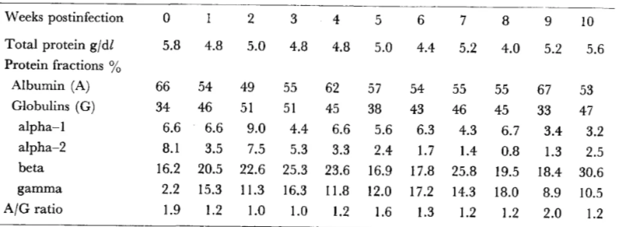
Abstract: Severe hemolytic anemia was observed in the peripheral blood of beagles infected with B. gibsoni, in the acute stage of infection, with an increase of parasitized erythrocytes beginning on the Ist week postinfection. Haptoglobin concentration in the serum decreased remarkably up to the 2nd week postinfection, showing little or no hapto‑
globin in the serum on the 3rd week to the 5th week postinfection. An abrupt increase of monocytes and neutrophils in the acute stage was noted, showing degenerated figures with some vacuoles in some cells. The beta and gamma globulins in the serum increased and albumin decreased remarkably in the acute stage. The gamma globulins remained at a level remarkably higher than the preinfection level until the 24th week postinfection.
Rapid increase of lgM and lgG fluorescent antibody titers was noted on the Ist week postinfection, showing a tendency for relatively higher activity to continue to the 24th week postinfection.
Recently, infection with Babesia (B.) gibsoni or B. sp. in dogs has been reported from various regions of Japan (Akashi et al., 1969; Kusunoki et al.. 1971 ; Noda, 1977). B. gibsoni is reported to be smaller than B. canis with delicate ring forms.
Aside from the size difference, it is known that hemoglobinuria, hemoglobinemia, icterus and consistent high fever, which are characteristic manifestations of acute B. canis infection, are not observed either in natural or in experimental infections with B. gibsoni (Ristic et al.. 1971 ; Weinman and Ristic, 1968). In some cases, infections of the Babesiae are complicated by concurrent infections with other hemotropic agents such as Ehrlichia canis. Haemobartonella canis, and Hepatozoon canis. lvlixed infections of Babesiae and E. canis often occur (Lewis and Ristic, 1977). For dogs in such conditions, the clinical signs, resulting frQm infection with B. gibsoni, which are less conspicuous than those observed in dogs infected with B. canis, are shown as different pathogenicity (Groves, 1972).
In spite of a large number of available reports on clinical and experimental canine babesiosis, information is very limited on the synthetic analysis of patho‑
physiological changes which are expressed as the body defense reaction of the host in babesial infection (Dorner, 1969 ; Schindler, 1970; Sibinovic, 1969). Consequent‑
ly, as the first step of experimental studies on canine babesiosis, young beagles and
Departments of Veterinary Physiology and Pathologyl , Obihiro University, 080 Obihiro, Hokkaido.
mongrel dogs were set up to determine some of the pathophysiological changes of blood occurring in these dogs after infection with the parasites, using the same strain of B. gibsoni (Lewis and Ristic, 1977).
MATERIALS AND METHODS
Babesia species: The strain of B. gibsoni (Lewis and Ristic, 1977) used in this study was originally obtained from a hunting dog that contracted the parasite in Hyogo Prefecture in Japan. This was maintained in the authors' Iaboratory in a deep freezer at ‑80 C and by dog passages during the experiments.
Experimental animals: Parasite free beagles from the colony maintained in a closed environment were used in the present study. Fourteen 90‑day‑old beagles from three litters produced by the same parent were used in the experiments. Four young mongrel dogs from the same parent, also 90‑day‑old, were used as control group. None of the beagles were vaccinated against distemper, rabies or canine hepatitis until the end of the experiments. Three healthy adult beagles, weighing approximately 15 kg, harboring no protozoa in the blood, and inoculated with B.
gibsoni four times in two years, were used as Babesia chronic beagles. To maintain regular conditions for breeding and feeding, each dog was put in separate cages, and was given a limited volume (250 g) of standard dog diet every day. Tap water was given freely during the experiment.
Experimental methods : The groups of beagles and mongrel dogs, all of which were 90‑day‑old, were all used in the experiments. Dogs were inoculated intravenously with approximately I x 109 parasitized erythrocytes which had been harvested from a splenectomized dog where in 30 per cent of the erythrocytes contained B.
gibsoni. The suspension was made in 3 ml physiological saline solution (PBS). As a diluent and anticoagulant, 0.005 mg of heparin per 0.5 ml of blood was used.
Three ml of blood were collected from each dog groups using the forearm vein. To collect serum samples, 2 ml of the blood were kept at 4 C for 30 minutes, and then centrifuged at 3,000 rpm for 15 minutes. The resulting sera were stored at ‑80 C until used.
Measurement methods : Body temperatures of each dog were examined daily at 6 O'clock in the evening for 6 months. Erythrocytes were counted in a I : 100 dilution of blood samples in Hayem's solution, and white blood cells in a I : 10 dilu‑
tion ofblood sample in Turk's solution, by using the Neubauer hemocytometer. The hematocrit was measured by the capillary tube method, centrifuging at 12,000 rpm for 5 minutes. Hemoglobin concentration was measured by the cyanmethemoglobin method. The hemogram was studied microscopically with smears fixed in methanol and stained with May‑Griinwald and Giemsa stains. The percentage of each type of leukocyte was calculated from a count of 200 cells. Reticulocytes were calculated from a count of 1,000 erythrocytes stained with 0.1 per cent brilliant cresyl blue.
Total protein in serum was measured by the biurette method using a photometer.
The protein fractions were ascertained by means of disc and acetate membrane electrophoreses. The staining of serum haptoglobin was estimated by the method described previously (Makimura and Suzuki, 1974a). Concentrations of immuno‑