*宮崎大学医学部放射線科
** 同 第二外科
*** 同 第一病理
**** 同 第二病理 受付:15 年 4 月 4 日
最終稿受付:15 年 10 月 14 日
別刷請求先:宮崎郡清武町木原 5200 (0 889–1692) 宮崎大学医学部放射線科
藤 田 晴 吾 E-mail: [email protected]
I. は じ め に
近年の悪性腫瘍に占める原発性肺癌の頻度は増 加傾向にあり,その術前検索としての縦隔リンパ 節転移の正確な診断は,手術適応や術式などの治
療方針を決定する上で重要である.原発性肺癌に おける縦隔リンパ節転移診断方法として,通常は X 線 CT 検査を行い,気管支鏡下または CT ガイ ド下生検後の病理組織診断により確定される1). しかし CT, MRI による形態計測に基づくリンパ 節転移の診断には限界があり,最大径 1 cm 以下 の N0 症例においてもその 13% にリンパ節転移 があることが知られている2).このことは,実際 の診療現場においては術前検査でリンパ節転移が 陰性と診断された症例でも,常に upstaging によ る術式の変更や化学療法,放射線治療等の追加治 療を念頭におく必要があることを示している.ま た upstaging の可能性を治療前に把握することは,
以後の診療方針決定に重要であると思われる.
《原 著》
原発性非小細胞肺癌における
201Tl SPECT の 縦隔リンパ節転移予測能の検討
藤田 晴吾* 長町 茂樹* 西井 龍一* 二見 繁美*
中田 博* 黒木 正臣* 小野 誠治* 田村 正三*
松崎 泰憲** 鬼塚 敏男** 浅田祐士郎*** 片岡 寛章****
要旨 原発性非小細胞肺癌 113 例 (腺癌 69 例,扁平上皮癌 31 例,大細胞癌 10 例,細気管支肺胞上 皮癌 2 例,神経内分泌細胞癌 1 例) を対象に術前 201Tl SPECT の摂取率 (ER, DR) および保持率 (RI) によ る縦隔リンパ節転移予測能を検討し,腫瘍径との関連および胸部 CT 検査にて縦隔リンパ節転移なしと 診断された N0 症例における 201Tl SPECT 保持率の最適閾値を算出した.113 例中,術後病理診断によ り 62 例に縦隔リンパ節転移が確認された.リンパ節転移陰性群と比較しリンパ節転移陽性群では ER,
DR では有意差は認めなかったが,RI は陽性群 91±67%, 陰性群 13±36% と有意差を認めた.また
原発病巣の最大腫瘍径に基づくリンパ節転移の予測は困難であった.次に 201Tl SPECT 検査および胸部 CT 検査のリンパ節転移診断能の指標として,それぞれ sensitivity (Sen), specificity (Spe), positive predictive value (PPV), negative predictive value (NPV), accuracy (Acc) を算出した.保持率 (RI) のリンパ節転移予測 における Cut-off 値を 35% とおくことで Sen: 82.2%, Spe: 82.3%, PPV: 85.0%, NPV: 79.2%, Acc: 82.3% と 胸部 CT による Sen: 72.6%, Spe: 82.4%, PPV: 83.3%, NPV: 71.2%, Acc: 77.0% と比較して同等か高い値が 得られた.原発性非小細胞肺癌患者で術前の胸部 CT 上,異常を認めない場合でも 201Tl SPECT 検査 で保持率が 35% を超える腫瘍に関しては常に Upstaging を疑う必要があると思われる.
(核医学 41: 1–7, 2004)
核医学診断領域で近年,有用とされている 18F- FDG PET 検査による縦隔リンパ節転移の診断に おける正診率は 68〜92% といわれている3〜5).し かし限られた施設でしか施行できないため,現状 では一般の病院で検査プロトコールに組み入れる ことは困難である.一方,多くの病院で施行可能 な 201Tl SPECT では,delayed scan を用いること で原発病巣の良悪性の鑑別6〜9)や組織学的分化
度10,11) と同時に縦隔リンパ節転移の診断について
も高い正診率を示すことが報告されている12,13). しかし,SPECT 装置には空間分解能に限界があ り,直径 1 cm 程度の病巣では検出されない可能 性がある.
これらに対して間接的な方法ではあるが,縦 隔 お よ び 肺 門 リ ン パ 節 転 移 の 推 定 法 と し て Takekawaらは,肺癌原発巣の 201Tl SPECT 保持率 が有用と報告している14).
今回われわれは,原発性非小細胞肺癌において
201Tl SPECT の保持率による縦隔リンパ節予測能
を再検討し,原発巣の最大腫瘍径との関連および 胸部 CT 検査上,縦隔リンパ節転移なしと診断さ れた N0 症例における 201Tl SPECT 保持率の最適 閾値を算出した.
II. 対象および方法
対象は宮崎大学医学部附属病院で 1994〜2002 年 9 月までに術前 201Tl SPECT が施行され早期像,
後期像共に異常集積を認めた原発性非小細胞肺癌 113 例で, 内訳は腺癌 69 例, 扁平上皮癌 31 例, 大 細胞癌 10 例, 細気管支肺胞上皮癌 2 例, 神経内 分泌細胞癌 1 例である. 年齢は 38〜83 歳 (平均 65 歳), 性別は男性 83 例, 女性 30 例であった.
全 症 例 に お い て 2検 出 器 型 ガ ン マ カ メ ラ Prism2000 (Picker 社) を使用し,コリメータは低 エネルギー高分解能コリメータを使用した.
SPECT 収集条件は 5° ステップ毎の 15 秒撮影を 72 フレーム収集し,フィルタ付逆投影法で再構 成を行った.
201Tl SPECT 検査は,塩化タリウム 111 MBq を 静注し,10 分後 (早期像) および 3 時間後 (後期
*
*
*
Table 1 Comparison of Tl indices between mediastinal lymph nodes metastasis (+) and (−) group (All cases)
lymph nodes
metastasis ER DR RI (%)
− (n=51) 1.5±0.53 1.8±0.65 13±36
+ (n=62) 1.3±0.75 2.2±1.1 91±67
(mean±s.d.)
*p<0.0001
Table 2 Comparison of Tl indices between mediastinal lymph nodes metastasis (+) and (−) group (Cases with chest CT negative)
lymph nodes
metastasis ER DR RI (%)
− (n=42) 1.4±0.42 1.7±0.59 15±39
+ (n=17) 1.6±0.50 2.7±0.77 72±31 (mean±s.d.)
*p<0.0001
Table 3 Comparison of maximal diameter of primary tumor between lymph nodes metastasis (+) and (−) group
lymph nodes
metastasis Tumor diameter (mm)
− (n=51) 29±15
n.s.
+ (n=62) 38±16
(mean±s.d.)
Table 4 Comparison of diagnostic ability in various cut- off value of RI on 201Tl SPECT
Cut-off value of RI
25% 35% 45% Chest CT
Sen 85.4% 82.2% 77.4% 72.6%
Spe 60.7% 82.3% 84.3% 82.4%
PPV 80.3% 85.0% 85.7% 83.3%
NPV 80.8% 79.2% 75.4% 71.2%
Acc 80.5% 82.3% 80.5% 77.0%
Sen: Sensitivity, Spe: Specificity, PPV: Positive pre- dictive value, NPV: Negative predictive value, Acc:
Accuracy, RI: retention index
3
像) を撮像した.集積の有無は核医学を専攻する 医師 3 名の合議で判定し,定量指標を対側肺野の 平均カウント (normal count: N) に対する腫瘍部平 均カウント (tumor count: T) をそれぞれ早期像摂 取率 (early ratio: ER),後期像摂取率 (delayed ra- tio: DR) として測定し,201Tl SPECT 保持率 (reten- tion index: RI) を (DR−ER)×100/ER の式にて算 出した.
胸部 CT 検査は X-Vigor (TOSHIBA), または
Aquilion (TOSHIBA) を用い,10 mm 厚 10 mm 間 隔の単純および造影像を行った.診断基準はリン パ節の短径 10 mm 以上をリンパ節転移陽性と し,放射線科専門医 2 名の合議により判定した.
201Tl SPECT と胸部 CT との撮影間隔は 0 日から 9 日 (平均 4.7 日) であり,検査終了後 2 週間以内 に手術を施行した.
検討項目は,(1) 201Tl SPECT 各定量指標の術後 病理診断にて診断された縦隔リンパ節転移陽性群 a
c
b
d
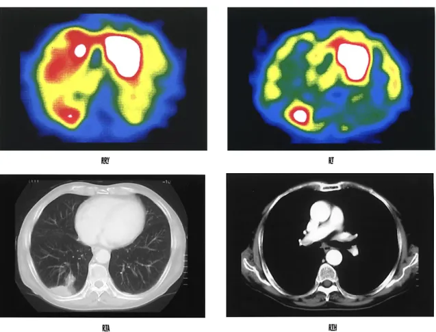
Fig. 1 The chest CT and SPECT images of a 79-year-old man with lung moderately differentiated adenocarcinoma and pathological lymph nodes metastasis represent a typical case in N0 case of the chest CT. (a) 201Tl SPECT early image shows abnormal accumulation in right lower lobe. The value of ER was 1.8. (b) 201Tl SPECT delayed image shows abnormal accumulation in right lower lobe. The value of ER was 3.0. The retention index is 67% for 201Tl chloride, that is higher than the optimal cut-off value of 35%. (c) The chest CT shows a mass of 30 mm in the right lower lobe with no lymph node metastasis larger than 1.0 cm. The chest CT was diagnosed as T2N0M0. (d) After operation, pathological lymph node metastasis was examined in #7 that less than 1.0 cm in the chest CT. Post operative diagnosis was pT2N2M0.
と陰性群との群間比較,(2) 胸部 CT 上,N0 と診 断された症例の術後病理診断リンパ節転移の有無 による 201Tl SPECT 定量指標の群間比較,(3) 原 発巣の最大腫瘍径について縦隔リンパ節転移陽性 群と陰性群の群間比較,(4) 縦隔リンパ節転移予 測における 201Tl SPECT 定量指標の最適閾値の算 出である.なお原発巣の最大腫瘍径は,胸部 CT 上最も腫瘍面積が大きく描出されているスライス 面にて測定した.群間比較は対応のない t 検定を 用い,統計処理ソフトは Stat View version 5 (SAS inc.) を用いた.
III. 結 果
術前 201Tl SPECT が施行され早期像,後期像と
もに異常集積を認めた全症例 113 例中,術後病理 診断により 62 例に縦隔リンパ節転移が確認され た.内訳は腺癌 34 例,扁平上皮癌 19 例,大細胞 癌 7 例,細気管支肺胞上皮癌 1 例,神経内分泌細 胞癌 1 例であった.術前 CT での stage 分類は stage IA 24 例,stage IB 23 例,stage IIA 9 例,
stage IIB 9 例,stage IIIA 18 例,stage IIIB 27 例,
stage IV 3 例であった.術後病理組織診断にて原 発巣および廓清したリンパ節により診断した術後 stage 分類は stage IA 20 例,stage IB 13 例,stage IIA 4 例,stage IIB 14 例,stage IIIA 26 例,stage IIIB 31 例,stage IV 5 例であった.術前 CT と術 後病理組織診断での stage 分類で比較すると,up- staging の症例が 21 例,down staging の症例は 12 例であった.全 113 症例中,術後病理組織診断に より 62 例に縦隔リンパ節転移が確認された.術 後病理診断により縦隔リンパ節転移陰性群 (n=
51) と比較して,縦隔リンパ節転移陽性群 (n=62) では ER, DR では有意な差はみられなかった が,RI は 91±67% であり,陰性群 13±36% と 比較して (p 値<0.0001) 有意差を認めた (Table 1).
なお胸部 CT 検査にて縦隔リンパ節転移なしと 診断した N0 症例 59 例における 201Tl SPECT 定量 指標の比較では,術後病理診断により縦隔リンパ 節転移陰性群 (n=42) と比較して,縦隔リンパ節 転移陽性群 (n=17) では DR, RI ともに有意に高
値を示した (Table 2).
また,縦隔リンパ節転移陽性群と陰性群におけ る最大腫瘍径の群間比較では両群間に有意差は認 めず,リンパ節転移の有無を原発巣の最大腫瘍径 から予測することは困難と考えられた (Table 3).
次に 201Tl SPECT 検査および胸部 CT 検査のリ ンパ節転移予測能として,それぞれ sensitivity, specificity, postive predictive value, negative predic- tive value, accuracy を算出し,201Tl SPECT 検査に おける縦隔リンパ節転移予測における最適閾値を 求めた.RI の cut-off 値を 5〜70% まで変化させ て検討したが,35% とおくことで,どの項目につ いても胸部 CT 検査と比較して同等か高い値が得 られた (Table 4).これに対して DR の縦隔リンパ 節転移の最適閾値は 1.5 であり,sen.=73.7%,
spe.=19.6%, PPV=48.3%, NPV=42.3%, acc.
=49.9% と CT の成績を下回る結果となった.
術後に縦隔リンパ節転移が確定した 62 例中,
術前の胸部 CT で診断し得たものが 45 例 (73%) であり,残りの 17 例 (27%) はリンパ節転移陰性 と診断された.このうち 15 例は術前に施行した
201Tl SPECT の RI の cut-off 値を 35% とおくこと で予測可能であった (Fig. 2).これらの内訳は腺 癌 8 例,扁平上皮癌 4 例,その他 3 例であった.
IV. 症 例
79 歳男性
右下葉背側 (S10) の胸膜直下に 5×3 cm 大の腫 Fig. 2 Positivity of 201Tl SPECT and chest CT in 62 lung
cancer patients.
5 瘤が認められ,縦隔に有意なリンパ節腫大はな
く, また胸膜にも播種, 浸潤を疑う所見はなかっ
たことより胸部 CT 検査上,T2N0M0 と診断した.
201Tl SPECT にて同部位に一致して早期像, 後期
像共に集積があり,定量値は ER: 1.8, DR: 3.0,
RI: 67% と cut-off 値である 35% 以上を示した.
右下葉切除およびリンパ節廓清術施行され,術後 病理診断にて中分化度腺癌,気管支分岐部リンパ 節 (#7) 転移, 病期 pT2N2M0 と診断された (Fig. 1).
V. 考 察
肺癌の病期分類とその 5 年生存率の報告によれ ば,原発病巣径に加えて,所属リンパ節への転移 が重要な予後因子であることが知られている15). 特に臨床病期 N2 症例では, 5 年生存率が極端に 低くなる.従来から用いられている CT による診 断は,リンパ節の大きさ (短径 1 cm 以下) が診断 基準であり,腫大のないリンパ節転移に関して は,偽陰性となる16〜19).梁らの報告2) によるとリ ンパ節の短径が 1 cm 未満の場合でもその 13% に リンパ節転移を認めるとされている.今回の検討 でも胸部 CT 検査では縦隔リンパ節転移陰性と診 断された症例のうち 27% に術後病理診断で縦隔 リンパ節転移が認められた.したがって 10〜30%
程度の症例では治療に際し,常に upstaging の可 能性を念頭におく必要があると思われる.
Takekawa らは縦隔リンパ節陽性症例では陰性 症例と比較して原発巣の RI が有意に高値を示す ことを報告している14) が,本検討においても術後 の病理診断でリンパ節転移陽性と確定した群にお いては 201Tl SPECT 保持率 RI が有意に高値を示 すことが確認された.RI が高値を示す腫瘍では,
その最大径に関係なく,悪性度が高く,腫瘍細胞 の viability が高いことが知られており6,20〜22),胸 部 CT 検査上腫大がなく,縦隔リンパ節転移の診 断基準を満たしていない場合でも転移をきたして いる可能性が高いと思われる.今回のわれわれの 検討において,胸部 CT では縦隔リンパ節転移を なしと診断された症例 (n=59) における術後病理 組織診断のリンパ節転移の有無の 201Tl SPECT 定
量指標の比較においては,RI だけでなく DR に おいても有意差を認めた.しかし,胸部 CT で有 意な縦隔リンパ節腫大を認めないという条件が付 くこと,また 113 症例全例を対象にした場合は DR に関しては 2 群間で有意差を認めないことよ り,縦隔リンパ節転移予測能に用いる指標として は RI が有効と思われた.
その基準であるが,RI 35% を cut-off 値とする ことにより,201Tl SPECT 検査における縦隔リン パ節転移予測能は CT 検査のリンパ節径の測定に 基づく診断能と比べて同等以上の数値を示した.
この保持率 (RI) 35% を用いることで,術前の胸 部 CT では診断できなかった縦隔リンパ節転移症 例 17 例中 15 例 (88.2%) までが,術前の時点で縦 隔リンパ節陽性の可能性がある群として扱いうる ようになった.このことから術前の縦隔リンパ節 転移診断において 201T SPECT は胸部 CT 検査に 対して,相補的な役割を担うものと考えられた.
また病理組織型との関連では,腺癌,扁平上皮 癌において各々 8/34 例 (23%), 4/19 例 (21%) に
CT 検査では偽陰性で,201Tl SPECT 保持率で予測
可能な縦隔リンパ節を認めた.このことから病理 組織型に関係なく,縦隔リンパ節転移の可能性は 常に考慮する必要があり,非侵襲的な予測法とし
ての 201Tl SPECT の有用性が確認された.した
がって原発巣の大きさにかかわらず,RI が高値 を示す症例においては stage を上げて治療方針を 考慮する必要があると思われる.
問題点であるが,本研究は,原発巣の集積強度 から,転移能を予測するものであり,縦隔部の集 積の有無に基づく縦隔転移の検出能は評価してい ない.しかし実際に縦隔部に集積があっても,レ トロスペクティブな研究では集積が病理標本のど のリンパ節に適合するかの詳細な判定は困難であ り,今回は施行しなかった.近年は形態画像との
Fusion が可能になっており23〜26),今後は縦隔集
積の有無に基づく縦隔転移の検出能評価のプロス ペクティブな study も必要と思われた.
VI. 結 語
原発性非小細胞肺癌 113 例を対象に縦隔リンパ 節転移予測能を,201Tl SPECT を用いて検討し た.術前 SPECT で RI が高い腫瘍では,胸部 CT 上は有意なリンパ節腫大がない場合でもリンパ節 転移をきたしている頻度が高く,201Tl SPECT の 定量指標はリンパ節転移予測に有用と思われた.
その最適閾値として RI 35% が推奨された.
文 献
1) 黒木正臣,中田 博,橋口典久,田村正三: Real- time Computed Tomography (CT) Fluoroscopy ガイ ド下生検の実際: IVR 会誌 2000; 15: 347–351.
2) 梁 尚志: 末梢小型肺癌――治療を行うことは有 益か――.綜合臨床 2001; 50: 2311–2314.
3) Kubota R, Yamada S, Kubota K, et al: Intramural distribution of F-18 fluorodeoxyglucose in vivo: High accumulation on macrophages and granulation tissue studied by microautography. J Nucl Med 1992; 33:
1972–1980.
4) 出村芳樹,水野史朗,若林聖伸,戸高嘉考,岡村 誠太郎,飯島慎吾,他: 肺癌臨床診断における FDG-PET の有用性についての検討―従来の核医 学検査との比較.日呼吸会誌 2000; 38: 676–681.
5) 北瀬正則,原 眞咲,Patz Jr EF, 加藤和子,佐藤 洋造,佐竹元治,他: cT1N0 肺癌症例の FDG-PET における N 因子診断能の検討.臨床放射線 2000;
45: 209–214.
6) Tonami N, Shuke N, Yokoyama K, Seki H, Takayama T, Kinuya S, et al: Thallium-201 single photon emission computed tomography in the evaluation of suspected lung cancer. J Nucl Med 1989; 30: 997–
1004.
7) Flores LG, Ochiai E, Nagamachi S, Jinnouchi S, Ohnishi T, Futami S, et al: The diagnostic role of
201Tl SPET imaging in patients with lung tumors:
Comparison with computed tomography. Nucl Med Commun 1996; 17: 493–499.
8) 長町茂樹,陣之内正史,西井龍一,二見繁美,田 村正三,松崎泰憲: 孤立性肺結節における 201Tl-
SPECT の診断能の再検討――生検検査,腫瘍
マーカー検査との比較――. 核医学 2001; 38: 737–
745.
9) Tonami N, Yokoyama K, Taki J, Hisada K, Watanabe Y, Takashima T, et al: Thallium-201 SPECT depicts radiologically occult lung cancer. J Nucl Med 1991;
32: 2284–2285.
10) Yamamoto Y, Nishiyama Y, Fukunaga K, Kobayashi T, Satoh K, Fujita J, et al: Evaluation of histopathol-
ogical differentiation in lung adenocarcinoma patients using 201Tl-chloride and 99Tcm-MIBI SPECT. Nucl Med Commun 2001; 22: 539–545.
11) Higashi K, Ueda Y, Sakuma T, Seki H, Oguchi M, Taniguchi M, et al: Comparison of 18F-FDG PET and
201Tl SPECT in evaluation of pulmonary nodules. J Nucl Med 2001; 42: 1489–1496.
12) Tonami N, Yokoyama K, Taki J, Shuke N, Kinuya S, Nakajima T, et al: 201Tl SPECT in the detection of mediastinal lymph node metastases from lung cancer.
Nucl Med Commun 1991; 12: 779–792.
13) Matsuno S, Tanabe M, Kawasaki Y, Satoh K, Urrutia AE, Ohkawa M, et al: Effectiveness of planar image and single photon emission tomography of thallium- 201 compared with gallium-67 in patients with primary lung cancer. Eur J Nucl Med 1992; 19: 86–95.
14) Takekawa H, Itoh K, Abe S, Ogura S, Isobe H, Sukou N, et al: Retention index of thallium-201 single photon emission computerized tomography (SPECT) as an indicator of metastasis in adenocarcinoma of the lung. Br J Cancer 1994; 70: 315–318.
15) Mountain CF: Revisions in the International System for the treatment of Staging Lung Cancer. Chest 1997;
111: 1710–1717.
16) Prenzel KL, Monig SP, Sinning JM, Baldus SE, Brochhagen HG, Schneider PM, et al: Lymph Node Size and Metastatic Infiltration in Non-small Cell Lung Cancer. Chest 2003; 123: 463–467.
17) Kamiyoshihara M, Kawashima O, Ishikawa S, Morishita Y: Mediastinal lymph node evaluation by computed tomographic scan in lung cancer. J Cardiovasc Surg (Torino) 2001; 42: 119–124.
18) Pirronti T, Macis G, Sallustio G, Minordi LM, Granone P, Vecchio FM, et al: Evaluation of the “N”
factor in nonsmall cell lung cancer. Correlation between computerized tomography and pathologic anatomy. Radiol Med (Torino) 2000; 99: 340–346.
19) Guyatt GH, Lefcoe M, Walter S, Cook D, Troyan S, Griffith L, et al: Interobserver variation in the computed tomographic evaluation of mediastinal lymph node size in patients with potentially resectable lung cancer. Canadian Lung Oncology Group. Chest 1995; 107: 116–119.
20) 山路 滋: 201Tl SPECT による原発性肺癌の治療効 果判定の有用性.核医学 1995; 32: 1333–1340.
21) Sehweil AM, McKillop JH, Milroy R, Wilson R, Abdel-Dayem HM, Omar YT: Mechanism of 201Tl uptake in tumours. Eur J Nucl Med 1989; 15: 376–
379.
22) Tonami N, Yokoyama K, Shuke N, Taki J, Kinuya S, Miyauchi T, et al: Evaluation of suspected malignant pulmonary lesions with 201Tl single photon emission computed tomography. Nucl Med Commun 1993; 14:
7 602–610.
23) Caldwell CB, Mah K, Ung YC, Danjoux CE, Balogh JM, Ganguli SN, et al: Observer variation in contour- ing gross tumor volume in patients with poorly defined non-small-cell lung tumors on CT: the impact of 18FDG-hybrid PET fusion. Int J Radiat Oncol Biol Phys 2001; 51: 923–931.
24) Ketai L, Hartshorne M: Potential uses of computed tomography-SPECT and computed tomography- coincidence fusion images of the chest. Clin Nucl Med
2001; 26: 433–441.
25) Suga K, Matsunaga N, Kawakami Y, Furukawa M:
Phantom study of fusion image of CT and SPECT with body-contour generated from external Compton scatter sources. Ann Nucl Med 2000; 14: 271–277.
26) D’Amico TA, Wong TZ, Harpole DH, Brown SD, Coleman RE: Impact of computed tomography- positron emission tomography fusion in staging patients with thoracic malignancies. Ann Thorac Sung 2002; 74: 160–163.
Summary
Usefulness of
201Tl SPECT in the Predication of Mediastinal Lymph Nodes Metastasis in Patients with Non Small Cell Lung Carcinoma (NSCLC)
Seigo F
UJITA*, Shigeki N
AGAMACHI*, Ryuichi N
ISHII*, Shigemi F
UTAMI*, Hiroshi N
AKADA*, Masaomi K
UROKI*, Seiji O
NO*, Shozo T
AMURA*, Yasunori M
ATSUZAKI**,
Toshio O
NIZUKA**, Yujiro A
SADA*** and Hiroaki K
ATAOKA****
*Department of Radiology, **Department of Second Surgery, ***Department of First Pathology,
****Department of Second Pathology, Miyazaki Medical College
Predictivity of mediastinal lymph nodes metastasis of 201Tl SPECT were examined before operation in 113 patients with non-small cell lung cancer (69 ad- enocarcinoma, 31 squamous cell carcinoma, 10 large cell carcinoma, 2 bronchiolo-alveolar carcinoma, 1 neuroendocrine cell carcinoma). Patients were clas- sified into two groups, with or without lymph nodes metastasis according to the pathological diagnosis.
We calculated parameters of 201Tl SPECT early ratio, delayed ratio, retention index (RI) and maximal diam- eters. In addition, we calculated optimal cut-off value of RI to estimate the mediastinal lymph nodes me- tastasis. Mediastinal lymph nodes metastasis was con- firmed pathologically in 62 patients. ER and DR did not show any statistical significance between two groups. Maximal diameters of primary tumor were also comparable between two groups. RI was signifi-
cantly higher in mediastinal lymph node metastasis positive group compared to that in mediastinal lymph node metastasis negative group.
The sensitivity (Sen), specificity (Spe), positive pre- dictive value (PPV), negative predictive value (NPV) and accuracy (Acc) of 201Tl SPECT were 82.2%, 82.3%, 85.0%, 79.2% and 82.3%. These parameters were similar of higher than 72.6%, 82.4%, 83.3%, 71.2% and 77.0% of chest CT.
The RI of 201Tl SPECT was useful tool for predict- ing lymph nodes metastasis in non-small cell lung can- cer. The optimum cut-off value of RI in the prediction of mediastinal lymph nodes metastasis was 35%. We should take into account of upstaging in cases with higher RI (>35%).
Key words: 201Tl SPECT, NSCLC, Mediastinal lymph nodes metastasis, Predictive value.