• 検索結果がありません。

Microcoleus pseudautumnalis sp. nov. (Cyanobacteria, Oscillatoriales) producing 2-methylisoborneol

N/A
N/A
Protected

Academic year: 2021

シェア "Microcoleus pseudautumnalis sp. nov. (Cyanobacteria, Oscillatoriales) producing 2-methylisoborneol"

Copied!
9
0
0

読み込み中.... (全文を見る)

全文

(1)

Microcoleus pseudautumnalis sp. nov. (Cyanobacteria, Oscillatoriales) producing 2-methylisoborneol

Yuko Niiyama * and Akihiro Tuji

Department of Botany, National Museum of Nature and Science, 4–1–1 Amakubo, Tsukuba, Ibaraki 305–0005, Japan

* E-mail: [email protected] (Received 13 May 2019; accepted 26 June 2019)

Abstract A new species, Microcoleus pseudautumnalis, producing both 2-methylisoborneol (2-MIB) and geosmin is described. We have conducted a systematic study of a bad-smelling, 2-MIB producing planktic Pseudanabaena species in Japan and described four new species (P.

foetida, P. subfoetida, P. cinerea, and P. yagii). In the course of this study, we found another kind of filamentous cyanobacteria with a bad smell in a plankton sample collected from a pond in Japan. The morphology of M. pseudautumnalis resembles that of M. autumnalis (Trevisan ex Gomont) Strunecký, Komárek et Johansen (basionym: Phormidium autumnale Trevisan ex Gomont). The sheath is thin and always contains only one trichome. Trichomes are immotile, gray- ish-green, not constricted at the cross-walls, not attenuated or attenuated towards the ends with truncated or capitated apical cells, and sometimes with calyptrae that are relatively wider (6.9–

7.6 μm) than those of M. autumnalis. The phylogeny of the 16S rRNA gene of M. pseudautumnalis revealed that it is in the clade of the genus Microcoleus and contains an 11-bp insert. Microcoleus autumnalis s. str. is said to lack this insert. Microcoleus pseudautumnalis has four kinds of 2-MIB genes, and the phylogeny of this taxon is different from those of Pseudanabaena sp. and Plankto- thricoides raciborskii.

Key words : 16S rRNA gene sequences, 2-MIB, 2-methylisoborneol, geosmin, Microcoleus autumnalis, Microcoleus vaginatus.

Introduction

Bad-smelling drinking water supplied from reservoirs and bad-smelling fish and shellfish cause genuine problems in Japan. The substance causing the odor was identified as 2-methyliso- borneol (2-MIB) or geosmin (Yagi, 1983), and odor-producing cyanobacteria have been reported in several lakes, reservoirs, and ponds (cf. Morii et al., 1982; Yamada et al., 1985, 1986; Oikawa et al., 2000, Oikawa and Ishibashi, 2004; Tsunoda et al., 2014). The authors con- ducted a systematic study of the odor-producing filamentous cyanobacteria that included a detailed morphological description, ecological observation, and phylogenetic analysis, and we

have clarified that thin filamentous planktic cya- nobacteria producing 2-MIB belong to the genus Pseudanabaena. Four new Pseudanabaena spe- cies were described (P. foetida Niiyama, Tuji et Ichise, P. subfoetida Niiyama et Tuji, P. cinerea Tuji et Niiyama and P. yagii Tuji et Niiyama) (Niiyama et al., 2016; Tuji and Niiyama, 2018).

In the course of the above-mentioned study,

we observed another kind of cyanobacteria spe-

cies producing 2-MIB, which showed clearly dif-

ferent morphological characteristics from Pseu-

danabaena but had some resemblance to

Phormidium or Microcoleus species. In Japan, it

has been reported that some benthic or periphytic

cyanobacteria with a Phormidium-like morphol-

ogy in streams or banks produce geosmin or

(2)

2-MIB. However, the classification of such organisms is unclear. This study focuses on the morphology and genetic characteristics of our newly found cyanobacteria that produces 2-MIB.

Materials and Methods Sampling site and cultured strain

The sample was collected from Naka-numa Pond, Ryugasaki City, Ibaraki Pref., Japan in September 2016, using a plankton net. Naka- numa Pond is surrounded by paddy fields, and no rivers or streams flow in or out of this pond.

Naka-numa Pond has an almost round shape with a diameter of about 100 m, and its maximum depth is about 14 m.

Isolation was done by the agar plate method (Tuji and Niiyama, 2014) with BG-11 medium (Waterbury and Stanier, 1981). Only one unialgal strain with a bad odor, Ak1609, was obtained.

For the maintenance of the strain, 10 ml of modi- fied C medium (Ichimura and Watanabe, 1977;

Niiyama et al., 2011) contained in a test tube was used. The culture was illuminated by cool-white fluorescent lamps, with a photon flux density of ca. 20 μmol/m

2

/sec, a photoperiod of 8 hours light and 16 hours dark, and a temperature of 18°C.

Morphological observation was performed for this cultured strain under a light microscope (BH-2, Olympus Corporation, Tokyo, Japan).

Microphotographs were taken with a Canon digi- tal camera EOS Kiss X5 (Canon Inc., Tokyo, Japan). The cultured strain is maintained in the Department of Botany, National Museum of Nature and Science. The specimens are housed in the herbarium of TNS (Department of Botany, National Museum of Nature and Science).

SPME-GC/MS analysis

The odor-producing substance in the culture medium of Ak1609 was analyzed using the gas chromatography/mass spectrometry combined with solid phase micro extraction (SPME-GC/

MS) method (JWWA, 2011) at Hiroshima Envi- ronment and Health Association.

Genomic DNA extraction, polymerase chain reaction (PCR) amplification, sequencing and assembling

A 1.5-ml volume of fresh cultured material was centrifuged at 10,000 rpm for 5 min at room temperature. The supernatant was removed, and the cell pellets were kept in a freezer at −20°C until extraction. Total genomic DNA was extracted using an extraction kit (DNeasy Plant Mini Kit, Qiagen) in accordance with the manu- facturerʼs instructions. The region between the 16S rRNA gene and the internal transcribed spacer (ITS) was amplified using four primers sets, set G (PLG1.3 and pits-CyanR), set J (PS- 16S-27f and pits-CyanR), set K (PS-16S-27f and 16S-1492R), and set L (PLG1.3 and 16S-1492R) (Table 1). The region between rbcL and rbcX was amplified using primer set M (cx-b and cw-b). The region of the gene cluster for 2-MIB was amplified using primer sets N (1609-mtf-r1 and MIB-cnbA-852f), O (1609-mtf-r2 and MIB- cnbA-736fb), P (PS-cnbA-start and 1609-mtf- r1), Q PS-cnbA-start and 1609-mtf-r2, R (MIB- SAM-F2 and 1609-mtf-r1), S (MIB-SAMF2 and 1609-mtf-r2), T (MIB-SAMF2 and MIB- SAMR1), and U (MIB-CnbA-852f and MIB- SAMR1). These primers for MIB genes were designed using Wang et al. (2011) and modified during this study.

PCR was performed on a thermal cycler (Ver- iti Thermal Cycler, Thermo Fisher Scientific) using 0.25 μl polymerase (Phusion Hot Start Flex DNA Polymerase, BioLabs), 5 μl GC Buffer in the polymerase kit, 2 μl 2.5 mM dNTPS, 14.25 μl sterile deionized water, 0.25 μl each of 10 pM concentrations of the forward and reverse prim- ers, and 1 μl of DNA template. The temperature cycling program used the following conditions:

16S rRNA to ITS regions; 98°C for 30 sec; 35 cycles of 98°C for 10 sec, 54°C for 15 sec, and 72°C for 60 sec, and the final elongation step was 72°C for 7 min; rbcLX regions; 98°C for 30 sec;

10 cycles of 98°C for 10 sec, 60°C to 50°C (−1.0°C per cycle) for 15 sec, 72°C for 60 sec;

35 cycles of 98°C for 10 sec, 50°C for 15 sec, and

72°C for 60 sec, and the final elongation step was

(3)

72°C for 7 min; MIB genes region; 98°C for 30 sec; 35 cycles of 98°C for 10 sec, 55°C for 15 sec, 72°C for 180 sec, and the final elongation step was 72°C for 7 min.

The concentrations of the amplified products were verified on a 1% agarose gel. Direct sequencing of the PCR products was undertaken using the primers presented in Table 1 with Big Dye Terminator Chemistry and an ABI 3130xl Genetic Analyzer (Applied Biosystems, Foster City, CA). The obtained sequences were assem- bled using Chromas PRO (Technelysium Pty Ltd, Tewantin, Australia). The assembled results were checked manually after automatic assem- bling by Chromas Pro.

Phylogenetic reconstruction

Phylogenetic and molecular evolutionary anal- yses for the sequence of Ak1609 and similar sequences retrieved from National Center for Biotechnology information (NCBI) were con- ducted using the MEGA 7 computer program (Kumar et al., 2016). The alignments were

checked manually. A maximum likelihood (ML) tree was calculated using MEGA software with the best fit model determined by Akaike Informa- tion Criterion (AIC) corrected scores, and the substitution nucleotide matrix parameters were calculated by the software. A tree using 1000 bootstrap replicates was generated. Each codon position was partitioned and analyzed for 16S rRNA. Neighbor-joining (NJ) trees with 1000 bootstrap replicates were also calculated using MEGA software. All positions containing gaps and missing data were partially deleted (site coverage cutoff 50%). Planktothricoides racibor- skii (strain: NIES-207) was used for the out- group.

Results and Discussion

The morphology of the cultured strain, Ak1609, resembles that of Microcoleus autum- nalis (Trevisan ex Gomont) Strunecký, Komárek et Johansen (≡Phormidium autumnale Trevisan ex Gomont) (Figs. 1–10, Table 2). Ak1609 and

Table 1. Primers for PCR and sequences used in this study.

Target Primer name Reference

16S rRNA-ITS 16S-27f AGAGTTTGATCMTGGCTCAG Lane 1991

PLG1.3 (CYA108F) ACGGGTGAGTAACRCGTRA Urbach et al. 1992

pits-CyanR CTCTGTGTGCCAAGGTATC Ernst et al. 2003

F3 L GTCCCGCAACGAGCGCAAC Hiraishi et al. 1994

16S-1492r GGTTACCTTGTTACGACTT Turner et al. 1999

rbcL–rbcX cx-b GGGGCARGTARGAAAGGRTTTCGTA Tuji & Niiyama 2018

cw-b CGTAGCTTCYGGTGGTATCCACGT Tuji & Niiyama 2018

MIB genes MIB-SAMF2 GAVTTCCTSVTGGRCCACCTCG Wang et al. 2011

cnba-852f GMRYTGCGBGARCGYCARGARYACGA This study

MIB-cnba-736fb TTGTCGAYTACGARACCTCKCCRCG This study

PS-mtf-start ATGTCAACGCCCCAAAMTATCACTGC This study

MIB-mtf-278f GGCGGTTCYGGTCGCGGCGG This study

MIB-mtf-446r CGCATGGCTCCCGTCTCGAAGCC This study

PS-mtf-end TTACCGAATGATGCGGTCAGCAACG This study

PS-mic-start ATGAAAGATACCAACYTGGATAATAC This study

MIB-mic-400f GACCCAKMTCGGCTGTTGAT This study

PS-mic-end TTAGGCTAGTGATTGTGAATCTGGC This study

PS-cnbB-start ATGACCCAAGACTTTAACTCCCATGG This study

MIB-cnbB-681f CGCCCGCCAAAAGCCCAAGATA This study

MIB-cnbB-890r CGCTCGCGCAACTCATGCACAGTC This study

1609-cnbB-1300fb TATCCTTGTCCCCGACGCCGTCGGCATTC This study

PS-cnbB-end CTACCGCCCGATCTCGACATCCTCG This study

MIB-SAMR1 TCSACGTACATGSTSGACTCGT Wang et al. 2011

1609-mtf-r1 TGGTGGTAATAGCCGTCAACTTGGCCGAGC This study

1609-mtf-r2 AGTTGGCAACCGACTTCTGATATTCGCTGCG This study

(4)

Figs. 1–10. Light microscopy photographs of Microcoleus pseudautumnalis sp. nov. Scale bars=10 μm.

(5)

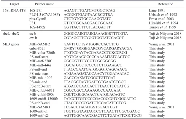
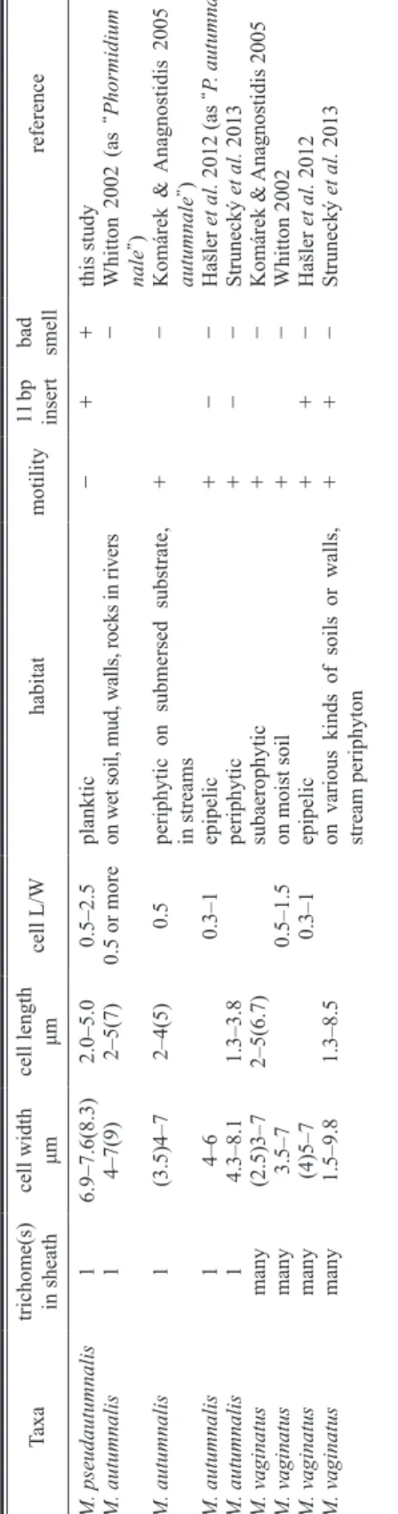
Table 2. Comparison of morphological, ecological and genetic characteristics of Micr ocoleus pseudautumnalis , M. autumnalis , and M. vaginatus . T axon names are iden - tical with thorse shown in Fig. 1 1. Taxa trichome(s) in sheath

cell width μm cell length μm cell L/W habitat motility

11 bp insert bad smell

reference M. pseudautumnalis 1 6.9–7.6(8.3) 2.0–5.0 0.5–2.5 planktic − + + this study M. autumnalis 1 4–7(9) 2–5(7) 0.5 or more on wet soil, mud, walls, rocks in rivers − Whitton 2002 (as “ Phormidium autum - nale ” ) M. autumnalis 1 (3.5)4–7 2–4(5) 0.5 periphytic on submersed substrate, in streams + − Komárek & Anagnostidis 2005 (as “ P. autumnale ” ) M. autumnalis 1 4–6 0.3–1 epipelic + − − Hašler et al. 2012 (as “ P. autumnale ” ) M. autumnalis 1 4.3–8.1 1.3–3.8 periphytic + − − Strunecký et al. 2013 M. vaginatus many (2.5)3–7 2–5(6.7) subaerophytic + − Komárek & Anagnostidis 2005 M. vaginatus many 3.5–7 0.5–1.5 on moist soil + − Whitton 2002 M. vaginatus many (4)5–7 0.3–1 epipelic + + − Hašler et al. 2012 M. vaginatus many 1.5–9.8 1.3–8.5

on various kinds of soils or walls, stream periphyton

+ + − Strunecký et al. 2013 M. vaginatus many (3.8)4.5–5.5 2–5(6.7) on arid soils ++ + − Boyer et al. 2002

Table 3. The similarity (%) and the number of base dif ferences per partial 16S rRNA sequence (1036 positons) from 26 strains between sequences are shown. All positions containing gaps and missing data were eliminated.

1234567891011121314151617181920212223242526 1Microcoleus pseudautumnalis Ak1609—99.9099.8199.8199.8199.8199.8199.8199.7199.7199.7199.7199.7199.5299.5299.5299.5299.4299.3299.2399.1398.9498.7598.6598.2691.99 2Microcoleus vaginatus ISBAL M61—99.9099.9099.7199.7199.9099.7199.8199.6199.7199.8199.8199.6199.6199.6199.4299.3299.2399.3299.2399.0398.6598.7598.1791.99 3Microcoleus vaginatus SRS1-KK221—100.0099.8199.81100.0099.8199.9099.7199.6199.9099.9099.5299.5299.7199.5299.2399.3299.2399.3299.1398.5598.6598.0792.08 4Microcoleus cf. vaginatus Ru-6-12210—99.8199.81100.0099.8199.9099.7199.6199.9099.9099.5299.5299.7199.5299.2399.3299.2399.3299.1398.5598.6598.0792.08 5Microcoleus vaginatus E1 12322—100.0099.81100.0099.7199.9099.5299.9099.7199.3299.3299.7199.7199.2399.5299.0399.3299.1398.5598.4698.0792.18 6Microcoleus vaginatus E1723220—99.81100.0099.7199.9099.5299.9099.7199.3299.3299.7199.7199.2399.5299.0399.3299.1398.5598.4698.0792.18 7Microcoleus vaginatus ISBAL M14210022—99.8199.9099.7199.6199.9099.9099.5299.5299.7199.5299.2399.3299.2399.3299.1398.5598.6598.0792.08 8Microcoleus vaginatus ISBAL M222322002—99.7199.9099.5299.9099.7199.3299.3299.7199.7199.2399.5299.0399.3299.1398.5598.4698.0792.18 9Microcoleus vaginatus ISBAL M232113313—99.6199.5299.8199.8199.4299.6199.6199.4299.1399.2399.1399.2399.2398.6598.5598.1791.99 10Microcoleus vaginatus KZ-23-1343311314—99.6199.8199.6199.2399.2399.6199.6199.1399.4299.1399.2399.0398.4698.3698.0792.18 11Microcoleus vaginatus K25 083344554554—99.5299.5299.3299.3299.3299.2399.1399.0399.2398.9498.7598.4698.4698.0792.08 12Phormidium autumnale sv3032111111225—99.8199.4299.4299.8199.6199.1399.4299.1399.4299.2398.4698.5597.9792.18 13Phormidium cf. autumnale CCALA 145321133132452—99.4299.4299.6199.4299.1399.2399.1399.2399.0398.4698.5597.9791.99 14Microcoleus vaginatus SNM1-KK15455775768766—99.2399.2399.0398.9498.8498.9498.8498.6598.2698.3697.7891.60 15Microcoleus sp. PET 11 754557757487668—99.2399.0399.1398.8499.3299.0399.0398.6598.5598.1791.99 16Microcoleus vaginatus ISBAL M10543333334472488—99.4299.3299.2398.9499.6199.4298.6598.7598.1791.99 17Oscillatoria amoena CCAP 1459/39565533536484610106—98.9499.8199.3299.0398.8498.4698.7598.3691.99 18Microcoleus sp. MUM 11 56788888899999119711—98.7598.6599.5299.3299.1399.0398.6591.80 19Phormidium autumnale JR127877557586106812128213—99.1398.8498.6598.4698.9498.5592.18 20Phormidium autumnale CCALA 1548788101081099899117117149—98.5598.4698.1798.7598.2691.99 21Phormidium sp. isolate: 200898777777881168121041051215—99.6199.0398.9498.5591.99 22Phormidium autumnale LCR Cyant3a1110999999810138101410612714164—98.8499.1398.7592.18 23Phormidium autumnale Arct-Ph51314151515151515141616161618141416916191012—98.7598.7591.31 24Phormidium autumnale LCR-CYANT11141314141616141615171615151715131310111311913—99.4291.99 25Phormidium autumnale SAG 78.7918192020202020201920202121231919171415181513136—91.80 26Planktothricoides raciborskii NIES-20783838282818182818381828183878383838581838381908385—

(6)

its cultured solution have a strong bad smell.

Both 2-MIB and geosmin were detected from the cultured solution by the SPME-GC/MS. Ak1609 is in the Microcoleus clade including M. autum- nalis and M. vaginatus for the phylogenetic tree of the 16S rRNA gene (Fig. 11, Table 3). How-

ever, it is immotile and planktic and produces 2-MIB and geosmin, and these characteristics differ from those of M. autumnalis and M. vagi- natus. Thus, we propose a new species, M. pseu- dautumnalis, as follows.

Fig. 11. Maximum likelihood tree based on 16S rRNA gene sequences. The general time reversible with gamma

distribution and invariable sites model (GTR+G+I) was used. Sequences retrieved from NCBI are used for

phylogenetic analysis, and the OTUs are shown with their registered name, culture number and accession

number. Numbers at nodes indicate NJ (Neighbor Joining)/ML bootstrap support values (only values higher

than 70 are shown). ◇: lacking11-bp insert.

(7)

Microcoleus pseudautumnalis Niiyama et Tuji, sp. nov.

Description: Filaments mostly straight, with- out any type of branching, combine to make a black colored thin membranous colony in a test tube. Sheath thin, firm, colorless, always contain- ing one trichome. Trichomes immotile, with or without sheath, grayish-green to pale yellowish- green colored, 6.9–7.6 μm in width, not con- stricted at the cross-walls, somewhat granulated at the cross-walls, not attenuated at the ends with rounded apical cells or attenuated towards the curved ends with truncated or capitated apical cells, sometimes with calyptra. Cells shorter or longer than wide, cell width to length ratio 0.5–

2.5, without gas vesicles. Thallus has the extreme musty odor that comes from 2-MIB and geosmin.

Holotype: A formalin-fixed specimen, TNS- AL-61750 in TNS (Department of Botany, National Museum of Nature and Science), from cultured strain Ak1609 maintained in TNS.

Isotype: A formalin-fixed specimen, TNS- AL61748 in TNS.

Type locality: Naka-numa pond, Ibaraki Pref., Japan.

Habitat: Plankton in ponds.

Table 2 shows a comparison of the morpho- logical, ecological, and genetic traits and the presence or absence of a bad smell for M. autum- nalis (P. autumnale), M. vaginatus, and the new species reported in this study, M. pseudautumna- lis. M. pseudautumnalis always has one trichome in the sheath, just like M. autumnalis. Microco- leus pseudautumanalis has very closed sequence to M. autumnalis and M. vaginatus in 16S rRNA (table 3). It is noted that M. vaginatus has 11 base pairs inserted into the 16S rRNA gene (Boyer et al., 2002; Siegesmund et al. 2008;

Hašler et al., 2012; Strunecký et al., 2013). On the other hand, M. autumnalis (P. autumnale s.

str.) lacks this 11-bp insert (Hašler et al., 2012;

Strunecký et al., 2013) and forms a monophy- letic group in the genus Microcoleus (Strunecký et al., 2013). Microcoleus pseudautumnalis is in the same clade as M. vaginatus based on an anal- ysis of the 16S rRNA gene and has the 11-bp

insert as does M. vaginatus (Fig. 12). Microco- leus autumnalis is said to be an epipelic or epi- phytic species in aquatic habitats, and M. vagina- tus is thought to be a soil or aquatic species, and both are motile, especially M. vaginatus (Boyer et al. 2002). However, M. pseudautumnalis is an immotile and planctic species. No studies have been reported on Microcoleus species producing 2-MIB.

The 4507-bp sequence of the gene cluster for 2-MIB including the cnbA, mtf, mic, and cnbB genes was obtained from M. pseudautumnalis (cultured strain Ak1609: accession no.:

LC486303). These genes and their order are the same as Pseudanabaena sp. dqh15 (HQ830028) and Planktothricoides raciborskii CHAB3331 (HQ830029) presented in Wang et al. (2011).

The gene cluster sequences excluding ITS regions (4079 bp) were compared. Microcoleus pseudautumnalis is closer to Pl. raciborskii (216 bp differences) than Pseudanabaena sp.

dqh15 (429 bp differences). Pseudanabaena sp.

dqh15 and Pl. raciborskii had 508 bp differences.

These large differences between M. pseudautum- nalis and other 2-MIB-producing taxa suggest that these 2-MIB-producing genes evolved sepa- rately and do not exhibit lateral gene transfer, which is found in many genes producing second- ary metabolites such as geosmin (Hayashi et al., 2019).

Tsunoda et al. (2014) reported a 2-MIB pro- ducing peryphytic Phormidium sp. from the Tama River in Japan, and they proposed that this species may be P. autumunale or P. favosum based on their microscopic observation. Because the 16S rRNA sequence of the 2-MIB-producing cultured strain presented in Tsunoda et al. (2014) (accession no.: AB820727) lacks the 11-bp insert (see Fig. 12) and is close to P. autumnale Arct- Ph5 in the phylogenetic tree (Fig. 11), it is differ- ent from M. pseudautumnalis and should be M.

autumnalis (P. autumnale s. str.) (Hašler et al., 2012; Strunecký et al., 2013).

Microcoleus pseudautumnalis produces not

only 2-MIB but also geosmin. Although many

musty-odor-producing cyanobacteria strains are

(8)

reported in Japan, each cultured strain produces only one kind of odor-producing substance (Yagi, 1983; Oikawa and Ishibashi, 2004). The present study is the first to report the production of both 2-MIB and geosmin by one strain of cya- nobacteria.

Acknowledgments

We express special thanks to the Hiroshima Environment and Health Association for analyz- ing off-odor substances using SPME-GC/MS.

References

Boyer, S. L., Johansen, J. R., Flechtner, V. R. and How- ard, G. L. 2002. Phylogeny and genetic variance in ter- restrial Microcoleus (Cyanophycea) species based on sequence analysis of the 16S rRNA gene and associated 16S-23S ITS region. Journal of Phycology 38: 1222–

1235.

Ernst, A., Becker, S., Wollenzien, U.-I.-A. and Postius, C.

2003. Ecosystem-dependent adaptive radiations of picocyanobacteria inferred from 16S rRNA and ITS-1 sequence analysis. Microbiology (Reading) 149: 217–

228.

Hašler, P., Dvořák, P., Johansen, J. R., Kinter, M., Ondřej, V. and Poulíčková, A. 2012. Morphological and molec- ular study of epipelic filamentous genera Phormidium, Microcoleus and Geitlerinema (Oscillatoriales, Cyanophyta/Cyanobacteria). Fottea 12: 341–356.

Hayashi, S., Ohtani, S., Godo, T., Nojiri, Y., Saki, Y., Esumi, T. and Kamiya, H. 2019. Identification of geos- min biosynthetic gene in geosmin-producing colonial cyanobacteria Coelosphaerium sp. and isolation of geosmin non-producing Coelosphaerium sp. from brackish Lake Shinji in Japan. Harmful Algae 84:

19–26.

Hiraishi, A. and Kaneko, M. 1994. Use of polymerase chain reaction-amplified 16S rRNA gene sequences to identify pink-pigmented bacteria found in a potable water treatment system. Bulletin of Japanese Society of Microbial Ecology 9: 55–65.

Fig. 12. Variable positions of the rRNA regions between Phormidium autumnale (=Microcoleus autumnalis), M.

pseudautumnalis and M. vaginatus showing 11-bp insert presented in Hašler et al. (2012) and Strunecký et al.

(2013).

(9)

Ichimura, T. and Watanabe, M. M. 1977. An axenic clone of Microcystis aeruginosa Kütz. emend. Elenkin from Lake Kasumigaura. The Bulletin of Japanese Society of Phycology 25:177–181.

JWWA 2011. Water Examination Methods (2011 Edition), vol. 4. Japan Water Works Association, Tokyo.

Komárek, J. and Anagnostidis, K. 2005. Cyanopro- karyota. 2. Teil: Oscillatoriales. In: Büdel. B., Gärdner, G., Krienitz, L. and Schagerl. M. (eds.), Süswasserflora von Mitteleuropa, vol. 19/2. 759 pp. Elsevier, München.

Kumar, S., Stecher, G. and Tamura, K. 2016. MEGA7:

Molecular Evolutionary Genetics Analysis Version 7.0 for Bigger Datasets. Molecular Biology and Evolution 33: 1870–1874.

Lane, D. J. 1991. 16S/23S rRNA sequencing. In: Stacke- brandt, E. and Goodfellow, M. (eds.), Nucleic acid techniques in bacterial systematics pp. 125–175. John Wiley and Sons, New York.

Morii, H., Kasama, K. and Zentani, B. 1982. Musty odor- production by the cyanobacteria Pseudanabaena spp.

isolated from reservoirs of water supply in Kure City.

Bulletin of Faculty of Fisheries, Nagasaki University 52: 47–54 (in Japanese with English abstract).

Niiyama, Y., Tuji, A. and Tsujimura, S. 2011. Umezakia natans M.Watan. does not belong to Stigonemataceae but Nostocaceae. Fottea 11: 163–169.

Niiyama, Y., Tuji, A., Takemoto, K. and Ichise S. 2016.

Pseudanabaena foetida sp. nov. and P. subfoetida sp.

nov. (Cyanophyta/Cyanobacteria) producing 2-methyl- isoborneol from Japan. Fottea 16: 1–11.

Oikawa, E. and Ishibashi, Y. 2004. Species specificity of musty odor producing Phormidium tenue in Lake Kamafusa. Water Science and Technology 49: 41–46.

Oikawa, E., Ishibashi, Y., Abe, T. and Umetsu, H. 2000.

Phylogenetic classification of musty odor and/or toxic compounds producing cyanobacteria. Environmental Engineering Research 37: 183–191 (in Japanese with English abstract).

Siegesmund, M. A., Johansen, J. R., Karsten, U. and Friedl, T. 2008. Coelofasciculus gen. nov. (Cyanobacte- ria): morphological and molecular criteria for revision of the genus Microcoloeus Gomont. Journal of Phycol- ogy 44: 1572–1585.

Strunecký, O., Komárek, J., Johansen, J., Lukešová, A.

and Elster, J. 2013. Molecular and morphological crite- ria of revision of the genus Microcoleus (Oscillatoriales ,Cyanobacteria). Journal of Phycology 49: 1167–1180.

Tsunoda, T., Nakahigashi, H., Kanami, T. and Oikawa, T.

2014. Genetic analysis of 2-methylisoborneol-produc- ing cyanobacterium sampled from the upper reaches of

the Tama river. Journal of Japan Society on Water Environment 37: 9–13 (in Japanese with English abstract).

Tuji, A. and Niiyama, Y. 2014. Freshwater diatom flora in the Imperial Palace, Tokyo (2010–2013). Memories of the National Museum of Nature and Science 49: 75–88 (in Japanese with English abstract).

Tuji, A. and Niiyama, Y. 2018. Two new Pseudanabaena (Cyanobacteria, Synechococcales) species from Japan, Pseudanabaena cinerea and Pseudanabaena yagii, which produce 2-methylisoborneol. Phycological Research 66: 291–299.

Turner, S., Pryer, K. M., Miao, V. P. W. and Palmer, J. D.

1999. Investigating deep phylogenetic relationships among cyanobacteria and plastids by small subunit rRNA sequence analysis. Journal of Eukaryotic Micro- biology 46: 327–338.

Urbach, E., Robertson, D. L. and Chisholm, S. W. 1992.

Multiple evolutionary origins of procholorphytes within the cyanobacterial radiation. Nature 355: 267–

Wang, Z., Xu, Y., Shao, J., Wang, J. and Li, R. 2011. 270.

Genes associated with 2-methylisoborneol biosynthesis in cyanobacteria: isolation, characterization, and expression in response to light. PLoS ONE 6: e18665–

e18665.

Waterbury, J. B. and Stanier, R. Y. 1981. Isolation and growth of cyanobacteria from marine and hypersaline environments. The Prokaryotes 7: 221–223.

Whitton, B. A. 2002. Phylum Cyanophyta (Cyanobacte- ria). John, D. M., Whitton, B. A. and Brook, A. J.

(eds.), The Freshwater Algal Flora of the British Isles.—An identification guide to freshwater and terres- trial algae, pp. 25–122. Cambridge University Press, Cambridge.

Yagi, M., 1983. Odor produced by blue green algae. Eisei Kagaku 29: 16–22 (in Japanese with English abstract).

Yamada, N., Aoyama, K., Yamada, M. and Hamamura, N.

1985. Studies on earthy-musty odor in natural water (1) Growth characteristics and 2-methylisoborneol produc- tion of Phormidium tenue. Japan Journal of Water Pol- lution Research 8: 515–521 (in Japanese with English abstract).

Yamada, N., Aoyama, K., Yamada, M. and Hamamura, N.

1986. Studies on earthy-musty odor in natural water (3) Isolation of bacteria-free Phormidium tenue and the effect of associated bacteria on the growth of axenic P.

tenue. Japan Journal of Water Pollution Research 9:

379–385 (in Japanese with English abstract).

Table  1.  Primers for PCR and sequences used in this study.
Table 3. The similarity (%) and the number of base differences per partial 16S rRNA sequence (1036 positons) from 26 strains between sequences are shown
Fig.  11.  Maximum likelihood tree based on 16S rRNA gene sequences. The general time reversible with gamma  distribution and invariable sites model (GTR+G+I) was used
Fig.  12.  Variable positions of the rRNA regions between Phormidium autumnale (=Microcoleus autumnalis), M

参照

関連したドキュメント

Use of the long-read sequencer with the CCS technique, which can provide numerous accu- rate full-length 16S rRNA gene sequences [17, 18] identified as many as 34 bacterial

Key words and phrases: Nonstandard Analysis, Compact Linear Opera- tors, Weak Cauchy Sequences, Weak

To confirm the discriminatory power of the 10 chosen concatenated hypervariable proteins (C10HKP), we constructed phylogenetic trees based on C10HKP, 4MLSA, and the 16S rRNA

(Fig. The nucleotide sequence of the 16S rRNA gene from the strain B2 has been deposited in DDBJ/EMBL/ GenBank databases under accession no. Strain P7 requires the

Similarly, bacterial abundance, which was estimated from 16S rRNA gene copy number using quantitative real time PCR (qPCR), increased with time for the methanol- and the

To understand the lichen-association of fungi, algae or cyanobacteria and the non-photosynthetic bacteria, 18S/16S rRNA gene sequences were compared among rock-dwelling lichens

similarity of 16S rRNA gene sequences; however, higher criteira (e.g., >99% similarity) resulted in narrower distributions (endemism) of certain Antarctic bacteria (Nakai et

The phylogenetic tree based on the neighbour-joining NJ generated com- parisons of the 16S rRNA gene sequences revealed that these isolates were clustered within the family