Disrupted tongue microbiota and detection of nonindigenous bacteria on the day of allogeneic hematopoietic stem cell transplantation
奥, 菜央理
http://hdl.handle.net/2324/4110457
出版情報:九州大学, 2020, 博士(歯学), 課程博士 バージョン:
権利関係:© 2020 Oku et al. This is an open access article distributed under the terms of the Creative Commons Attribution License.
RESEARCH ARTICLE
Disrupted tongue microbiota and detection of nonindigenous bacteria on the day of
allogeneic hematopoietic stem cell transplantation
Saori Oku1,2, Toru TakeshitaID1,3, Toshiko Futatsuki2, Shinya Kageyama1, Mikari Asakawa1, Yasuo MoriID4, Toshihiro Miyamoto4, Jun HataID5,6, Toshiharu Ninomiya5,6, Haruhiko Kashiwazaki2, Yoshihisa Yamashita1*
1 Section of Preventive and Public Health Dentistry, Division of Oral Health, Growth and Development, Faculty of Dental Science, Kyushu University, Fukuoka, Japan, 2 Section of Geriatric Dentistry and Perioperative Medicine in Dentistry, Faculty of Dental Science, Kyushu University, Fukuoka, Japan, 3 OBT Research Center, Faculty of Dental Science, Kyushu University, Fukuoka, Japan, 4 Department of Medicine and Biosystemic Science, Kyushu University Graduate School of Medical Sciences, Fukuoka, Japan, 5 Department of Epidemiology and Public Health, Graduate School of Medical Sciences, Kyushu University, Fukuoka, Japan, 6 Center for Cohort Studies, Graduate School of Medical Sciences, Kyushu University, Fukuoka, Japan
*yoshi@dent.kyushu-u.ac.jp
Abstract
Disruption of the intestinal microbiota caused by intensive chemotherapy, irradiation and antibiotics can result in development of severe gut graft-versus-host disease and infectious complications, leading to poorer outcomes among allogeneic hematopoietic stem cell trans- plantation (allo-HSCT) recipients. Although the oral cavity is also densely colonized by indig- enous microorganisms, the bacterial composition in allo-HSCT recipients remains unclear.
We determined the tongue microbiota composition of 45 patients with hematological disor- ders on the day of transplantation and compared them to 164 community-dwelling adults.
The V1–V2 regions of the 16S rRNA gene sequences demonstrated that the allo-HSCT recipients had less diverse and distinct microbiota from that of community-dwelling adults.
The full-length 16S rRNA gene sequences identified 146 bacterial taxa in the microbiota of allo-HSCT recipients, of which 34 bacterial taxa did not correspond to bacteria primarily inhabiting the oral cavity deposited in the expanded Human Oral Microbiome Database.
Notably, the detection of Staphylococcus haemolyticus and/or Ralstonia pickettii was signifi- cantly associated with a higher risk of mortality during the follow-up period. These results demonstrate that the oral cavity of allo-HSCT recipients is colonized by a disrupted micro- biota on the day of transplantation and suggest that detection of specific nonindigenous taxa could be a predictor of transplant outcome.
a1111111111 a1111111111 a1111111111 a1111111111 a1111111111
OPEN ACCESS
Citation: Oku S, Takeshita T, Futatsuki T, Kageyama S, Asakawa M, Mori Y, et al. (2020) Disrupted tongue microbiota and detection of nonindigenous bacteria on the day of allogeneic hematopoietic stem cell transplantation. PLoS Pathog 16(3): e1008348.https://doi.org/10.1371/
journal.ppat.1008348
Editor: Dana J. Philpott, University of Toronto, CANADA
Received: September 11, 2019 Accepted: January 23, 2020 Published: March 9, 2020
Copyright:©2020 Oku et al. This is an open access article distributed under the terms of the Creative Commons Attribution License, which permits unrestricted use, distribution, and reproduction in any medium, provided the original author and source are credited.
Data Availability Statement: The sequence data obtained in this study have been deposited in the DDBJ Sequence Read Archive under accession no.
DRA009550 and DRA009551.
Funding: This study was supported in part by Grants-in Aid for by JSPS KAKENHI Grant numbers JP19H03863 (T. T.), JP19K22722 (T. T.), JP16H02692 (Y. Y.), and JP16H05850 (Y. Y.) from Japan Society for the Promotion of Science (JP).
The funders had no role in study design, data
Author summary
Allogeneic hematopoietic stem cell transplantation (allo-HSCT) recipients are subjected to intensive chemotherapy, irradiation and antibiotics which could affect the intestinal as well as oral microbiota. We employed full-length 16S rRNA gene sequencing analysis with high taxonomic resolution using a third-generation sequencer, PacBio Sequel, and deter- mined the bacterial composition of the tongue microbiota of allo-HSCT recipients after conditioning regimens. This comprehensive molecular approach identified 34 taxa uncommon in the oral cavity, which constituted 0–99.4% (median, 0.27%) of each tongue microbiota. Of them,Staphylococcus haemolyticusandRalstonia pickettiiwere frequently found in allo-HSCT recipients, and their detection was significantly associated with a higher risk of mortality during the follow-up period. These results suggest that careful attention should be given to the bacterial composition of the disrupted oral microbiota in allo-HSCT recipients.
Introduction
Allogeneic hematopoietic stem cell transplantation (allo-HSCT) is a curative treatment option for various hematological malignancies and inherited hematopoietic disorders [1,2]. In order to eradicate residual malignant cells, as well as immunocompetent cells to ensure engraftment of infused donor cells, allo-HSCT recipients undergo a conditioning regimen including inten- sive chemotherapy and/or total body irradiation [3], resulting in mucosal injury. They require broad-spectrum antibiotics until neutrophil recovery in order to prevent and treat bacterial penetration into the bloodstream through the damaged mucosal barrier. Long-term use of broad-spectrum antibiotics can seriously affect the indigenous microbiota, which in the steady-state contributes to maintaining homeostasis among microorganisms or between the microorganisms and the host [4]. Dysbiosis of the gastrointestinal tract in allo-HSCT recipi- ents was reportedly associated with the increased risk of bacteremia [5]; severe graft-versus- host disease (GVHD)[6,7]; disease relapse [8]; and, ultimately, inferior overall survival [9].
The oral cavity is also densely colonized by diverse microorganisms constituting the indige- nous microbiota, which serves as a defensive barrier against the establishment of pathogenic bacteria [10]; thus, disruption of the oral microbiota could lead to colonization by exogenous pathogenic bacteria in the oral cavity. The bacterial populations shed from intraoral surfaces are constantly transported to the gastrointestinal and respiratory tracts via saliva and could contribute to systemic health. The preparative regimen prior to allo-HSCT and broad-spec- trum antibiotics could also alter the oral microbiota. In fact, a recent study reported that the denaturing gradient gel electrophoresis profiles of oral mucosal microbiota drastically changed after allo-HSCT in six recipients, especially in recipients who received glycopeptide antibiotics in combination with ß-lactam [11]. Another recent study using 16S rRNA gene sequencing analysis indicated a decrease in identified bacterial taxa and the presences of unique bacterial taxa in oral microbiota in allo-HSCT based on longitudinal observation at three timepoints of four aplastic anemia patients who received allo-HSCT [12]. However, the oral bacterial com- position of allo-HSCT recipients and its association with transplant outcome have not been fully elucidated.
In this study, we collected the tongue microbiota, a dominant source of the bacterial popu- lation in saliva [13–15], from 45 allo-HSCT recipients suffering from hematological disorders on the day of transplantation. The composition of the microbiota was compared with that of community-dwelling adults by using 16S rRNA gene sequencing analysis with a next-
collection and analysis, decision to publish, or preparation of the manuscript.
Competing interests: The authors have declared that no competing interests exist.
generation sequencer, and further determined based on full-length 16S rRNA gene sequences obtained from a third-generation long-read sequencer with a high taxonomic resolution. We also sought to elucidate the bacterial taxa that produce an impact on the transplant outcome.
Results
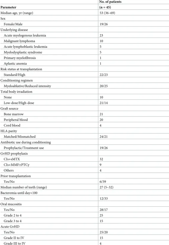
This study investigated the bacterial composition of the tongue microbiota collected from 45 adult allo-HSCT recipients [19 females and 26 males aged 36–69 years; mean±standard devia- tion (SD): 52.7±10.3 years] on the day of transplantation. Underlying hematological diseases were as follows: 23 patients with acute myelogenous leukemia, 10 with malignant lymphoma, 5 with acute lymphoblastic leukemia, 5 with myelodysplastic syndrome, and 2 with other condi- tions (Table 1). Of 45 patients, 20 patients underwent myeloablative conditioning regimens that contained 12 Gy total body irradiation or 12.8–mg/kg busulfan, whereas the remaining 25 patients underwent fludarabine-based reduced-intensity conditioning regimens that contained low-dose irradiation (2–4 Gy). Prophylactic oral levofloxacin was initiated at least 6 days before transplantation and was switched to other broad-spectrum antibiotics (e.g., cefepime, tazobactam/piperacillin, carbapenems) if the patients developed febrile neutropenia.
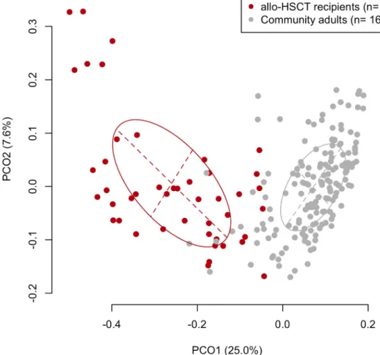
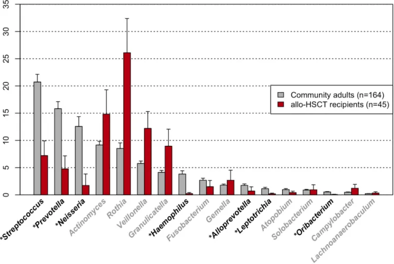
First, the overall composition of the oral microbiota of allo-HSCT recipients was compared with that of 164 age-matched (mean±SD: 55.0±10.8 years) community-dwelling adults including 93 females and 71 males based on the results of partial 16S rRNA gene sequencing (V1–V2 region) following rarefaction to 2,000 reads per sample to correct the unequal number of sequences per sample. A principal coordinate analysis (PCoA) plot based on the unweighted UniFrac metric demonstrated that the bacterial composition of the allo-HSCT recipients was distinct from that of the community-dwelling adults (Fig 1). Although the predominant bacte- rial genera in the community-dwelling adults were also present in the allo-HSCT recipients, those genera, such asStreptococcus,Prevotella, andNeisseria, were present at a significantly lower relative abundance in the allo-HSCT recipients (Fig 2). All three alpha diversity indices indicated that the microbiota of the allo-HSCT recipients was much less diverse than that of the community-dwelling adults (Table 2). In contrast, a substantial number of species-level operational taxonomic units (OTUs), absent in community-dwelling adults, were found in the microbiota of the allo-HSCT recipients (S1 Table). These OTUs were barely detectable even in the large number (3,077,343) of all obtained reads of the community-dwelling adults (S1 Table) which almost covers the bacterial taxa in their microbiota (S1 Fig). Most of these OTUs did not correspond to bacterial taxa for which the primary body site is denoted as “Oral” in the expanded Human Oral Microbiome Database (eHOMD) [16].
To identify the nonindigenous taxa detected in the oral cavity of the allo-HSCT recipients, we further employed full-length 16S rRNA gene sequencing analysis with a high taxonomic resolution by using a third-generation sequencer, PacBio Sequel (Pacific Biosciences, Menlo Park, CA, USA). A number of accurate full-length 16S rRNA gene sequences (about 1,500 bases) were obtained by the use of the circular consensus sequence (CCS) technique, which repeatedly determines the template DNA sequences based on long sequencing potential, although the lower throughput (as compared with the short-read sequencers) only allowed us to determine the microbiota of allo-HSCT recipients. A total of 152,040 high-quality CCS reads corresponding to 1,014 distinct full-length 16S rRNA gene sequences were obtained for the 45 recipients (mean±SD, 3378±1533 reads; range, 770–7198 reads), after a quality filter- ing and denoising procedure using R and DADA2. The rarefaction curve for the number of unique sequences almost approached a plateau in each sample (S2 Fig). The relative abun- dances of each bacteria and alpha diversity in full-length 16S rRNA gene sequencing analysis
Table 1. Baseline characteristics of the study population.
No. of patients
Parameter (n = 45)
Median age, yr (range) 53 (36–69)
Sex
Female/Male 19/26
Underlying disease
Acute myelogenous leukemia 23
Malignant lymphoma 10
Acute lymphoblastic leukemia 5
Myelodysplastic syndrome 5
Primary myelofibrosis 1
Aplastic anemia 1
Risk status at transplantation
Standard/High 22/23
Conditioning regimen
Myeloablative/Reduced intensity 20/25
Total body irradiation
None 10
Low-dose/High-dose 21/14
Graft source
Bone marrow 21
Peripheral blood 20
Cord blood 4
HLA parity
Matched/Mismatched 24/21
Antibiotic use during conditioning
Prophylactic/Treatment use 19/26
GvHD prophylaxis
CIs+sMTX 32
CIs+MMF±PTCy 9
Others 4
Prior transplantation
Yes/No 6/39
Median number of teeth (range) 27 (5–32)
Bacteremia until day+100
Yes/No 12/33
Oral mucositis
Yes/No 28/17
Grade 2 to 4 25
Grade 3 to 4 15
Acute GvHD
Yes/No 25/20
Grade II to IV 15
Grade III to IV 4
Abbreviation: GvHD, graft-versus host disease, CIs, calcineurin inhibitors; MTX, methotrexate; MMF, mycophenolate mofetil; PTCy, post-transplant cyclophosphamide
https://doi.org/10.1371/journal.ppat.1008348.t001
differed but highly correlated with those in V1–V2 region of 16S rRNA gene sequencing analy- sis (S3 Fig).
Of the obtained 16S rRNA gene sequences, 934 distinct sequences containing 139,923 reads (92.0%) were assigned to the 112 oral indigenous taxa (“Oral” was included in their “Body Site” status in eHOMD) deposited in the eHOMD [16], with�98.5% identity. Most of the microbiota on the tongue of allo-HSCT recipients were composed of taxa common in the oral cavity, includingRothia mucilaginosa,Granulicatella adiascensandVeillonella dispar(S4 Fig).
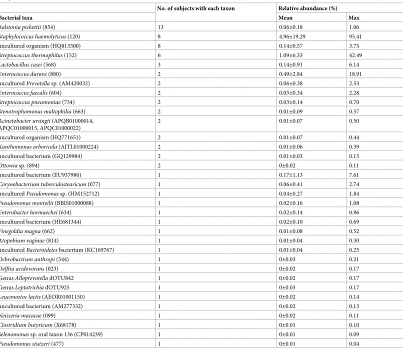
On the other hand, as many as 12,101 reads corresponding to 78 sequences, were unassigned and corresponded to 17 non-oral taxa in eHOMD and 17 bacterial taxa deposited in the SILVA database. The remaining 2 sequences containing 16 reads could not be identified using this database, so we determined their taxonomies up to the genus level. Of the 34 identified non-oral taxa, 15 were found in the microbiota of multiple allo-HSCT recipients (Table 3).
These non-oral bacteria obtained predominance in the disrupted oral microbiota of some allo- HSCT recipients (Table 3); in particular,Staphylococcus haemolyticuscomprised more than 40% of the microbiota in three patients.
The detection of the top 4 non-oral bacteria (observed in more than 5 cases) present in the oral cavity on the day of stem cell infusion, was not associated with clinical parameters,
Fig 1. Unweighted UniFrac plot of microbiota composition between community-dwelling adults and allo-HSCT patients. The ellipses cover 67% of the samples belonging to each sample type.
https://doi.org/10.1371/journal.ppat.1008348.g001
including incidence of oral mucositis, bacteremia through 100 days after transplantation, or acute GvHD (S2 Table). In contrast,Staphylococcus haemolyticusorRalstonia pickettii, as well as either or both taxa were associated with a significantly higher risk of all-cause mortality at 692 days of the median follow-up period (P= 0.009,P= 0.02 andP= 0.003, respectively, log- rank test;Fig 3). The estimated one-year overall survival rate among recipients positive for Staphylococcus haemolyticusand/orRalstonia pickettiiwas 37.8% (95% confidence interval
Fig 2. Relative abundances of predominant bacterial genera in the tongue microbiota of community-dwelling adults and allo-HSCT patients. Only 17 bacterial genera commonly (>98%) present in community-dwelling adults are shown.�P<0.05 in a Wilcoxon rank-sum test followed byPvalue adjustment for multiple comparison.
https://doi.org/10.1371/journal.ppat.1008348.g002
Table 2. Alpha diversity indices of the tongue microbiota of 164 community-dwelling adults and 45 allogeneic hematopoietic stem cell transplantation (allo-HSCT) recipients on transplantation date in V1-V2 regions of 16S rRNA gene sequencing analysis.
Community-dwelling adults (n = 164)
allo-HSCT recipients (n = 45)
P-valuea
Number of OTU 90.9±18.3 41.1±15.6 <0.001
Phylogenetic diversity 6.9±1.1 4.0±1.1 <0.001
Shannon diversity index 3.2±0.3 2.1±0.6 <0.001
Values with errors are mean±SD.
aWilcoxon rank-sum test.
https://doi.org/10.1371/journal.ppat.1008348.t002
(CI), 19.7%–72.3%), whereas that of patients without detection of either of these taxa, was 78.2% (95% CI, 64.2%–95.3%). Of 17 patients positive forStaphylococcus haemolyticusand/or Ralstonia pickettii, 12 had died of progressive disease (n = 8), GvHD (n = 3), or infection (n = 1), whereas 8 of 28 patients without detection of these taxa, had died of progressive disease (n = 5), GvHD (n = 1), or infection (n = 2).
Table 3. Nonindigenous bacteria identified from the tongue microbiota found in allo-HSCT recipients on the transplantation date in full-length 16S rRNA gene analysis.
No. of subjects with each taxon Relative abundance (%)
Bacterial taxa Mean Max
Ralstonia pickettii(854) 13 0.06±0.18 1.06
Staphylococcus haemolyticus(120) 8 4.96±19.29 95.41
uncultured organism (HQ813300) 8 0.14±0.57 3.75
Streptococcus thermophilus(152) 6 1.09±6.33 42.49
Lactobacillus casei(568) 3 0.14±0.91 6.14
Enterococcus durans(880) 2 0.49±2.84 18.91
unculturedPrevotellasp. (AM420032) 2 0.06±0.38 2.53
Enterococcus faecalis(604) 2 0.05±0.34 2.28
Streptococcus pneumoniae(734) 2 0.03±0.14 0.70
Stenotrophomonas maltophilia(663) 2 0.01±0.09 0.57
Acinetobacter ursingii(APQB01000014, APQC01000015, APQC01000022)
2 0.01±0.07 0.50
uncultured organism (HQ771651) 2 0.01±0.07 0.44
Xanthomonas arboricola(AJTL01000224) 2 0.01±0.06 0.39
uncultured bacterium (GQ129984) 2 0.01±0.03 0.15
Ottowiasp. (894) 2 0±0.02 0.11
uncultured bacterium (EU937980) 1 0.17±1.13 7.61
Corynebacterium tuberculostearicum(077) 1 0.06±0.41 2.74
unculturedPseudomonassp. (HM152712) 1 0.04±0.27 1.84
Pseudomonas monteilii(BBIS01000088) 1 0.02±0.16 1.08
Enterobacter hormaechei(634) 1 0.02±0.14 0.96
uncultured bacterium (HE681344) 1 0.02±0.10 0.69
Finegoldia magna(662) 1 0.01±0.08 0.52
Atopobium vaginae(814) 1 0.01±0.04 0.30
unculturedBacteroidetesbacterium (KC169767) 1 0.01±0.04 0.25
Ochrobactrum anthropi(544) 1 0±0.03 0.21
Delftia acidovorans(023) 1 0±0.02 0.17
GenusAlloprevotelladOTU842 1 0±0.02 0.17
GenusLeptotrichiadOTU925 1 0±0.03 0.17
Leuconostoc lactis(AEOR01001150) 1 0±0.02 0.14
uncultured bacterium (AM277332) 1 0±0.02 0.13
Neisseria macacae(099) 1 0±0.02 0.11
Clostridium butyricum(X68178) 1 0±0.01 0.10
Selenomonassp. oral taxon 136 (CP014239) 1 0±0.01 0.09
Pseudomonas stutzeri(477) 1 0±0.01 0.04
Values with errors are mean±SD.
aTaxon ID in expanded Human Oral Microbiome database (triple digits) or GenBank number is given in parentheses following bacterial name.
https://doi.org/10.1371/journal.ppat.1008348.t003
Among other potential covariates, disease risk stratification prior to transplantation was significantly associated with the survival rate during the follow-up period in a univariate Cox proportional hazards regression analysis (Hazard Ratio (HR) = 5.1, 95% CI = 1.8–14.2;
P= 0.001), along with detection of either/both of the two taxa (HR = 3.5, 95% CI = 1.4–8.6;
P= 0.006) (Table 4). A multivariate model incorporating these two factors demonstrated that detection of either/both of the two taxa remained statistically significant independent of the risk status (adjusted HR = 2.5, 95% CI = 1.0–6.4;P= 0.04:Table 4). We constructed another model incorporating additional use of antibiotics with the detection of either/both of the two taxa considering their potential relationship. Detection of either/both of the two taxa was also significantly associated with a higher risk of mortality in this model, while no significant rela- tionship was observed between additional use of antibiotics and higher risk of mortality after the adjustment (Table 4).
We further analyzed the characteristics of patients with and without detection of these bac- teria and sought causative variables associated with this difference in transplant outcome between the two groups (Table 5). Additional use of antibiotics for febrile neutropenia and/or infections to prophylactic oral levofloxacin was significantly relevant for the detection of these bacteria. Furthermore, a higher detection rate was observed in patients receiving multidrug regimens, rather than those receiving single cephem for the treatment of febrile neutropenia and/or infections (S3 Table). The PCoA plot based on unweighted UniFrac distance also showed that the additional antibiotics use, rather than conditioning regimens and underlying diseases, had an impact on the overall bacterial composition (S5 Fig).
Fig 3. Survival plots of the patients with detection or no detection ofStaphylococcus haemolyticusandRalstonia pickettiion the tongue generated by the Kaplan–
Meier method. Significant differences were determined using a log-rank test.
https://doi.org/10.1371/journal.ppat.1008348.g003
Discussion
This study demonstrated that the indigenous oral microbiota of allo-HSCT recipients was dis- rupted at the time point of stem cell transplantation after receiving intensive chemotherapy, radiation, and antibiotics. The lower diversity of the oral microbiota, as well as the change in components, among allo-HSCT recipients as compared with that in community-dwelling adults was confirmed (Table 2andFig 1). The bacterial genera predominantly observed in the oral microbiota of community-dwelling adults such asStreptococcus,NeisseriaandPrevotella, occupied a significantly lower proportion of the microbiota in allo-HSCT recipients (Fig 2).
Instead, taxa uncommon in the oral cavity were detected or even dominated in the microbiota of several allo-HSCT recipients (Table 3andS1 Table).
Use of the long-read sequencer with the CCS technique, which can provide numerous accu- rate full-length 16S rRNA gene sequences [17,18] identified as many as 34 bacterial taxa not corresponding to bacteria primarily inhabiting the oral cavity deposited in eHOMD [16] in the tongue microbiota of allo-HSCT recipients (Table 3). Nonetheless, there remains the possi- bility that they were merely transient bacteria present in the oral cavity at the time of sample
Table 4. Univariate and multivariate associations of the detection ofStaphylococcus haemolyticusand/orRalstonia pickettiiwith the high risk of mortality at the fol- low-up period in Cox proportional hazard regression models.
Univariate Multivariate
Model 1 Model 2
HR (95% CI) P-value aHR (95% CI) P-value aHR (95% CI) P-value
Detection of either/both of the two taxa 3.5 (1.4–8.6) .006 2.5 (1.0–6.4) .04 3.1 (1.0–9.2) .03
Age 1.0 (0.9–1.0) .26
Underlying disease (leukemia vs other) 1.2 (0.4–3.2) .66
Risk status at transplantation 5.1 (1.8–14.2) .001 4.1 (1.4–11.7) .008
Conditioning regimens
Myeloablative (reference)
Reduced intensity 1.3 (0.5–3.2) .54
Total body irradiation
None (reference)
Low dose 1.2 (0.2–2.1) .62
High dose 1.8 (0.1–1.7) .30
Antibiotics use during conditioning
Prophylactic use (reference)
Additional use for treatment 2.3 (0.8–6.5) .09 1.2 (0.3–4.2) .70
Graft source
Peripheral blood (reference)
Bone marrow 1.3 (0.5–3.4) .53
Cord blood 1.1 (0.2–5.4) .86
HLA parity
Matched (reference)
Mismatched 1.3 (0.5–3.1) .53
GvHD prophylaxis
CIs+sMTX (reference)
CIs+MMF±PTCy 1.4 (0.5–4.0) .47
Others 1.5 (0.3–7.0) .54
Abbreviation: HR, hazard ratio; aHR, adjusted hazard ratio; GvHD, graft-versus host disease; CIs, calcineurin inhibitors; MTX, methotrexate; MMF, mycophenolate mofetil; PTCy, post-transplant cyclophosphamide.
https://doi.org/10.1371/journal.ppat.1008348.t004
collection. However, although this cross-sectional study could evaluate only a snapshot of the tongue microbiota of allo-HSCT recipients on the day of transplantation, 15 of 34 non-oral bacteria were detected in the tongue swab samples of multiple allo-HSCT recipients (Table 3).
In addition, 12 taxa were identified as predominant in the tongue microbiota with maximum relative abundances>1% (Table 3), suggesting their detection was not just contamination.
Many of these taxa, such asRalstonia pickettii[19],Staphylococcus haemolyticus[20],
Table 5. Relationship between the detection ofStaphylococcus haemolyticusand/orRalstonia pickettiiand the baseline characteristics of the recipients.
Either/both of the two taxa
Detected Not detected
(n = 17) (n = 28) P-valuea
Age (Mean±SD) 54±10 51±10 0.46a
No. (%) of female 9 (52.9) 10 (35.7) 0.35b
Underlying disease 0.73b
Acute myelogenous leukemia 8 (47.0) 15 (53.5)
Malignant lymphoma 5 (29.4) 5 (17.8)
Acute lymphoblastic leukemia 1 (5.8) 4 (14.2)
Myelodysplastic syndrome 2 (11.7) 3 (10.7)
Primary myelofibrosis 0 (0) 1 (3.5)
Aplastic anemia 1 (5.8) 0 (0)
Prior transplantation 2 (11.7) 4 (14.2) 1.00b
Risk status at transplantation 0.06b
Standard 5 (29.4) 17 (60.7)
High 12 (70.5) 11 (39.2)
Conditioning regimens 0.37b
Myeloablative 6 (35.2) 14 (50.0)
Reduced intensity 11 (64.7) 14 (50.0)
Antibiotic use during conditioning 0.001b
Prophylactic use 2 (11.7) 17 (60.7)
Additional use for treatment 15 (88.2) 11 (39.2)
Total body irradiation 0.72b
None 4 (23.5) 6 (21.4)
Low dose 9 (52.9) 12 (42.8)
High dose 4 (23.5) 10 (35.7)
Graft source 0.07b
Peripheral blood 11 (64.7) 10 (35.7)
Bone marrow 4 (23.5) 16 (57.1)
Cord blood 2 (11.7) 2 (7.1)
HLA parity 0.23b
Matched 7 (41.1) 17 (60.7)
Mismatched 10 (58.8) 11 (39.2)
GvHD prophylaxis 0.41b
CIs+sMTX 10 (58.8) 22 (78.5)
CIs+MMF±PTCy 5 (29.4) 4 (14.2)
Others 2 (11.7) 2 (7.1)
aStudent’s t-test.
bFisher’s exact test.
Abbreviation: GvHD, graft-versus host disease; CIs, calcineurin inhibitors; MTX, methotrexate; MMF, mycophenolate mofetil; PTCy, post-transplant cyclophosphamide.
https://doi.org/10.1371/journal.ppat.1008348.t005
Enterococcus faecalis[21],Stenotrophomonas maltophilia[22], andAcinetobacter ursingii[23]
reportedly had natural or acquired resistance to antibiotics. Thus, detection of these pathologi- cal bacteria was quite reasonable in the microbiota of allo-HSCT recipients treated with long- term broad-spectrum antibiotics and/or antifungal agents during the peritransplant period.
Whether their detection results from their overgrowth or drastic decrease of other indigenous bacteria or both remains unknown in this study, because the 16S rRNA gene sequencing analy- sis only allows qualitative consideration on the bacterial community, or relative abundances of bacteria. Nevertheless, in any of these cases, it is a remarkable phenomenon indicating disrup- tion of indigenous microbiota which serves as a defensive barrier against infection.
Of the uncommon taxa found in allo-HSCT patients, the detection ofStaphylococcus hae- molyticusandRalstonia pickettiion the day of transplantation could be a powerful predictor of transplant outcome (Fig 3).Staphylococcus haemolyticus, a coagulase-negative staphylococci (CNS), is a member of the normal skin microbiota [20]. With its ability to acquire multidrug resistance, this microorganism has reportedly been the second most frequently isolated CNS, followingStaphylococcus epidermidis, especially as sourced from the bloodstream [24].Ralst- noia pickettii, a non-fermenting Gram-negative bacillus, is not a major pathogen but has been isolated from a wide variety of clinical specimens, including blood and sputum, or even from contaminated fluids used for patient care [19]. Aspiration or ingestion of these bacteria might be associated with poorer outcome of allo-HSCT recipients. However, the major cause of mor- tality was progressive diseases, and no patient died of the complications associated with detectedStaphylococcus haemolyticusorRalstonia pickettiiin the present study. The incidence of bacteremia until 100 days after transplantation, mucositis, or acute GvHD was comparable between allo-HSCT recipients with and without the detection of these two taxa. Further studies are required to clarify whether they are involved in death of recipients.
Another possible explanation for the higher mortality rate among patients positive forStaphy- lococcus haemolyticusorRalstonia pickettiiis that oral dysbiosis is a surrogate marker for a dysbio- tic pattern of the intestinal microbiota. Intensive chemotherapy and irradiation allow bacteria to enter the systemic circulation through the disrupted mucosal barrier of the GI tract and/or oral cavity. This then provides a cytokine milieu fit for T-cell proliferation and activation and leads to the development of GvHD after donor–cell engraftment. As a previous mouse model indicated, intestinal GvHD targeted intestinal stem cells as well as their niche Paneth cells play a pivotal role in the regeneration of the damaged mucosal epithelium. The loss of Paneth cells resulted in reduced production ofα-defensins, antimicrobial peptides required for maintaining intestinal microbial ecology; less diversity of the intestinal microbiome in turn had an impact on the mortal- ity of allo-HSCT recipients [9]. In this study, we found no significant relationship between dysbio- sis of the oral cavity and incidence or severity of intestinal GvHD (S4 Table). To clarify these issues, a direct comparison of the microbiome diversity, and their components, between the oral cavity and gastrointestinal tract in each case, should be performed in future studies.
These two taxa were detected from 2 of 19 patients receiving only prophylactic antibiotics, whereas they were also detected from over half of the patients receiving additional antibiotics for the treatment of neutropenic fever/infections (Table 5). Furthermore, higher detection rates were observed in the patients receiving multidrug regimens relative to those receiving additional treatment by single cephem (S3 Table). Although we could not specify the causative antibiotic/s, our results suggest that heavy treatment with multiple antibiotics during the pre- transplant conditioning period strongly correlated with detection of the two taxa in the dis- rupted tongue microbiota.
We confirm that detection of either/both of the two taxa was significantly associated with the high risk of mortality, independent of other relevant factors such as risk status at transplan- tation or use of additional antibiotics. However, we could not construct a further complex
model incorporating all potential confounding factors, because more than two exposures (1/10 of the number of events, 20 events in this study) should not be incorporated into the Cox pro- portional hazards regression model in order to avoid over-adjustment [25]. A future study with a larger sample size is needed to show that the detection of the two taxa is a completely independent predictor for mortality risk.
The tongue coating of community-dwelling adults was collected using a sampling device based on an electric toothbrush developed in our previous study [26], which enabled us to col- lect bacteria attached to a bonded-fiber fabric on a brush head from a 15 mm-diameter circular area on the center of the tongue dorsum. On the other hand, we had already commenced with sample collection from allo-HSCT patients before development of this device, thus their sam- ples were collected by using a conventional cotton swab. There is little difference in the bacte- rial composition between the samples collected by the two devices. However, a quantity of bacteria, collected using a cotton swab, remained trapped in the swab after centrifugation.
Hence, use of a cotton swab resulted in lower DNA recovery than the newly developed method. This difference in the sampling device prevents the comparison of absolute bacterial quantity between the community-dwelling adults and allo-HSCT patients in this study. A future study using the quantitative sampling device, with quantitative PCR analysis, is required to clarify whether these bacteria actually colonize and grow in allo-HSCT or only survive on the tongue dorsum of the allo-HSCT recipients.
Low diversity in the intestinal microbiota was reported to be a good predictor of high risk of mortality following allo-HSCT [9]. In contrast, no significant relationship was observed between alpha diversity (Shannon diversity index) of the tongue microbiota and incidence of transplant complications, although overall survival rate tended to be lower in subjects with lower diverse microbiota (S5 Table). This result implies that detection of specific bacterial taxa in less diverse microbiota of allo-HSCT patients would be more helpful for the prediction of mortality risk rather than microbial diversity itself.
This comprehensive analysis revealed that altered indigenous microbiota of allo-HSCT recipients was observed in the oral cavity, as previously reported in the gastrointestinal tract [5]. Lowered diversity of the oral microbiota with an increased proportion of drug-resistant bacteria uncommon in the oral cavity was observed on the day of stem cell transplantation.
Among such bacterial taxa, allo-HSCT recipients with the detection ofStaphylococcus haemo- lyticusand/orRalstonia pickettiishowed inferior transplant outcome, although the mecha- nisms require further investigation. Careful attention should be given to bacterial composition of the disrupted oral microbiota in allo-HSCT recipients.
Materials and methods Ethics statement
All participants understood the nature of the study and provided written informed consent.
The ethics committee of Kyushu University Hospital approved this study (reference no. 2019–
246). All experiments were conducted in accordance with approved guidelines.
Study population and sample collection
The study population consisted of adult patients aged 36–69 years who underwent allo-HSCT at Kyushu University Hospital from February 2016 to October 2018. Oral microbiota samples were collected from the center of tongue dorsum on the day of transplantation with a cotton swab. The collected samples were placed in 200μl of lysis buffer containing 10 mM Tris-HCl, 1 mM EDTA, and 1% sodium dodecyl sulfate. After the swab was discarded following centrifu- gation, DNA extraction was performed using a bead-beating method as described previously
[27]. Of the 75 patients who provided samples, 30 were excluded from the analysis due to the low quantity and/or quality of the extracted DNA causing an inability to obtain sufficient amounts of PCR amplicons for sequencing.
The microbiota composition of community-dwelling adults of a similar generation was determined in order to compare the overall microbiota composition with that of allo-HSCT recipients. This population consisted of 164 adult residents of Hisayama Town, Fukuoka, Japan who received a health examination as well as periodontal disease screenings for adults aged 40, 50, 60 and 70 years in 2016 (n = 41, 35, 53, and 35 subjects, respectively). The subjects aged 70 years were part of the study population of our previous study on the tongue microbiota of community-dwelling elderly adults aged 70 to 80 years [26]. Sample collection using a sam- pling device based on an electric toothbrush with a bonded-fiber fabric and DNA extraction were conducted as previously described [26]. The samples from the subjects aged 40, 50 and 60 years were collected in the same health examination and processed using the same procedure.
Ion Torrent 16S rRNA gene sequencing analysis
The V1–V2 region of the 16S rRNA gene of 45 allo-HSCT recipients was amplified using the following primers: 8F (5’- AGA GTT TGA TYM TGG CTC AG -3’) with Ion Torrent adaptor A and the sample-specific 8-base tag sequence and 338R (5’- TGC TGC CTC CCG TAG GAG T -3’) with the Ion Torrent trP1 adaptor sequence and the sample-specific 8-base tag sequence.
PCR amplification, purification, quantification of each amplicon and amplicon pooling were performed as previously described [28]. Emulsion PCR and enrichment of template-positive particles were performed using an Ion PGM Hi-Q View OT2 kit (Thermo Fisher Scientific, Waltham, MA), and sequencing was performed with the Ion PGM (Thermo Fisher Scientific) using an Ion PGM Hi-Q View Sequencing kit (Thermo Fisher Scientific).
The V1–V2 region of 16S rRNA gene of the 35 community-dwelling adults aged 70 years was determined in our previous study [26] with the Ion PGM (Thermo Fisher Scientific) and the same reagents used in this study, except that the previous reverse primer did not contain an 8-base tag sequence. The V1–V2 regions of 16S rRNA gene sequences of the remaining 130 residents aged 40, 50 and 60 years were determined via the same procedure.
The quality of the raw sequence reads was checked using a script written in R as previously described [26]. The quality-filtered reads were assigned to the appropriate samples based on their tag sequences, followed by trimming their adaptor, tag, and forward primer sequences.
Similar sequences were assigned into operational taxonomic units (OTUs) using UPARSE [29], with a minimum pairwise identity of 97%. Following rarefaction to 2,000 reads per sam- ple to correct for the unequal number of sequences per sample (a minimum of 2,182 reads), alpha diversity indices were calculated using the diversity function in the vegan library of R.
The dissimilarity between all bacterial communities was assessed using the unweighted Uni- Frac metric [30], and the similarity relationship was represented in a principal coordinate anal- ysis (PCoA) plot drawn by R. The taxonomy of representative sequences was determined using the RDP classifier with a minimum support threshold of 80% and the RDP taxonomic nomenclature (to the genus level). The taxonomy of the OTUs which were only present in allo-HSCT recipients was further searched using BLAST against 998 bacterial 16S rRNA gene sequences (HOMD 16S rRNA RefSeq version 15.1) in the expanded Human Oral Microbiome Database (eHOMD) [16] with a threshold identity of 98.5%.
Full length 16S rRNA gene sequencing analysis using Pacbio Sequel
The 16S rRNA genes containing all variable regions were amplified using the following prim- ers: 8F (5’- AGA GTT TGA TYM TGG CTC AG -3’) with Ion Torrent adaptor A and the
sample-specific 8-base tag sequence and 1492R (5’- GGY TAC CTT GTT ACG ACT T -3’).
PCR amplification was carried out using KOD DNA polymerase (Toyobo, Osaka, Japan) under the following cycling conditions: 98˚C for 2 min, followed by 30 cycles of 98˚C for 15 s, 60˚C for 20 s, and 74˚C for 90 s. Equal amounts of DNA were pooled following purification using an Agencourt AMPure XP kit (Beckman Coulter, Brea, CA, USA). The pooled DNA was gel purified using a Wizard SV gel and PCR cleanup system (Promega, Madison, WI, USA).
The purified amplicons were sequenced using the Sequel Sequencing kit 2.1 (Pacific Biosci- ences, Menlo Park, CA, USA) on a PacBio Sequel (Pacific BioSciences). The obtained long sequence reads, which repeatedly determined the template DNA, were processed using SMRT Link software version 5.1.0.26412 (Pacific Biosciences), which provided circular consensus sequence (CCS) reads with high accuracy.
The CCS reads were excluded from the analysis if they were�1,000 bases, if they were
�1,700 bases, if they had an average quality score of�40, or if they did not include the correct forward and reverse primer sequences. The remaining CCS reads were assigned to the appro- priate sample by examining tag sequences using R, followed by trimming of tag and primer sequences. The 211,164 quality-checked CCS reads were further processed by using the DADA2 pipeline [31] including the quality-filtering, denoising and chimera-filtering proce- dures with default settings for PacBio reads (version 1.9.1). The taxonomy of each denoised CCS sequence was determined using BLAST against 998 oral bacterial 16S rRNA gene sequences in the eHOMD (eHOMD 16S rRNA RefSeq version 15.1) [16]. Nearest neighbor taxons with�98.5% identity were selected as candidates for each sequence. The denoised sequences with no hit were further compared against 695,172 bacterial sequences in SILVA database (SILVA_132_SSURef_Nr99_tax_silva.fasta) and nearest neighbors with�98.5%
identity were selected as candidates for each sequence. The taxonomy of the remaining unde- fined sequences was determined up to the genus level using the RDP classifier with a minimum support threshold of 80%.Pseudomonas fluorescensHOT-612 was uniquely identified in a slight amount of PCR amplicon obtained from negative control in our preliminary analysis, thus we excluded the sequences corresponding to this taxon from the analysis, as PCR contam- inant. We also discarded the sequences corresponding to phylumCyanobacteria/Chloroplast, because all these sequences matched with the chloroplast’s 16S rRNA genes in plants. The numbers of the denoised CCS sequences corresponding to the same taxa were combined, and the relative abundance and detection frequencies of each taxon were calculated in R. The sequence data obtained in this study have been deposited in the DDBJ Sequence Read Archive under accession no. DRA009550 and DRA009551.
Statistical analysis
All statistical analyses were conducted with R version 3.5.1. The Wilcoxon rank-sum test was conducted to compare the alpha diversity indices between the community-dwelling adults and the allo-HSCT recipients. The relative abundance of bacterial genera were compared using a Wilcoxon rank-sum test followed byPvalue adjustment for multiple comparison. Fisher’s exact test was conducted to analyze the relationship between the presence of non-oral bacteria and incidence of transplant complications (oral mucositis, bacteremia until day 100, acute GVHD and death) during the follow-up period (by July 21, 2019; 283–1258 days). The survival curves were generated by the Kaplan–Meier method and a log-rank test was conducted to examine significant differences in survival curves between the recipients with and without spe- cific bacterial taxa. A Cox proportional hazard regression analysis was performed to estimate adjusted hazard ratio and 95% CIs. The risk status at transplantation was classified based on the criteria described previously [32].
Supporting information
S1 Fig. Rarefaction curve for a number of OTUs in allo-HSCT patients and community- dwelling adults in the V1–V2 regions of 16S rRNA gene analysis by Ion PGM.
(TIF)
S2 Fig. Rarefaction curve for a number of unique sequences per samples in the full-length 16S rRNA gene analysis by PacBio Sequel.
(TIF)
S3 Fig. Comparison between the results of V1-V2 regions and full-length 16S rRNA gene analyses. Alpha diversity (number of identified sequences) and the relative abundances of seven predominant bacterial taxa (mean relative abundance>5%) in each analysis are shown.
(TIF)
S4 Fig. Relative abundance of predominant bacterial taxa with mean relative abundances
>1% and 34 non-oral taxa in the tongue microbiota of 45 allo-HSCT patients on the trans- plantation date in full-length 16S rRNA gene analysis using PacBio Sequel.
(TIF)
S5 Fig. A principal coordinate analysis plot showing similarity relationship among tongue microbiota of allo-HSCT patients who received different antibiotic use and conditioning regimens, and have different underlying diseases using an unweighted UniFrac metric, respectively. The points corresponding to different groups are depicted in different colors in each diagram. The microbiota difference between the groups were investigated statistically by permutational multivariate analysis of variance (perMANOVA) test. The ellipses cover 67% of the samples belonging to each sample type.
(TIF)
S1 Table. Bacterial taxa corresponding to 12 OTUs present in the tongue microbiota of multiple allo-HSCT recipients on the transplantation date but absent in 164 community- dwelling adults (CDA) in V1-V2 regions of 16S rRNA gene sequencing data which rarified 2000 reads per sample.
(PDF)
S2 Table. Incidence of transplant complications in the recipients with the detection of four non-oral bacterial taxa.
(PDF)
S3 Table. Relationship between the detection ofStaphylococcus haemolyticusand/orRal- stonia pickettiiand antibiotics used during pretransplant conditioning.
(PDF)
S4 Table. Relationship between the detection ofStaphylococcus haemolyticusand/orRal- stonia pickettiiand the severity of intestinal GvHD.
(PDF)
S5 Table. Incidence of transplant complications in the recipients with the microbiota with different alpha diversity (Shannon diversity index).
(PDF)
Acknowledgments
We thank Yoshiko Imamura and Megumi Sugihara for assistance in sample collection and all patients who participated in this study.
Author Contributions
Conceptualization: Saori Oku, Toru Takeshita, Toshiko Futatsuki, Haruhiko Kashiwazaki, Yoshihisa Yamashita.
Data curation: Saori Oku, Toru Takeshita, Toshiko Futatsuki, Shinya Kageyama, Yasuo Mori, Toshihiro Miyamoto, Jun Hata, Toshiharu Ninomiya.
Funding acquisition: Toru Takeshita, Yoshihisa Yamashita.
Investigation: Saori Oku, Toru Takeshita, Toshiko Futatsuki, Shinya Kageyama, Mikari Asa- kawa, Yoshihisa Yamashita.
Supervision: Toshihiro Miyamoto, Jun Hata, Toshiharu Ninomiya, Haruhiko Kashiwazaki, Yoshihisa Yamashita.
Validation: Saori Oku, Toru Takeshita, Shinya Kageyama, Yasuo Mori, Toshihiro Miyamoto, Toshiharu Ninomiya, Haruhiko Kashiwazaki, Yoshihisa Yamashita.
Visualization: Saori Oku, Toru Takeshita, Shinya Kageyama, Yoshihisa Yamashita.
Writing – original draft: Saori Oku, Toru Takeshita, Yasuo Mori.
Writing – review & editing: Saori Oku, Toru Takeshita, Toshiko Futatsuki, Shinya Kageyama, Mikari Asakawa, Yasuo Mori, Toshihiro Miyamoto, Jun Hata, Toshiharu Ninomiya, Haru- hiko Kashiwazaki, Yoshihisa Yamashita.
References
1. Jenq RR, van den Brink MR. Allogeneic haematopoietic stem cell transplantation: individualized stem cell and immune therapy of cancer. Nat Rev Cancer. 2010; 10(3):213–21. Epub 2010/02/20.https://doi.
org/10.1038/nrc2804PMID:20168320.
2. Copelan EA, Chojecki A, Lazarus HM, Avalos BR. Allogeneic hematopoietic cell transplantation; the current renaissance. Blood Rev. 2019; 34:34–44. Epub 2018/11/24.https://doi.org/10.1016/j.blre.2018.
11.001PMID:30467067.
3. Copelan EA. Hematopoietic stem-cell transplantation. N Engl J Med. 2006; 354(17):1813–26. Epub 2006/04/28.https://doi.org/10.1056/NEJMra052638PMID:16641398.
4. Becattini S, Taur Y, Pamer EG. Antibiotic-Induced Changes in the Intestinal Microbiota and Disease.
Trends Mol Med. 2016; 22(6):458–78. Epub 2016/05/15.https://doi.org/10.1016/j.molmed.2016.04.003 PMID:27178527; PubMed Central PMCID: PMC4885777.
5. Taur Y, Xavier JB, Lipuma L, Ubeda C, Goldberg J, Gobourne A, et al. Intestinal domination and the risk of bacteremia in patients undergoing allogeneic hematopoietic stem cell transplantation. Clin Infect Dis.
2012; 55(7):905–14. Epub 2012/06/22.https://doi.org/10.1093/cid/cis580PMID:22718773; PubMed Central PMCID: PMC3657523.
6. Shono Y, Docampo MD, Peled JU, Perobelli SM, Velardi E, Tsai JJ, et al. Increased GVHD-related mor- tality with broad-spectrum antibiotic use after allogeneic hematopoietic stem cell transplantation in human patients and mice. Sci Transl Med. 2016; 8(339):339ra71. Epub 2016/05/20.https://doi.org/10.
1126/scitranslmed.aaf2311PMID:27194729; PubMed Central PMCID: PMC4991773.
7. Golob JL, Pergam SA, Srinivasan S, Fiedler TL, Liu C, Garcia K, et al. Stool Microbiota at Neutrophil Recovery Is Predictive for Severe Acute Graft vs Host Disease After Hematopoietic Cell Transplanta- tion. Clin Infect Dis. 2017; 65(12):1984–91. Epub 2017/10/12.https://doi.org/10.1093/cid/cix699PMID:
29020185; PubMed Central PMCID: PMC5850019.
8. Peled JU, Devlin SM, Staffas A, Lumish M, Khanin R, Littmann ER, et al. Intestinal Microbiota and Relapse After Hematopoietic-Cell Transplantation. J Clin Oncol. 2017; 35(15):1650–9. Epub 2017/03/
16.https://doi.org/10.1200/JCO.2016.70.3348PMID:28296584; PubMed Central PMCID:
PMC5455763.
9. Taur Y, Jenq RR, Perales MA, Littmann ER, Morjaria S, Ling L, et al. The effects of intestinal tract bacte- rial diversity on mortality following allogeneic hematopoietic stem cell transplantation. Blood. 2014; 124 (7):1174–82. Epub 2014/06/19.https://doi.org/10.1182/blood-2014-02-554725PMID:24939656;
PubMed Central PMCID: PMC4133489.
10. Dahlen G. Bacterial infections of the oral mucosa. Periodontol 2000. 2009; 49:13–38. Epub 2009/01/21.
https://doi.org/10.1111/j.1600-0757.2008.00295.xPMID:19152524.
11. Muro M, Soga Y, Higuchi T, Kataoka K, Ekuni D, Maeda Y, et al. Unusual oral mucosal microbiota after hematopoietic cell transplantation with glycopeptide antibiotics: potential association with pathophysiol- ogy of oral mucositis. Folia Microbiol (Praha). 2018; 63(5):587–97. Epub 2018/03/14.https://doi.org/10.
1007/s12223-018-0596-1PMID:29532421.
12. Ames NJ, Barb JJ, Ranucci A, Kim H, Mudra SE, Cashion AK, et al. The oral microbiome of patients undergoing treatment for severe aplastic anemia: a pilot study. Ann Hematol. 2019; 98(6):1351–65.
Epub 2019/03/29.https://doi.org/10.1007/s00277-019-03599-wPMID:30919073.
13. Segata N, Haake SK, Mannon P, Lemon KP, Waldron L, Gevers D, et al. Composition of the adult digestive tract bacterial microbiome based on seven mouth surfaces, tonsils, throat and stool samples.
Genome biology. 2012; 13(6):R42. Epub 2012/06/16.https://doi.org/10.1186/gb-2012-13-6-r42PMID:
22698087; PubMed Central PMCID: PMC3446314.
14. Mager DL, Ximenez-Fyvie LA, Haffajee AD, Socransky SS. Distribution of selected bacterial species on intraoral surfaces. J Clin Periodontol. 2003; 30(7):644–54. Epub 2003/07/02. 376 [pii].https://doi.org/
10.1034/j.1600-051x.2003.00376.xPMID:12834503.
15. Zhou Y, Gao H, Mihindukulasuriya KA, La Rosa PS, Wylie KM, Vishnivetskaya T, et al. Biogeography of the ecosystems of the healthy human body. Genome biology. 2013; 14(1):R1. Epub 2013/01/16.
https://doi.org/10.1186/gb-2013-14-1-r1PMID:23316946; PubMed Central PMCID: PMC4054670.
16. Chen T, Yu WH, Izard J, Baranova OV, Lakshmanan A, Dewhirst FE. The Human Oral Microbiome Database: a web accessible resource for investigating oral microbe taxonomic and genomic informa- tion. Database (Oxford). 2010:baq013. Epub 2010/07/14.https://doi.org/10.1093/database/baq013 baq013 [pii]. PMID:20624719; PubMed Central PMCID: PMC2911848.
17. Schloss PD, Jenior ML, Koumpouras CC, Westcott SL, Highlander SK. Sequencing 16S rRNA gene fragments using the PacBio SMRT DNA sequencing system. PeerJ. 2016; 4:e1869.https://doi.org/10.
7717/peerj.1869PMID:27069806; PubMed Central PMCID: PMC4824876.
18. Wagner J, Coupland P, Browne HP, Lawley TD, Francis SC, Parkhill J. Evaluation of PacBio sequenc- ing for full-length bacterial 16S rRNA gene classification. BMC Microbiol. 2016; 16(1):274.https://doi.
org/10.1186/s12866-016-0891-4PMID:27842515; PubMed Central PMCID: PMC5109829.
19. Stelzmueller I, Biebl M, Wiesmayr S, Eller M, Hoeller E, Fille M, et al. Ralstonia pickettii-innocent bystander or a potential threat? Clin Microbiol Infect. 2006; 12(2):99–101. Epub 2006/01/31.https://doi.
org/10.1111/j.1469-0691.2005.01309.xPMID:16441445.
20. Czekaj T, Ciszewski M, Szewczyk EM. Staphylococcus haemolyticus—an emerging threat in the twi- light of the antibiotics age. Microbiology. 2015; 161(11):2061–8. Epub 2015/09/14.https://doi.org/10.
1099/mic.0.000178PMID:26363644.
21. Kristich CJ, Rice LB, Arias CA. Enterococcal Infection-Treatment and Antibiotic Resistance. In: Gilmore MS, Clewell DB, Ike Y, Shankar N, editors. Enterococci: From Commensals to Leading Causes of Drug Resistant Infection. Boston2014.
22. Brooke JS. Stenotrophomonas maltophilia: an emerging global opportunistic pathogen. Clinical microbi- ology reviews. 2012; 25(1):2–41. Epub 2012/01/11.https://doi.org/10.1128/CMR.00019-11PMID:
22232370; PubMed Central PMCID: PMC3255966.
23. Dortet L, Legrand P, Soussy CJ, Cattoir V. Bacterial identification, clinical significance, and antimicrobial susceptibilities of Acinetobacter ursingii and Acinetobacter schindleri, two frequently misidentified opportunistic pathogens. J Clin Microbiol. 2006; 44(12):4471–8. Epub 2006/10/20.https://doi.org/10.
1128/JCM.01535-06PMID:17050816; PubMed Central PMCID: PMC1698419.
24. Takeuchi F, Watanabe S, Baba T, Yuzawa H, Ito T, Morimoto Y, et al. Whole-genome sequencing of staphylococcus haemolyticus uncovers the extreme plasticity of its genome and the evolution of human-colonizing staphylococcal species. J Bacteriol. 2005; 187(21):7292–308. Epub 2005/10/21.
https://doi.org/10.1128/JB.187.21.7292-7308.2005PMID:16237012; PubMed Central PMCID:
PMC1272970.
25. Peduzzi P, Concato J, Feinstein AR, Holford TR. Importance of events per independent variable in pro- portional hazards regression analysis. II. Accuracy and precision of regression estimates. Journal of clinical epidemiology. 1995; 48(12):1503–10. Epub 1995/12/01.https://doi.org/10.1016/0895-4356(95) 00048-8PMID:8543964.
26. Asakawa M, Takeshita T, Furuta M, Kageyama S, Takeuchi K, Hata J, et al. Tongue Microbiota and Oral Health Status in Community-Dwelling Elderly Adults. mSphere. 2018; 3(4).https://doi.org/10.1128/
mSphere.00332-18PMID:30111628; PubMed Central PMCID: PMC6094060.
27. Takeshita T, Nakano Y, Yamashita Y. Improved accuracy in terminal restriction fragment length poly- morphism phylogenetic analysis using a novel internal size standard definition. Oral Microbiol Immunol.
2007; 22(6):419–28. Epub 2007/10/24. OMI384 [pii]https://doi.org/10.1111/j.1399-302X.2007.00384.x PMID:17949346.
28. Takeshita T, Kageyama S, Furuta M, Tsuboi H, Takeuchi K, Shibata Y, et al. Bacterial diversity in saliva and oral health-related conditions: the Hisayama Study. Scientific reports. 2016; 6:22164.https://doi.
org/10.1038/srep22164PMID:26907866; PubMed Central PMCID: PMC4764907.
29. Edgar RC. UPARSE: highly accurate OTU sequences from microbial amplicon reads. Nature methods.
2013; 10(10):996–8. Epub 2013/08/21.https://doi.org/10.1038/nmeth.2604PMID:23955772.
30. Lozupone C, Knight R. UniFrac: a new phylogenetic method for comparing microbial communities. Appl Environ Microbiol. 2005; 71(12):8228–35. Epub 2005/12/08. 71/12/8228 [pii]https://doi.org/10.1128/
AEM.71.12.8228-8235.2005PMID:16332807; PubMed Central PMCID: PMC1317376.
31. Callahan BJ, McMurdie PJ, Rosen MJ, Han AW, Johnson AJ, Holmes SP. DADA2: High-resolution sample inference from Illumina amplicon data. Nature methods. 2016; 13(7):581–3.https://doi.org/10.
1038/nmeth.3869PMID:27214047; PubMed Central PMCID: PMC4927377.
32. Mori Y, Yoshimoto G, Nishida R, Sugio T, Miyawaki K, Shima T, et al. Gastrointestinal Graft-versus- Host Disease Is a Risk Factor for Postengraftment Bloodstream Infection in Allogeneic Hematopoietic Stem Cell Transplant Recipients. Biol Blood Marrow Transplant. 2018; 24(11):2302–9. Epub 2018/06/
18.https://doi.org/10.1016/j.bbmt.2018.06.002PMID:29909153.