首都大学東京 博士(理学)学位論文(課程博士)
論 文 名 マトリクス支援レーザー脱離イオン化の機構研究と 応用手法の提案(英文)
著 者
許 家瑋 審査担当者
主 査 委 員 委 員 委 員
上記の論文を合格と判定する 平成 年 月 日
首都大学東京大学院理工学研究科教授会
研究科長
DISSERTATION FOR A DEGREE OF DOCTOR OF PHILOSOPHY IN SCIENCE
TOKYO METROPOLITAN UNIVERSITY
TITLE
:
Study of ionization process and research on application in matrix-assisted laser desorption/ionizationAUTHOR
:
Xu JiaweiEXAMINED BY
Examiner in chief Examiner
Examiner
Examiner
QUALIFIED BY THE GRADUATE SCHOOL OF SCIENCE AND ENGINEERING TOKYO METROPOLITAN UNIVERSITY Dean
Date
1
Study of ionization process and research on application in matrix-assisted laser desorption/ionization
Xu Jiawei
Department of Chemistry, Graduated School of Science, Tokyo Metropolitan University, 1-1 Minami-Osawa, Hachioji, Tokyo 192-0397, Japan
2014
マトリクス支援レーザー脱離イオン化の機構研究と応用 手法の提案
許
シュ
家
ジャ
瑋
ウェイ
2
Contents
Chapter One ... 4
General Introduction 1. Mass spectrometry ... 4
2. Matrix assisted laser desorption/ionization (MALDI) time-of-flight (MALDI- TOF) mass spectrometry ... 10
3. Mechanisms of ionization process in MALDI ... 12
4. Cyclodextrins and the their application in MALDI ... 15
5. Chiral molecules, chiral separation and the usage of Cyclodextrins ... 18
6. This study ... 22
Figure captions ... 24
References ... 25
Chapter Two ... 35
Spectroscopic evidence of analyte-support mechanism in matrix-assisted laser desorption/ionization Abstract: ... 35
1. Introduction ... 37
2. Experimental section ... 40
3. Results and discussion ... 41
4. Conclusion ... 51
Figure captions ... 52
References ... 62
Chapter 3 ... 65
Using cyclodextrin for the detection of chiral molecules with matrix-assisted laser desorption ionization Abstract ... 65
1. Introduction ... 66
3
2. Experimental section ... 68
3. Results and discussion ... 69
Conclusion ... 76
Figure captions ... 78
References ... 84
General conclusion ... 87
Acknowledgement ... 89
4
Chapter One
General Introduction
1. Mass spectrometry
Mass spectrometry is a kind of essential tool for analytical research. The mass of compounds in samples can be measured, which makes mass spectrometry one of the most important and effective methods for qualifying materials. Mass spectrometry can provide large amount of information of a sample during one analysis by producing, separating and detecting the compounds in gas phase [1]. By ionizing the chemical compounds, the ions are separated and detected according to their mass to charge ratios. Among all the analytical methods, mass spectrometry is always considered to be highly specific and sensitive [2].
A mass spectrometry instrument always contains three main parts, which are ionization source, analyzer and detector [3]. For each part, there are several methods that exist. The samples introduced to mass spectrometry will be ionized by the ionization source first, and then be separated by the analyzer and detected by the detector at last [4].
The first mass spectrometer was made in 1919, which won the Noble prize in 1922.
The mass to charge ratio (m/z) of ions is measured in mass spectrometry analysis.
Compounds in samples get ionized by the ionization source to generate positively charged ions which hold different m/z. With the acceleration in electric field, the ions will generate an ion beam and enters the analyzer, where the same ions get focused and different ions are separated. The ions reach the detector at different times and their molecular weights can be measured. To get rid of the interference from the molecules in air, which may inhibit ions from traveling to the detector, the whole mass spectrometer system is usually kept under high vacuum [5].
5
By combining mass spectrometry with other separation techniques, a great progress was achieved in analytical science. Mass spectrometry is mostly used together as the detector with gas chromatography (GC), high performance liquid chromatography (HPLC) and capillary electrophoresis (CE). The mixture samples are separated by these methods following the mass spectrometry detection. In this way, the separation, qualification and on some degree, quantification can be achieved at the same time, with high accuracy and efficiency [6-7].
Ionization source
There are many kinds of ionization sources developed for mass spectrometry, for example, traditional ionization methods such as electron ionization (EI) and chemical ionization (CI) and newly developed soft ionization methods such as matrix assisted laser desorption/ionization (MALDI), electrospray ionization (ESI) and fast atom bombardment (FAB). Since detailed introduction on MALDI will be provided later, the other methods will be shortly introduced here.
Electron ionization
Electron ionization is also called electron impact. In EI, the electrons generated from a filament are accelerated to 70 eV first. After concentrating these energized electrons into a beam, the sample in gas phase is bombarded by this beam. Under this condition, the interaction between energized electrons and gas phase molecules will take place, which will result in the ions to be generated [8].
In real applications, EI has many restrictions. First, the energy of the electrons affects the ionization efficiency strongly. If electrons do not have enough energy, the ionization efficiency will decrease significantly. This feature makes us to keep the electron energy high, which causes the second restriction that the samples must be thermally stable, which means that the molecular weight of samples cannot be too large. Actually EI is usually used for the molecules under 2000 Da [9-10].
6 Chemical ionization
Chemical ionization is also a traditional ionization method used in mass spectrometry.
In CI, reagent gas is necessary [11]. Reagent gas first gets preferentially ionization be the electrons entering ionization source and ionization plasma will be generated from this reaction [12]. Some widely used reagent gases are CH4, NH3 and isobutene. This ionization plasma will react with analyte molecules and cause the analyte to be ionized. Energy which takes part in the ionization process is much lower than EI, which result in less fragments and simpler spectrum [13]. There are mainly two variations of CI according to the reactions during the ionization, positive chemical ionization and negative chemical ionization [14].
Electrospray ionization
ESI is a kind of soft ionization method, which means that the analyte molecules generate much less fragments during the ionization process, since the reaction conditions are much milder than the methods that are introduced above [15].
According to this feature, ESI has been widely used for the detection of large biological molecules, such as protein and DNA [16].
In ESI, samples are dissolved in the solvent that is usually prepared by mixing water and volatile organic compounds like methanol or acetonitrile, which are also capable to provide protons during the ionization process. Some other compounds like acetic acid are also added into the solvent to increase the conductivity of the solvent. The sample solution goes through a capillary and is injected into a vacuum chamber by a needle. Solvent of this solution will evaporate to form small droplets and the droplets will be charged during the traveling and evaporation process. During the evaporation of solvent, the droplets will explode and generate more small droplets, which have small weigh and strong charge [17-18]. Because of solution samples can be directly introduced as sample, ESI can be easily combined with other separation methods,
7
such as HPLC, GC and CE to achieve highly automatic separation and detection for large molecules [19-21]. Meanwhile, ESI is one of the most essential tools for the research on the fold of proteins [22-24].
Different with other ionization methods, multiply charged ions can be generated with ESI, the mass range is largely extended. However, because of this feature, structural information obtained becomes poor for the ESI mass spectrometry detection. To overcome this drawback, ESI can be coupled with tandem mass spectrometry (ESI- MS/MS). Another limitation of ESI is that since the solvent used must have low boiling point, such as acetonitrile and methanol, if the sample cannot dissolve in such solvents, this technique is difficult to be applied [25].
Fast atom bombardment
FAB mass spectrometry is also known as liquid secondary ion mass spectrometry [26- 28]. Samples are dissolved in a non-volatile solvent, like glycerol, thioglycerol and diethanolamine, which is called a matrix. The matrix will protect the analyte molecules. Under high vacuum, a beam of neutral atoms with high energy, which is produced by accelerating ions from an ion source through a charge-exchange cell, is used to bombard the mixture of matrix and analyte. Ar and Xe are commonly used as the neutral atoms. Proton exchange will occur during the bombardment so that the analyte molecules can be protonated [29].
FAB is also one kind of soft ionization techniques. Fragments generated during the ionization process is also very few. As a result, FAB is useful for the detection of large molecules as well and it was firstly used to elucidate the amino acid sequence of the oligopeptide [30]. There are also some drawbacks of FAB. The most significant one is that FAB is difficult to be applied for the detection of low molecular weight compounds, since in the spectrum of FAB, too many signals which derive from the matrix exist in the low molecular region, and these signals are less reproducible [31].
8
There are several kinds of analyzers used in mass spectrometry. The most common and being widely used ones are ion trap, quadrupole and time-of flight (TOF) mass analyzer. TOF will be introduced in other part so ion trap and quadrupole will be introduced here.
Ion trap
Ion trap is a traditional technique and firstly it was used for optimizing the detection accuracy of optical spectrometry. An ion trap is made up by a pair of ring electrode and two end cap electrodes. A fundamental radio frequency (RF) voltage is applied to the ring electrodes then a direct current voltage is applied. When ions generated by the ionization source enter the ion trap, by adjusting the RF and direct current voltages, the ions can be stably stored in the ion trap. By increasing the RF voltage, ions become unstable and are thrown out. With the increase of the RF voltage, the m/z of ions which is thrown out also increases. Using this phenomenon, ions can be separated and detected [32-33].
Furthermore, if an alternating current voltage is applied to the end cap electrodes, when the frequency consistent with the ion oscillation frequency, resonance will occur and increase the amplitude of the ions. At the same time, He is always added in the ions trap to decrease the kinetic energy of the ions generated from the ionization source so that the ion trap can capture ions more efficiently, and He also acts as collision gas. When the ions whose amplitudes are amplified by the resonance collide with He molecules, fragments of the ions will be generated and this phenomenon will help to the structural analysis of the sample ions.
Compared with other kinds of analyzers for mass spectrometry, commonly ion trap is cheaper than the other ones. Moreover, it can easily be combined to triple mass spectrometry while the it becomes much harder for other techniques.
Quadrupole mass analyzer
9
Just as the name indicates, quadrupole is made up by four cylindrical rods, which are set parallel to each other. In mass spectrometry, samples ions will be selected in the quadrupole according to their mass-to charge ratios (m/z) [34].
For the four parallel metal rods in quadrupole, each two rods which is opposite to each other are connected together to make a pair so the four rods will be separated into two pairs. Then an RF voltage is applied between one pair of rods and another RF for the other pair. Meanwhile, a direct current voltage is applied onto the RF voltage.
In this case, for the ions which travel between the rods in the quadropole, only ions which have a certain m/z are able to reach the detector at the end of quadrupole according to the voltages applied. On the hand, the trajectories of the ions which do not have suitable mass-to charge ratio will be become unstable and then those ions will collide with the rods. Because of this principle, quadrupole can be used for the selection of ions with a particular m/z and it is possible to scan for a range of m/z by changing the applied voltage continuously [35-36].
By setting three quadrupoles linearly, a triple quadropole can be formed. The first and third quadrupoles will perform the mass selection and the second quadrupole is used as a collision cell. Using the triple quadropole, the fragments which may containing structural information of the analyte molecules, because it is possible to perform an artificial fragmentation of the analyte molecule in the second quadrupole, after the analyte molecule is selected by the first one, and the fragments will be selected and detected by the third quadropole again. This technique can perform the qualification and quantification at the same time and has been widely used for the detection and determination of the structure of large molecules like proteins [37-38].
The drawbacks of quadropole is that compared with TOF, quadropole has lower and sensitivity and the mass range is narrower. Comparing with ion trap, it is thought that ion trap has better performance on the qualification while quadropole has a better effect on the quantification.
10
2. Matrix assisted laser desorption/ionization (MALDI) time-of-flight (MALDI-TOF) mass spectrometry
The technique of MALDI was firstly developed by Franz Hillenkamp, et al, in 1985 and great progress was made by Tanaka in 1987, which leads him to the Nobel Prize in 2002 [39]. Compared with other soft ionization methods, the ionization conditions are even milder. This allows MALDI to be used for the detection of large biomolecules such as protein and DNA and for large organic molecules, MALDI is also available. MALDI is always combined with time-of-flight (TOF) analyzer so the term MALDI-TOF is the mostly used name for this technique.
In MALDI, as the name tells, matrix is one of the most important materials which consist of crystallized molecules. Matrix used in MALDI always has the following features: (1) they have low molecular weight but they cannot evaporate during the sample preparation; (2) they have strong absorption in ultra violet (UV)-visible range;
(3) they are usually acidic so that they are able to provide protons during the ionization process and (4) they always have polarized groups, allowing the application in aqueous solutions [40-42].
For MALDI-TOF detection, sample and matrix are firstly dissolved in a mixture of water and organic solvent, such as ethanol and acetonitrile, so that both hydrophobic and hydrophilic molecules can dissolve in this solution. Then sample and matrix are mixed on a plate which is used for the detection of MALDI. After the solvent evaporates, the plate is introduced into the MALDI instrument and a beam of laser will be irradiated onto the plate to induce the ionization process to occur.
When the laser is irradiated onto the mixture of matrix and sample, matrix molecules will absorb the laser energy and transform the laser energy into vibration energy. This energy will be passed from one matrix molecule to another and at last to the analyte molecules in the samples. At the same time, a proton can be passed as well. If the energy that analyte molecule received is strong enough, it will transit to a very high
11
energy level which will make it to be desorbed and ionized from the mixture (Figure 1-1). Compared with other ionization sources, the instrument is simpler and the ions generated are usually singly charged. This allows the analyte molecules to be detected in the terms of their molecular weights. On the other hand, because of the existence of matrix molecules, they are ionized at the same time during the ionization process.
Moreover, since the matrix needs to have strong absorption in the UV-visible range, in order to transfer energy to the analyte molecules, they always absorb too much energy so that the fragmentation of matrix occurs frequently. As a result, the low mass range is always occupied by the fragment peaks, which makes the low molecular weight molecules difficult to be detected.
After the molecules in the sample are ionized in the MALDI ionization source, they need to be separated by the mass analyzer. TOF is the most commonly used technique combined with MALDI. Ions generated by MALDI are accelerated by an acceleration electric field and enter the TOF analyzer for separation. The ions will be accelerated again by an electric field with certain voltage [43-45]. The potential energy of an ion in this electric field will be:
At the same time, the kinetic energy the ion obtained during the acceleration is:
Since equals to , we can combine these two equations:
When the ion travels in the TOF tube, the time it costs to reach the end of the tube is (l is the length of the tube):
12 If we change the equation into this form:
( )
Since the length of the tube, voltage and we suppose that all ions are singly charged, they can be considered to be constant values. So we can rearrange the equation into the following shape:
√ √
√ √ √
From this equation we can see that the time that an ion takes to travel through the TOF tube is only determined by its molecular weight. As a result, ions of molecules with low molecular weights will reach the detector earlier while large molecules take longer time, so that the molecules can be separated according to their m/z and since in MALDI, ions are mostly singly charged, the molecules are actually separated according to their molecular weights.
Compared with other analyzers, TOF has a much larger ranger of detectable molecular weights and the accuracy is higher. This is also the reason that TOF is always combined with MALDI since the variety of the ions that generated by MALDI is large and the molecular weights of analyte molecules are high.
3. Mechanisms of ionization process in MALDI
After about 30 years since MALDI was developed, many researches have been done on revealing the mechanism of the desorption/ionization process. According to the data obtained in these studies, there are mainly three representative models of the ionization process have been raised: (1) the photochemical model, (2) the cluster ionization model, and (3) the pseudo proton transfer model.
13
In the photochemical model of Ehring et al., the generation of a matrix cation, [m]+, by multi-photon matrix excitation is considered the initial process of MALDI. In this model, the processes of energy pooling and multiphoton absorption are explained to be the most important processes leading to the ionization of matrix molecule, while ions of analyte are produced by protonation or deprotonation from a collision process with a matrix ion to form a positive or a negative analyte ion [46]. However, compared with the ionization potential of most matrix molecules, the energy of two nitrogen laser photons is 7.36 eV, which is insufficient for the photoionization. So the two-photon ionization of a matrix molecule was considered to be doubtful [47] and the ionization efficiency should be quite low for a three photon ionization process.
Because of the low efficiency which is caused by the limited number of excited matrix molecules, energy pooling processes should not be able to produce a large number of matrix ions. Furthermore, many phenomena observed in MALDI mass spectrometry detection cannot be explained with this model, such as “sweet spots”
and matrix suppression effect [48].
To solve the problems in the photochemical ionization model, the cluster ionization model was introduced by Karas et al [49]. In the cluster ionization model, clusters between matrix and analyte are desorbed first by irradiation with an excitation laser.
Upon multi-photon ionization of matrix in the clusters, the evaporation of neutral molecules, the dissociation of matrix molecules, and chemical reactions including ionization take place. This model supposes that in the matrix environment which is acidic, protonated analyte polymers should exist. Desorption of these clusters takes place during the laser irradiation. Then, the analyte ions are produced in the gas phase by the desolvation of neutral matrix molecules. This process is similar to the water desolvation for a typical ESI mass spectrometer. It has also been deduced with a molecular beam experiment that matrix cluster ions can be generated by laser excitation, which indicates that cluster ions could be the precursors of the analyte ions [50]. However, it cannot be proved that most analyte ions are generated from cluster
14
ions. Also, it is difficult to explain the reason that singly charged ions are the major products from the ionization process with this model, since multiply charged ions for large biomolecules exist extensively in the product of ESI, whose mechanism is similar with this model. Meanwhile, a clear explanation on sweet spots and the matrix suppression effect is still not introduced.
Meanwhile, Chang et al. reported a model of pseudo proton transfer that occurs between a matrix and an analyte during crystallization [51]. It is assumed in this model that two analyte molecules share a proton through hydrogen bond in the crystal.
Meanwhile, a matrix molecule close to this dimer is coupled to the bonding proton.
When the laser is irradiated to this matrix molecule and the matrix molecule absorbs laser energy, it will be excited matrix molecule and it can transfer its energy to the analyte dimer through short range energy transfer mechanism, which causes the generation of one protonated and one deprotonated analyte ion at the same time.
Furthermore, in order to explain the reason that always an equal amount of positive and negative ions were produced in the ionization process, they proposed an energy transfer induced disproportionation (ETID) model. Polymers of analyte molecules can be generated during the crystallization process in the sample preparation step. If two analyte polymers share a proton, energy can be transferred from a nearby excited matrix molecule to the polymer and results in the ionization of the polymer. Within the two polymers sharing a proton, it was considered that each polymer has the same possibility to receive that proton, so that the production of positive and negative polymer ions should be the same because of this heterogeneous sharing of the proton.
Meanwhile, for monomer–dimer complex, which refers to the real case such as protein molecules form a monomer while matrix molecules form a dimer, the number of protons which are transferred from a monomer to a dimmer should be similar to the number of protons transferred from dimmer to a monomer. As a result, equal number of positive and negative polymer ions can be generated.
15
However, the effect of solvent during sample preparation is not considered in that model. Solvent has been proven to greatly influence the ionization process: an unsuitable solvent sometimes yields no result. Hazama et al. reported that they used H2O, methanol, ethanol, tetrahydrofuran and acetonitrile (ACN) as solvent separately and only H2O and ACN returned reasonable peak intensity while the other solvents results in very low peak intensities of all ions. Sadly, such influence from solvent was almost not discussed in the above three models.
In addition to these models, we recently proposed (4) the analyte-support mechanism that shows that the stabilization of matrix molecule by an analyte having a large dipole moment is important for the ionization of the matrix molecule [52]. MALDI is generally used to detect non-volatile compounds, such as biopolymers. Biopolymers are non-volatile because their large dipole moments enhance the interaction between molecules. Our proposed model has following mechanism: After the photoexcitation of a matrix molecule, intramolecular proton transfer takes place in the matrix molecule in the electronic excited state (ESIPT reaction [53-54]). The polarity of the analyte stabilizes the intramolecular proton transferred matrix molecule. Then, a proton from surrounding water molecules attaches to matrix to stabilize the charge- separated species in the electronic excited state or the ground state. On the other hand, the mechanism of the desorption process remains unclear. In this regard, we proposed a possible desorption process based on exciton migration and vibrational energy transfer in organic crystals [55].
4. Cyclodextrins and the their application in MALDI
Cyclodextrins (CDs) were discovered as the product of amylose under the function of cyclodextrin glycosyltransferase which is produced in Bacillus [56]. They have been widely used for many purposes, such as food industrial and pharmaceutical
16
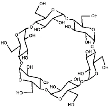
production[57]. The fundamental research on CDs started from 1930s and the industrial production of CDs was firstly achieved in Japan in 1960 [58]. CDs contain six to twelve α-D-glucopyranoside units and within these different kinds of CDs, the ones which have 6, 7 and 8 units were mostly studied and they were considered to be essential for application. They are called as α, β and γ-cyclodextrin, respectively. The structure of α-cyclodextrin is shown in Figure 1-2.
According to the results of X-ray diffraction (XRD), infrared ray spectrometry (IR) and nuclear magnetic resonance (NMR), it was discovered that the units which make up the CDs are all in the chair conformation. Units combine with each other by the 1,4-glycosidic bond to form the ring structure with a cavity inside. Since the units cannot spin freely, the structure of CDs is not cylindrical but conical [59-61].
In the structure of CDs, both rims of the cavity are made up by hydroxyl groups, which make these areas hydrophilic, while the inside of the cavity is shielded by the C-H bonds so that a hydrophobic area is formed. Because of this structure, CDs can work as containers to keep many kinds of molecules in the cavity, including organic, inorganic and gas molecules. The hydrophilicity of the rims and the hydrophobicity in the cavity enables CDs to selectively form clathrates and molecular assembly systems with those molecules under the effect of Van der Waals force, hydrophobic interactions and the matching effect between CDs and molecules in the cavity. This selectivity results in that CDs have some features like enzymes so they are considered to be a kind of ideal host molecules. Thus, in catalytic research, analytical research, food production and pharmaceutical research, CDs have been paid much attention to and widely used [62-63]. Furthermore, to improve the application and adjust the chemical feature of CDs, modification of CDs has become one of the most popular directions on the study of CDs [64].
In our research group, we have tried to use CDs in the detection of MALDI, but the purpose was not for chiral separation [65-67]. As it has been introduced above, detection of molecules with low molecular weight by MALDI mass spectrometry is
17
quite difficult because the matrix will generates a lot of fragments during the ionization process and these fragment peaks make the analysis of the low molecular region impossible. In order to overcome this drawback, many studies on suppressing the fragmentation have been performed. So far, there have been three kinds of methods introduced to achieve the purpose. They are: (1) applications of additives; (2) organic matrix free methods and (3) the combination of (1) and (2). Mixing additives with traditional matrix molecules to make a co-matrix is the most popular method.
Many kinds of compounds have been tried to mix with matrix and carry out the effect on simplifying the mass spectrometry spectrum. Such compounds include phosphoric acid, ammonium salts, saccharides and many other compounds [68-70]. At the same times, the studies on matrix free methods were performed. It has been proved that the application of desorption/ionization on silicon (DIOS), silicon nanowires (SiNW) and nanostructure-initialtor mass spectrometry (NIMS) are effective methods for the purpose of smoothing the spectrum and suppressing the fragmentation [71-73].
Moreover, metal nanoparticles are considered to be effective as well, because of their special physicochemical properties, including large surface areas, thermal and electrical conductivities and size-dependent absorption. Nanoparticles of platinum, gold, silver, copper and iron and their oxides have been used to achieve the goal [74- 76]. Moreover, the combination of additives and organic matrix free methods was studied. In previous study of our group, we have reported the application of zeolite with silver and gold nanoparticles and the effect on suppressing fragment peaks and selective ionization on certain kind of compounds has been proved [77-78].
So, the application of CDs in our group was as an additive aiming the suppression of fragment peaks and enhancing the peak of analyte molecules. We have studied α and β-CDs together with traditional organic matrices. It was considered that matrix molecules can enter the cavity of CDs so that they are protected during the ionization process, which results in the fewer fragment peaks. At the same time, CDs have very strong interactions with alkali metal ions. In the spectra of mass spectrometry with
18
common matrix, the intensities of alkali metal related peaks are always strong. With the application of CDs, these peaks are also suppressed significantly. The effectiveness of using CDs as an additive has been proved. However, the chiral separation was not studied at that time.
5. Chiral molecules, chiral separation and the usage of Cyclodextrins
Asymmetry means the absence of symmetry. When an object is under certain conversions, the same part of it repeats regularly, in another word, some parts are not changed, this object is called symmetric. Symmetry exist widely in the world, so at the same time, asymmetric is the same. Asymmetry in molecules is very common.
Asymmetric molecules turn out to have isomers, which have the same molecular formula but different features. As a result, the term “chiral” in chemistry rise up from this phenomenon.
Chirality was first discovered in 1815 when a beam of polarized light was irradiated onto different compounds, the light was rotated to different directions by molecules with different chirality. The term chirality was firstly introduced in 1894 and different enantiomers or diastereomers of the same compound were called as enantiomers because they have different optical properties. If rotated, including a rotation and a reflection in a plane, a molecule turns out to be the same molecule, then this molecule called to be achiral (not chiral). In another word, if the same molecule cannot be achieved after such rotation, this molecule is chiral [79].
Chiral molecules have some special characteristics in reactions, especially for the enzyme reactions. The different behaviors of enantiomers in human bodies were discovered in 1960s. After some pregnant women took a kind of medicine, they gave birth to many deformed babies. The result of investigation on this medicine showed that the chemical in this medicine had two enantiomers. The R-type of this chemical
19
is medical effective, while the S-type will result in the deformation of babies. Also, from other studies it was understood that for amino acids, only the L-type can be used in the metabolism in biosomes, because the spatial structure decided that only L-type can be stabilized and catalyzed by the enzymes. From the knowledge, people started to realize that enantiomers may have great influence on human body and separation on chiral molecules has been paid lots of attention [80-81].
The most popular method for chiral separation now is to use some chiral selectors.
These selectors need to be able to interact with target molecules and at the same time, since the structures of enantiomers are slightly different with each other, the strength of interaction with the selectors would not be the same. According to this phenomenon, many kinds of selectors have been applied and several kinds of techniques have been employed [82-83]. The most efficient methods among these are considered to be GC, HPLC and CE [84-85]. The selector can be dissolved together with the samples of in the buffer so that they can interact with each other during the separation process. Or the selector can be fixed onto the stationary phase of the capillary wall so that the isomer which has higher affinity with the selector will be slowed down during the separation. This technique has been widely used and validated and most of chiral separation in real study is achieved by these techniques.
Based on their own features which are introduced above, CDs were thought to be available for chiral separations, since enantiomers do not have the same structure, so that after mixed with CDs, the interaction with CDs would not be the same. Then the isomers can be separated by selecting according to that difference. The study on the application of CDs in chiral separation has been carried out for many years and now they are being widely used in several separation techniques to achieve chiral separation, including HPLC and CE. In HPLC, CDs can be added into the sample solution to interact with sample molecules during the separation or be stabilized on the stationary phase in order to interact with sample molecules when they pass the stationary phase. In CE, similar with HPLC, CDs can be added into the running buffer
20
or stabilized on the capillary wall to perform the enantioselectivity (chiral selectivity) [86-89]. The application of CDs for chiral separation in these techniques has been proved to be effective and trustable.
The mechanism of CDs for chiral separation is still under study but common accepted opinion is that the enantioselectivity of CDs are from their structures. So far, there are four possible mechanisms introduced by previous research. They are: (1) inclusion mechanism; (2) external association mechanism; (3) confirmation inducing mechanism and (4) interaction mechanism.
For the inclusion mechanism, Armstrong et al. suggested that the mechanism of chiral separation using CDs is that the chiral points in CDs form inclusion complex with target molecules under the effect of π-π interaction and hydrogen bond interaction [90]. It was considered that this interaction could be rather strong, and such effect can occur not only in the cavity, but the surface of CD molecules as well, if the confirmations of CD molecules and the target molecules and interactions between them can occur.
For the external association mechanism, the mechanism was also introduced by Armstrong [90]. In this mechanism, it was explained as that target molecules form incomplete inclusion complex with CDs. The condition for the interaction to occur is that a strong interaction, including hydrogen bond and dipole moment interactions, between these molecules is necessary. Compared with HPLC, the interactions are weaker in GC and many thermodynamics data were obtained based on the inclusion effect.
Venema et al. proposed the confirmation inducing mechanism [91]. In this model, it was considered that when the target molecule gets close to a CD molecule, the target molecule would have inductive effects on the CD molecule, which makes the confirmation of CD to change to a state which is more suitable for the target molecule, so that the interaction between CD and target molecule is enhanced.
21
Also from Venema et al., the interaction mechanism was raised [92]. CD molecules were considered as host molecules and the target molecules as guest molecules. The interaction between the host and the guest achieve the separation of the guest molecule. This interaction includes Van der Waals force, hydrophobic interactions, dipole moment interactions and the matching effect between CDs and target molecules.
Above all, there has been no consensus on the mechanism deducing the enantioselectivity of CDs. According to the data so far, the mechanism of enantioselectivity differs with the type and derivative of CDs and the target chiral molecules. However, it has been revealed that the adaptation between the conformation of CDs and chiral molecules and the strength of the interaction play the most important role for the separation of chiral molecules.
To improve the enantioselectivity of CDs, modifications on CD molecules have been carried out for many years. Each α-D-glucopyranoside unit has 5 chiral carbon atoms and this structure is thought to be the key factor for the enantioselectivity and by performing the derivatization on the C-2, C-3 and C-6 –OH group, the diameters of the two rims and the size of the cavity can be modified. This feature enables CDs to be stabilized to the stationary phase for HPLC or the capillary wall for GC and CE.
Furthermore, the derivatization on the –OH groups by other groups have been paid more attention. There are two directions of the modification. One is that all –OH groups are replaced by the same group, such as alkyl groups including methyl, ethyl, butyl and amyl groups. Dai et al. performed such research and discovered that ethyl β- CD has the best separation effect [93]. The other direction is that the –OH groups on the C-2 and C-3 are replaced by one group and –OH group on C-6 is modified by another, or C-2 and C-6 the same, C-3 different. By using this method, Ramos et al.
reported that the enantioselectivity was decided by the space confirmation of the compounds by using 2,3-dimethyl-6-tertbutyldimethylsilyl-β-CD as the chiral selector for the separation of derivatives of γ-butyrolactone and polarity change after the
22
modification of the alkyl groups almost had no effect. It was concluded as that the inclusion effect is more important than the polar interaction outside the cavity of CD under this situation [94].
6. This study
In this study, there are mainly two parts. The first part is about the mechanism of ionization process in MALDI. As it has been introduced above, a new “analyte- support” mechanism explaining this process was raised by our group, but the relationship between matrix and analyte molecules was not fully studied. At the same time, the effect of solvent was not well understood. So this work was focused on the validation of this model and tried to provide more experimental evidence supporting this mechanism and trying to have a clearer view on this model, accompanied with the discussion on the effect of solvent. In this study, after the recrystallization of the mixed crystal of matrix and analyte, scanning electron microscopy (SEM), diffuse reflectance spectroscopy, Raman spectroscopy and MALDI mass spectrometry were employed, and according to the data obtain using those methods, a more complete explanation on this new model was achieved. Meanwhile, the effect of solvents on MALDI detection was also studied and discussed, with a conclusion that existence of proton donor in the solvent is necessary.
The second part is about the application of MALDI for the chiral separation. Since no research has been done on such field, we want to make some breakthrough effort in this application. By using CDs as additive, chiral separation of amino acids with different chirality was assayed. α and β-CDs were employed in this study. Several mixing ratios of matrix/CDs/analyte were studied and different pretreatment methods were compared. By analyzing the spectra obtained, the difference on the behaviors of amino acids with different chirality was found. From this result, the possibility of
23
using CDs to achieve chiral separation in MALDI was proved. Furthermore, by calculating the size of matrix molecules, analyte molecules and the cavity of CDs, the possible reason on the enantioselectivity was also revealed. It was considered that after determining the interaction efficiency of amino acids with different chirality, the application method of CDs for chiral separation in MALDI could be set up.
24
Figure captions
Figure 1-1: A simple model of the working scheme of MALDI.
Figure 1-2: The structures of α-cyclodextrin [65].
25
References
1. Sparkman, O. David, Mass spectrometry desk reference. Pittsburgh: Global View Pub. 2000.
2. K. Downard, “Mass Spectrometry: A Foundation Course”, Royal Society of Chemistry, UK, 2004.
3. C. Dass, “Fundamentals of Contemporary Mass Spectrometry”, Wiley, USA, 2007 4. C. Dass, “An Introduction to Biological Mass Spectrometry”, Wiley, USA, 2002 5. K, Downard, "William Aston – the man behind the mass spectrograph". Eur. J.
Mass Spectrom., 2007, 13: 177-190.
6. He Meiyu, “Organic and Biological Mass Spectrometry”, Beijing University Press, 2002.
7. R. Davis, M. Frearson, “Mass Spectrometry – Analytical Chemistry by Open Learning”, Wiley, UK, 1987.
8. T.D. Maerk, G.H. Dunn, “Electron impact ionization”, Springer, Austria, 1984.
9. L. Wolfgang, Electron-impact ionization Cross-Sections and Ionization Rate Coefficient for Atoms and Ions from Hydrogen to Calcium, Zeitschrift für Physik, 1968, 216:241-247.
10. Yong-Ki Kim, M. Eugene Rudd, Binary-encounter-dipole model for electron- impact ionization, Phys. Rev. A: At., Mol., Opt. Phys., 1994, 50:3954-3967.
11. M. S. B. Munson, F.H.J. Field, Journal of the American Chemical Society, 1966, 88:2621-2630.
26
12. H.M. Fales, G.W. Milne, J.J. Pisano, et al., Biological applications of electron ionization and chemical ionization mass spectrometry, Recent Progress in Hormone Research, 1972, 28:591–626.
13. de Hoffmann, Edmond, S. Vincent, “Mass Spectrometry: Principles and Applications” (Second ed.), Wiley, 2003, Canada.
14. R.C. Dougherty, Negative chemical ionization mass spectrometry: applications in environmental analytical chemistry, Biomedical Mass Spectrometry, 1981, 8:283-292.
15. J.B. Fenn, M. Mann, C.K. Meng, et al., Electrospray ionization for mass spectrometry of large biomolecules, Science, 1989, 246:64-71.
16. Pitt, James J, Principles and Applications of Liquid Chromatography-Mass Spectrometry in Clinical Biochemistry, The Clinical Biochemist Reviews, 2009, 30:19-34.
17. P. Kebarle, U.H. Verkerk, Electrospray: From ions in solution to ions in the gas phase, what we know now, Mass Spectrom. Rev., 2009, 28:898-917.
18. K.Y. Li, H.H. Tu, A.K. Ray, Charge Limits on Droplets during Evaporation, Langmuir, 2005, 21:3786-3794.
19. M.C. Garcia, The effect of the mobile phase additives on sensitivity in the analysis of peptides and proteins by high-performance liquid chromatography–
electrospray mass spectrometry, J. Chromato. B, 2005, 825:111-123.
20. N. Czuczy, M. Katona, Z. Takats, Selective detection of specific protein-ligand complexes by electrosonic spray-precursor ion scan tandem mass spectrometry, J. Am.
Soc. Mass Spectrom. 2009, 20:227-237.
21. D. Touboul, L. Maillard, A. Grässlin, et al., How to deal with weak interactions in noncovalent complexes analyzed by electrospray mass spectrometry: cyclopeptidic
27
inhibitors of the nuclear receptor coactivator 1-STAT6, J. Am. Soc. Mass Spectrom.
2009, 20:303-311.
22. P. Nemes, S. Goyal, A. Vertes, Conformational and Noncovalent Complexation Changes in Proteins during Electrospray Ionization, Anal. Chem, 2008, 80:387-395.
23. Sobott, Robinson, Characterising electrosprayed biomolecules using tandem- MS—the noncovalent GroEL chaperonin assembly, Int. J. Mass Spectrom, 2004, 236:25-32.
24. L. Konermann, D.J. Douglas, Equilibrium unfolding of proteins monitored by electrospray ionization mass spectrometry: Distinguishing two-state from multi-state transitions, Rapid Commun. Mass Spectrom, 1998, 12:435-442.
25. A. Li, ; Q. Luo, S. Park, R.G. Cooks, Synthesis and Catalytic Reactions of Nanoparticles formed by Electrospray Ionization of Coinage Metals, Angewandte Chemie International Edition, 2014, 53:3147-3150.
26. M. Barber, R.S. Bordoli, R.D. Sedgiyick, et al., Fast Atom Bombardment of Solids (F.A.B.) : A New Ion Source for Mass Spectrometry, J.C.S. CHEM. COMM., 1981, 7:325-327.
27. M. Barber, R.S. Bordoli, R.D. Sedgiyick, et al., Fast atom bombardment of solids as an ion source in mass spectrometry, Nature, 1981, 293:270-275
28. M. Barber, R.S. Bordoli, G.J. Elliott, et al., Anal. Chem., 1982, 54:645-657.
29. K.B. Tomer, The development of fast atom bombardment combined with tandem mass spectrometry for the determination of biomolecules, Mass Spectrometry Reviews, 1989. 8: 445-482.
30. W.D. Lehmann1, M. Kessler1, W.A. König, Investigations on basic aspects of fast atom bombardment mass spectrometry: Matrix effects, sample effects, sensitivity and quantification, Biol. Mass Spectrom., 1984, 11:217-222.
28
31. M. Barber, R.S. Bordoli, R.D. Sedgiyick, et al., Fast atom bombardment mass spectrometry of the angiotensin peptides, Biol. Mass Spectrom., 1982, 9:208-214.
32. D.J. Douglas, A.J. Frank, D.M. Mao, Linear ion traps in mass spectrometry, Mass Spectrom. Rev., 2005, 24:1-29.
33. Blaum, Klaus, High-accuracy mass spectrometry with stored ions, Physics Reports, 2006, 425:1-78.
34. P.E. Miller, M.B. Denton, The quadrupole mass filter: Basic operating concepts, J.
Chem. Educ., 1986, 63: 617.
35. G.C. Stafford, P.E. Kelley, J.E.P. Syka, et al., Recent improvements in and analytical applications of advanced ion trap technology, Int. J. Mass Spectrom. Ion Processes, 1984, 60:85-98.
36. Z. Ouyang, G. Wu, Y. Song, H. Li, et al., Rectilinear ion trap: concepts, calculations, and analytical performance of a new mass analyser, Anal. Chem. 2004, 76: 4595-605.
37. R.A. Yost, C.G. Enke, Selected ion fragmentation with a tandem quadrupole mass spectrometer, J. Am. Chem. Soc., 1978, 100: 2274-2275.
38. Chernushevich, V. Igor, An introduction to quadrupole–time-of-flight mass spectrometry, J. Mass Spectrom., 2001, 36: 849-865.
39. K. Tanaka, H. Waki, Y. Ido, et al., Protein and polymer analysis up to m/z 100,000 by laser ionization time-of-flight mass spectrometry, Rapid Commun. Mass Spectrom, 1988, 2: 151-153.
40. M.W.F. Nielen, MALDI time-of-flight mass spectrometry of synthetic polymers, Mass Spectrom. Reviews, 1999, 18: 309-344.
41. A.N. Krutchinsky, W.Z. Zhang, B.T. Chait, Rapidly Switchable Matrix-Assisted Laser Desorption/Ionization and Electrospray Quadrupole-Time-of-Flight Mass
29
Spectrometry for Protein Identification, J. Am. Soc. Mass. Spectrom. 2000, 11: 493- 504.
42. F. Hillenkamp, M. Karas, Matrix-assisted laser desorption/ionisation, an experience, Int. J. Mass. Spectrom, 2000, 200: 71–77.
43. J.E. Campana, Time-of-Flight Mass Spectrometry: a Historical Overview, Instrum Sci. Technol., 1987, 16: 1-14.
44. N.M. Kohan, W.D. Robertson, R.N. Compton, Electron ionization time-of-flight mass spectrometry: historical review and current applications, Mass Spectrom. Rev., 2008, 27: 237-285.
45. W.E. Stephens, A Pulsed Mass Spectrometer with Time Dispersion, Phys. Rev., 1946, 69: 691.
46. H. Ehring, M. Karas, F. Hillenkamp, Role of photoionization and photochemistry in ionization processes of organic molecules and relevance for matrix-assisted laser desorption lonization mass spectrometry, Org. Mass Spectrom, 1992, 27: 427-480.
47. V. Karbach, R. Knochenmuss, Do single matrix molecules generate primary ions in ultraviolet matrix-assisted laser desorption/ionization, Rapid Commun. Mass Spectrom, 1998, 12: 968-974.
48. Y. Liu, X. Sun, B. Guo, Matrix-assisted laser desorption/ionization time-of-flight analysis of low-concentration oligonucleotides and mini-sequencing products, Rapid Commun. Mass Spectrom, 2003, 17: 2354-2360.
49. M. Karas, M. Gluckmann, J. Schafer, Ionization in matrix-assisted laser desorption/ionization: singly charged molecular ions are the lucky survivors, J. Mass Spectrom, 2000, 35: 1-12.
30
50. A.N. Krutchinsky, A.I. Dolgunine, M.A. Khodorkovski, Matrix-Assisted Laser Desorption Ionization of Neutral Clusters Composed of Matrix and Analyte Molecules, Anal. Chem, 1995, 67: 1963-1967.
51. W.C. Chang, L.C.L. Huang, Y.-S. Wang, et al., Matrix-assisted laser desorption/ionization (MALDI) mechanism revisited, Anal. Chem. Acta. 2007, 582:
1-9.
52. K. Murakami, A. Sato, K. Hashimoto, et al., Study of ionization process of matrix molecules in matrix-assisted laser desorption ionization. Chem. Phys. 2013, 419: 37- 43.
53. A. Douhal, F. Lahmani, A. H. Zewail, Proton Transfer Reaction Dynamics, Chem.
Phys. 1996, 207: 477-498.
54. S. Nagaoka, U. Nagashima, Effects of node of wave function upon excited-state intramolecular proton transfer of hydroxyanthraquinones and aminoanthraquinones, Chem. Phys. 1996, 206: 353-362.
55. Y. Minegishi, D. Morimoto, J. Matsumoto, et al., Desorption Dynamics of Tetracene Ion from Tetracene-Doped Anthracene Crystals Studied by Femtosecond Time-Resolved Mass Spectrometry, J. Phys. Chem. C., 2012, 116: 3059-3064.
56. A. Biwer, G. Antranikian, E. Heinzle, Enzymatic production of cyclodextrins, Appl Microbiol Biotechnol, 2002, 59:609-625.
57. S. Menuel, J.P. Joly, B. Courcot, et al., Synthesis and inclusion ability of a bis-β- cyclodextrin pseudo-cryptand towards Busulfan anticancer agent, Tetrahedron, 2007,63: 1706-1714.
58. M.E. Davis, M.E. Brewster, Cyclodextrin-based pharmaceutics: past, present and future, Nature Reviews Drug Discovery, 2004, 3: 1023-1035.
31
59. C. Klein, J. Hollender, H. Bender, et al., Catalytic center of cyclodextrin glycosyltransferase derived from x-ray structure analysis combined with site-directed mutagenesis, Biochemistry, 1992, 31: 8740-8746.
60. S.M. Botellaa, M.A. Martína, B. Castillo, et al., Analytical applications of retinoid—cyclodextrin inclusion complexes: Characterization of a retinal—β—
cyclodextrin complex, J. Pharm. Biomed. Anal., 1996, 14: 909–915.
61. H.J. Schneider, F. Hacket, V. Rüdiger, NMR Studies of Cyclodextrins and Cyclodextrin Complexes, Chem. Rev., 1998, 98: 1755-1786.
62. S. Ishiwata, M. Kamiya, Cyclodextrin inclusion: catalytic effects on the degradation of organophosphorus pesticides in neutral aqueous solution, Chemosphere, 1999, 39: 1595-1600.
63. T. Loftsson, M.E. Brewster, Pharmaceutical applications of cyclodextrins. 1. Drug solubilization and stabilization, J. Pharm. Sci., 1996, 85: 1017-1025.
64. E.M.Martin, Cyclodextrins and their uses: a review, Process Biochem., 2004, 39:
1033-1046.
65. S. Yamaguchi, T. Fujita, T. Fujino, et al., Suppression of Matrix-related Ions Using Cyclodextrin in MALDI Mass Spectrometry, Anal. Sci., 2008, 24:1497-1500.
66. T. Fujita, T. Fujino, K. Hirabayashi, et al., MALDI Mass Spectrometry Using 2,4,6-Trihydroxyacetophenone and 2,4-Dihydroxyacetophenone with Cyclodextrins:
Suppression of Matrix-related Ions in Low-molecular-weight Region, Anal. Sci., 2010, 26: 743-748.
67. T. Yonezawa, T. Asano, T. Fujino, et al., Cyclodextrin-supported organic matrix for application of MALDI-MS for forensics. Soft-ionization to obtain protonated molecules of low molecular weight compounds, Chem. Phys., 2013, 419: 17-22.
32
68. S. Kjellstroem, O. N. Jensen, Phosphoric Acid as a Matrix Additive for MALDI MS Analysis of Phosphopeptides and Phosphoproteins, Anal. Chem., 2004, 76: 5109- 5117.
69. J. Asara, J. Allison, Enhanced detection of phosphopeptides in matrix-assisted laser desorption/ionization mass spectrometry using ammonium salts, J. Am. Soc.
Mass Spectrom., 1999, 10: 35-44.
70. T. Nishikaze, M. Takayama, Cooperative effect of factors governing molecular ion yields in desorption/ionization mass spectrometry, Rapid Commun. Mass Spectrom., 2006, 20:376-382.
71. J. Wei, J.M. Buriak, G. Siuzdak, Desorption–ionization mass spectrometry on porous silicon, Nature, 1999, 399: 243-246.
72. E.P. Go, J.V. Apon, G. Luo, et al., Desorption/Ionization on Silicon Nanowires, Anal. Chem., 2005, 77: 1641-1646.
73. T.R. Northen, O. Yanes, M.T. Northen, et al., Clathrate nanostructures for mass spectrometry, Nature, 2007, 449: 1033-1036.
74. H. Kawasaki, T. Yonezawa, T. Watanabe, et al., Platinum Nanoflowers for Surface-Assisted Laser Desorption/Ionization Mass Spectrometry of Biomolecules, J.
Phys. Chem. C., 2007, 111: 16278-16283.
75. Y.F. Huang, H.T. Chang, W. Tan, Nile Red-Adsorbed Gold Nanoparticle Matrixes for Determining Aminothiols through Surface-Assisted Laser Desorption/Ionization Mass Spectrometry, Anal. Chem., 2006, 78: 1485-1493.
76. K. Shrivas, H.F. Wu, Applications of silver nanoparticles capped with different functional groups as the matrix and affinity probes in surface-assisted laser desorption/ionization time-of-flight and atmospheric pressure matrix-assisted laser desorption/ionization ion trap mass spectrometry for rapid analysis of sulfur drugs and biothiols in human urine, Rapid Commun. Mass Spectrom., 2008, 22: 2863-2872.
33
77. M. Yang, T. Fujino, Silver nanoparticles on zeolite surface for laser desorption/ionization mass spectrometry of low molecular weight compounds, Chem.
Phys. Lett., 2013, 576: 61–64.
78. M. Yang, T. Fujino, Gold nanoparticles loaded on zeolite as inorganic matrix for laser desorption/ionization mass spectrometry of small molecules, Chem. Phys. Lett., 2014, 592: 16–163.
79. R. Bentley, From optical activity in quartz to chiral drugs: molecular handedness in biology and medicine, Perspectives in Biology and Medicine, 1995, 38: 188-229.
80. E.J. Ariens, S.W. Timmermans, Stereochemistry and Biological Activity of Drugs.
Blackwell Scientific Publications, 1983, UK.
81. M. Eichelbaum, A.S. Gross, Stereochemical aspects of drug action and disposition, Adv. Drug Res., 1996, 28: 1-64.
82. M. Novotny, H. Soini, M. Stefansson, Chiral separation through, Anal. Chem., 1994, 66: 646-655.
83. B. Chankvetadzea, L. Chankvetadzea, S. Sidamonidzea, et al., 3-Fluoro-, 3- chloro- and 3-bromo-5-methylphenylcarbamates of cellulose and amylose as chiral stationary phases for high-performance liquid chromatographic enantioseparation, J.
Chromatogr. A., 1997, 787: 67-77.
84. G. Gübitz, M.G. Schmid, Chiral Separation by Chromatographic and Electromigration Techniques. A Review, Biopharm. Drug Dispos., 2001, 22: 291-336.
85. S. Fanali, Enantioselective determination by capillary electrophoresis with cyclodextrins as chiral selectors, J. Chromatogr. A., 2000, 875: 89-122.
86. A.M. Stalcup, K.H. Gahm, A Sulfated Cyclodextrin Chiral Stationary Phase for High-Performance Liquid Chromatography, Anal. Chem., 1996, 68: 1369-1374.