Kei ko SEKI
Department of Microbiology (II), The Jikei University School of Medicine
Staphylococcus aureus
(
S. aureus)i s known as t he caus at i ve agent of pur ul ent i nf l ammat i on or f ood poi s oni ng,whi l e i t i s known as one of t he i ndi genous mi cr oor gani s ms on f ace or mucos a of nas al cavi t y. The pur pos e of pr es ent s t udy i s t o demons t r at e t he s ubs t ances i nvol ved i n t he bact er i al adher ence,and al s o t o cons i der whet her apopt os i s of bact er i a‑i nf ect ed cel l woul d be i mpor t ant f or t he hos t ani mal or not .
Two cl i ni cal i s ol at es of S. aur
eus,OK1 and OK11,and L929 f i br obl as t wer e us ed. I n an
in vivo experi ment wi t h mi ce,OK11 s howed hi gher vi r ul ent t han OK1. L929 cel l s i nges t ed mor e number s of OK11 t han OK1 dur i ng t he 6‑hr i ncubat i on. Mor phol ogi cal ver i f i cat i ons us i ng i nhi bi t or s of act i n‑pol ymer i zat i on r eveal ed t hat i nt r acel l ul ar F‑act i n bundl e pl ays an i mpor t ant r ol e on t he i nges t i on of
S. aureus. Apopt os i s was i nduced i nt o t he cel l s i nf ect ed wi t h ei t her OK1 or OK11. The t umor ‑necr os i s ‑f act or ‑al pha(TNF‑α)cont ent i n t he medi um i ncr eas ed as t he i ncubat i on t i me. TNF‑α r el eas ed by t he i nf ect ed cel l mi ght pl ay a r ol e as t he ext r i ns i c i nducer of apopt os i s f or t hat cel l . Chr omat i n condens at i on,one of t he mor phol ogi cal cr i t er i a of apopt os i s ,was gr eat l y di f f er ent bet ween OK1‑and OK11‑i nf ect ed cel l s . Chr omat i n wi t hi n a nucl eus heavi l y condens ed t o f or m l ar ge cl umps and t he nucl ear membr ane was not f ound i n an OK1‑i nf ect ed cel l ,whi l e s ome s mal l er condens ed cl umps of chr omat i n wer e beneat h t he nucl ear membr ane and many dens e s pot s wer e obs er ved i n t he nucl eopl as m of an OK11‑
i nf ect ed cel l . Al l above event s i ncl udi ng t he i nges t i on of bact er i a wer e s t r ongl y pr es ent ed i n cas e of OK11.
By t hes e r es ul t s ,t he bact er i al s t r ai n wi t h hi gher vi r ul ence appear ed t o be i nges t ed by and i nduce apopt os i s t o t he f i br obl as t . Fr om t he vi ewpoi nt of t he hos t def ens e mechani s m,t he cel l s whi ch wer e i nvaded by or act i vel y i ncor por at ed many bact er i a need t o be apopt ot i c t o pr event t he f ur t her expans i on of t he i nf ect i ous f oci .
(Tokyo Jikeikai Medical Journal 2007;122:123‑35) Key words:Staphylococcus aureus,fibroblast,host defense mechanism,apoptosis,F‑actin
I.は じ め に
黄色ブドウ球菌は伝染性膿痂疹,蜂巣炎,癰あ るいは などの化膿性皮膚疾患,骨髄炎,髄膜炎,
ブドウ球菌性熱傷様皮膚症候群(SSSS),トキ シックショック症候群(TSS),および食中毒など 多くの疾患の起因菌として知られている.SSSS,
TSS,あるいは食中毒のように特定の毒素との関
係が明確にされているものもあるが,ほとんどの
場合において発症のメカニズムは明らかにされて
いない.これまで筆者らの研究室では黄色ブドウ
球菌に関する研究を長年行ってきており,マウス
を用いた感染実験で,静脈内接種された黄色ブド
ウ 球 菌 は 腎 に 長 く 留 ま り 膿 瘍 を 形 成 す る こ
いない.一方,疾患とは関わりなく,30〜40% の 健常者の鼻腔内には黄色ブドウ球菌が定着し , 除菌を行っても同一人に頻回に定着することが明 らかにされており ,この事実は菌の定着し易さ に宿主の要因が大きく関わっていることを示唆し ている.以上のように,感染症の発症には菌が産 生する毒素や分解酵素が関与することはもちろん であるが,防御機能も含めた宿主側の因子が大き な影響を及ぼしていると考え,筆者らはこれまで 黄色ブドウ球菌の病原因子の解析とともに,宿主 との相互作用を明らかにする目的でマウスや培養 細胞を用いて検討を重ねてきた.本稿ではこれま でに得られた結果を述べ,今後の展望などについ て考えてみたい.
II.黄色ブドウ球菌菌株とその性質
おもに使用 し て い る 黄 色 ブ ド ウ 球 菌 菌 株 は OK1および OK11株で,これは岡山大学皮膚科で アトピー性皮膚炎患部から採取して東京慈恵会医 科大学微生物学講座第 2に分与され,分離した株 である.亜テルル酸カリウムと卵黄を含むスタン プ用培地に採取された検体をマンニット食塩培地 上に広げて分離し,得られた孤立コロニーのコア グラーゼ産生性 を確認の上,黄色ブドウ球菌菌 株とした.病原性を BALB/cマウスの尾静脈内接
性を示し,マウスに対する病原性が OK1で著し く低いことを除き,その他の性質についてはあま り大きな違いは認められなかった.
III.線維芽細胞による黄色ブドウ球菌の 取り込み
好中球やマクロファージが黄色ブドウ球菌を始 めとする多くの細菌を取り込み,細胞内で殺菌処 理することは周知の事実であり,これは生体の防 御機能のひとつである.好中球などのようなプロ フェッショナルな貪食細胞とは別に,細菌を取り 込む細胞の存在がいくつか報告されている . 筆者らも培養細胞のいくつかについて黄色ブドウ 球菌を取り込むか否かを検討し,腎細胞や線維芽 細胞が黄色ブドウ球菌を活発に取り込むことを明 らかにした .これらの細胞を用いた実験で は,ブドウ球菌属のうち,ヒトに常在的に存在す る表皮ブドウ球菌のような病原性の低い菌種は取 り込まれず,概して黄色ブドウ球菌はよく取り込 まれた.また,黄色ブドウ球菌の中でも取り込ま れ方は菌株に依り,病原性の強い株の方が取り込 まれやすい傾向にあった.この,低病原性のブド ウ球菌が取り込まれないという事実は,マクロ ファージ や好中球 においても同様で(Fi g.
Table 1. Characterization of two staphylococcal strains,OK1 and OK11
coagultype ase α‑toxin γ‑hemolysin PV‑
leukocidin enterotoxin exftoloxiiat nive LD (CFU/head)
OK1 I + + − D − 2.7×10
OK11 V + + − − − 4.8×10
1),一見矛盾しているようにも思われるが,細胞 内で菌が殺菌処理されない仕組みがあるならば,
それは疾患の発症機構と関連するのではないかと 考えている.
では,黄色ブドウ球菌が線維芽細胞に取り込ま れるならば,アドヘジンとしての役割を何が果た しているのであろうか この疑問を解決するた めに,生体内物質のうちで細菌が接触する可能性 のあるもののひとつとして細胞外マトリックスを 想定し,さらにその中のフィブロネクチンに焦点 を絞った.進士らがフィブロネクチン結合タンパ ク(FnBP)について検討したところ,OK1,OK11 のいずれも FnBPを保有することが明白になっ たが,フィブロネクチンの結合量は OK11の方が 多く,OK11の菌細胞表面でフィブロネクチンが 多量体を形成して結合するのではないかと推測さ れた .マクロファージと同様に L929細胞は低 病原性の OK1より高病原性の OK11をはるかに 多量に取り込んだ(Tabl e 2).これは上述の OK11 におけるフィブロネクチン多量体形成に関連する と考えられた.L929細胞の菌取り込みにフィブロ ネクチンが大きく関与していることは,抗体を添 加した条件下で菌の取り込みが大きく低下したこ と,フィブロネクチンを添加した際には取り込み
数が少し増加したことなどから示唆された(Fi g.
2‑Aおよび D).フィブロネクチン添加による取 り込み数の増加の割合はマクロファージに比べる と小さい.これは,L929細胞が産生したフィブロ ネクチンが細胞表面上にネットワーク状に分布し (Fi g.3),菌はここに FnBPを介して結合するた め,添加されたフィブロネクチンの影響を受けな いからであろう.菌取り込みへのフィブロネクチ ンの関与は,FnBP遺伝子欠損株や本来フィブロ
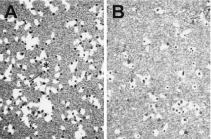
Fig.1. Phagocytosis of human polymorphonuclear cells. Phagocytosis of polymorphonuclear cell(PMN)was observed using phagocytic plaque method . After a PMN (a black granule,arrow) ingested a lot of staphylococci,white part(phagocytic plaque,*)was formed around the PMN.
Background is a staphylococcal thin‑layer formed on a plastic dish. A,S. aureus;B,S. epider midis.
-
Table 2. Ingestion of Staphylococcus aureus cells by L929 f ibro- blasts
OK1
(CFU/well) OK11 (CFU/well) (1.4±1.9)×10 (6.1±1.3)×10 S. aureus and L929 cells were incubat- ed in a 24‑well culture plate for 1 h.
L929 cells were lysed with sterile water after the r emoval of the extracellular bact eria with lyso- staphin. Then,bacterial suspension was diluted and pl ated onto the nor- mal agar plates,and the number of colony was counted on t he next day.
Values are expressed as mean±SD.
Difference between two values is sig- nificant(p<0.001).
ネクチンを産生しない血管内皮細胞などを用いた 進士らの検討でも明らかにされている .黄色ブ ドウ球菌においては FnBPのほかにラミニン結 合タンパク やビトロネクチン結合タンパク の存在も明らかにされているので,抗ラミニン抗 体や抗ビトロネクチン抗体を添加した際の取り込 みを検討した.この場合にも抗フィブロネクチン 抗体添加時ほどではないが,取り込み数の減少が 認められており(Fi g.2‑Bおよび C),これらの細
胞外マトリックスも菌の取り込みに関与している と考えられた.
次に,細胞が積極的に菌を取り込んでいるのか どうか,ということについて考えてみたい.黄色 ブドウ球菌を取り込ませた L929細胞を化学固定 し,電子顕微鏡で観察した.走査電子顕微鏡法に よる細胞表面構造観察(Fi g.4)や凍結割断レプリ カ法による細胞膜二次元構造の観察(Fi g.5)を 行ったところ,菌を取り込ませた直後には菌直下 の細胞膜が窪み,その後,菌周囲の細胞膜が盛り 上がり襞を生じた.これによって菌は細胞内に取 り込まれ,ファゴソーム内に菌が観察された.つ いで,細胞形態とくに細胞骨格系の構造保存に優 れているといわれる急速加圧凍結を行って細胞内 の微細構造を詳細に観察したところ(Fi g.6),菌 が細胞膜に接着している直下や,細胞内に取り込 まれた菌の周囲には F‑アクチンを主体とした細 胞骨格と思われる構造物が明瞭に認められた.細 胞には細胞質内に三次元的に細胞骨格のネット ワークが存在し,細胞の移動,強化,可溶性物質 や顆粒状物質の取り込みに際したエンドソームや
Fig.2. Effects of extracellular matrices on ingestion of staphylococci. L929 and staphylococcal cellswere incubated with(A)anti‑fibronectin,(B)ant i‑vitronectin,(C)anti‑laminin,and(D)fi bronectin,respectively,at 37°C for 1 h. ʻContr olʼin each graph is the result obtained from a reaction mixture without each additive.
-
Fig.3. Fibronectin‑network formed on L929 cells. FITC‑labeled anti‑mouse cellular fi bronectin was used.
-
ピノソームの形成と移動,あるいは細胞の生合成 で生じた物質や分解物が入った小胞の移動,細胞 外マトリックスへの付着などの機能を果たしてい ることが知られている.そのおもな機能構成分子 はアクチンやチューブリンであり,さらにそれら のタンパク質に結合するミオシン,カルモデュリ ン,スペクトリンなどがある.そこで,筆者らは G‑アクチンの重合体であるアクチンフィラメン ト(F‑アクチン)に注目して実験を行った.F‑ア クチンへの G‑アクチン重合を阻害するサイトカ ラシン D (Cyt D),および F‑アクチンの脱重合剤 として知られるラトランキュリン A(LTA)によ
る取り込み阻害を見たところ,いずれの薬剤を用 いても L929細胞による黄色ブドウ球菌の取り込 み数が著しく減少し,LTAによる取り込み数は 特に低値を示した(Fi g.7).F‑アクチンをローダ ミン標識ファロイジンで染色すると Fi g.8のよ うな蛍光顕微鏡像が得られた.すなわち,非感染 の無処理 L929細胞はディッシュによく接着して おり,細胞内によく伸展した F‑アクチンのストレ スファイバーを観察することができたが,Cyt D あるいは LTA処理により細胞の接着性は失われ て形が丸みを帯び,F‑アクチンのストレスファイ バーは消失してアクチンの小さい塊が細胞質内に
Fig.4. Scanning electron micrographs of L929 cells. Fibroblasts were fixed(A)15 and(B)60 minutesafter the infection. In earlier time(A),plasma membrane was dented(arrowheads)by attach ment of bacteria(*). Then,plasma membr ane swelled up to form pseudopodia(arrows) around the bacteria(B). Bar=1μm.
-
Fig.5. Freeze fracture replica images. *,S. aureus;↑,cell membrane;▲,phagosomal membrane.
分散していた.また,黄色ブドウ球菌のプロテイ ン Aが免疫グロブリン G (I gG)の Fc部位と非 特異的に結合する性質を利用して,FI TC標識ウ サギ I gGとローダミン標識ファロイジンで感染 細胞を二重染色すると,F‑アクチンの細胞質内伸 展も認められるが,非感染細胞とは異なって細胞 辺縁に非常に多く集積し,その一端に FI TCの緑 色蛍光が認められ,F‑アクチンに黄色ブドウ球菌 が結合していることを示唆させる像が得られた.
さ ら に,GFP融 合 ア ク チ ン 発 現 ベ ク ター
(Cl ont ech)を導入した L929細胞(GFP‑act i n‑
L929)を作 成 し,こ の 細 胞 に よ る,BacLi ght
Red bact er i al s t ai n(Mol ecul ar Pr obe)標識黄 色ブドウ球菌の取り込みを共焦点レーザー顕微鏡 下で数分間にわたり動画像として記録した.アク チン束が伸びた先に存在していた菌が消失した り,細胞表面上に存在していた菌が短時間のうち に消失したりする様子が観察され,これは菌が細 胞内に取り込まれたことを示唆する画像であっ た.一方,この細胞を LTAで処理した場合にはア クチン束が伸びることも,また,細胞表面の菌が 消失することもなかった.以上のことより,L929 細胞は F‑アクチンを駆使して非常に活発に菌を 取り込むことが明らかとなった.
Fig.6. Transmission electron micrographs of L929 cells. Cells were fixed with high pressure rapid freezing method. Bundles of actin filaments(ar rows)are recognized under the cell membrane beneath a bacterium as well as around a bacter ium ingested by L929 cell. *,S. aureus.
Fig.7. Effect of F‑actin‑depolymerizing agents on the ingestion of S. aureus. L929 and staphylococcal cells were incubated in the presence of the inhi bitor,cytochalasin D (CytD)or latrunculin A (LTA). * ;significant(p<0.0001).
IV.菌を取り込んだ線維芽細胞における アポトーシスの誘導
L929細胞に菌を感染させると 3時間後には細 胞がディッシュか ら 徐々に 剥 が れ 始 め,ディッ シュへの接着をかろうじて保持している細胞も次 第に丸みを帯び,6時間後には接着している細胞 はわずかになった(Fi g.9).そこで,ディッシュ に残った細胞と剥がれて浮き上がった細胞につい てアポトーシスのひとつの指標となる Ter mi nal
deoxynucl eot i dyl t r ans f er as e‑medi at ed dUTP‑
bi ot i n ni ck end l abel i ng(TUNEL)染色を行っ たところ,感染 3時間後で,剥がれた細胞の 90%
は TUNEL陽性であり,接着して残っている細胞 も約半数は陽性であった.接着して残った細胞に ついて透過型電子顕微鏡による観察を行うと,
OK1感染細胞と OK11感染細胞とでは核の様相 が明らかに異なっていた(Fi g.10).大半の OK1 感染細胞は傷害を受けていなかったが,アポトー シスを起こしているものもあった.そのような細
Fig.8. Actin filaments of L929 cells with or without latrunculin A‑treatment. Well‑developed stressfibers are recognized within fibroblasts(A). Str ess fibers are not recognized in the latrunculin A‑
treated fibroblasts(B). ʻBʼis an image using Nomarski optics merged with the fluorescent filter. In the S. aureus‑infected cells(C),actin fiber‑assemblages are near the rim of the cell,and the ingested bacteria(arrows)are at the end of the fiber.
Fig.9. Alteration of the cell shape after bacterial infection.
胞では核膜が消失し,核はクロマチンが強く凝縮 していくつかの断片を形成しており,細胞質中の ミトコンドリアなどの構造が比較的よく保存され ていた.一方,OK11感染細胞では傷害を受けたも のが多く認められた.それらの細胞ではあまり大 きくないクロマチン凝集塊が核質内や核膜に沿っ た核の周縁部に存在していた.また,細胞質には 空胞が多く,細胞内小器官は膨潤していたり破壊 されていたりした.これらの像から,OK1感染で は典型的なアポトーシスが誘導され,OK11感染 ではアポトーシスも誘導されるがネクローシスに いたる細胞がかなり多いと考えられた.
阻害ペプチドを用いた検討においてはカスパー ゼ 8およびカスパーゼ 3の活性阻害ペプチド(40 μM I ETDおよび 40μM DEVD)の添加によりア ポトーシスが阻止されたので,デスレセプターを 介し,これらのプロテアーゼを経てアポトーシス が誘導される可能性が考えられた.一方,カスパー ゼ 9の阻害ペプチド(40μM LEHD)ではアポ トーシスが阻害されなかったため,細胞内に取り 込まれた菌産生物のミトコンドリア傷害によるア ポトーシス誘導の可能性は低いのではないかと考 えた.OK11感染後 3時間から 6時間にかけて細 胞からの TNF‑αおよび I L‑6放出が増加し,こ れは感染細胞の破壊と関連しているのではないか と思われた.また,I L‑1βは OK1感染により多く 放出され,I L‑8は逆に時間経過とともに減少し た.さらに,培地中に TNF‑αの中和抗体を添加
しておくとアポトーシスが誘導されにくくなると いうことからも,L929細胞のアポトーシスは細胞 内からよりも細胞から放出された TNF‑αによ り誘導されるのではないかと考えている.
一方,細胞内に取り込まれた菌がアポトーシス 誘導に全く関与しないかどうかを調べるために,
培養時間の異なる菌,すなわち代謝活性の異なる 菌を用いて検討した.Fi g.11は 3時間培養 OK11 あるいは 20時間培養 OK11を感染させ,細胞質 中のカスパーゼ 3をウェスタンブロットで検出し たものである.早期対数増殖期(3時間培養)の菌 を感染させるとカスパーゼ 3が誘導されるのに対
Fig.10. Transmission electron micrographs of S. aureus‑infected L929 cells. A,OK1‑infected cell;B,OK11‑infected cell. Bar=2μm.
Fig.11. Western blot analysis of caspase 3 in OK11‑infected L929 cel ls. Caspase 3 with the molecular weight of 35 kD was obvi ously detected only when 3 h‑cul tured bacteria were used.
-
健 康 成 人 の 好 中 球 を デ キ ス ト ラ ン と Fi col l
‑Paqueを用いて分離し,菌の取り込みと ア ポ トーシス誘導との関連を検討した.好中球も OK1 より OK11を多く取り込んだが,線維芽細胞とは 異なり,対数増殖期の菌よりも静止期の菌を多く 取り込んだ(Fi g.12).静止期の菌を取り込んだ好 中球では細胞内菌数が多いにもかかわらず分葉核 の形状が比較的よく保たれていたが,対数増殖期 の菌を取り込んだ好中球では核は丸くなるかある いは核が消失しており,細胞の傷害が著しいと考 えられた(Fi g.13).この事実は,対数増殖期の菌 の代謝が活発であり,代謝産物により細胞の傷害 が起きている可能性があることを示唆する.対数 増殖期の菌を貪食した好中球では DNAの断片化 が生じており(Fi g.14),アポトーシスを起こして
導性が強く(Fi g.15‑C),L929線維芽細胞でもほ
Fig.13. Variation of nuclear shape in S. aureus‑ingesting polymorphonuclear cells. PMNs were classi fied into three groups due to the nuclear shape,and the results were translated in a pie chart. As illustrated in the legend box,the ratio of the cel l with normal nucleus is shown as dark area,the cell with condensed chromatin as hatched area,and t he cell with nucleus damaged and dyed in pale blue or pink with Giemsaʼs solution as whi te area.
- Fig.12. Phagocytosis of bacteria cultured for dif
ferent hours by polymor phonuclear cells. -
ショナルな貪食細胞とは異なり,線維芽細胞ある バリアーとなろう.一方,細胞がネクローシスを 起こすと細胞の崩壊とともに細菌は細胞外に出る が,そのような細菌には宿主細胞の細胞質の一部 が付着している.この付着物は細菌にとって保護 膜のような役割を果たし,菌が好中球などに貪食 されにくくなるばかりでなく血中の抗菌物質に対 しても抵抗できる状況をつくり,分裂・増殖して 感染巣を拡大できる,と推測される.また,クロ マチン凝縮と核の断片化が生じていながら細胞質 は膨潤して菌が飛び出しかけている,といったア ポトーシスとネクローシスの中間型のような感染 線維芽細胞の電子顕微鏡像も得ているが,これも 菌が増殖できる状況であると思われる.この中間 型はタイプ 2アポトーシスと呼ばれ,EBウイル スで Bリンパ球に誘導されるもの やアンチマ
Fig.15. Transmission electron micrographs of polymorphonuclear cells. PMNs phagocytosed lots of OK11 in a logarithmic phase(A and B). Condens ed chromatin are recognized. On the other hand,PMNs were incubated in culture supernat ant of OK11(C). *,S. aurers.
Fig.14. DNA laddering of S. aureus‑ingesting polymorphonuclear cel ls.
に運ばれて定着・増殖し,腎内菌数が加熱死菌非 投与マウスよりも高値を示したのであろう,と推 測した.もうひとつはリゾスタフィン投与マウス を用いた実験で得られた .マウスにリゾスタ フィンを投与すると好中球に貪食されていない菌 は溶菌するが,すでに貪食されていた菌はリゾス タフィンの作用を受けにくく,好中球内で増殖し た状態で腎に運ばれ感染巣を形成したのであろ う,と推測した.これらの結果は,好中球が黄色 ブドウ球菌にとってある種の隠れ家になり得るこ とを示唆している.
一方,細菌を取り込んだ細胞の産生物質が誘導 したアポトーシス細胞をマクロファージなどの貪 食細胞が処理する,さらに貪食細胞から種々のサ イトカインが放出される,などの一連の応答反応 から考えると,アポトーシスが感染阻止に役立っ ている,ともいえる .
アポトーシスになるかネクローシスになるか,
また,これらのことが生体にとって感染防御に繋 がるのか病巣拡大に繋がるのかは,菌種や菌数,あ るいは細胞の種類によって異なることが明らかで ある.細胞内で生ずる実に微妙なアンバランスに 伴って代謝経路がわずかに変化し,その先行きが 決定されると考えられる.ここに影響を及ぼして いるものは,冒頭でも述べたように,菌が産生す る多くの毒素や種々の酵素であり,また,宿主細 胞が保有する食菌作用や抗菌物質の産生性,常在 細菌叢を構成する細菌などである.毒素や酵素が 細胞や組織に作用すると感染症へと発展するが,
食菌作用,抗菌物質,および常在細菌は感染防御 に大きな影響を及ぼしている.感染症に罹患しな い,感染症に罹患して回復する,あるいは重篤に なる,というさまざまな過程における重要な因子
部門・佐々木博之助教授はじめ関係諸氏のご指導とご 協力を,また,遺伝子組換えに関しては(株)KAN 研 究所・松井毅主任研究員(現 大阪大学)のご指導をい ただきました.ここに御礼申し上げます.
文 献
1) Hasegawa N,Kondo I,Hoshina S,Kurosaka K. Comparison of vir ulence and immunity of Staphylococcus aureus i n mouse kidney. Jikei- kai Med J 1987;34:195‑203.
2) Seki K,Nishihara S,Ikigai H,Mausda S.
Effect of intravenous administration of heat‑ killed bacterial cells on blood clearance and kidney lodgement proper ty of Staphylococcus aureus organisms subs equently injected to mice. Jikeikai Med J 1988;35:275‑84.
3) Seki K,Ogasawara M,Sakurada J,Murai M, Masuda S. Altered virulence of a pleiotropic Staphylococcus aureus mut ant with a low producibility of coagul ase and other factors in mice. Microbiol Immunol 1989;33:981‑90.
4) 関啓子,櫻田純次,進士ひとみ,田嶌亜紀子,益 田昭吾.黄色ブドウ球菌の腎創傷部位への定着お よびその後の増殖に関与する因子の解析.Bacte- rial Adherence研究 1999;13:30‑3.
5) Seki K,Shinji H,Murai M,Tajima A,Masuda S. Effect of lysostaphi n on establishment of staphylococcal infectious foci in mouse kidney.
Jikeikai Med J 2005;52:21‑9.
6) Kondo I,Masuda S,Kimura K,Kurosaka K, Hasegawa N. Effects of intrarenal inocula- tion of Staphylococcus aureus on mice. Infect Immun 1971;4:103‑9.
7) Ishida Y,Seki K. Effects of intrasplenic inoc- ulation of live Staphylococcus aureus organ- isms on resistance of mice against intravenous- ly and intrarenally inoculated S. aureus organisms. Jikeikai Med J 1992;39:21‑31.
部 に お け るStaphylococcus aureusの 定 量 的 検 討.日皮会誌 1994;104:1249‑57.
11) 関啓子,益田昭吾.アトピー由来黄色ブドウ球菌 の病原性に関する検討 :特に Cowan I株との比 較による.臨床と微生物 1996;23:371.
12) Terada M,Tsutsui H,Imai Y,Yasuda K, Mizutani H,Yamanishi K,et al. Contribution of IL‑18 to atopic‑dermat itis‑like skin inflam- mation induced by Staphylococcus aureus prod- uct in mice. Proc Natl Acad Sci USA 2006;
103:8816‑21.
13) Cardona ID,Cho SH,Leung DY. Role of bacterial superantigens i n atopic dermatitis:
implications for future therapeutic strategies. Am J Clin Dermatol 2006;7:273‑9.
14) Lomholt H,Andersen KE,Kilian M.Staphy- lococcus aureus clonal dynamics and virulence factors in children wit h atopic dermatitis. J Invest Dermatol 2005;125:977‑82.
15) 関啓子,櫻田純次,益田昭吾.医学生の鼻腔より 分離した黄色ブドウ球菌の性状について.臨床と 微生物 1992;19:377.
16) 梅田昭子,呼蘭,天児和暢.健康人における黄色 ブドウ球菌の 感 染 動 態 の 調 査.臨 床 と 微 生 物 1992;19:376‑7.
17) Hwang S,Seki K,Sakurada J,Ogasawara M, Murai M, Ohmayu S, et al. Improved methods for detection and s erotyping of coagulase from Staphylococcus aur eus. Mi- crobiol Immunol 1989;33:175‑82.
18) Lina G,Piemont Y,Godall‑Gamot F,Bes M, Peter MO,Gauduchon V,et al. Involvement of Panton‑Valentine l eukocidin‑producing Staphylococcus aureus i n primary skin infec- tions and pneumonia. Clin Infect Dis 1999;
29:1128‑32.
22) Menzies BE,Kourteva I. Internalization of Staphylococcus aureus by endot helial cells induces apoptosis. Inf ect Immun 1998;66:
5994‑8.
23) Hensen SM,Pavicic MJ,Lohuis JA,Poutrel B.
Use of bovine primary mammary epithelial cells for the compari son of adherence and invasion ability of Staphylococcus aur eus strains. J Dairy Sci 2000;83:418‑29.
24) Wesson CA,Derlinger J,Liou LE,Bayles KW, Bohach GA,Trumble WR. Apoptosis induced by Staphylococcus aur eus in epithelial cells utilizes a mechanism i nvolving caspase 8 and 3. Infect Immun 2000;68:2998‑3001.
25) Hess DJ,Henry‑Stanley MJ,Erickson EA, Wells CL. Intracellular survival of Staphylo- coccus aureus within cultured enterocytes. J Surg Res 2003;114:42‑9.
26) Usui A,Murai M,Seki K,Sakurada J,Masuda S. Conspicuous inges tion of Staphylococcus aureus organisms by mur in fibroblasts in vitro. Microbiol Immunol 1992;36:545‑50.
27) Murai M,Seki K,Sakurada J,Usui A,Masuda S. Effects of cytochal asins B and D on Staph- ylococcus aureus adherence to and ingestion by mouse renal cells from pr imary culture. Mi- crobiol Immunol 1993;37:69‑73.
28) Shinji H,Sakurada J,Seki K,Murai M, Masuda S. Different effects of fibronectin on the phagocytosis of Staphylococcus aur eus and coagulase‑negative staphyl ococci by murine peritoneal macrophages . Microbiol Immunol 1998;42:851‑61.
29) Murai M,Sakurada J,Seki K,Shinji H,Hirota Y,Masuda S.Apoptos is observed in BALB/
3T3 cells having ingested Staphylococcus au-
Koabayashi N, Hwang S, et al. Simple method for observation of phagocyt osis on bacterial thinlayer. Mi crobiol Immunol 1989;
33:81‑5.
33) Shinji H,Kamada M,Seki K,Tajima A,Iwase T,Masuda S. Expres sion and distribution of very late antigen 5 in mous e peritoneal macro- phage upon ingestion of fibronectin‑bound Staphylococcus aureus. Microbiol Immunol 2007;51:63‑71.
34) 吉沢幸夫,Gong Z,近藤勇.黄色ブドウ球菌のラ ミニン結合タンパク.臨床と微生物 1992;19:
398‑9.
35) Liang OD,Flock JI,Wadstrom T. Isolation and characterization of vi tronectin‑binding surface protein from Staphylococcus aureus.
Biochim Biophys Acta 1995;1250:110‑6.
36) Bayles KW,Wesson CA,Liou LE,Bohach GA, Trumble WR. Intracellular Staphylococcus
immortalization and senescence. Cell Struc Funct 2003;28:61‑70.
39) Papucci L,Formigli L,Schiavone N,Tani A, Donnini M,Lapucci A,et al. Apoptosis shifts to necrosis via intermedi ate types of cell death by a mechanism dependi ng on c‑myc and bcl‑
2 expression. Cell Tissue Res 2004;316:197
‑209.
40) Watson RWG,Redmond P,Wang JH,Condron C,Bouchier‑Hayes D. Neut rophils undergo apoptosis following i ngestion of Escherichia coli. J Immunol 1996;3986‑92.
41) Grassme H,Jendrossek V,Gulbins E. Molecu- lar mechanisms of bacteria induced apoptosis. Apoptosis 2001;6:441‑5.
42) DeLeo FR. Modulation of phagocyte apoptosis by bacterial pathogens. Apoptosis 2004;9:399‑413.