Phosphatase activity of soluble Epoxide
Hydrolase (sEH)
著者(英)
Rinawati Purba Endang
学位名
博士(理学)
学位授与機関
関西学院大学
学位授与番号
34504甲第539号
Phosphatase activity of soluble Epoxide Hydrolase (sEH)
Thesis Submitted to School of Science and Technology, Kwansei Gakuin University,
for the Doctor of Science Degree By
Endang Rinawati Purba 2014
[CONTENTS] Page Abstract -‐-‐-‐-‐-‐-‐-‐-‐-‐-‐-‐-‐-‐-‐-‐-‐-‐-‐-‐-‐-‐-‐-‐-‐-‐-‐-‐-‐-‐-‐-‐-‐-‐-‐-‐-‐-‐-‐-‐-‐-‐-‐-‐-‐-‐-‐-‐-‐-‐-‐-‐-‐-‐-‐-‐-‐-‐-‐-‐-‐-‐-‐-‐-‐-‐-‐-‐-‐-‐-‐-‐-‐-‐-‐-‐-‐-‐-‐-‐-‐-‐-‐-‐-‐-‐-‐ 3 General Introduction -‐-‐-‐-‐-‐-‐-‐-‐-‐-‐-‐-‐-‐-‐-‐-‐-‐-‐-‐-‐-‐-‐-‐-‐-‐-‐-‐-‐-‐-‐-‐-‐-‐-‐-‐-‐-‐-‐-‐-‐-‐-‐-‐-‐-‐-‐-‐-‐-‐-‐-‐-‐-‐-‐-‐-‐-‐-‐-‐-‐-‐-‐-‐-‐-‐-‐-‐-‐-‐-‐ 4 CHAPTER I -‐-‐-‐-‐-‐-‐-‐-‐-‐-‐-‐-‐-‐-‐-‐-‐-‐-‐-‐-‐-‐-‐-‐-‐-‐-‐-‐-‐-‐-‐-‐-‐-‐-‐-‐-‐-‐-‐-‐-‐-‐-‐-‐-‐-‐-‐-‐-‐-‐-‐-‐-‐-‐-‐-‐-‐-‐-‐-‐-‐-‐-‐-‐-‐-‐-‐-‐-‐-‐-‐-‐-‐-‐-‐-‐-‐-‐-‐-‐-‐-‐-‐ 7 Isolation and characterization of Xenopus soluble epoxide hydrolase
I.1 Introduction -‐-‐-‐-‐-‐-‐-‐-‐-‐-‐-‐-‐-‐-‐-‐-‐-‐-‐-‐-‐-‐-‐-‐-‐-‐-‐-‐-‐-‐-‐-‐-‐-‐-‐-‐-‐-‐-‐-‐-‐-‐-‐-‐-‐-‐-‐-‐-‐-‐-‐-‐-‐-‐-‐-‐-‐-‐-‐-‐-‐-‐-‐-‐-‐-‐-‐-‐-‐-‐-‐-‐-‐-‐-‐-‐-‐ 8 I.2 Materials and Methods -‐-‐-‐-‐-‐-‐-‐-‐-‐-‐-‐-‐-‐-‐-‐-‐-‐-‐-‐-‐-‐-‐-‐-‐-‐-‐-‐-‐-‐-‐-‐-‐-‐-‐-‐-‐-‐-‐-‐-‐-‐-‐-‐-‐-‐-‐-‐-‐-‐-‐-‐-‐-‐-‐-‐-‐-‐-‐-‐-‐-‐-‐ 10 I.3 Results -‐-‐-‐-‐-‐-‐-‐-‐-‐-‐-‐-‐-‐-‐-‐-‐-‐-‐-‐-‐-‐-‐-‐-‐-‐-‐-‐-‐-‐-‐-‐-‐-‐-‐-‐-‐-‐-‐-‐-‐-‐-‐-‐-‐-‐-‐-‐-‐-‐-‐-‐-‐-‐-‐-‐-‐-‐-‐-‐-‐-‐-‐-‐-‐-‐-‐-‐-‐-‐-‐-‐-‐-‐-‐-‐-‐-‐-‐-‐-‐-‐-‐ 17 I.4 Discussion -‐-‐-‐-‐-‐-‐-‐-‐-‐-‐-‐-‐-‐-‐-‐-‐-‐-‐-‐-‐-‐-‐-‐-‐-‐-‐-‐-‐-‐-‐-‐-‐-‐-‐-‐-‐-‐-‐-‐-‐-‐-‐-‐-‐-‐-‐-‐-‐-‐-‐-‐-‐-‐-‐-‐-‐-‐-‐-‐-‐-‐-‐-‐-‐-‐-‐-‐-‐-‐-‐-‐-‐-‐-‐-‐-‐-‐-‐ 28
CHAPTER II -‐-‐-‐-‐-‐-‐-‐-‐-‐-‐-‐-‐-‐-‐-‐-‐-‐-‐-‐-‐-‐-‐-‐-‐-‐-‐-‐-‐-‐-‐-‐-‐-‐-‐-‐-‐-‐-‐-‐-‐-‐-‐-‐-‐-‐-‐-‐-‐-‐-‐-‐-‐-‐-‐-‐-‐-‐-‐-‐-‐-‐-‐-‐-‐-‐-‐-‐-‐-‐-‐-‐-‐-‐-‐-‐-‐-‐-‐-‐-‐ 30 The Metabolism of lysophosphatidic acids by allelic variants of human soluble epoxide hydrolase
II.1 Introduction -‐-‐-‐-‐-‐-‐-‐-‐-‐-‐-‐-‐-‐-‐-‐-‐-‐-‐-‐-‐-‐-‐-‐-‐-‐-‐-‐-‐-‐-‐-‐-‐-‐-‐-‐-‐-‐-‐-‐-‐-‐-‐-‐-‐-‐-‐-‐-‐-‐-‐-‐-‐-‐-‐-‐-‐-‐-‐-‐-‐-‐-‐-‐-‐-‐-‐-‐-‐-‐-‐-‐-‐-‐-‐ 31 II.2 Materials and Methods -‐-‐-‐-‐-‐-‐-‐-‐-‐-‐-‐-‐-‐-‐-‐-‐-‐-‐-‐-‐-‐-‐-‐-‐-‐-‐-‐-‐-‐-‐-‐-‐-‐-‐-‐-‐-‐-‐-‐-‐-‐-‐-‐-‐-‐-‐-‐-‐-‐-‐-‐-‐-‐-‐-‐-‐-‐-‐-‐-‐-‐ 33 II.3 Results -‐-‐-‐-‐-‐-‐-‐-‐-‐-‐-‐-‐-‐-‐-‐-‐-‐-‐-‐-‐-‐-‐-‐-‐-‐-‐-‐-‐-‐-‐-‐-‐-‐-‐-‐-‐-‐-‐-‐-‐-‐-‐-‐-‐-‐-‐-‐-‐-‐-‐-‐-‐-‐-‐-‐-‐-‐-‐-‐-‐-‐-‐-‐-‐-‐-‐-‐-‐-‐-‐-‐-‐-‐-‐-‐-‐-‐-‐-‐-‐-‐ 38 II.4 Discussion -‐-‐-‐-‐-‐-‐-‐-‐-‐-‐-‐-‐-‐-‐-‐-‐-‐-‐-‐-‐-‐-‐-‐-‐-‐-‐-‐-‐-‐-‐-‐-‐-‐-‐-‐-‐-‐-‐-‐-‐-‐-‐-‐-‐-‐-‐-‐-‐-‐-‐-‐-‐-‐-‐-‐-‐-‐-‐-‐-‐-‐-‐-‐-‐-‐-‐-‐-‐-‐-‐-‐-‐-‐-‐-‐-‐-‐ 46 General conclusion -‐-‐-‐-‐-‐-‐-‐-‐-‐-‐-‐-‐-‐-‐-‐-‐-‐-‐-‐-‐-‐-‐-‐-‐-‐-‐-‐-‐-‐-‐-‐-‐-‐-‐-‐-‐-‐-‐-‐-‐-‐-‐-‐-‐-‐-‐-‐-‐-‐-‐-‐-‐-‐-‐-‐-‐-‐-‐-‐-‐-‐-‐-‐-‐-‐-‐-‐-‐-‐-‐-‐49 References -‐-‐-‐-‐-‐-‐-‐-‐-‐-‐-‐-‐-‐-‐-‐-‐-‐-‐-‐-‐-‐-‐-‐-‐-‐-‐-‐-‐-‐-‐-‐-‐-‐-‐-‐-‐-‐-‐-‐-‐-‐-‐-‐-‐-‐-‐-‐-‐-‐-‐-‐-‐-‐-‐-‐-‐-‐-‐-‐-‐-‐-‐-‐-‐-‐-‐-‐-‐-‐-‐-‐-‐-‐-‐-‐-‐-‐-‐-‐-‐-‐50 Abbreviations -‐-‐-‐-‐-‐-‐-‐-‐-‐-‐-‐-‐-‐-‐-‐-‐-‐-‐-‐-‐-‐-‐-‐-‐-‐-‐-‐-‐-‐-‐-‐-‐-‐-‐-‐-‐-‐-‐-‐-‐-‐-‐-‐-‐-‐-‐-‐-‐-‐-‐-‐-‐-‐-‐-‐-‐-‐-‐-‐-‐-‐-‐-‐-‐-‐-‐-‐-‐-‐-‐-‐-‐-‐-‐-‐-‐-‐58 Bibliography -‐-‐-‐-‐-‐-‐-‐-‐-‐-‐-‐-‐-‐-‐-‐-‐-‐-‐-‐-‐-‐-‐-‐-‐-‐-‐-‐-‐-‐-‐-‐-‐-‐-‐-‐-‐-‐-‐-‐-‐-‐-‐-‐-‐-‐-‐-‐-‐-‐-‐-‐-‐-‐-‐-‐-‐-‐-‐-‐-‐-‐-‐-‐-‐-‐-‐-‐-‐-‐-‐-‐-‐-‐-‐-‐-‐-‐-‐ 59 Acknowledgement -‐-‐-‐-‐-‐-‐-‐-‐-‐-‐-‐-‐-‐-‐-‐-‐-‐-‐-‐-‐-‐-‐-‐-‐-‐-‐-‐-‐-‐-‐-‐-‐-‐-‐-‐-‐-‐-‐-‐-‐-‐-‐-‐-‐-‐-‐-‐-‐-‐-‐-‐-‐-‐-‐-‐-‐-‐-‐-‐-‐-‐-‐-‐-‐-‐-‐-‐-‐-‐-‐-‐-‐ 60
3
Abstract
Soluble epoxide hydrolase (sEH) is an enzyme with multiple functions that has two distinct enzyme activities: epoxide hydrolase (C-‐terminal domain) and phosphatase (N-‐terminal domain). The endogenous substrates of epoxide hydrolase are epoxyeicosatrienoic acids (EETs) that are hydrolyzed by sEH to corresponding diols, dihydroxyeicosatrienoic acids (DHETs). The N-‐terminal domain metabolizes lysophosphatidic acids (LPAs). In this study, I investigated the catalytic activity of sEH isolated from Xenopus laevis and the metabolism of lysophosphatidic acids (LPAs) by allelic variants of human sEH. Firstly the catalytic activities of both N/C terminal domains of sEH were investigated. Xenopus sEH cDNA was isolated from embryos of Xenopus laevis. The Xenopus sEH was expressed in Escherichia coli and was purified. The purified Xenopus sEH did not show phosphatase activity toward 4-‐methylumbelliferyl phosphate (4-‐MUP) or several LPAs although it had EH activity. The epoxide hydrolase activity of sEH seemed to be similar to that of human sEH, while Xenopus sEH did not have phosphatase activity toward several substrates that human sEH metabolizes. In contrast, to elucidate the sEH phosphatase activity that metabolizes LPAs, the human sEH were used. A purified wild-‐type (WT) and six allelic variants of sEH (K55R, R103C, C154Y, R287Q, V422A, and E470G) were used in this study. The R103C and R287Q variants revealed significant lower activity than WT sEH. The kinetic study indicated that R103C and R287Q variants had lower Vmax/Km ratio toward stearoyl-‐LPA than other variants. Regarding the effect of sEH allelic variants on VEGF expression, all variants except V442A revealed suppressed VEGF mRNA levels in Hep3B cells. These results suggest that the R103C and R287Q variants have lower phosphatase activity, however, all allelic variants except V442A have similar effect to the VEGF suppression.
General Introduction
sEH enzymes are widely present in all living organisms, such as mammal, bacteria, yeast, and fungi. The sEH enzyme plays a significant role in the detoxification, catabolism, and regulation of signaling molecules. sEH has two domains: N-‐terminal phosphatase domain and C-‐terminal epoxide hydrolase domain. The C-‐terminal domain is connected via a proline-‐rich linker to a smaller N-‐terminal domain. The C-‐terminal epoxide hydrolase catalytic activity has been well studied. The epoxide hydrolase (EH) are enzymes that catalyze the hydrolysis of exogenous and endogenous epoxide to their corresponding diols by addition of water. The C-‐terminal epoxide hydrolase sequences are member of the α/β hydrolase fold superfamily. In the C-‐terminal domain, D335, D496, and H524 are catalytic active sites that are together called the catalytic triad for epoxide hydrolase activity 1). In contrast, a function of N-‐ terminal phosphatase that has high homology to the haloacid dehalogenase family of phosphatases, remain less studied. In this study, characterization of sEH catalytic activity of Xenopus laevis was investigated. On the other hand, the mutated residues of human sEH allelic variants were examined to understand the effect of these variants on the metabolism of lysophosphatidic acids (LPAs). These investigations may useful for further clarifying the role of sEH phosphatases functions.
The endogenous substrate of EH is epoxyeicosatrienoic acids (EETs). EETs are generated by the activity of both selective and also more general cytochrome p450 (CYP) enzymes on arachidonic acid and inactivated largely by sEH, which converts them to their corresponding dihydroxyeicosatrienoic acids (DHETs). The biological effects of EETs are terminated through its metabolism by sEH to DHETs. sEH is largely critical in the control of EET levels because of its ability to catalyze the degradation of EETs into diols 2). In mice and Zebrafish, inactivation of sEH decreased progenitor cell proliferation 3). On the other hand, in previous study LPAs found to be the endogenous
5
substrates for the phosphatase activity 4). Isoprenoid phosphatase was also found to be a phosphatase substrate of sEH 5). Several lines of evidence indicate a biological role for sEH phosphatase activity (N-‐terminal domain). In mice, it seems that the N-‐terminal domain play a role in the development of
hypoxia-‐induced pulmonary hypertension 6). In this study, the
characterization of important phosphatase activity in Xenopus sEH was evaluated by the comparison with human sEH.
In comparison with mammal sEH, little is known about sEH of Xenopus laevis. Xenopus sEH displays high homology to the C-‐terminal domain of the human sEH that contains the amino acid residue critical to catalytic activity. However, several residues of catalytic active site on the N-‐terminus of human sEH is not conserved in Xenopus sEH. The previous investigation in animal models showed sEH catalytic activity in the sea urchin (Strongylocentrotus purpuratus) 7), Caenorhabditis elegans 8), and chicken (Gallus gallus) 7). However, the EH and phosphatase activities in frog sEH were not investigated. In Chapter I, the isolation and characterization of Xenopus sEH will provide information on the role of sEH enzymatic activity. For our knowledge, this is the first study to characterize Xenopus sEH.
In mammalian sEH, single nucleotide polymorphisms (SNPs) of sEH were
found and several allelic variants displays different epoxide hydrolase activity 9,10). Most of the biological roles of sEH allelic variants have been attributed to its more well defined on EH activity. Endogenous EH substrates include arachidonic acid and linoleic acid epoxide, which have been shown to regulated blood pressure 11) and inflammation 12). The R103C amino acid substitution was associated with increased cell death induced in cortical neuron by oxygen-‐glucose deprivation and re-‐oxygenation 13). The R287Q variant is associated with increased plasma cholesterol levels in familial hypercholesterolemia 14), the onset on coronary artery calcification in Africa-‐ American individuals 15), and insulin resistance in type 2 diabetic 16). However,
the effect of allelic variants of human sEH on the phosphatase activities toward other LPAs remains unknown. In Chapter II, the catalytic activities of human sEH allelic variants were examined to understand the effect of these variants on the metabolism of LPAs.
LPA is a bioactive phospholipid with diverse physiological action on many of cells types. LPA signaling are large views on the potential pathway in human diseases that include cardiovascular and cancer 17,18). Previously we found the LPAs are endogenous substrate of sEH. In addition, phosphatase domain contributed to the expression of vascular endothelial growth factor (VEGF) and cell growth 19). The variants R103 and R287 of sEH are promising research area in the direction of signaling molecule associated with the human diseases such as cardiovascular and cancer. In the present experiment, six allelic variants of sEH were expressed in the Hep3B cells to understand the effect of these variants on VEGF suppression. The major disease areas associated with LPA as described above and sEH allelic variant may reveal a linkage between LPA and phosphatase activity. In Chapter II, the action of sEH on several LPAs could explain the biological functions associated with the phosphatase activity. The metabolism of LPA, phosphatases, and sEH allelic variant might contribute to provide new insight on the role of this matter in the near future.
7
CHAPTER I
Isolation and characterization of Xenopus soluble epoxide hydrolase
I.1 Introduction
Soluble epoxide hydrolase (sEH) is a ubiquitous enzyme in vertebrates that transform epoxides to their corresponding diols 20,21). Human sEH has 555 amino acid residues containing N-‐terminal and C-‐terminal domains. The N-‐ terminal domain has 1-‐209 amino acid residues and the C-‐terminal domain has 217-‐555 amino acid residues. The C-‐terminal domain is connected via a proline-‐rich linker to a smaller N-‐terminal domain. sEH has two distinct enzyme activities: N-‐terminal phosphatase activity and C-‐terminal epoxide hydrolase activity 6,22). In the N-‐terminal domain, a substitution of D11, T123, N124 or D185 leads to sEH mutant protein having altered kinetic properties. In contrast, substitution of D9, K160, D184 or N189 resulted in a complete loss of phosphatase activity consistent with an essential function for catalytic activity 23). In the C-‐terminal domain, D335, D496, and H524 are catalytic active sites that are together called the catalytic triad for epoxide hydrolase activity 1). The catalytic activity of human sEH has been found and explained by the action of the C-‐terminal domain alone. Therefore, the recombinant human sEH lacking the C-‐terminal domain also displays phosphatase activity 6,24). Mammalian soluble epoxide hydrolase consisting of EH and phosphatase domain is though to be a product of the fusion of two ancestral bacteria: haloalkane dehalogenase and haloacid dehalogenase. The N-‐terminal domain of sea urchin (Strongylocentrotus purpuratus) lacks residues thought to be important for sEH phosphatase activity 7). No predicted enzymes correspond to full-‐length sEH in the genome of Caenorhabditis elegans 8). Chicken (Gallus gallus) sEH has high homology of the N-‐terminal domain with the human sEH but lack phosphatase activity 7). However, EH and phosphatase activities in amphibian sEH were not investigated. In this study, the characterization of Xenopus sEH will provide information on the role of sEH enzymatic activity.
9
sEH is a key enzyme in the metabolic conversion or degradation of epoxyeicosatrienoic acids 25) which are produced by cytochrome P450 (CYP) epoxygenase from arachidonic acid 25,26). The biological effects of EETs are terminated through its metabolism by sEH to dihydroxyeicosatrienoic acids (DHETs), a process that serves as a key regulator of tissue EET levels 27). In mice, sEH inactivation attenuated progenitor cell proliferation but the sEH products 12, 13-‐dihydroxyoctadacenoic acid (12, 13-‐DiHOME) and 11, 12-‐ dihydroxyeicosatrienoic acid stimulated canonical Wnt signaling and rescued the effect of sEH inhibition. In Zebrafish, sEH downregulation/inhibition impaired the development of the caudal vein plexus and decreased the number of progenitor cells 3).
Previously it was found that lysophosphatidic acids (LPAs) are substrates for the phosphatase activity of human sEH 4). Isoprenoid phosphatase was also found to be a substrate of sEH 5). Several lines of evidence indicate a biological role for sEH phosphatase activity (N-‐terminal domain). The sEH-‐null mice that lack both epoxide hydrolase and phosphatase activities have lower cholesterol and steroid levels 28). In mice, it seems that the N-‐terminal domain play a role
in the development of hypoxia-‐induced pulmonary hypertension 6).
Furthermore, our previous study found the phosphatase domain contributed to the expression of vascular endothelial growth factor (VEGF) and cell growth 19).
The aim of this study is to clarify the characterization of Xenopus sEH catalytic activity. To our knowledge, this is the first study to characterize Xenopus sEH.
I.2 Materials and methods
Eggs and embryos of Xenopus laevis
Eggs were obtained from female Xenopus laevis (Watanabe Zoushoku, Hyogo, Japan) by human chorionic gonadotropin injection. Eggs were raised in chestnuts suspended in 1.0 x Modified Birth’s Solution (MBS), 0.5 mM HEPES, pH 7.5, containing 10 mM NaCl, 0.2 mM KCl, 0.1 mM MgCl2, 0.2 mM CaCl2. The chestnuts were isolated from a male by surgical operation. The fertilized embryos were dejellied using 2% cysteine and washed with 0.1 x MBS several times. Embryos were staged according to Nieuwkoop and Feber’s normal table 29). The embryos were cultured in 60 mm glass dishes containing 15 mL medium at 180C.
Isolation of RNA and Reverse transcription-‐PCR
Total RNA was extracted from eggs at various stages (0, 10.5, 18, 23, 26, 30 and 38) and converted to cDNA using reverse transcriptase (Fermentas, Burlington, Ontario, Canada) according to the manufacturer’s instructions as follows: incubation at 250C for 15 min and at 420C for 60 min, followed by heating at 700C for 10 min. The DNA sequences of the primers used in this study are shown in Table I-‐1. PCR was performed with primers 1 and 2 for Xenopus sEH and with primers 3 and 4 for histone-‐H4 under the following conditions: denaturation at 940C for 2 min and 27 and 23 cycles, respectively, 940C for 30 sec, 550C for 30 sec, and 720C for 30 sec. The reaction mixtures contained 10 pmol specific primers and Go Taq Green Master Mix was acquired from Promega (Madison, WI). DNA fragments were separated on an agarose gel and visualized with ethidium bromide staining. Bands of gel images were quantified by Scion Image software version 4.03 (National Institutes of Health, Bethesda, MD).
11
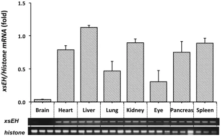
Detection of Xenopus sEH mRNA in various tissues and embryos
Expression of Xenopus sEH mRNA was detected by RT-‐PCR at various Xenopus embryo stages and in several tissues. Tissues were collected from brain, heart, liver, lung, kidney, eye, pancreas and spleen of adult Xenopus. Total RNA was extracted from the tissues and converted to cDNA by reverse transcriptase reaction as described above. PCR was performed by the same method described above.
Whole-‐mount in situ hybridization
Whole-‐mount in situ hybridization (WISH) was performed using albino Xenopus embryos at stage 18 (middle neurula). Thirty embryos were fixed with fully dehydrated ethanol. A DNA fragment for the probe was constructed by primers 5 and 6. PCR was performed as follows: denaturation at 940C for 3 min, then 30 cycles of 940C for 1 min, 520C for 2 min, and 680C for 1 min. The amplified Xenopus sEH cDNA fragment included 887 bp nucleotides and was ligated into pBluescripIISK+ vector. The sense probe for sEH was prepared by linearization with HindIII and transcribed with T3 RNA polymerase. The anti-‐ sense probe for sEH was prepared by linearization with BamHI and by transcription with T7 RNA polymerase. This probe was hybridized and visualized according to the protocol for Roche Diagnostics DIG with minor modification 30,31). Images of in situ hybridizations were taken using an Olympus SZX16 Stereo Microscope equipped with an Olympus DP71 microscope digital camera.
Isolation of Xenopus sEH cDNA and preparation of sEH mutant
Full-‐length Xenopus sEH cDNA was isolated by PCR using primers 7 and 8 in Table I-‐1. These primers were designed from the nucleotide sequence reported as Xenopus sEH (GenBank accession no. NM_001093674). PCR was
performed using cDNA synthesized from total RNA of Xenopus tailbud embryos (st.38) with GeneAmp high fidelity Taq DNA polymerase: denaturation at 940C for 3 min, then 30 cycles of 940C for 1 min, 550C for 2 min, and 680C for 1 min. The amplified sEH cDNA included 1683 bp nucleotides, from which the deduced protein had an open reading frame of 561 amino acid residues. The amino acid sequence deduced from the DNA sequence of the isolated Xenopus sEH cDNA in this study was found to have two amino acid substitutions, Thr to Asn at 29 and Arg to His at 146 (N29T/H146R), compared with the amino acid sequence of Xenopus sEH reported in GenBank (accession no. NM_001093674). The nucleotide sequences of several cDNAs from different individuals were analyzed and found to have same substitution. Therefore, it may be a native mutation. The clone of Xenopus sEH cDNA reported in GenBank was prepared by substitution of N29T/H146R.
Table I-‐1. Primers used in this study.
Primer No.
Sequences of primers
1 5’-‐CGGGATAACATTCAGGGTATCACT-‐3’ 2 5’-‐ATCCATGGCGGTAACTGTCTTCCT-‐3’ 3 5’-‐GGTTGTAGAGTCGTGTCGTA-‐3’ 4 5’-‐CTCCAGGAATACCAACTCTC-‐3’ 5 5’-‐GGAATTCCATATGGCTGGGAAGCGCTTCGT-‐3’ 6 5’-‐AAGGATCCATGGCTTCCCAGAGAGTT-‐3’ 7 5’-‐GGAATTCCATATGGCTGGGAAGCGCTTCGT-‐3’ 8 5’-‐TGCAAGCTTCAGTTTGGATGTTACGGGCA-‐3’ 9 5’-‐TTAATACGACTCACTATAGG-‐3’ 10 5’-‐GGGAGATGTAGGCTAGTTTCTAGCCGTTGAA-‐3’ 11 5’-‐TAGCCTACATCTCCCCAGTG-‐3’ 12 5’-‐TCTGCAACAAACTTGGAGAA-‐3’ 13 5’-‐TTCTCCAAGTTTGTTGCAGA-‐3’ 14 5’-‐ACCAGGTCAAAATGGCGACTTAGTGAAGAGA-‐3’ 15 5’-‐CCATTTTGACCTGGTTGTAG-‐3’ 16 5’-‐CTCCAGGAATACCAACTCTC-‐3’ 17 5’-‐TTAATACGACTCACTATAGG-‐3’ 18 5’-‐CTCCAGGAATACCAACTCTC-‐3’ 19 5’-‐GGGGTCCTGCTCACCCCTGGG-‐3’ 20 5’-‐CTTCCAGGAATACCAACTCTC-‐3’ 21 5’-‐TTAATACGACTCACTATAGG-‐3’ 22 5’-‐TTCAATCAACGGAACTCCGGTCACTTTCTC-‐3’ 23 5’-‐GAGAAAGTGACCGGAGTTCCGTTGATTGAA-‐3’ 24 5’-‐TGATGATGATGCGGCCGCCAGTTTGGATGTTACGGGCAAGTTA-‐3’
13
Fragment I was amplified by PCR with primers 9 and 10 and fragment II with primers 11 and 12. Fragment III was amplified using primers 13 and 14. Fragment IV was amplified using primers 15 and 16. To obtain fragment V, fragments I and II was used as templates with primers 17 and 13. Fragment VI was made using fragments III and IV with primers 17 and 12. The last step was to obtain mutant 1 by fragments V and VI, used as templates with primers 18 and 12. By comparing our Xenopus sEH with human sEH, I found that Asp residues at 11 were substituted by Gly, which is an active site of phosphatase, and sEH G11D was designated xsEH mutant 2. PCR with primers 19 and 20 was carried out as above. A chimera, having a phosphatase domain of human sEH and epoxide hydrolase domain of Xenopus sEH, was also prepared. Primers for the phosphatase domain (primers 21 and 22) amplifies fragments corresponding to human sEH amino acids 1-‐334. Primers for epoxide hydrolase domain (primers 23 and 24) amplifies fragments corresponding to Xenopus sEH amino acids 334-‐563. To combine both of these domains, primers 21 and 24 were used to amplify fragments. All fragments were ligated into pET-‐21a(+) (Novagen).
Purification of Xenopus sEH
Full-‐length cDNAs of Xenopus wild-‐type, mutant 1, mutant 2 and chimera sEH were subcloned into pET-‐21a(+) vector with BamHI and XbaI enzymes sites (Takara Bio, Shiga, Japan). The recombinant His-‐tagged sEH proteins expressed in E. coli, BL21-‐CodonPlus (DE3) (Stratagene, La Jolla, CA) were purified with a Ni-‐NTA agarose column (Qiagen, Hilden, Germany). The purified proteins were dialyzed in 10 mM Tris Buffer, pH 7.5, overnight and their concentrations were measured with the Bradford method (Protein Assay, Bio-‐Rad, Hercules, CA).
Assay of epoxide hydrolase activity
Epoxide hydrolase activity of sEH was measured using a fluorescent substrate, 3-‐phenyl-‐cyano (6-‐methoxy-‐2-‐naphthalenyl) methyl ester-‐2-‐ oxiraneacetic acid (PHOME) purchased from Cayman Chemical (Ann Arbor, MI). Purified Xenopus sEH and human sEH (0.5 µg) were reacted with 25 µM PHOME in 25 mM Bis Tris-‐HCl, pH 7.0, containing 0.01% Bovine Serum Albumin (BSA). The effect of pH on EH activity was evaluated with Bis Tris buffer (pH 5.5-‐7.5). The effect of ionic strength was evaluated with 10, 25, 50, and 100 mM Bis Tris buffer (pH 7.0). The catalytic activity of purified Xenopus sEH and human sEH was investigated for temperatures ranging from 20 to 370C. For inhibition assay, Xenopus sEH or human sEH was incubated with 25 µM PHOME in 25 mM Bis Tris buffer, pH 7.0, containing 0.01% BSA in the absence or presence of 0 to 0.2 µM of N, N’-‐dicyclohexyl urea (DCU) (Wako, Osaka, Japan). Fluorescence of the reaction product, 6-‐methoxy-‐2-‐ naphthaldehyde (6-‐MNA), was measured every 5 min for 60 min by the EnVision 2104 Multilabel Reader (Perkin Elmer, Waltham, MA) at an excitation wavelength of 330 nm and an emission wavelength of 465 nm at 300C. The concentration of 6-‐MNA produced by sEH was determined with a calibration curve prepared with authentic 6-‐MNA.
Assay of phosphatase activity
Phosphatase activity was measured using 4-‐Methylumbelliferyl
Phosphate (Wako, Osaka, Japan) in 25 mM Bis Tris buffer, pH 7.0, containing 1 mM MgCl2 and 0.01% BSA. The reaction was started by the addition of the enzyme, purified Xenopus sEH or human sEH (5.0 µg). The effects of ionic strength, pH and temperature were assessed as described above. The reaction was performed at 370C for up to 60 min and the fluorescence intensity of the produced 4-‐methylumbelliferone was measured every 5 min by the EnVision 2104 Multilabel Reader at an excitation wavelength of 330 nm and an emission wavelength of 465 nm.
15
Phosphatase assay of Xenopus sEH using malachite green
The phosphatase activity of sEH toward LPA (stearoyl L-‐α-‐
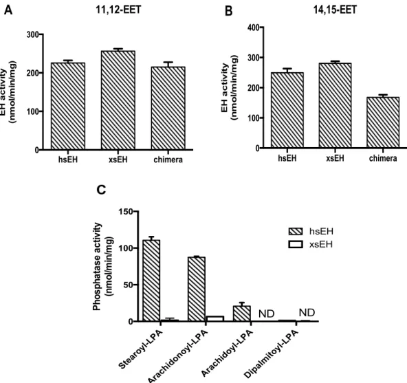
lysophosphatidic acid (1-‐octadecanoyl-‐sn glycerol-‐3-‐phosphate) sodium salt) (Avanti Polar Lipids, Birmingham, AL) was detected by the Biomol green assay (Enzo Life Science, Plymouth Meeting, PA) according to the manufacturer’s instructions. Arachidonoyl L-‐α-‐lysophosphatidic acid sodium salt and arachidoyl L-‐α-‐lysophosphatidic acid sodium salt were purchased from Echelon Bioscience (Salt Lake City, UT). Dipalmitoyl L-‐α-‐lysophosphatidic acid sodium salt was from Wako. Purified human sEH, Xenopus sEH, and chimera (0.6 µg) were pre-‐incubated for 5 min at 370C in 25 mM Bis Tris buffer, pH 7.0, containing 1 mM MgCl2 and 0.01% BSA. LPAs were added at a final concentration of 10 µM and incubated for 5 min at 370C. The reaction was stopped by the addition of Biomol green reagent and held at room temperature for 60 min. The resulting green color was measured by the EnVision 2104 Multilabel Reader at 630 nm.
Separation and quantitation of EET metabolites
The epoxide hydrolase activities of purified recombinant Xenopus sEH, human sEH and chimera toward 10 µM EETs (11,12 EET or 14,15 EET) were assayed in incubation mixture (final volume 0.5 ml) consisting of 100 mM potassium phosphate buffer, pH 7.4, and 2 nmol stearic acid as an internal control. The reaction mixture containing purified recombinant protein (6.0 µg) was incubated at 370C for 5 min and the reaction was stopped by the addition of 0.4 M citric acid and I mL of ethyl acetate. After centrifugation at 4000 rpm for 10 min, the upper organic layer was transferred to a new tube and evaporated under nitrogen. The resulting residue was dissolved in ethanol (20 µl) and analyzed by HPLC equipped with an Evaporative Light Scattering Detector (ELSD) system (Prominence-‐Shimadzu, Kyoto, Japan) using a TSKgel ODS-‐100Z 5μm column (4.4 mm I.D x 15 cm; Tosoh, Tokyo, Japan). To detect
EET metabolites, mobile phase A (water: acetic acid, 100:0.1), and mobile phase B (acetonitrile: acetic acid, 100:0.1) were used. The metabolites were separated at a flow rate of 1 ml/min, with a linear gradient of A to B from 50% to 100% for 30 min. DHET production was measured by a calibration curve prepared with authentic DHETs.
Sample preparation and analysis of endogenous substrate
Eighty milligrams of Xenopus liver was homogenized and 200 µl of methanol and 2 µl of formic acid were added. The homogenates were centrifuged at 14,000 rpm for 10 min at 40C. The supernatants were applied to the SPE cartridge and washed with 3 ml of water, and 1 ml of 10% methanol. The sample was eluted in 0.5 ml acetonitrile followed by 1.5 ml ethyl acetate. These fractions were combined and dried under N2 32,33). The residue was dissolved with 40 µl of ethanol for analysis by UPLC/electrospray ionization (ESI)/MS. The chromatography was performed with a C18 reversed-‐phase column (TSK-‐GEL ODS-‐140HTP, 4.6 x 250 mm, 5 µm) and the UPLC system (Acquity UPLC system, Waters, Milford, MA). Mobile phase A consisted of 50% water, 30% acetonitrile, 20% methanol and 1% acetic acid. Mobile phase B consisted of 80% acetonitrile, 20% methanol and 1% acetic acid. A flow rate of 0.2 ml/min and 5 µl injection volume was used to deliver the mobile phases A and B with a linear gradient from 40% A for 20 min to 40-‐100% B for 27 min. Mass spectrometry was carried out using a Nanofrontier LD mass spectrometer (Hitachi, Tokyo, Japan) and ESI. The analytes were detected by tandem TOF monitored by total ion, m/z 319 (EETs).
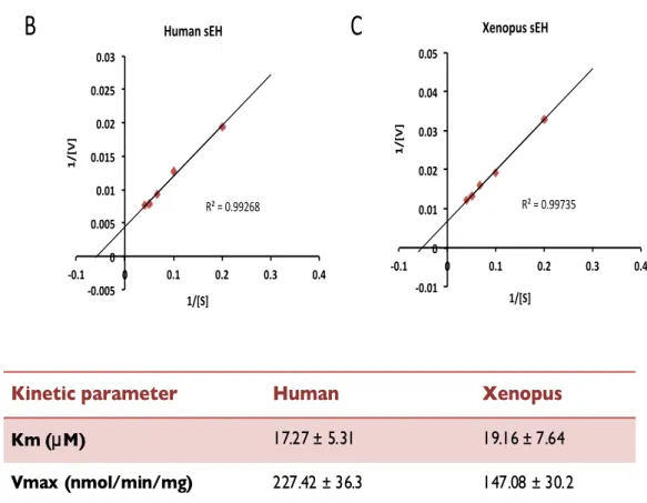
Statistics and kinetics analysis
The kinetic parameters Km and Vmax were obtained using Prism enzyme kinetic software (Graphpad Software, La Jolla, CA). Statistical analysis was performed with Student’s t-‐test and p<0.05 were considered significant.
17
I.3 Results
Expression and purification of Xenopus sEH and its catalytic activity
I found two substitutions of amino acid in the sequence deduced from our Xenopus sEH cDNA compared with that of the original Xenopus sEH reported in GenBank. I prepared Xenopus sEH with substitutions of amino acid residues with N29T and H146R (designated mutant 1). Both amino acid exchanges occurred in the phosphatase domain. The purified Xenopus sEH was reacted with the generic substrate 4-‐Methylumbelliferyl Phosphate for phosphatase activity and with PHOME as a substrate for EH activity. The Xenopus sEH revealed significant EH activity (Fig. I-‐1A) but no phosphatase activity (Fig. I-‐1B). Also, another substrate for phosphatase, p-‐nitrophenyl phosphate, was used, but mutant 1 (N29T/H146R) and wild-‐type Xenopus sEH still had no phosphatase activity (data not shown).
Fig. I-‐1 The epoxide hydrolase and phosphatase activities of Xenopus and human sEH. (A) EH activities of Xenopus wild-‐type, Mutant 1, Mutant 2, and Chimera sEH were assessed using purified protein (0.5 µg) toward 25 µM PHOME. (B) Purified sEH (5 µg) was incubated with 0.5 mM 4-‐Methylumbelliferyl Phosphate. The fluorescence of 4-‐methylumbelliferone was measured every 1 min for 60 min at 330 nm (exitation) and 465 nm (emission).
hsEH xsEH Mut 1 Mut 2 chimera 0.0 0.2 0.4 0.6 0.8 1.0 Ph o s p h a ta s e a c ti v it y (n m o l/m in /m g )
B
ND NDhsEH xsEH Mut 1 Mut 2 chimera 0 20 40 60 80 EH a c ti v it y (n m o l/m in /m g )
A
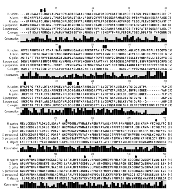
To investigate whether mutant 1 and wild-‐type Xenopus sEH lack phosphatase activity, a homology search of the phosphatase domain was performed with other members of haloacid dehalogenase (HAD), phosphonoacetaldehyde hydrolase (Phos), and phosphoserine phosphatase (PSP). Several amino acid residues have important functions in the two-‐step catalytic mechanism of phosphatase compared with HAD, Phos and PSP 34). Based on the sequence homology with human sEH phosphatase domain, almost all residues important for phosphatase activity are conserved in Xenopus sEH, but the amino acid residue of the 11th aspartic acid was not conserved (Fig. I-‐2).
This amino acid residue is thought to be important for phosphatase activity. Wild-‐type Xenopus sEH has a glycine residue at the 11th position and sEH G11D (designated mutant 2) was constructed by exchanging the 11th glycine with aspartic acid. The EH activity of Xenopus sEH G11D was lower than that of human sEH, and Xenopus sEH G11D lacked phosphatase activity (Fig. I-‐1). These results suggest that other amino acid substitutions or regions of the peptide chain are required for phosphatase activity. The Xenopus sEH chimera was constructed by combining the human sEH phosphatase domain and Xenopus sEH epoxide hydrolase domain. The N-‐terminal domain of Xenopus sEH (1st to 232th amino acids) was exchanged for human sEH (1st to 229th amino acids). The EH activity of the chimera was measured and was found to be similar to that of wild-‐type Xenopus sEH. The chimera also had phosphatase activity, suggesting the C-‐terminal domain of Xenopus sEH was not a cause of the lack of phosphatase activity (Fig. I-‐1B).