石川県立看護大学 大学院 看護学研究科
博士論文
Increasing Cerebral Oxyhemoglobin by Ankle Exercise:
An Attempt at Preventing Symptoms of Orthostatic Hypotension
足関節運動による脳内酸素化ヘモグロビンの増加
— 起立性低血圧を予防するための試み —
永谷 幸子
2015
Increasing Cerebral Oxyhemoglobin by Ankle Exercise:
An Attempt at Preventing Symptoms of Orthostatic Hypotension
by
Sachiko NAGAYA
A Dissertation Submitted in Partial Fulfilment of the Requirements for the Degree of Doctor of Nursing
Graduate School of Nursing
Ishikawa Prefectural Nursing University
March 2015
和文概要
頻度や程度の差はあるが、多くの人が、しゃがんだ姿勢から急に立ち上がった場合など に、立ちくらみを経験している。この立ちくらみは、姿勢を変える時に、重力の影響を受け て循環血液の分布が変動することが原因で生じる。これは、起立性低血圧として、よく知ら れた症状である。起立時だけでなくベッド上での体位変換においてもこのような症状が生じ る場合がある。特に、圧受容器反射の機能が低下している高齢者や、自律神経障害を伴う疾 患(糖尿病、パーキンソン病など)の患者は、体位変換時に大幅な血圧低下を示すことがあ る。血圧の低下に付随した脳虚血の程度によっては、めまいやもうろう感、失神などが生じ る場合もある。またこれらの症状は転倒の原因にもなることから、これに対処するための看 護介入が求められている。
この起立性低血圧の症状を予防するための看護的な介入として、スクワットなどが有効 だと報告されている。しかしこれらの方法は、主に長時間立位をとった後に起こる血圧低下 への対処方法であり、体位変換直後に起こる症状の予防には適さない。体位変換直後の症状 を予防するためには、体位変換の前から、事前に症状を予測し対応できるような介入が求め られる。
下肢の筋は、筋ポンプとして循環動態の調節に重要な役割を担っている。足関節運動(背 底屈運動)は、下肢の静脈血の流速を上げる効果的な方法であることが証明されている。こ の足関節運動を体位変換前に実施し、下肢の静脈血を押し出すことで静脈還流を増加できれ ば、体位変換後の脳循環の変動を軽減できる可能性がある。しかし、足関節運動が、脳循環 にまで効果を示すかどうかは明らかにされていない。
この論文の目的は、足関節運動の脳循環への効果を検証することである。この目的を達成す るために、第2章では、仰臥位で足関節運動を行い、前額部の酸素化ヘモグロビン濃度(OxyHb)
の変化を確認した。第 3 章では、仰臥位で足関節運動を行った後に、実際に体位変換を実施 し、運動の効果を検討した。
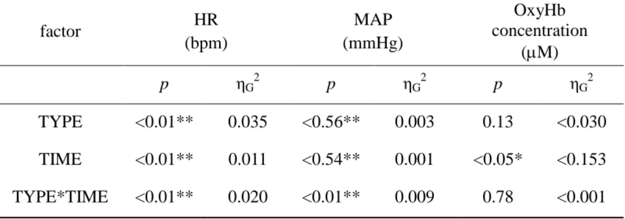
第2章では、17名の高齢女性が研究に参加した。参加者は、仰臥位で足関節の背底屈運 動を1分間実施した。心拍数、血圧と前額部のOxyHbを測定した。運動の種類は、能動的と 受動的運動の 2 条件であった。足関節運動は、能動的、受動的運動の両者ともに、前額部の OxyHbを有意に上昇させることが明らかになった(p < 0.05)。
第3章では、健康な成人と高齢者11名を対象に、3条件(能動的/受動的足関節運動および、
コントロール)を比較した。参加者は、仰臥位で能動的、受動的な足関節運動を1分間実施 した後、端座位に体位を変換された。体位変換はギャッチベッドを用い受動的に実施した。
加えてコントロールとして足関節運動を行わない場合も実施した。これらの条件で脈拍数、
血圧、前額部のOxyHbを測定した。体位変換開始時から90秒間のOxyHbの平均値を比較し たところ、受動的運動とコントロール(運動なし)の間に有意な差は確認されなかったが、
能動的運動は、受動的運動およびコントロールよりもOxyHbが有意に高いことが確認された
(p < 0.05)。この結果から、能動的な足関節運動は、体位変換を行った際の初期のOxyHbの 低下を緩和する効果を持ちうることが示唆された。
本研究の結果を解釈する上でいくつかの限界がある。前額部のOxyHbが維持されたこと と、起立性低血圧の症状が予防できることを同様に考えることはできない。足関節運動を実 際の症状緩和に結び付けるためには、より効果的な運動の強さや運動の継続時間を検討する こと、足関節運動を行った場合の症状の変化について確認するなど、さらなる研究が必要で ある。これらの限界はあるものの、本研究によって足関節運動(特に能動的運動)が起立性 低血圧の症状緩和に効果があると考える一つの根拠を示すことができたと考えられる。
体位変換は、患者の活動を拡大することにつながる重要な看護介入である。一方で、体 位変換によって循環血液の分布が変動することも事実である。体位変換によって引き起こさ れる循環動態の変動を懸念して、看護師は保守的な介入方法を選択する傾向があるとも考え られる。患者の体位の変換に関して、これまでの看護介入は、体位変換後の循環動態の変動 に対応するか、あるいは、変動を起こすような体位変換を避ける、という内容に限られてい た。足関節運動が看護介入法として確立されれば、より積極的に患者の活動を拡大できるよ うな看護を展開することが可能になると考えられる。
Abstract
Standing up quickly sometimes results in a transient lightheadedness. This is a common symptom for most people. The main cause of lightheadedness is the blood distribution changes induced by the gravity effect. A similar phenomenon sometimes occurs when nurses change a patient’s posture, even if this is carried out on a bed. The cardiovascular system promptly adjusts the blood distribution; therefore, severe hemodynamic fluctuation is usually not observed. However, elderly people with lower baroreceptor sensitivity and patients with dysfunctions of the autonomic nervous system (e.g., diabetic mellitus, Parkinson’s disease) have problems when they stand up. This phenomenon is known as
“orthostatic hypotension (OH).” The OH is sometimes accompanied by symptoms such as dizziness, faintness and syncope, depending on the degree of cerebral hypoperfusion.
Furthermore, these symptoms could cause falling. Thus nursing intervention is required to prevent OH.
To prevent this phenomenon, some physical counter-maneuvers such as squatting have been proposed as nursing interventions. However, these maneuvers address symptoms that occur after standing for a long time and do not seem to be effective in preventing the hypotension experienced in the early phase after postural change. Prior intervention is required to prevent the symptoms that occur immediately after postural change.
The skeletal muscle pump plays an important role in adjusting hemodynamics. Ankle exercise (i.e., ankle plantar flexion and dorsiflexion movements) has been proven to be an effective intervention to increase venous velocity. If ankle exercise performed in a supine position is proven to have the effect of maintaining cerebral hemodynamics, it can prevent the symptoms of the early phase after postural change. However, the role of ankle exercises in cerebral hemodynamics has been less well established.
The goal of this dissertation is to confirm the efficacy of ankle exercises for cerebral hemodynamics. To achieve this goal, the first study, with an experiment, was conducted to identify the effect of active and passive ankle exercises on the cerebral oxyhemoglobin (OxyHb) in a supine position (Chapter 2). Following the results of the first experiment, the
second experiment studied the effect of ankle exercises on postural changes (Chapter 3).
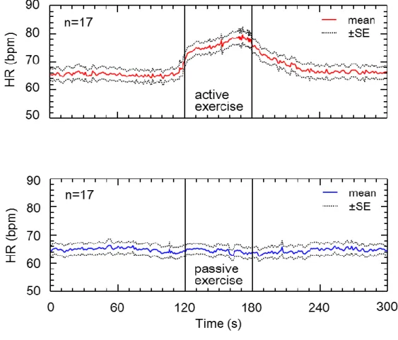
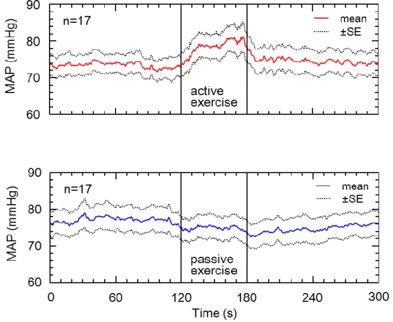
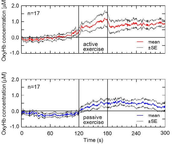
In the first study, discussed in Chapter 2, there were seventeen elderly women who participated. The participants repeated ankle plantar flexion and dorsiflexion movements for 60 s. Heart rate, blood pressure, and cerebral OxyHb levels were recorded in a supine position.
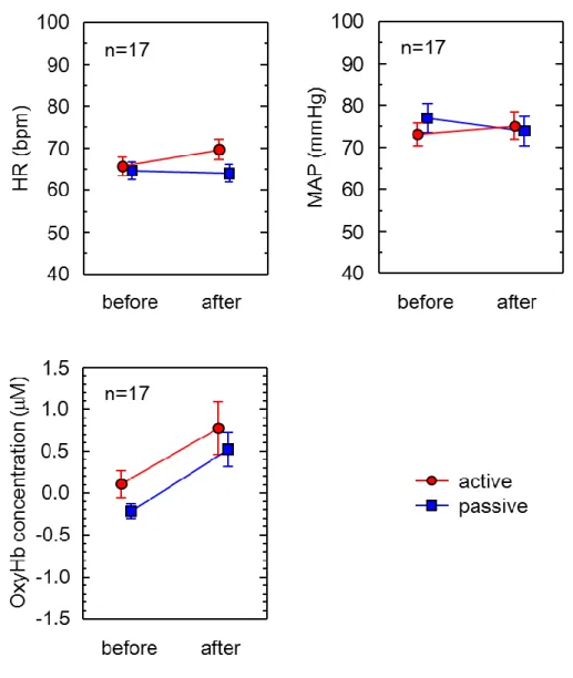
Two types of exercises were used, active movement and passive movement. Both active and passive ankle exercises could increase cerebral OxyHb (p < 0.05).
Eleven healthy adults and elderlies participated in the experiment in Chapter 3. Three conditions (active/passive ankle exercise, and control condition) were examined for each participant. The participants performed ankle exercise for 60 s before postural change. The postural change was conducted passively by using the electric motor of a hospital bed.
Additionally, as a control condition, data was taken without ankle exercise. Pulse rate, blood pressure, and cerebral OxyHb levels were recorded in a supine position. No significant difference was observed between passive exercise and the control condition. Conversely, active ankle exercise demonstrated significantly higher cerebral OxyHb than passive exercise and the control during and after postural change (p < 0.05). From the results in Chapter 3, active ankle exercise which was performed before postural change has the possibility of attenuating the initial changes in cerebral OxyHb.
There are some limitations to be observed when interpreting this study. The demonstration that there is an effect of maintaining cerebral OxyHb does not fully support the idea that such exercise will improve the symptoms of OH. Further studies on such aspects as changing the degree of the exercise, or its duration, and confirming the improvement of symptoms may answer this question. In spite of these limitations, the author believes that this study provides some evidence for considering that ankle exercise (especially active ankle exercise) can prevent symptoms of OH.
Postural change is an important nursing intervention for expanding the patient’s mobilization. On the other hand, it is a fact that postural change has the possibility of inducing changes in blood distribution. Nurses may tend to avoid postural change when they foresee the likelihood of hemodynamic instability after postural change. If ankle exercises before postural change are validated as a nursing intervention, we can expect them to expand the
mobility of patients.
List of Table
Title page
Table 2.1. Characteristics of 17 participants 25
Table 2.2. Results of ANOVA for effects of the TYPE factor and
TIME factor on ankle exercises 35
Table 3.1. Pulse rate and mean arterial blood pressure response to postural change from supine to sitting (n = 11) 46
List of Figures
Title page
Figure 1.1. Examples of physical counter maneuvers 9
Figure 1.2. Near infrared spectroscopy (NIRO-200; Hamamatsu
Photonics, Shizuoka, Japan) 13
Figure 1.3. Absorption spectra of oxyhemoglobin and
deoxyhemoglobin 14
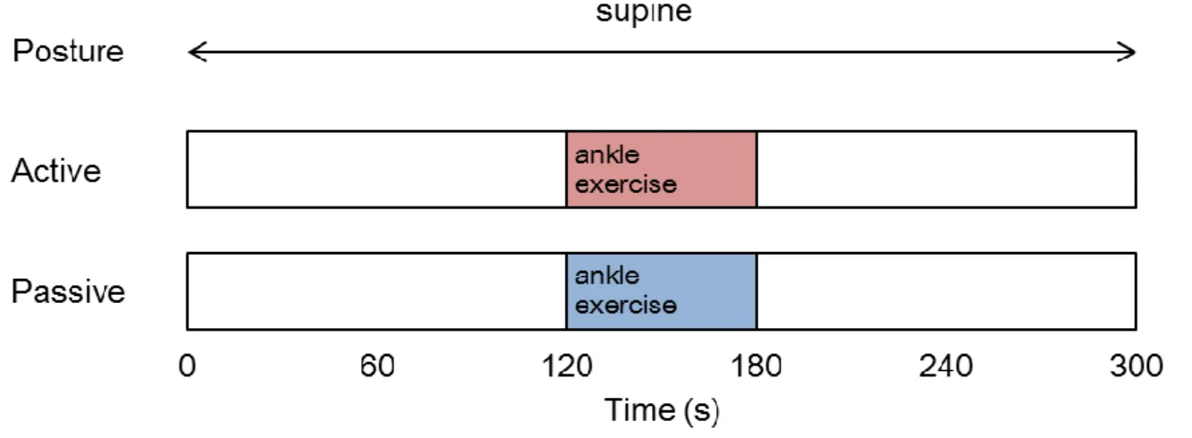
Figure 2.1. Schematic representation of the experimental protocol
(Chapter 2) 27
Figure 2.2. Heart rate responses to active and passive ankle exercises
(n = 17) 31
Figure 2.3. Mean arterial blood pressure responses to active and
passive ankle exercises (n = 17) 32
Figure 2.4. Oxyhemoglobin responses to active and passive ankle
exercises (n = 17) 33
Figure 2.5.
Effects of active and passive ankle exercises on 60 s averages of heart rate, mean arterial blood pressure, and oxyhemoglobin (n = 17)
34
Figure 3.1. Schematic representation of the experimental protocol
(Chapter 3) 43
Figure 3.2. Pulse rate and mean arterial blood pressure response to postural change following ankle exercises (n = 11) 47 Figure 3.3. Oxyhemoglobin responses to ankle exercises and postural
change (n = 11) 48
Figure 3.4. Oxyhemoglobin during each ankle exercises (n = 11) 49 Figure 3.5. Oxyhemoglobin response to postural change following
ankle exercises (n = 11) 50
List of Abbreviations
ANOVA Analysis of variance
BP Blood pressure
CBF Cerebral blood flow
CBF-V Cerebral blood flow velocity CBV Cerebral blood volume
CO Cardiac output
CT Computed tomography
DBP Diastolic blood pressure
fNIRS Functional near infrared spectroscopy HHb Deoxyhemoglobin concentration
HR Heart rate
MAP Mean arterial blood pressure NIRS Near infrared spectroscopy OH Orthostatic hypotension OxyHb Oxyhemoglobin concentration PET Positron emission tomography
PR Pulse rate
rCBF Regional cerebral blood flows rOEF Regional oxygen extraction fraction SBP Systolic blood pressure
SPECT Single photon emission computed tomography
SE Standard error
SV Stroke volume
TCD Transcranial Doppler
tHb Total hemoglobin
TPR Total peripheral resistance
Xe-CT Xenon 133 computed tomography ηG2 Generalized eta squared
Table of Contents
Chapter 1: Backgrounds 1
1.1 Physiological Background 2
1.1.1 Cardiovascular responses to postural change 2 1.1.2 Hypotension is induced by postural change 4
1.2 Postural Change as Nursing Intervention 6
1.2.1 Nursing problems in postural changes 6
1.2.2 Treatments for orthostatic hypotension 7
1.3 Methodological Background 9
1.3.1 Measurement of cerebral hemodynamics 9
1.3.2 Principles of near infrared spectroscopy 11
1.4 Aims of This Dissertation 15
1.5 Overview of This Dissertation 15
1.6 References 16
Chapter 2: Ankle Exercise Increases Cerebral Blood Oxygenation in the Elderly 23
2.1 Introduction 24
2.2 Methods 24
2.2.1 Participants 24
2.2.2 Experimental procedures 25
2.2.3 Measuring physiological responses 27
2.2.4 Statistical analysis 28
2.2.5 Ethical considerations 28
2.3 Results 29
2.4 Discussion 35
2.4.1 Responses of HR and MAP to ankle exercises 35 2.4.2 Responses of cerebral hemodynamics to ankle exercises 36
2.5 Conclusions 37
2.6 References 38
Chapter 3: Ankle Exercise Increases Cerebral Blood Oxygenation During 40 and After Postural Change
3.1 Introduction 41
3.2 Methods 42
3.2.1 Participants 42
3.2.2 Experimental procedures 42
3.2.3 Measuring physiological responses 43
3.2.4 Statistical analysis 44
3.2.5 Ethical considerations 44
3.3 Results 45
3.4 Discussion 50
3.4.1 Differences in the effect on OxyHb of different ankle exercises 50 3.4.2 The effect of ankle exercise to postural change 51
3.5 Conclusions 53
3.6 References 54
Chapter 4: General Discussion 56
Funding 60
Acknowledgements 60
Appendices 61
Appendix A: Individual data of heart rate, mean blood pressure, and oxyhemoglobin before and after ankle exercises (Chapter 2)
62
Appendix B: Individual data of oxyhemoglobin in the experiment of Chapter 2
64
Appendix C: Individual data of pulse rate, mean blood pressure, and oxyhemoglobin before and after postural change (Chapter 3)
65
Appendix D: Individual data of oxyhemoglobin in the experiment of Chapter 3
68
Chapter 1
Backgrounds
1.1 Physiological Background
1.1.1 Cardiovascular responses to postural change
Human beings acquired upright posture and bipedalism in the process of evolution.
Upright posture and bipedalism brought great advantages to humans because it allowed them to acquire large brains and to use their upper extremities freely. However, having evolved in the limited environment of the planet Earth, humans sometimes experienced a trade-off when a change took place in their physical makeup.
To develop bipedalism from quadrupedalism, the human skeleton had to change dramatically. Quadrupedalism puts the center of gravity at or near the point that is central to the four legs (near the center of the torso), but it is located between two legs with bipedalism.
The human pelvis changed its shape to support the abdominal viscera and to maintain balance during bipedal locomotion. In exchange, the size of the human birth canal was reduced, while the fetus has a big brain (Lovejoy, 1988). Childbirth in humans is very difficult compared to that of other animals. A human is prone to lumbar pain, hernias, hemorrhoids and varices;
these diseases are rarely observed in the quadrupedal animals, including, of course, the other primates that are our relatives. These diseases are attributed to maintaining an upright posture against the pull of gravity.
The human cardiovascular system also faced the challenges of regulating the circulation during various movements. Compared with apes, humans have larger brains, which require more blood. The human brain accounts for only 2% of the body mass, but it needs about 14% of the resting cardiac outputs. In addition, the grey matter has a very high rate of oxidative metabolism (about 7 ml O2/min/100g), which causes a loss of consciousness after a few seconds of cerebral ischemia (Levick, 2010, p.294). Even worse, the human brain is located above the heart; considering the hydrostatic aspect, this is a very disadvantageous position for the brain. The human cardiovascular system has to pump blood upward to sustain this expensive organ. Of course, there are many enormous animals that have longer necks than do humans: giraffes, for example. The brain of a giraffe is about 2 m above its heart.
Therefore, giraffes have a unique problem in the adjustment of circulation. They have powerful hearts and very high blood pressure. Moreover, they have extremely muscular
arterioles and very tight skin and fascia in their extremities (Hargens, Millard, Pettersson, &
Johansen, 1987; Rowell, 1993, pp. 1–36). These physical characteristics maintain a giraffe’s cerebral circulation over a distance greater than the 2 m that separate the heart and brain.
However, the human body is completely different from that of a giraffe, so the human cardiovascular system is forced to use other strategies to adjust their circulation.
The principal mechanism underlying the adjustment for movement and postural changes is a system of adaptation to gravity. Thus, the postural effect on circulation has more impact when the movement is greater in a vertical direction. When a human being moves from a supine to upright posture, blood is immediately shifted from the thorax to the lower part of the body. The amount of blood thus affected is about 500 to 700 ml (8 to 10 ml/kg) (Smith, & Kampine, 1990, p. 97). The walls of the veins contain little elastic tissue, creating a large capacity for holding blood. Humans have large legs compared to those of other animals;
this allows the shifted blood to pool in the veins of the lower limbs. This blood pooling in the veins leads reduction in venous return to the heart. As a result, the cardiac output decreases (Frank-Staring mechanism), and subsequently, the mean arterial blood pressure (MAP) falls.
If there is an extreme decrease in MAP, an adequate cerebral blood pressure cannot be maintained, causing syncope (Costanzo, 2014, pp. 174–184).
When people stand up, severe hypotension usually does not occur because an immediately compensatory response works to adjust the shift in blood volume. The baroreceptor reflex plays an important role in these adjustments. The baroreceptors are mechanoreceptors located in the carotid sinus. The baroreceptor senses a drop in the MAP by the degree of stretching in the blood vessel walls and sends signals to the medullary vasomotor center. The vasomotor center directs an increase in sympathetic activity to the heart and blood vessels. Simultaneously, orders from the vasomotor center decrease parasympathetic activity to the heart. As a result, the heart rate (HR), total peripheral resistance (TPR) and venoconstriction are increased; then the MAP goes back to normal (Pocock, & Richards, 2006, pp. 262–310). This prompt response by the baroreceptor works on the circulation to facilitate dynamic postural changes in humans.
The skeletal muscle pump is another mechanism that acts quickly to adjust the
circulation in humans. In upright humans, the skeletal muscles of the lower extremities soon begin rhythmic cycles of reflex contraction and relaxation. This muscle contraction and relaxation are due to the unconscious swaying motion of the body while standing. Rhythmic cycles of muscle contraction help squeeze the pooled blood from the lower extremities, aiding it in returning to the heart so that the blood pressure (BP) is maintained at an adequate level (Smith, & Kampine, 1990, pp. 99–107). Because of this compensatory mechanism, the leg muscles are considered to be a “second heart.”
1.1.2 Hypotension is induced by postural change
The hemodynamics changes associated with changes in posture are a very natural response. Normally, a postural change does not have a serious effect on blood pressure in healthy people. However, some people have symptoms of hypotension such as dizziness, syncope, and falling, when they change their posture because their cardiovascular system cannot act immediately to maintain the BP. These symptoms are often observed when such people stand up; therefore, this condition is known as “orthostatic hypotension (OH).” The consensus definition of OH is a reduction of systolic blood pressure (SBP) of at least 20 mmHg or a reduction of digastric blood pressure (DBP) of at least 10 mmHg within 3 min of erect standing (The Consensus Committee of the American Autonomic Society and the American Academy of Neurology, 1996). OH can be divided into subclasses. Initial OH occurs immediately after standing (within 30s) and many healthy people are familiar with the symptoms of initial OH (Wieling, Krediet, van Dijk, Linzer, & Tschakovsky, 2007). Delayed OH produces symptoms within 3 to 30 min of standing (Gibbons, & Freeman, 2006; Streeten,
& Anderson, 1992). As for a hospital situation, it is considered that initial OH is a situation more frequently encountered by nurses because patients rarely maintain a standing posture for 30min.
OH is observed in all age groups. However, the prevalence of OH increases with age (Lipsitz, 1989; Masaki, et al., 1998) because baroreceptor sensitivity decreases with aging (Rutan, et al., 1992). The prevalence of OH in the community-dwelling elderly is reported to be 6 to 34 %, depending on the methodology used and differences in population (Hiitola,
Enlund, Kettunen, Sulkava, & Hartikainen, 2009; Kobayashi, & Yamada, 2012; Luukinen, Koski, Laippala, & Kivelä, 1999; Matsubayashi, et al., 1997). In hospitalized elderly people, OH is a relatively common condition. The prevalence is over 50% in patients attending geriatric clinics (Poon, & Braun, 2005) and those admitted to acute care hospitals (Gorelik, et al., 2005; Shibao, Grijalva, Raj, Biaggioni, & Griffin, 2007; Weiss, Grossman, Beloosesky, &
Grinblat, 2002; Vloet, Pel-Little, Jansen, & Jansen, 2005).
OH may result from neurogenic and nonneurogenic conditions. Diabetes mellitus (Van Hateren, et al., 2012; Wu, Yang, Lu, Wu, & Chang, 2008) and Parkinson’s disease (Allcock, Ullyart, Kenny, & Burn, 2004; Senard, et al., 1997; Velseboer, de Haan, Wieling, Goldstein, & de Bie, 2011) are well-known causes of neurogenic OH. Nonneurogenic factors, such as cardiac impairment, reduced intravascular volume and vasodilation are related to OH.
Some drugs may cause OH. Antihypertensives, diuretics and vasodilators are recognized as causes of OH (Hajjar, 2005; Kamaruzzaman, Watt, Carson, & Ebrahim, 2010). There is a significant relationship between OH and the number of drugs being taken (Poon, & Braun, 2005). Especially for hospitalized patients, bed rest is one of the problems associated with OH.
Within 24 to 48 hours, bed rest results in a decrease in plasma volume (Creditor, 1993;
Fortney, Hyatt, Davis, & Vogel, 1991; Olson, 1967/1990). Bed rest can also lead to baroreceptor dysfunction. The signs of orthostatic intolerance begin to appear within 3 to 4 days after bed rest (Convertino, Hung, Goldwater, & DeBusk, 1982; Greenleaf, & Kozlowski, 1982). These physical complications related to bed rest can cause OH by themselves.
Baroreceptor sensitivity decreases with aging; in addition, the elderly usually have multiple medical problems and take several drugs. Because of these factors, OH in the elderly population tends to have multiple causes. Some studies have been reported that in the elderly, OH is associated with difficulty walking (Rutan, et al., 1992), cognitive impairment (Mehrabian, et al., 2010), and falling (Allan, Ballard, Rowan, & Kenny, 2009; Gangavati, et al., 2011; Ooi, Hossain, & Lipsitz, 2000; Rutan, et al., 1992). Preventing and treating OH is one of the important challenges in improving the quality of life for the elderly.
1.2 Postural Change as Nursing Intervention 1.2.1 Nursing problems in postural changes
At the end of the day, humans go to their beds to rest. Rest and sleep are essential functions. For patients, being in a supine position on a bed has some benefits because it requires less oxygen consumption than other positions (e.g., a sitting position) (Jones, & Dean, 2004). The benefits of rest were emphasized as treatment in the 19th century. Bed rest was strongly recommended for diseases such as myocardial infarction and after orthopedic surgery (Allen, Glasziou, & Del Mar, 1999). However, it is very unnatural for humans to continue bed rest for a long time because usually they lie down only while they are asleep. Prolonged bed rest sometimes does severe harm. Many previous studies have revealed negative effects of bed rest and recommend the early initiation of patient mobilization (Perme, & Chandrashekar, 2009; Stiller, & Phillips, 2003; Timmerman, 2007).
Bed rest leads to many complications such as pneumonia (Drakulovic, et al., 1999), pressure ulcers (Lahmann, Kottner, Dassen, & Tannen, 2012), deep venous thrombosis (Cook, et al., 2000), and joint contracture (Clavet, Hébert, Fergusson, Doucette, & Trudel, 2008).
When patients cannot move by themselves or their movements are restricted by some medical problem and treatment, nurses regularly change their patients’ posture. However, as discussed in an earlier part of this chapter, carrying out postural change for patients means that the patient’s blood is more or less shifted by the effect of gravity, even if the body does not move much. Therefore postural changes in patients can result in potential adverse hemodynamic effects, such as reduced cardiac output, hypotension, and arrhythmia (Evans, 1994).
Leaving the hospital bed and standing up on his/her own feet is the first step in a patient’s return to daily life. On the other hand, difficulties can easily be predicted when nurses promote early mobilization in critically ill and elderly patients because those patients sometimes experience hemodynamic instability that includes OH (Bundgaard-Nielsen, et al., 2009; Feldstein, & Weder, 2012; Jans, Bundgaard-Nielsen, Solgaard, Johansson, & Kehlet, 2012). It is reported that a patient’s hemodynamics instability can complicate a nurse’s attempts at early mobilization that includes postural change (Garzon-serrano et al., 2011;
Haines, Skinner, & Berney, 2013). Thus, nurses observe the patient’s BP carefully when they
carry out postural change, especially the first one (Nagaya, Fujimoto, & Kobayashi, 2014).
Postural change is a basic nursing care, but that does not mean it is easy. Nurses are required to have the ability to assess whether the patient’s circulatory system can tolerate a change in posture. In addition, postural change can become more effective means of care if a way is proposed to actively promote an adjustment in hemodynamics in order to expand mobilization.
1.2.2 Treatments for orthostatic hypotension
OH can be induced by a variety of factors, sometimes by multiple ones at the same time. Therefore, some different approaches have been proposed for managing each factor. As a pharmacological approach, the European Federation of Neurological Societies recommends fludrocortisone and midodrine to manage OH (Moya, et al., 2009). In Japan, the guidelines for the diagnosis and management of syncope recommend fludrocortisone, erythropoietin, midodrine hydrochloride, and etilefrine hydrochloride (Inoue, et al., 2012). The effectiveness of pharmacological and nonpharmacological approaches to OH has been confirmed through previous studies. The important and difficult issue in OH symptom management is how to treat the condition without excessive supine hypertension. Therefore, a nonpharmacological approach, which would have no adverse effects, is considered to be a better first line defense.
If OH is primarily caused by hypovolemia, then oral giving salt and adequate amounts of water is a safe approach because it can expand plasma volume (El-Sayed, &
Hainsworth, 1996). In Japan, if patients do not have hypertension, 10 g of salt and 2 to 3 L of water per day are recommended (Inoue, et al., 2012).
For patients with autonomic failure, drinking about 500 ml of water at room temperature improves their condition in less than 5 min (Shannon et al., 2002). Drinking water can raise sympathetic activity and increase the BP. After drinking 500ml of water, the BP increases and this effect is sustained for more than 60 min (Jordan, Shannon, Grogan, Biaggioni, & Robertson, 1999; Jordan et al., 2000).
Some nonpharmacological approaches work to return pooling blood from the legs to the heart. Compression garments or tightly fitting body stockings are useful for treating OH
because they can reduce the venous capacitance bed (Podoleanu, & Maggi, 2006). However, it sis difficult for elderly patients to put on tightly fitting stockings by themselves. In addition, patients have to wear the stockings all the time if they want to reduce the symptoms of OH.
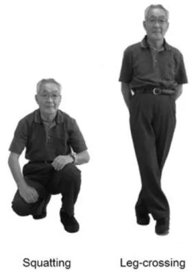
Leg-crossing and squatting are representative examples of physical counter maneuvers (Figure 1.1.). Leg-crossing can contribute to the mechanical compression of veins in the lower extremities by stimulating the muscles. This stimulus reduces venous capacity and increases the TPR (Ten Harkel, van Lieshout, & Wieling, 1994). Consequently, the increased venous return raises the mean blood pressure to a high enough level to maintain cerebral blood flow (CBF) (Van Lieshout, ten Harkel, & Wieling, 1992). Squatting has the same effect as leg-crossing. To bend the legs can increase muscle tone, which promotes venous return. In addition, in a squat posture, the distance between the legs and the heart is shorter than when upright posture: therefore, it is considered a more advantageous posture for the heart when we consider the hydrostatic involved. Leg-crossing and squatting are easy and economical approaches because they do not need special aids like compression garments or salt tablets, or even a glass of water. However, if patients want to prevent orthostatic symptoms they have to perform these maneuvers before the BP falls. It is difficult for elderly patients to always do these maneuvers with the appropriate timing, especially in cases in which OH occurs immediately after a patients stands up.
Ankle exercises in the supine positon have been proven to be an effective intervention to increase venous velocity by squeezing accumulated blood from the lower part of the body (Kwon, Jung, Kim, Cho, & Yi, 2003; Sochart, & Hardinge, 1999; Stein, et al., 2009). Studies have been conducted on ankle exercises for preventing deep vein thrombosis.
Ankle exercises are simple to execute, even for elderly patients. Therefore, if this intervention is proven to be effective in preventing postural change-induced hypotension, particularly OH, it could be particularly helpful for elderly people.
Figure 1.1. Examples of physical counter maneuvers
The pictures show examples of physical counter maneuvers recommended to patients with orthostatic hypotension. Physical counter maneuvers using muscle contractions of lower limbs. These maneuvers can help maintain blood pressure to compensate for continued orthostatic stress.
1.3 Methodological Background
1.3.1 Measurement of cerebral hemodynamics
The primary function of the cardiovascular system is to carry blood to cells throughout the body. It transports nutrients and oxygen to the cells and takes away waste products. The most important destination for the blood is the brain because it is very susceptible to hypoxemia. It is no wonder that researchers’ interests are directed toward cerebral hemodynamic changes when they discuss how postural change effects cardiovascular system.
Several devices have been developed to estimate cerebral hemodynamics. The main techniques are positron emission tomography (PET), single photon emission computed tomography (SPECT), Xenon 133 computed tomography (Xe-CT), transcranial Doppler (TCD), and near infrared spectroscopy (NIRS) (Dhar, & Diringer, 2014, pp. 20–36). PET can provide information on the cerebral blood flow (CBF), cerebral blood volume (CBV),
regional oxygen extraction fraction (rOEF), and glucose metabolism. This information can be obtained by injecting a radioisotope into the body (Buzung, 2011, pp. 311–342). SPECT can also measure the CBF. Like PET, SPECT uses radioactive tracer materials and scanners.
Nonradioactive Xenon is used as a contrast material to measure the CBF for Xe-CT. Xenon gas dissolves in blood and can pass through the blood-brain barrier. The concentration of Xenon in the brain is quantitated and visualized by computed tomography (CT) (Wintermark, et al., 2005). These three techniques can be used to detect and provide significant information about diseases, such as cerebrovascular disease, Alzheimer’s disease, Parkinson’s disease, and brain tumors. However, the physical burden on the patients is not negligible, as specific tracers are needed to measure the CBF. In addition, patients must lie completely still while the tracers circulate throughout the body and also during the subsequent scan. Therefore, these techniques are not suitable for measuring the CBF changes of postural change. When the focus is placed on studies of postural change and OH, there are only a few devices that researchers can use: TCD and NIRS for example.
TCD and NIRS are used to measure different aspect of cerebral hemodynamics. TCD uses 2MHz ultrasound to insonate the basal cerebral arteries (Aaslid, Markwalder, & Nornes, 1982). The TCD method measures changes in the velocity of the cerebral blood flow (CBF-V) and vessel pulsatility. NIRS, on the other hand, measures changes in cerebral oxygenation in the cerebral cortical tissue, using near infrared light. Although these techniques are different, previous studies have revealed that both CBF-V and cerebral oxygenation showed similar changes with orthostatic stress (Kim, Bogert, Immink, Harms, Colier, & van Lieshout, 2011;
Krakow, Ries, Daffertshofer, & Hennerici, 2000; Madsen, et al., 1998). From these studies, it can be concluded that both techniques are suitable for studies of postural change. However, TCD requires advanced techniques to create accurate images. In addition, insonation of the middle cerebral artery is inadequate or impossible in 5–20% of all patients (White, et al., 2001). These obstacles sometimes limit the usefulness of TCD for researchers. It is considered that NIRS may be the better technique for measuring the cerebral hemodynamics associated with postural changes.
1.3.2 Principles of near infrared spectroscopy
NIRS is one of the major continuous and noninvasive methods available for measuring cerebral hemodynamics (Figure 1.2.). In 1977, Jöbsis (1977) reported that in vivo hemoglobin can be detected by using near infrared. Visible light (wavelength 390–700 nm) does not penetrate biological tissue. However, near infrared light (wavelength 700–1,000 nm) can penetrate biological tissue because biological tissue has an optical window for specific wavelengths. Near infrared light is absorbed by chromophores (oxyhemoglobin, deoxyhemoglobin and myoglobin). The absorption spectra of oxyhemoglobin (OxyHb) and deoxyhemoglobin (HHb) are different. These two absorption spectra cross at a wavelength of about 800 nm (Figure 1.3.). The optical intensity of near infrared that penetrates biological tissue changes with the oxygen status of hemoglobin. NIRS measures the optical intensity changes between emission and detection probes. By calculating this change in optical intensity, NIRS shows us the change in concentration of OxyHb and HHb. (Yamashita, &
Niwayama. 2013, pp. 1–19). The relationship between total hemoglobin (tHb) measured by NIRS and CBF measured by PET revealed a significant correlation when NIRS assumed a penetration depth of 0.9 cm into the brain (Hock, et.al., 1997). According to this result, NIRS is considered capable of measuring phenomenon occurring in the cerebral cortex when the optical probes are attached to the scalp.
The application of NIRS has been expanded to various fields. Simultaneously, there have been some discussions about how researchers interpret the information from NIRS.
Depending on the researchers’ interests, information from NIRS has been widely applied to explain various parameters such as CBF, cerebral oxygenation, and cerebral function.
In infants, the data obtained by NIRS showed a significant correlation with CBF as determined by 133 Xenon clearance (Bucher, Edwards, Lipp, & Duc, 1993). In healthy subjects, OxyHb and tHb measured by NIRS are correlated with changes in regional cerebral blood flow (rCBF) detected by PET (Hoshi, & Tamura, 1993). These results have led to NIRS’
being considered an acceptable monitor of CBF.
Doctors have been interested in using NIRS in clinical setting. Oxygen exchange in the brain is essential for patients receiving intensive care and/or surgery; therefore, NIRS has
been used as a helpful real time monitor to observe the cerebral oxygenation (Talpahewa, Ascione, Angelini, & Lovell, 2003). NIRS is widely used to a monitor of cerebral oxygenation from newborns to adults (Munro, Walker, & Barfield, 2004; Wyatt, 1993;
Zweifel, et al., 2010).
NIRS also has been used as a method of measuring the brain responses associated with various stimuli (Tanida, Sakatani, Takano, & Tagai, 2004; Tsunetsugu, Miyazaki, & Sato, 2005; Tsunetsugu, Miyazaki, & Sato, 2006). Putting probes on the surface of the skull, NIRS measures the concentration changes in OxyHb and HHb that are evoked by nervous activities.
These signals can be used to interpret the cerebral functions of each area of the brain. The development of functional NIRS (fNIRS) has allowed researchers to detect activities of the whole brain noninvasively. fNIRS has been used to understand human cerebral function by giving specific tasks to the subjects (e.g. doing arithmetic, speaking, driving) (Dieler, Tupak,
& Fallgatter, 2012; Tanida, Sakatani, Takano, & Tagai, 2004; Walter, et al., 2001).
Applications of NIRS are not confined to the task of estimating the brain condition.
NIRS allows the measurement of changes in the concentration of OxyHb and HHb where an oxygen transfer to tissues takes place within the reach of the near infrared. NIRS allows taking measurements of a natural condition because fiber optics is well-suited to any posture or movement. These strong advantages of NIRS have been utilized in various types of research on the human body (Truijen, et al., 2012).
NIRS is also used in studies of the effect of orthostatic stress on hemodynamic changes. Previous studies have shown that orthostatic stress causes a significant reduction in frontal cortical oxygenation in patients with autonomic failure (Hunt, et al., 2006) and healthy elderly people (Edlow, et al., 2010; Mehagnoul-Schipper, Vloet, Colier, Hoefnagels, & Jansen, et al., 2000; Mehagnoul-Schipper, Colier, & Jansen, 2001). Measuring the cerebral hemodynamics of children is relatively easy with NIRS. Several studies have estimated orthostatic effects on the cerebral hemodynamics of children who have orthostatic dysregulation (Kim, et al., 2009; Tanaka, Matsushima, Tamai, & Kajimoto, 2002).
In this way, NIRS is a very useful apparatus for measuring the cerebral oxygenation changes related with postural changes. However, it has some limitations, as do other
techniques. The data provided by NIRS have to do with changes in concentration. NIRS displays the divergence of values from the point that researchers have set to zero. In other words, the data obtained from NIRS depend on the time when zero is set. If this limitation is not well understood, the analysis and discussion of research can become misleading.
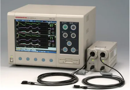
Figure 1.2. Near infrared spectroscopy
(NIRO-200; Hamamatsu Photonics, Shizuoka, Japan)
The figure shows an example of near infrared spectroscopy (NIRS). The basic NIRS has one set of optodes (emission and detection). By increasing the number of optodes, NIRS can measure the values of different body regions simultaneously.
The optode is attached to human body with double-stick tape. NIRO-200 is dual optods NIRS. The picture is NIRO-200 which is manufactured by Hamamatsu photonics.
Figure 1.3. Absorption spectra of oxyhemoglobin and deoxyhemoglobin This figure was drawn using the data which were taken from on the Oregon medical Laser Center website:
(http://omlc.ogi.edu/spectra/hemoglobin/summary.html).
The figure shows the absorption spectra of oxyhemoglobin (OxyHb) and deoxyhemoglobin (HHb) in the near infrared region of the spectrum. The wavelength of visible light is from about 390 to 700 nm. Light with wavelengths shorter than visible light (but longer than X-ray) is ultraviolet. Infrared has a longer wavelength than visible light. OxyHb and HHb have different absorption spectra that cross at about 800 nm. This wavelength region falls within near infrared portion of the spectrum. Therefore, we can use the near infrared to measure the oxygenation level of human body because it can detect the differences between OxyHb and HHb.
1.4 Aims of This Dissertation
Previous studies have revealed that physical counter-maneuvers can help to maintain blood pressure by increasing venous return. Physical counter-maneuvers are easy approaches for various patients. However, these maneuvers do not seem to be effective in preventing the hypotension that occurs in the early phase after postural change. There are some patients who experienced symptoms immediately after postural change. Nurses sometimes face and have to treat these situations. Ankle plantar flexion and dorsiflexion movements have been proven to be an effective intervention to increase venous velocity. However, the question has remained unanswered as to whether this simple exercise can attenuate the impact of postural change on cerebral hemodynamics. If ankle exercise is confirmed as having an effect similar to physical counter-maneuvers, this exercise may be able to contribute to nursing. The main purpose of this dissertation is to confirm the efficacy of ankle exercise for cerebral oxygenation.
1.5 Overview of This Dissertation
The dissertation consists of four chapters. This chapter is an overview of the current status of knowledge regarding physiological responses to postural changes, nursing interventions or treatments, and methodological backgrounds.
Following this introductory chapter, the relationship between ankle exercises and cerebral oxygenation in the supine position is studied in Chapter 2. Both active and passive ankle exercises increased cerebral blood oxygenation in the supine position.
In Chapter 3, the evaluation of the efficacy of ankle exercises is focused on the cerebral oxygenation changes induced by postural changes. Active ankle exercise resulted in significantly higher cerebral OxyHb than passive ankle exercise and the control condition during and after postural change.
In the final chapter, Chapter4, the findings of Chapter 2 and 3 are discussed together.
The strengths and limitations of this dissertation are mentioned. The contributions of this dissertation to nursing and suggestions for the direction of further research are described in Chapter 4.
1.6 References
Aaslid, R., Markwalder, T. M., & Nornes, H. (1982). Noninvasive transcranial Doppler ultrasound recording of flow velocity in basal cerebral arteries. Journal of Neurosurgery, 57(6), 769–74.
Allan, L. M., Ballard, C. G., Rowan, E. N., & Kenny, R. A. (2009). Incidence and prediction of falls in dementia: a prospective study in older people. PLoS One, 4(5), e5521.
Allcock, L. M., Ullyart, K., Kenny, R. A., & Burn, D. J. (2004). Frequency of orthostatic hypotension in a community based cohort of patients with Parkinson’s disease. Journal of Neurology, 75(10), 1470–1.
Allen, C., Glasziou, P., & Del Mar, C. (1999). Bed rest: a potentially harmful treatment needing more careful evaluation. Lancet, 354(9186), 1229–33.
Bucher, H. U., Edwards, A. D., Lipp, A. E., & Duc, G. (1993). Comparison between near infrared spectroscopy and 133Xenon clearance for estimation of cerebral blood flow in critically ill preterm infants. Pediatric Research, 33(1), 56–60.
Bundgaard-Nielsen, M., Jørgensen, C. C., Jørgensen, T. B., Ruhnau, B., Secher, N. H., &
Kehlet, H. (2009). Orthostatic intolerance and the cardiovascular response to early postoperative mobilization. British Journal of Anaesthesia, 102(6), 756–62.
Buzung, T. M. (2011). Computed tomography. In R. Kramme, K. P. Hoffmann, R. S. Pozos (Eds.), Handbook of medical technology (pp. 311–342). Boston, MA: Springer.
Clavet, H., Hébert, P. C., Fergusson, D., Doucette, S., & Trudel, G. (2008). Joint contracture following prolonged stay in the intensive care unit. Canadian Medical Association Journal, 178(6), 691–7.
Convertino, V., Hung, J., Goldwater, D., & DeBusk, R. F. (1982). Cardiovascular responses to exercise in middle-aged men after 10 days of bedrest. Circulation, 65(1), 134–140.
Cook, D., Attia, J., Weaver, B., McDonald, E., Meade, M., & Crowther, M. (2000). Venous thromboembolic disease: an observational study in medical-surgical intensive care unit patients. Journal of Critical Care, 15(4), 127–32.
Costanzo L.S. (2014). Physiology (5th ed.). Philadelphia, PA: Saunders Elsevier.
Creditor, M. C. (1993). Hazards of hospitalization of the elderly. Annals of Internal Medicine, 118(3), 219–23.
Dhar, R., & Diringer, M. C. (2014). Cerebral blood flow. In S. Schwab, D. Hanley, A. D.
Mendelow (Eds.), Critical care of the stroke patient (pp. 20–36). Cambridge: Cambridge University Press.
Dieler, A. C., Tupak, S. V., & Fallgatter, A. J. (2012). Functional near-infrared spectroscopy for the assessment of speech related tasks. Brain and Language, 121(2), 90–109.
Drakulovic, M. B., Torres, A., Bauer, T. T., Nicolas, J. M., Nogué, S., & Ferrer, M. (1999).
Supine body position as a risk factor for nosocomial pneumonia in mechanically ventilated patients: a randomized trial. Lancet, 354(9193), 1851–8.
Edlow, B. L., Kim, M. N., Durduran, T., Zhou, C., Putt, M. E., Yodh, A. G., et al. (2010). The effects of healthy aging on cerebral hemodynamic responses to posture change.
Physiological Measurement, 31(4), 477–95.
El-Sayed, H., & Hainsworth, R. (1996). Salt supplement increases plasma volume and orthostatic tolerance in patients with unexplained syncope. Heart, 75(2), 134–40.
Evans, D. (1994). The use of position during critical illness: current practice and review of the literature. Australian Critical Care , 7(3), 16–21.
Feldstein, C., & Weder, A. B. (2012). Orthostatic hypotension: a common, serious and underrecognized problem in hospitalized patients. Hypertension , 6(1), 27–39.
Fortney, S. M., Hyatt, K. H., Davis, J. E., & Vogel, J. M. (1991). Changes in body fluid compartments during a 28-day bed rest. Aviation, Space, and Environmental Medicine, 62(2), 97–104.
Gangavati, A., Hajjar, I., Quach, L., Jones, R. N., Kiely, D. K., Gagnon, P., et al. (2011).
Hypertension, orthostatic hypotension, and the risk of falls in a community-dwelling elderly population: the maintenance of balance, independent living, intellect, and zest in the elderly of Boston study. Journal of the American Geriatrics Society, 59(3), 383–9.
Garzon-Serrano, J., Ryan, C., Waak, K., Hirschberg, R., Tully, S., Bittner, E. A., et al. (2011).
Early mobilization in critically ill patients: patients’ mobilization level depends on health care provider's profession. PM & R , 3(4), 307–13.
Gibbons, C. H., & Freeman, R. (2006). Delayed orthostatic hypotension: a frequent cause of orthostatic intolerance. Neurology, 67(1), 28–32.
Gorelik, O., Fishlev, G., Litvinov, V., Almoznino-Sarafian, D., Alon, I., Shteinshnaider, M., et al. (2005). First morning standing up may be risky in acutely ill older inpatients. Blood Pressure, 14(3), 139–43.
Greenleaf, J., & Kozlowski, S. (1982). Physiological consequences of reduced physical activity during bed rest. Exercise and Sport Sciences Reviews, 10(1), 84–119.
Haines, K. J., Skinner, E. H., & Berney, S. (2013). Association of postoperative pulmonary complications with delayed mobilisation following major abdominal surgery: an observational cohort study. Physiotherapy, 99(2), 119–25.
Hajjar, I. (2005). Postural blood pressure changes and orthostatic hypotension in the elderly patient. Drugs & Aging, 22(1), 55–68.
Hargens, A. R., Millard, R. W., Pettersson, K., & Johansen, K. (1987). Gravitational haemodynamics and oedema prevention in the giraffe. Nature, 329(6134), 59–60.
Hiitola, P., Enlund, H., Kettunen, R., Sulkava, R., & Hartikainen, S. (2009). Postural changes in blood pressure and the prevalence of orthostatic hypotension among home-dwelling elderly aged 75 years or older. Journal of Human Hypertension, 23(1), 33–9.
Hock, C., Villringer, K., Müller-Spahn, F., Wenzel, R., Heekeren, H., Schuh-Hofer, S., et al., (1997). Decrease in parietal cerebral hemoglobin oxygenation during performance of a verbal fluency task in patients with Alzheimer’s disease monitored by means of near-infrared spectroscopy (NIRS) — correlation with simultaneous rCBF-PET measurements. Brain Research, 755(2), 293–303.
Hoshi, Y., & Tamura, M. (1993). Detection of dynamic changes in cerebral oxygenation coupled to neuronal function during mental work in man. Neuroscience Letters, 150(1), 5–
8.
Hunt, K., Tachtsidis, I., Bleasdale-Barr, K., Elwell, C., Mathias, C., & Smith, M. (2006).
Changes in cerebral oxygenation and haemodynamics during postural blood pressure changes in patients with autonomic failure. Physiological Measurement, 27(9), 777–85.
Inoue, H., Abe, N., Otsuji, Y., Kobayashi, Y., Sumitomo, N., Takase, B., et al. (2012).
Guidelines for diagnosis and management of syncope (in Japanese). 1–63.
http://www.j-circ.or.jp/guideline/pdf/JCS2012_inoue_h.pdf
Jans, Ø., Bundgaard-Nielsen, M., Solgaard, S., Johansson, P. I., & Kehlet, H. (2012).
Orthostatic intolerance during early mobilization after fast-track hip arthroplasty. British Journal of Anaesthesia, 108(3), 436–43.
Jöbsis, F. (1977). Noninvasive, infrared monitoring of cerebral and myocardial oxygen sufficiency and circulatory parameters. Science, 198(4323), 1264–1267.
Jones, A. Y. M., & Dean, E. (2004). Body position change and its effect on hemodynamic and metabolic status. Heart & Lung, 33(5), 281–90.
Jordan, J., Shannon, J. R., Black, B. K., Ali, Y., Farley, M., Costa, F., et al. (2000). The pressor response to water drinking in humans : a sympathetic reflex? Circulation, 101(5), 504–9.
Jordan, J., Shannon, J. R., Grogan, E., Biaggioni, I., & Robertson, D. (1999). A potent pressor response elicited by drinking water. Lancet, 353(9154), 723.
Kamaruzzaman, S., Watt, H., Carson, C., & Ebrahim, S. (2010). The association between orthostatic hypotension and medication use in the British Women’s Heart and Health Study.
Age and Ageing, 39(1), 51–6.
Kim, Y. S., Bogert, L. W. J., Immink, R. V, Harms, M. P. M., Colier, W. N. J. M., & van Lieshout, J. J. (2011). Effects of aging on the cerebrovascular orthostatic response.
Neurobiology of Aging, 32(2), 344–53.
Kim, Y. T., Tanaka, H., Takaya, R., Kajiura, M., Tamai, H., & Arita, M. (2009). Quantitative study on cerebral blood volume determined by a near-infrared spectroscopy during postural change in children. Acta Paediatrica, 98(3), 466–71.
Kobayashi, K., & Yamada, S. (2012). Development of a simple index, calf mass index, for screening for orthostatic hypotension in community-dwelling elderly. Archives of Gerontology and Geriatrics, 54(2), 293–7.
Krakow, K., Ries, S., Daffertshofer, M., & Hennerici, M. (2000). Simultaneous assessment of brain tissue oxygenation and cerebral perfusion during orthostatic stress. European
Neurology, 43(1), 39–46.
Kwon, O. Y., Jung, D. Y., Kim, Y., Cho, S. H., & Yi, C. H. (2003). Effects of ankle exercise combined with deep breathing on blood flow velocity in the femoral vein. The Australian Journal of Physiotherapy, 49(4), 253–8.
Lahmann, N. A., Kottner, J., Dassen, T., & Tannen, A. (2012). Higher pressure ulcer risk on intensive care? — comparison between general wards and intensive care units. Journal of Clinical Nursing, 21(3–4), 354–61.
Levick, J. R. (2010). An introduction to cardiovascular physiology (5th ed.). London: CRC Press.
Lipsitz, L. A. (1989). Orthostatic hypotension in the elderly. The New England Journal of Medicine, 321(14), 952–7.
Lovejoy, C. O. (1988). Evolution of human walking. Scientific American, 259(5), 118–25.
Luukinen, H., Koski, K., Laippala, P., & Kivelä, S. L. (1999). Prognosis of diastolic and systolic orthostatic hypotension in older persons. Archives of Internal Medicine, 159(3), 273–80.
Madsen, P., Pott, F., Olsen, S. B., Nielsen, H. B., Burcev, I., & Secher, N. H. (1998).
Near-infrared spectrophotometry determined brain oxygenation during fainting. Acta Physiologica Scandinavica, 162(4), 501–7.
Masaki, K. H., Schatz, I. J., Burchfiel, C. M., Sharp, D. S., Chiu, D., Foley, D., et al. (1998).
Orthostatic hypotension predicts mortality in elderly men: the Honolulu Heart Program.
Circulation, 98(21), 2290–5.
Matsubayashi, K., Okumiya, K., Wada, T., Osaki, Y., Fujisawa, M., Doi, Y., et al. (1997).
Postural dysregulation in systolic blood pressure is associated with worsened scoring on neurobehavioral function tests and leukoaraiosis in the older elderly living in a community.
Stroke, 28(11), 2169–73.
Mehagnoul-Schipper, D. J., Colier, W. N. J. M., & Jansen, R. W. M. M. (2001).
Reproducibility of orthostatic changes in cerebral oxygenation in healthy subjects aged 70 years or older. Clinical Physiology, 21(1), 77–84.
Mehagnoul-Schipper, D. J., Vloet, L. C. M., Colier, W. N. J. M., Hoefnagels, W. H. L., &
Jansen, R. W. M. M. (2000). Cerebral oxygenation declines in healthy elderly subjects in response to assuming the upright position. Stroke, 31(7), 1615–1620.
Mehrabian, S., Duron, E., Labouree, F., Rollot, F., Bune, A., Traykov, L., et al. (2010).
Relationship between orthostatic hypotension and cognitive impairment in the elderly.
Journal of the Neurological Sciences, 299(1–2), 45–8.
Moya, A., Sutton, R., Ammirati, F., Blanc, J. J., Brignole, M., Dahm, J. B., et al. (2009).
Guidelines for the diagnosis and management of syncope (version 2009). European Heart Journal, 30(21), 2631–71.
Munro, M. J., Walker, A. M., & Barfield, C. P. (2004). Hypotensive extremely low birth
weight infants have reduced cerebral blood flow. Pediatrics, 114(6), 1591–6.
Nagaya, S., Fujimoto, F., & Kobayashi, H. (2014). Assessment of blood pressure for determining the time to perform first postural change in patients after cardiac surgery in the intensive care unit. Journal of Nursing & Care, 3, 177.
Olson, E. (1967/1990). The hazards of immobility. The American Journal of Nursing. 90(3), 43–49.
Ooi, W. L., Hossain, M., & Lipsitz, L. A. (2000). The association between orthostatic hypotension and recurrent falls in nursing home residents. The American Journal of Medicine, 108(2), 106–111.
Perme, C., & Chandrashekar, R. (2009). Early mobility and walking program for patients in intensive care units: creating a standard of care. American Journal of Critical Care , 18(3), 212–21.
Pocock, G., & Richards, C. D. (2006). Human physiology: The basis of medicine (3rd ed.).
Oxford, NY: Oxford University Press.
Podoleanu, C., & Maggi, R. (2006). Lower limb and abdominal compression bandages prevent progressive orthostatic hypotension in elderly persons: a randomized single-blind controlled study. Journal of the American College of Cardiology, 48(7),1425–32.
Poon, I. O., & Braun, U. (2005). High prevalence of orthostatic hypotension and its correlation with potentially causative medications among elderly veterans. Journal of Clinical Pharmacy and Therapeutics, 30(2), 173–8.
Rowell, L. B. (1993). Human cardiovascular control. Oxford, NY: Oxford University Press.
Rutan, G. H., Hermanson, B., Bild, D. E., Kittner, S. J., LaBaw, F., & Tell, G. S. (1992).
Orthostatic hypotension in older adults. The Cardiovascular Health Study. CHS Collaborative Research Group. Hypertension, 19(6 Pt 1), 508–19.
Senard, J. M., Raï, S., Lapeyre-Mestre, M., Brefel, C., Rascol, O., Rascol, A., et al. (1997).
Prevalence of orthostatic hypotension in Parkinson’s disease. Journal of Neurology, 63(5), 584–9.
Shannon, J. R., Diedrich, A., Biaggioni, I., Tank, J., Robertson, R. M., Robertson, D., &
Jordan, J. (2002). Water drinking as a treatment for orthostatic syndromes. The American Journal of Medicine, 112(5), 355–60.
Shibao, C., Grijalva, C. G., Raj, S. R., Biaggioni, I., & Griffin, M. R. (2007). Orthostatic hypotension-related hospitalizations in the United States. The American Journal of Medicine, 120(11), 975–80.
Smith, J. J., & Kampine, J. P. (1990). Circulatory physiology: The essentials (3rd ed.).
Baltimore, MD: Williams & Wilkins.
Sochart, D. H., & Hardinge, K. (1999). The relationship of foot and ankle movements to venous return in the lower limb. The Journal of Bone and Joint Surgery, 81(4), 700–4.
Stein, P. D., Yaekoub, A. Y., Ahsan, S. T., Matta, F., Lala, M. M., Mirza, B., et al. (2009).
Ankle exercise and venous blood velocity. Thrombosis and Haemostasis, 101(6), 1100–3.
Stiller, K., & Phillips, A. (2003). Safety aspects of mobilising acutely ill inpatients.
Physiotherapy Theory and Practice, 19(4), 239–57.
Streeten, D. H., & Anderson, G. H. (1992). Delayed orthostatic intolerance. Archives of Internal Medicine, 152(5), 1066–72.
Talpahewa, S. P., Ascione, R., Angelini, G. D., & Lovell, A. T. (2003). Cerebral cortical oxygenation changes during OPCAB surgery. The Annals of Thoracic Surgery, 76(5), 1516–22.
Tanaka, H., Matsushima, R., Tamai, H., & Kajimoto, Y. (2002). Impaired postural cerebral hemodynamics in young patients with chronic fatigue with and without orthostatic intolerance. The Journal of Pediatrics, 140(4), 412–7.
Tanida, M., Sakatani, K., Takano, R., & Tagai, K. (2004). Relation between asymmetry of prefrontal cortex activities and the autonomic nervous system during a mental arithmetic task: near infrared spectroscopy study. Neuroscience Letters, 369(1), 69–74.
Ten Harkel, A. D., van Lieshout, J. J., & Wieling, W. (1994). Effects of leg muscle pumping and tensing on orthostatic arterial pressure: a study in normal subjects and patients with autonomic failure. Clinical Science, 87(5), 553–8.
The Consensus Committee of the American Autonomic Society and the American Academy of Neurology. (1996). Consensus statement on the definition of orthostatic hypotension, pure autonomic failure, and multiple system atrophy. Journal of the Neurological Sciences, 144(1–2), 218–9.
Timmerman, R. A. (2007). A mobility protocol for critically ill adults. Dimensions of Critical Care Nursing , 26(5), 175–9.
Truijen, J., Kim, Y. S., Krediet, C. T. P., Stok, W. J., Kölgen, R. S., Colier, W. N., et al. (2012).
Orthostatic leg blood volume changes assessed by near-infrared spectroscopy.
Experimental Physiology, 97(3), 353–61.
Tsunetsugu, Y., Miyazaki, Y., & Sato, H. (2005). Visual effects of interior design in actual-size living rooms on physiological responses. Building and Environment, 40(10), 1341–1346.
Tsunetsugu, Y., Miyazaki, Y., & Sato, H. (2006). Physiological effects in humans induced by the visual stimulation of room interiors with different wood quantities. Journal of Wood Science, 53(1), 11–16.
Van Hateren, K. J. J., Kleefstra, N., Blanker, M. H., Ubink-Veltmaat, L. J., Groenier, K. H., Houweling, S. T., et al. (2012). Orthostatic hypotension, diabetes, and falling in older patients: a cross-sectional study. The British Journal of General Practice : The Journal of the Royal College of General Practitioners, 62(603), e696–702.
Van Lieshout, J. J., ten Harkel, A. D., & Wieling, W. (1992). Physical manoeuvres for combating orthostatic dizziness in autonomic failure. Lancet, 339(8798), 897–8.
Velseboer, D. C., de Haan, R. J., Wieling, W., Goldstein, D. S., & de Bie, R. M. A. (2011).
Prevalence of orthostatic hypotension in Parkinson’s disease: a systematic review and meta-analysis. Parkinsonism & Related Disorders, 17(10), 724–9.
Vloet, L. C. M., Pel-Little, R. E., Jansen, P. A. F., & Jansen, R. W. M. M. (2005). High prevalence of postprandial and orthostatic hypotension among geriatric patients admitted to Dutch hospitals. The Journals of Gerontology. Series A, 60(10), 1271–7.
Walter, H., Vetter, S. C., Grothe, J., Wunderlich, A. P., Hahn, S., & Spitzer, M. (2001). The neural correlates of driving. Neuroreport, 12(8), 1763–7.
Weiss, A., Grossman, E., Beloosesky, Y., & Grinblat, J. (2002). Orthostatic hypotension in acute geriatric ward: is it a consistent finding? Archives of Internal Medicine, 162(20), 2369–74.
White, P. M., Wardlaw, J. M., Teasdale, E., Sloss, S., Cannon, J., & Easton, V. (2001). Power transcranial Doppler ultrasound in the detection of intracranial aneurysms. Stroke, 32(6), 1291–7.
Wieling, W., Krediet, C. T. P., van Dijk, N., Linzer, M., & Tschakovsky, M. E. (2007). Initial orthostatic hypotension: review of a forgotten condition. Clinical Science, 112(3), 157–65.
Wintermark, M., Sesay, M., Barbier, E., Borbély, K., Dillon, W. P., Eastwood, J. D., et al.
(2005). Comparative overview of brain perfusion imaging techniques. Stroke, 36(9), e83–
99.
Wu, J. S., Yang, Y. C., Lu, F. H., Wu, C. H., & Chang, C. J. (2008). Population-based study on the prevalence and correlates of orthostatic hypotension/hypertension and orthostatic dizziness. Hypertension Research , 31(5), 897–904.
Wyatt, J. S. (1993). Near-infrared spectroscopy in asphyxial brain injury. Clinics in Perinatology, 20(2), 369–78.
Yamashita, Y., & Niwayama, M., (2013). Principles and instrumentation. In T. Jue & K.
Masuda (Ed.), Application of Near Infrared Spectroscopy in Biomedicine (pp. 1–19).
Boston, MA: Springer.
Zweifel, C., Castellani, G., Czosnyka, M., Carrera, E., Brady, K. M., Kirkpatrick, P. J., et al.
(2010). Continuous assessment of cerebral autoregulation with near-infrared spectroscopy in adults after subarachnoid hemorrhage. Stroke, 41(9), 1963–8.