Fukushima Medical University
福島県立医科大学 学術機関リポジトリ
This document is downloaded at: 2021-11-08T00:33:54Z
Title The BCR/ABL tyrosine kinase inhibitor, nilotinib, stimulates expression of IL-1β in vascular endothelium in association with downregulation of miR-3121-3p( 本文 )
Author(s) 助川, 真純
Citation
Issue Date 2017-09-27
URL http://ir.fmu.ac.jp/dspace/handle/123456789/728
Rights © 2017. This accepted manuscript version is made available under the CC-BY-NC-ND 4.0 license. Published version: Leuk Res. 2017 Jul;58:83-90. doi: 10.1016/j.leukres.2017.05.005 DOI
Text Version ETD
1
The BCR/ABL tyrosine kinase inhibitor, nilotinib, stimulates expression of IL-1βin vascular endothelium in association with downregulation of miR--3p
Masumi Sukegawa1, Xiangmin Wang1, 2, Chie Nishioka3, Bin Pan1, 2,Kailin Xu2, Hiroshi Ohkawara1,Yoichi Hamasaki4, Masayuki Mita5, Kenichi Nakamura5, Masatoshi Okamoto6, Hiromi Shimura7, Masatsugu Ohta8, Takayuki Ikezoe1
1Department of Hematology, Fukushima Medical University, Hikarigaoka-1, Fukushima 960-1295, Japan.2Department of Hematology, The Affiliated Hospital of Xuzhou Medical University, No. 99, West Huaihai Road, 221002 Xuzhou, China.3Department of Hematology and Respiratory Medicine, Kochi University, Oko-cho, Nankoku, Kochi 783-8505, Japan.4Department of Hematology, Iwaki Kyoritsu General Hospital, Iwaki, Fukushima 973-8402 Japan. 5Department of Hematology, Shirakawa Kosei General Hospital, Shirakawa, 961-0005 Fukushima, Japan. 6Department of Hematology, YUASA Foundation Jusendo General Hospital, Koriyama, Fukushima 963-8585, Japan.
7Department of Laboratory Medicine, Fukushima Medical University, Hikarigaoka-1, Fukushima 960-1295, Japan. 8Department of Hematology, Fukushima Medical University Aizu Medical Center, Aizuwakamatsu, 969-3492 Fukushima, Japan.
Correspondence should be addressed to Takayuki Ikezoe ([email protected]).
Key words: ABL tyrosine kinase inhibitor, endothelial cell damage, microRNA, inflammatory cytokines
*Manuscript
Click here to download Manuscript: EC TKIs R28936170429(23145) (32022).doc Click here to view linked References
2
Abstract
BCR/ABL tyrosine kinase inhibitors (TKIs) have significantly improved the prognosis for in dividuals with chronic myeloid leukemia (CML). However, many patients treated with TKIs suffer from TKI-related complications. In particular, vascular events such as peripheral artery occlusive disease have become aserious clinical problem for patients who receive the TKI, nilotinib. At present, the molecular mechanisms by which TKIs cause vascular endothelial cell insults remain unknown.This study explored the effects of the TKIs, imatinib, nilotinib and dasatinib, on vascular endothelial cells in vitro, and found that only nilotinib induced expression of interleukin-1β (IL-1β) by vascular endothelial cells. Nilotinib-induced IL-1β expression stimulated the adhesion of monocytes to vascular endothelial cells in association with an increase in levels of adhesion molecules. MicroRNA database searching identified miR-3121-3p binding sites in the 3’-UTR of the IL-1β gene.
Exposure of endothelial cells to nilotinib caused downregulation of miR-3121-3p in these cells. Importantly, forced-expression of miR-3121-3p counteracted nilotinib-induced expression of IL-1β. Importantly, serum levels if IL-1β were significantly elevated in CML patients receiving nilotinib (n=14) compared to those receiving other TKIs (n=16) (3.76±1.22 pg/ml vs 0.27±0.77 pg/ml, p<0.05). Taken together, our data suggest that nilotinib decreases levels of miR-3121-3p resulting in an
3
increase in expression of IL-1β and adhesion molecules in vascular endothelial cells.
The miR-3121-3p/IL-1β axis could be a potential target to prevent vascular events in CML patients with high risk of vascular events.
4
1. Introduction
Imatinib, a BCR/ABL tyrosine kinase inhibitor (TKI), has revolutionized the treatment of individuals with chronic myeloid leukemia (CML).1 The second generation TKIs, nilotinib and dasatinib, inhibit BCR/ABL tyrosine kinase activity more potently than imatinib. Clinical trials comparing the efficacy of either nilotinib or dasatinib with that of imatinib in newly diagnosed CML patients found that the use of second generation TKIs produced a significantly stronger molecular response than imatinib.2,3 However, unexpectedly, these and other clinical trials found that the use of nilotinib was associated with a higher incidence of cardiovascular adverse events, including myocardial infarction and peripheral arterial occlusive disease (PAOD), compared with imatinib and dasatinib.2-6 In addition, recently approved TKI ponatinib more frequently causes cardiovascular adverse events in CML patients.7 These cardiovascular events preferentially occurred in nilotinib-treated patients with base line cardiovascular risk factors, although the precise mechanisms by which nilotinib causes cardiovascular events remain unknown.
Pro-inflammatory cytokines such as tumor necrosis factor (TNF) and interleukin-1(IL-1) cause vascular endothelial cell damage in association with an increase in levels of adhesion molecules that recruit leukocytes and platelets to vascular
5
endothelium.8,9 The injured vascular endothelial cells lose cell surface expression of thrombomodulin (TM), resulting in disruption of the anticoagulant TM/activated protein C system, and thrombus formation.10,11
Mature microRNAs (miRNAs) are non-protein-coding small RNAs comprising 19 to 25 nucleotides. MiRNAs suppress gene expression by binding to the 3’-untranslated region (3’-UTR) of target mRNAs resulting in target degradation or transcriptional repression.12,13 Dysregulated miRNA expression occurs in various types of inflammatory diseases, cancers and cardiovascular diseases.14
This study examined the effect of BCR/ABL TKIs on cytokine production, and miRNA and adhesion molecule expression in vascular endothelial cells to verify the mechanisms by which this class of agents causes vascular insult.
6
2. Materials and Methods
2.1. Cells. Human umbilical vein derived EA.hy926 cells and human umbilical vein endothelial cells (HUVECs) were purchased from Lonza Walkersville Inc. (Walkersville, MD, USA). EA.hy926 cells were cultured with DMEM culture medium supplemented with 10% FBS. HUVECs were cultured in EGM-2 medium (Lonza). Human acute monocytic leukemia THP-1 cells were obtained from ATCC (Manassas, VA, USA) and cultured with RPMI 1640 supplemented with 10% FCS.
2.2. Drugs. Imatinib, nilotinib, ponatinib, and dasatinib were obtained from Sigma (St.
Louis, MO., USA). These agents were diluted (10-2M) with DMSO and stored at -80℃ until use. RPMI 1640 was used to dilute TKIs for the in vitro study. The final concentration of DMSO in culture media was less than 0.001%, which did not affect the expression of cytokines in EA.hy926 cells (Supplementary Figure 1).
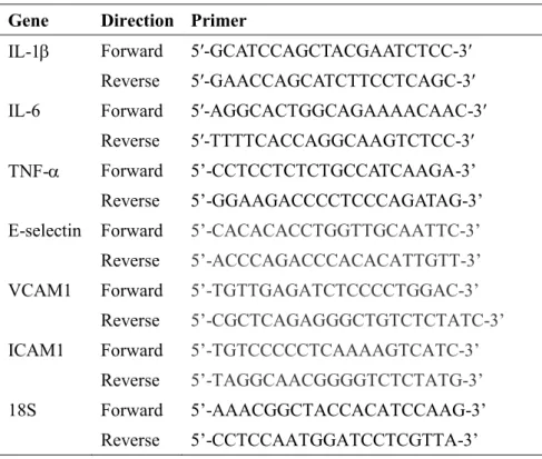
2.3. RNA isolation and quantitative real-time RT-PCR (qRT-PCR). RNA isolation and cDNA preparation were performed as described previously,15 and we measured 18SrRNA expression for normalization.15qRT-PCR was performed with Power SYBR Green PCRMaster Mix (Applied Biosystems, Carlsbad, CA, USA). Primers for PCR are shown in Table 1. PCR conditions for all genes were as follows: initial activation at 95°C for 10 min, followed by 45 cycles of 95°C for 15 s and 60°C for 1min, and fluorescence determination at the melting temperature of the product for 20s on an ABI
7
PRISM 7300 (Applied Biosystems).
2.4. Enzyme-linked immunosorbent assay (ELISA). Culture media of EA.hy926 cells was collected and assayed using an ELISA kit to measure the levels of IL-1 according to the manufacturer’s protocol (R&D systems, Minneapolis, MN, USA).
2.5. Western blot analysis. Immunoblot analysis was performed as described previously.16 Anti-ICAM1 (CST, 4915), anti-VCM1 (Abcam, ab134047, Cambridge, UK), anti-GAPDH (CST, 5174) and anti-E-selectin (Proteintech, 20894-1-AP,
Rosemont, IL, USA) antibodies were used.
2.6. Immunofluorescent staining. EA.hy926 cells were cultured in the presence of TKIs and fixed on glass slides by Autosmear CF120 (Sakura Fine-technical Co. Ltd, Tokyo, Japan). The fixed cells were incubated with primary antibodies against IL-1(Abcam, ab8320, 1:100) and IL-1R (Abcam, ab106278, 1:100) followed by fluorophore conjugated secondary antibodies, goat anti-rabbit IgG Alexa Fluor 488 (ThermoFisher, A11070, 1:200, Waltham, MA USA) and goat anti-mouse IgG Alexa Fluor 647(Abcam, ab150115, 1:200), respectively. The nuclei were stained with 4',6-Diamidino-2-phenylindole dihydrochloride (Roche,Basel, Switzerland).
2.7. Flowcytometry analysis. EA.hy926 cells were cultured in the presence of nilotinib.
After 5 days, cells were harvested and stained with phycoerythrin (PE)-conjugated
8
anti-CD54, anti-CD106 and anti-CD62E antibodies. All antibodies were purchased from BioLegend (San Diego, CA).
2.8. Adhesion assay. Adhesion of THP-1 acute monocytic leukemia cells to EA.hy926 cells was assessed by CBL CBA-210 CytoSelectTM 96-well Leukocyte-endothelium Adhesion Kit (Cell Biolabs Inc., San Diego, CA, USA). Briefly, EA.hy926 cells were cultured in a gelatin-coated 48-well culture plate for five days to allow them to form a monolayer. THP-1 cells were labeled by LeukoTracker™ (Cell BiolabsInc.) and co-cultured with the EA.hy926 monolayer for 90 min. The culture plate was washed three times with washing buffer. The adherent cells were lysed and subjected to
fluorescence measurement using a Fluoroskan Ascent FLplate reader (ThermoFisher).
2.9. miRNA target prediction. TargetScan (http://www.targetscan.org/) and miRBase (http://www.mirbase.org/) were used to predict the putative miRNAs that regulate expression of IL-1.
2.10. miRNA expression. Expression of miRNA was analyzed using a Mir-X miRNA qRT-PCR SYBR Kit (638314, Clontech Laboratories Inc., Mountain View, CA, USA) according to the supplier’s protocol. miRNA levels were normalized using U6 snRNA (638314, Clontech Laboratories Inc.) and relative quantities were determined by the ΔΔCt method. The miR-3121-3 primer was as follows: 5′-
9
TAAATAGAGTAGGCAAAGGACA -3′.
2.11. Overexpression of miR-3121 precursor. The precursor miR-3121-3p mimic (Sigma) was transfected into EA.hy926 cells, using INTERFER in transfection reagent according to the manufacturer’s instructions (Polyplus-transfection Inc., NY, USA). A non-targeting oligonucleotide mimic was used as a negative control.
2.12. Reporter assay. The human IL-1 3’-UTR was synthesized by PCR and cloned into the pGL4.10 [Luc2] vector (E6651, Promega, Madison, WI., USA). EA.hy926 cells were transfected with this reporter construct using Lipofectamine3000 (ThermoFisher).
The reporter assay was performed as described previously.17
2.13. Measurement of IL-1levels in vivo. Serum was withdrawn from leukemia patients receiving TKIs after obtaining written informed consent. Levels of IL-1 were measured using a human IL-1 ELISA kit (R&D systems). This study was approved by ethical committee of Fukushima Medical University.
2.14. Statistical analysis. Student’s t test was used when comparing results between two groups. Statistical analysis of difference between multiple groups was done by one-way ANOVA followed by Bonferroni multiple comparison tests. All statistical analyses were performed using PRISM statistical analysis software (GraphPad Software Inc., San Diego, CA, USA). P < 0.05 was considered statistically significant.
10
3. Results
3.1. Nilotinib increases levels of IL-1. We first examined the effects of BCR/ABL TKIs on cytokine production by vascular endothelial EA.hy926 cells. Exposure of these cells to nilotinib, but not imatinib and dasatinib, for five days significantly increased the levels of IL-1mRNA, as measured by qRT-PCR (Figure 1a). Nilotinib-mediated induction of IL-1 expression was dose-dependent up to 100 nM (Figure 1b). However, when cells were cultured in the presence of a higher dose of nilotinib (200 nM), the ability of nilotinib to induce expression of IL-1mRNAwas suppressed by nearly half compared with 100 nM nilotinib (Figure 1b). Likewise, nilotinib increased levels of IL-1 in HUVECs in a dose-dependent manner up to 100 nM (supplementary Figure 2a).
On the other hand, even the highest dose of imatinib and dasatinib (100 nM for seven days) did not increase levels of IL-1 (data not shown). Nilotinib also significantly increased levels of IL-6 in EA.hy926 cells; however induction was merely less than 2-fold of control diluent treated cells (Figure 1a). None of the TKIs significantly
stimulated the expression of TNF (Figure 1a). Moreover, neither tumor growth factor
, granulocyte-colony stimulating factor, granulocyte macrophage-colony stimulating factor, vessel endothelial growth factor, nor stem cell factor was induced in EA.hy926 cells after exposure to nilotinib (Supplementary Figure 3).The ELISA data indicated
11
that nilotinib, but not other TKIs, stimulated production of IL-1 protein in EA.hy926 cells, consistent with the results obtained by qRT-PCR (Figure 1c). Immunofluorescent staining showed that EA.hy926 cells express both IL-1 and the receptor for IL-1
(Figure 1d). Exposure of EA.hy926 cells to nilotinib, but not to other TKIs, increased the expression of IL-1and stimulated the formation of the IL-1/IL-1 receptorbinding complex (Figure 1d). These observations suggested that IL-1 mediates down-stream signaling in EA.hy926 cells by an autocrine mechanism in the presence of nilotinib.
3.2. Nilotinib increases levels of adhesion molecules in an IL-1 dependent manner.
We examined the effects of TKIs on the expression of IL-1-target genes such as adhesion molecules in EA.hy926 cells. Interestingly, only nilotinib significantly increased expression of ICAM1, VCAM1 and E-selectin mRNAs in EA.hy926 cells (Figure 2a).Consistent with these results, FACS found that cell surface expressed these adhesion molecules were increased in EA.hy926 cells after exposure to nilotinib (Figure 2b). Nilotinib-induced expression of adhesion molecules was also noted in HUVECs
(Supplementary Figure 2b). As expected, IL-1 receptor blocking antibody hampered the ability of nilotinib to induce expression of adhesion molecules at the protein level (Figure 2c).
3.3. Nilotinib enhances adhesion of hematopoietic cells to EA.hy926 cells. We
12
examined whether nilotinib-induced expression of adhesion molecules would play a role in adhesion of hematopoietic cells to vascular ECs. Adhesion assays utilizing monocytic leukemia THP-1 cells revealed that adherent THP-1 cells increased when EA.hy926 cells were cultured in the presence of nilotinib. Pre-incubation of EA.hy926 cells with an anti-IL-1 receptor antibody abrogated the ability of nilotinib to stimulate adhesion of THP-1 cells (Figure 2d). Neither imatinib nor dasatinib increased the number of THP-1 cells adherent to EA.hy926 cells.
3.4. Nilotinib decreases expression of miR-3121-3p. We next explored the molecular mechanisms by which nilotinib increases expression of IL-1 in EA.hy926 cells.
Database searches identified miR-3121-3p as a candidate miRNA that binds to the 3’-UTR of IL-1(Figure 3a). Nilotinib, but not imatinib and dasatinib, decreased levels of miR-3121-3p expression by approximately half in EA.hy926 cells (Figure 3b).
Likewise, nilotinib decreased levels of miR-3121-3p in HUVECs (Supplementary Figure 2c). Importantly, forced-expression of miR-3121-3p in EA.hy926 cells
counteracted nilotinib-mediated increases in levels of IL-1(Figure 3c).
3.5. miR-3121-3p binds to the 3’-UTR of IL-1. Further experiments utilizing the IL-1 3’-UTR luciferase reporter gene construct showed that forced expression of miR-3121-3p decreased transcriptional activity by approximately half (Figure 3d).
13
When miR-3121-3p binding sites in the 3’-UTR of the reporter gene construct were mutated, the presence of miR-3121-3p did not decrease transcriptional activity,
consistent with a direct interaction between miR-3121-3p and the IL-13’-UTR (Figure 3d).
3.6. Nilotinib increases serum levels of IL-1in vivo. We measured serum levels of IL-1 in CML patients who were treated with TKI. As shown in Figure 4, levels of IL-1 were significantly elevated in patients receiving nilotinib (n=14) compared to those who were treated with other TKI (imatinib, n=5;dasatinib, n=6; bosutinib, n=4;
ponatinib, n=1) (a mean 3.76±1.22 pg/ml vs a mean 0.27±0.77 pg/ml, p<0.05).
14
4. Discussion
The adhesion of inflammatory cells to vascular endothelium is a critical step in the development of atherosclerosis, including PAOD.20 Adhesion molecules such as ICAM-1, VCAM-1 and E-selectin are intimately involved in the pathogenesis of atherosclerosis; these adhesion molecules recruit monocytes and neutrophils to vascular ECs (ECs) and mediate plaque formation in blood vessels. The elevated serum levels of ICAM-1, VCAM-1 and E-selectin were associated with cardiovascular mortality in patients with stable carotid atherosclerosis.21The present study clearly demonstrated that exposing human vascular ECs to nilotinib, but not imatinib and dasatinib, increased the levels of a spectrum of adhesion molecules (Figure 1e). Exposing vascular ECs to nilotinib also significantly stimulated the production of IL-1 (Figure 1a-c). We found that exposing EA.hy926 cells to IL-1 (100 ng/ml, 24 hrs) significantly increased levels of ICAM-1, VCAM-1 and E-selectin (Supplementary Figure 4). In other studies, IL-1
induced expression of adhesion molecules in smooth muscle cells and glioblastoma cells,20, 22consistent with the possibility that nilotinib might induce the expression of adhesion molecules via IL-1in a variety of contexts. As expected, abrogation of IL-1
signaling by a IL-1 receptor blocking antibody attenuated the ability of nilotinib to induce expression of adhesion molecules (Figure 2a).
Of note, serum levels of IL-1 were detectable in all CML patients receiving nilotinib
15
(n=14). And their levels were significantly higher than those in patients who were treated with other TKI (Figure 4). These observations were consistent with in vitro results showing that exposure of ECs to nilotinib, but not to other TKIs, increased expression levels of IL-1.
Other researchers also investigated the effects of nilotinib in vascular endothelial cells. They demonstrated that nilotinib, but not imatinib induced expression of adhesion molecules on HUVECs. Furthermore, they found that nilotinib inhibited angiogenesis in vitro and in vivo.23,24
A recent clinical trial evaluating the efficacy and safety of ponatinib, a recently developed BCR/ABL TKI, also demonstrated an increase in the risk of cardiovascular events following treatment with this agent.25 We speculated that ponatinib also
stimulates expression of IL-1 and adhesion molecules in vascular ECs. However, contrary to our expectations, neither cytokines nor adhesion molecules were induced in EA.hy926 cells after exposure to ponatinib (10-50 nM for five days) (data not shown).
Ponatinib may havedistinct effectson vascular ECs, inflammatory cells and platelets. A recent in vitro study found that ponatinib decreased the viability and tube formation of human umbilical vein endothelial cells (HUVECs).26 Interestingly, forced expression of vascular endothelial growth factor receptor 2 (VEGFR2) in HUVECs attenuated these
16
negative effects mediated by ponatinib.22These observations suggested that ponatinib probably causes vascular insults via inhibition of VEGFR2.
We found that miR-3121-3p negatively regulated the expression of IL-1 mRNA (Figure 3). Interestingly, nilotinib decreased levels of miR-3121-3p in EA.hy926 cells, suggesting that nilotinib-mediated downregulation of miR-3121-3p contributed to upregulation of IL-1, leading to an augmented expression of adhesion molecules in vascular ECs (Figure 5). Importantly, forced-expression of miR-3121-3p in EA.hy926 cells effectively counteracted the elevation of IL-1 expression (Figure 3c).
Taken together, nilotinib has marked effects on vascular ECs, including the induction of IL-1 and adhesion molecule production in association with down-regulation of miR-3121-3p expression in vascular ECs. This may ultimately cause adhesion of inflammatory cells to vascular endothelium, resulting in plaque formation. These data suggest that therapeutic approaches aimed at boosting miR-3121-3p levels may have potential for the prevention of cardiovascular events in CML patients receiving nilotinib.
17
References
1. O'Brien SG, Guilhot F, Larson RA, Gathmann I, Baccarani M, Cervantes F, et al.
Imatinib compared with interferon and low-dose cytarabine for newly diagnosed chronic-phase chronic myeloid leukemia.N Engl J Med. 2003;348:994-1004.
2. Hochhaus A, Saglio G, Hughes TP, Larson RA, Kim DW, Issaragrisil S,et al.Long-term benefits and risks of frontline nilotinib vs imatinib for chronic myeloid leukemia in chronic phase: 5-year update of the randomized ENESTnd trial.Leukemia. 2016;30:1044-54.
3. Cortes JE, Saglio G, Kantarjian HM, Baccarani M, Mayer J, Boqué C, et al. Final 5-Year Study Results of DASISION: The Dasatinib Versus Imatinib Study in Treatment-Naïve Chronic Myeloid Leukemia Patients Trial.J Clin Oncol. 2016;34:
2333-40.
4. Gugliotta G, Castagnetti F, Breccia M, Levato L, D'Adda M, Stagno F, et al;
GIMEMA CML Working Party.Long-term outcome of a phase 2 trial with nilotinib 400 mg twice daily in first-line treatment of chronic myeloid leukemia.
Haematologica. 2015;100:1146-50.
5. Giles FJ, Mauro MJ, Hong F, Ortmann CE, McNeill C, Woodman RC, et al.Rates of peripheral arterial occlusive disease in patients with chronic myeloid leukemia in the chronic phase treated with imatinib, nilotinib, or non-tyrosine kinase therapy: a retrospective cohort analysis.Leukemia. 2013; 27:1310-5.
6. Aichberger KJ, Herndlhofer S, Schernthaner GH, Schillinger M,
Mitterbauer-Hohendanner G, Sillaber C, et al. Progressive peripheral arterial occlusive disease and other vascular events during nilotinib therapy in CML.Am J Hematol. 2011; 86:533-9.
18
7. Valent P, Hadzijusufovic E, Schernthaner GH, Wolf D, Rea D, le Coutre P.Vascular safety issues in CML patients treated with BCR/ABL1 kinase inhibitors.Blood.
2015;125:901-6.
8. Franscini N, Bachli EB, Blau N, Leikauf MS, Schaffner A, Schoedon G.Gene expression profiling of inflamed human endothelial cells and influence of activated protein C.Circulation. 2004; 110: 2903-9.
9. Seynhaeve AL, Rens JA, Schipper D, Eggermont AM, Ten Hagen TL.Exposing endothelial cells to tumor necrosis factor-α and peripheral blood mononuclear cells damage endothelial integrity via interleukin-1ß by degradation of vascular endothelial-cadherin.Surgery. 2014;155: 545-53.
10. Ikezoe T. Pathogenesis of disseminated intravascular coagulation in patients with acute promyelocytic leukemia, and its treatment using recombinant human soluble thrombomodulin.Int J Hematol. 2014;100: 27-37.
11. Ikezoe T. Thrombomodulin/activated protein C system in septic disseminated intravascular coagulation.J Intensive Care. 2015; 3:1.
12. Krol J, Loedige I, Filipowicz W. The widespread regulation of microRNA biogenesis, function and decay. Nat Rev Genet. 2010;11:597–610.
13. Schober A, Nazari-Jahantigh M, Weber C.MicroRNA-mediated mechanisms of the cellular stress response in atherosclerosis.Nat Rev Cardiol. 2015;12:361-74.
14. Esteller M.Non-coding RNAs in human disease.Nat Rev Genet. 2011;12:861-74.
15. Ikezoe T, Tanosaki S, Krug U, Liu B, Cohen P, Taguchi H, et al. Insulin-like growth factor binding protein-3 antagonizes the effects of retinoids in myeloid leukemia cells. Blood. 2004;104:237-42.
16. Ikezoe T, Yang Y, Heber D, Taguchi H, Koeffler HP. PC-SPES: potent inhibitor of
19
nuclear factor-kappa B rescues mice from lipopolysaccharide-induced septic shock.
Mol Pharmacol. 2003; 64:1521-9.
17. Takeuchi A, Nishioka C, Ikezoe T, Yang J, Yokoyama A.STAT5A regulates DNMT3A in CD34(+)/CD38(-) AML cells.Leuk Res. 2015;39:897-905.
18. Weisberg E, Manley PW, Breitenstein W, Brüggen J, Cowan-Jacob SW, Ray A, et al.
Characterization of AMN107, a selective inhibitor of native and mutant Bcr-Abl.Cancer Cell. 2005;7:129-41.
19. Coluccia AM, Cirulli T, Neri P, Mangieri D, Colanardi MC, Gnoni A, et al.
Validation of PDGFRbeta and c-Src tyrosine kinases as tumor/vessel targets in patients with multiple myeloma: preclinical efficacy of the novel, orally available inhibitor dasatinib.Blood. 2008;112:1346-56.
20. Wang X, Feuerstein GZ, Gu JL, Lysko PG, Yue TL.Interleukin-1 beta induces expression of adhesion molecules in human vascular smooth muscle cells and enhances adhesion of leukocytes to smooth muscle cells.Atherosclerosis.
1995;115:89-98.
21. Hoke M, Winter MP, Wagner O, Exner M, Schillinger M, Arnold Z, et al.The impact of selectins on mortality in stable carotid atherosclerosis.Thromb Haemost.
2015;114:632-8.
22. Hong L, Imeri L, Opp MR, Postlethwaite AE, Seyer JM, Krueger JM.Intercellular adhesion molecule-1 expression induced by interleukin (IL)-1 beta or an IL-1 beta fragment is blocked by an IL-1 receptor antagonist and a soluble IL-1 receptor.J Neuroimmunol. 1993;44:163-70.
23. Hadzijusufovic E, Albrecht-Schgoer K, Huber K, Grebien F, Eisenwort G, Schgoer W, et al. Nilotinib Exerts Direct Pro-Atherogenic and Anti-Angiogenic Effects on
20
Vascular Endothelial Cells: A Potential Explanation For Drug-Induced Vasculopathy In CML. ASH abstract. Blood. 2013 122:257.
24. Hadzijusufovic E, Albrecht-Schgoer K, Huber K, Grebien F, Eisenwort G, Schgoer W, et al. Further Evaluation of Pro-Atherogenic and Anti-Angiogenic Effects of Nilotinib in Mice and in Patients with Ph-Chromosome+ CML. ASH abstract.
Blood. 2014 124:1800.
25. Jain P, Kantarjian H, Jabbour E, Gonzalez GN, Borthakur G, Pemmaraju N, et al.Ponatinib as first-line treatment for patients with chronic myeloid leukaemia in chronic phase: a phase 2 study.Lancet Haematol. 2015; 2:e376-e383.
26. Gover-Proaktor A, Granot G, Shapira S, Raz O, Pasvolsky O, Nagler A, et al.
Ponatinib reduces viability, migration, and functionality of human endothelial cells.
Leuk Lymphoma. 2016 Oct 12:1-13. [Epub ahead of print]
21
Figure legends.
Figure 1. Effects of BCR/ABL TKIs on expression of cytokines.
(a) EA.hy926 cells were cultured in the presence of the indicated TKI. After five days, cells were harvested and expression levels of the indicated genes were measured by qRT-PCR. (b) EA.hy926 cells were cultured in the presence of various concentrations of nilotinib. At the indicated time point, cells were harvested and expression levels of IL-1 were measured by qRT-PCR. (c) EA.hy926 cells were cultured in the presence of
nilotinib for fivedays and IL-1 proteins released into culture media were measured by ELISA. (d) EA.hy926 cells were cultured in the presence of the indicated TKI for five days. Cells were fixed on glass slides by Autosmear. The fixed cells were screened with anti-IL-1receptor and IL-1antibodies followed by incubation with secondary antibodies conjugated to Alexa Fluor 488 (green) and Alexa Fluor 647 (red). The nuclei were stained with 4',6-Diamidino-2-phenylindole dihydrochloride (blue). The images were obtained under a fluorescence microscope. Statistical analysis was performed by one-way ANOVA followed by Bonferroni multiple comparison tests. The results represent the mean SD. * p<0.05, N.S., not significant.
Figure 2. Effects of BCR/ABL TKIs on expression of adhesion molecules.
(a) qRT-PCR. EA.hy926 cells were cultured in the presence of the indicated TKI for.
22
five days. Cells were harvested and expression levels of the indicated genes were measured by qRT-PCR. Statistical analysis was performed by one-way ANOVA followed by Bonferroni multiple comparison tests. (b) FACS. EA.hy926 cells were cultured in the presence of nilotinib. After five days cells were harvested and subjected to FACS to detect the expression of the indicated proteins. The mean fluorescence intensity of each experiment was summarized as a bar graph. Statistical analysis was performed by Student’s t test. (c) Western blot analyses. EA.hy926 cells were cultured in the presence of the indicated TKI with or without anti-IL-1 receptor antibody. After five days, proteins were extracted from cells and subjected to Western blot analyses.
The membrane was sequentially screened with the indicated antibodies. The results represent one of the three experiments performed independently. (d) Adhesion assays.
EA.hy926 monolayers were cultured for five days in the presence of the indicated TKI with or without anti-IL-1 receptor antibody. THP-1 cells labeled by LeukoTracker™
were subjected to co-culture with EA.hy926 cells. After 90 mins, adherent THP-1 cells were lysed and subjected to fluorescence measurement using a plate reader. Statistical analysis was performed by Student’s t test. * p<0.05. The results represent the mean SD. IL-1 receptor antibody, IL-1Rab.
23
Figure 3. (a) The sequence of miR-3121-3p and its potential binding site in the IL-1
3′-UTR. (b)The expression of miR-3121-3p in EA.hy926 cells treated with nilotinib was analyzed using a Mir-X miRNA qRT-PCR SYBR Kit. The results represent the mean SD of three experiments performed in triplicate. Statistical analysis was performed by one-way ANOVA followed by Bonferroni multiple comparison tests.
(c) The expression levels of miR-3121-3p and IL-1in untreated or nilotinib treated EA.hy926 cells transfected with either a control mimic or an miR-3121-3p mimic were analyzed using a Mir-X miRNA qRT-PCR SYBR Kit. The results shown represent the mean ± SD of three experiments performed in triplicate. Statistical analysis was performed by Student’s t test. (d) Effect of miR-3121-3p on IL-1 transcriptional activity. EA.hy926 cells were transfected with either a control mimic or a miR-3121-3p mimic. After 72 h, these cells were transfected with IL-1 3’-UTR-Luc (left panel) or IL-1 3’-UTR-mutant-Luc (right panel) together with a Renilla luciferase reporter
(pRL) for normalization. After 48 h, cells were harvested and subjected to the reporter gene assay. Experiments were repeated three times. Statistical analysis was performed by Student’s t test. * p<0.05.
Figure 4. Serum levels of IL-1 in CML patients. Serum levels of IL-1 in CML patients who were treated with TKI were measured by human IL-1 ELISA kit.
24
Statistical analysis was performed by one-way ANOVA followed by Bonferroni multiple comparison tests. * p<0.05.
.
Figure 5. Schematic depiction of the putative molecular mechanisms by which IL-1
increases expression of adhesion molecules in vascular endothelial cells. VCAM-1, vascular cell adhesion molecule 1; ICAM-1, Intercellular Adhesion Molecule 1.
1
The BCR/ABL tyrosine kinase inhibitor, nilotinib, stimulates expression of IL-1βin vascular endothelium in association with downregulation of miR--3p
Masumi Sukegawa1, Xiangmin Wang1, 2, Chie Nishioka3, Bin Pan1, 2,Kailin Xu2, Hiroshi Ohkawara1,Yoichi Hamasaki4, Masayuki Mita5, Kenichi Nakamura5, Masatoshi Okamoto6, Hiromi Shimura7, Masatsugu Ohta8, Takayuki Ikezoe1
1Department of Hematology, Fukushima Medical University, Hikarigaoka-1, Fukushima 960-1295, Japan.2Department of Hematology, The Affiliated Hospital of Xuzhou Medical University, No. 99, West Huaihai Road, 221002 Xuzhou, China.3Department of Hematology and Respiratory Medicine, Kochi University, Oko-cho, Nankoku, Kochi 783-8505, Japan.4Department of Hematology, Iwaki Kyoritsu General Hospital, Iwaki, Fukushima 973-8402 Japan. 5Department of Hematology, Shirakawa Kosei General Hospital, Shirakawa, 961-0005 Fukushima, Japan. 6Department of Hematology, YUASA Foundation Jusendo General Hospital, Koriyama, Fukushima 963-8585, Japan.
7Department of Laboratory Medicine, Fukushima Medical University, Hikarigaoka-1, Fukushima 960-1295, Japan. 8Department of Hematology, Fukushima Medical University Aizu Medical Center, Aizuwakamatsu, 969-3492 Fukushima, Japan.
Correspondence should be addressed to Takayuki Ikezoe ([email protected]).
Key words: ABL tyrosine kinase inhibitor, endothelial cell damage, microRNA, inflammatory cytokines
*Manuscript
Click here to download Manuscript: EC TKIs R28936170429(23145) (32022) - clear version.docClick here to view linked References
2
Abstract
BCR/ABL tyrosine kinase inhibitors (TKIs) have significantly improved the prognosis for in dividuals with chronic myeloid leukemia (CML). However, many patients treated with TKIs suffer from TKI-related complications. In particular, vascular events such as peripheral artery occlusive disease have become aserious clinical problem for patients who receive the TKI, nilotinib. At present, the molecular mechanisms by which TKIs cause vascular endothelial cell insults remain unknown.This study explored the effects of the TKIs, imatinib, nilotinib and dasatinib, on vascular endothelial cells in vitro, and found that only nilotinib induced expression of interleukin-1β (IL-1β) by vascular endothelial cells. Nilotinib-induced IL-1β expression stimulated the adhesion of monocytes to vascular endothelial cells in association with an increase in levels of adhesion molecules. MicroRNA database searching identified miR-3121-3p binding sites in the 3’-UTR of the IL-1β gene.
Exposure of endothelial cells to nilotinib caused downregulation of miR-3121-3p in these cells. Importantly, forced-expression of miR-3121-3p counteracted nilotinib-induced expression of IL-1β. Importantly, serum levels if IL-1β were significantly elevated in CML patients receiving nilotinib (n=14) compared to those receiving other TKIs (n=16) (3.76±1.22 pg/ml vs 0.27±0.77 pg/ml, p<0.05). Taken together, our data suggest that nilotinib decreases levels of miR-3121-3p resulting in an
3
increase in expression of IL-1β and adhesion molecules in vascular endothelial cells.
The miR-3121-3p/IL-1β axis could be a potential target to prevent vascular events in CML patients with high risk of vascular events.
4
1. Introduction
Imatinib, a BCR/ABL tyrosine kinase inhibitor (TKI), has revolutionized the treatment of individuals with chronic myeloid leukemia (CML).1 The second generation TKIs, nilotinib and dasatinib, inhibit BCR/ABL tyrosine kinase activity more potently than imatinib. Clinical trials comparing the efficacy of either nilotinib or dasatinib with that of imatinib in newly diagnosed CML patients found that the use of second generation TKIs produced a significantly stronger molecular response than imatinib.2,3 However, unexpectedly, these and other clinical trials found that the use of nilotinib was associated with a higher incidence of cardiovascular adverse events, including myocardial infarction and peripheral arterial occlusive disease (PAOD), compared with imatinib and dasatinib.2-6 In addition, recently approved TKI ponatinib more frequently causes cardiovascular adverse events in CML patients.7 These cardiovascular events preferentially occurred in nilotinib-treated patients with base line cardiovascular risk factors, although the precise mechanisms by which nilotinib causes cardiovascular events remain unknown.
Pro-inflammatory cytokines such as tumor necrosis factor (TNF) and interleukin-1(IL-1) cause vascular endothelial cell damage in association with an increase in levels of adhesion molecules that recruit leukocytes and platelets to vascular
5
endothelium.8,9 The injured vascular endothelial cells lose cell surface expression of thrombomodulin (TM), resulting in disruption of the anticoagulant TM/activated protein C system, and thrombus formation.10,11
Mature microRNAs (miRNAs) are non-protein-coding small RNAs comprising 19 to 25 nucleotides. MiRNAs suppress gene expression by binding to the 3’-untranslated region (3’-UTR) of target mRNAs resulting in target degradation or transcriptional repression.12,13 Dysregulated miRNA expression occurs in various types of inflammatory diseases, cancers and cardiovascular diseases.14
This study examined the effect of BCR/ABL TKIs on cytokine production, and miRNA and adhesion molecule expression in vascular endothelial cells to verify the mechanisms by which this class of agents causes vascular insult.
6
2. Materials and Methods
2.1. Cells. Human umbilical vein derived EA.hy926 cells and human umbilical vein endothelial cells (HUVECs) were purchased from Lonza Walkersville Inc. (Walkersville, MD, USA). EA.hy926 cells were cultured with DMEM culture medium supplemented with 10% FBS. HUVECs were cultured in EGM-2 medium (Lonza). Human acute monocytic leukemia THP-1 cells were obtained from ATCC (Manassas, VA, USA) and cultured with RPMI 1640 supplemented with 10% FCS.
2.2. Drugs. Imatinib, nilotinib, ponatinib, and dasatinib were obtained from Sigma (St.
Louis, MO., USA). These agents were diluted (10-2M) with DMSO and stored at -80℃ until use. RPMI 1640 was used to dilute TKIs for the in vitro study. The final concentration of DMSO in culture media was less than 0.001%, which did not affect the expression of cytokines in EA.hy926 cells (Supplementary Figure 1).
2.3. RNA isolation and quantitative real-time RT-PCR (qRT-PCR). RNA isolation and cDNA preparation were performed as described previously,15 and we measured 18SrRNA expression for normalization.15qRT-PCR was performed with Power SYBR Green PCRMaster Mix (Applied Biosystems, Carlsbad, CA, USA). Primers for PCR are shown in Table 1. PCR conditions for all genes were as follows: initial activation at 95°C for 10 min, followed by 45 cycles of 95°C for 15 s and 60°C for 1min, and fluorescence determination at the melting temperature of the product for 20s on an ABI
7
PRISM 7300 (Applied Biosystems).
2.4. Enzyme-linked immunosorbent assay (ELISA). Culture media of EA.hy926 cells was collected and assayed using an ELISA kit to measure the levels of IL-1 according to the manufacturer’s protocol (R&D systems, Minneapolis, MN, USA).
2.5. Western blot analysis. Immunoblot analysis was performed as described previously.16 Anti-ICAM1 (CST, 4915), anti-VCM1 (Abcam, ab134047, Cambridge, UK), anti-GAPDH (CST, 5174) and anti-E-selectin (Proteintech, 20894-1-AP,
Rosemont, IL, USA) antibodies were used.
2.6. Immunofluorescent staining. EA.hy926 cells were cultured in the presence of TKIs and fixed on glass slides by Autosmear CF120 (Sakura Fine-technical Co. Ltd, Tokyo, Japan). The fixed cells were incubated with primary antibodies against IL-1(Abcam, ab8320, 1:100) and IL-1R (Abcam, ab106278, 1:100) followed by fluorophore conjugated secondary antibodies, goat anti-rabbit IgG Alexa Fluor 488 (ThermoFisher, A11070, 1:200, Waltham, MA USA) and goat anti-mouse IgG Alexa Fluor 647(Abcam, ab150115, 1:200), respectively. The nuclei were stained with 4',6-Diamidino-2-phenylindole dihydrochloride (Roche,Basel, Switzerland).
2.7. Flowcytometry analysis. EA.hy926 cells were cultured in the presence of nilotinib.
After 5 days, cells were harvested and stained with phycoerythrin (PE)-conjugated
8
anti-CD54, anti-CD106 and anti-CD62E antibodies. All antibodies were purchased from BioLegend (San Diego, CA).
2.8. Adhesion assay. Adhesion of THP-1 acute monocytic leukemia cells to EA.hy926 cells was assessed by CBL CBA-210 CytoSelectTM 96-well Leukocyte-endothelium Adhesion Kit (Cell Biolabs Inc., San Diego, CA, USA). Briefly, EA.hy926 cells were cultured in a gelatin-coated 48-well culture plate for five days to allow them to form a monolayer. THP-1 cells were labeled by LeukoTracker™ (Cell BiolabsInc.) and co-cultured with the EA.hy926 monolayer for 90 min. The culture plate was washed three times with washing buffer. The adherent cells were lysed and subjected to
fluorescence measurement using a Fluoroskan Ascent FLplate reader (ThermoFisher).
2.9. miRNA target prediction. TargetScan (http://www.targetscan.org/) and miRBase (http://www.mirbase.org/) were used to predict the putative miRNAs that regulate expression of IL-1.
2.10. miRNA expression. Expression of miRNA was analyzed using a Mir-X miRNA qRT-PCR SYBR Kit (638314, Clontech Laboratories Inc., Mountain View, CA, USA) according to the supplier’s protocol. miRNA levels were normalized using U6 snRNA (638314, Clontech Laboratories Inc.) and relative quantities were determined by the ΔΔCt method. The miR-3121-3 primer was as follows: 5′-
9
TAAATAGAGTAGGCAAAGGACA -3′.
2.11. Overexpression of miR-3121 precursor. The precursor miR-3121-3p mimic (Sigma) was transfected into EA.hy926 cells, using INTERFER in transfection reagent according to the manufacturer’s instructions (Polyplus-transfection Inc., NY, USA). A non-targeting oligonucleotide mimic was used as a negative control.
2.12. Reporter assay. The human IL-1 3’-UTR was synthesized by PCR and cloned into the pGL4.10 [Luc2] vector (E6651, Promega, Madison, WI., USA). EA.hy926 cells were transfected with this reporter construct using Lipofectamine3000 (ThermoFisher).
The reporter assay was performed as described previously.17
2.13. Measurement of IL-1levels in vivo. Serum was withdrawn from leukemia patients receiving TKIs after obtaining written informed consent. Levels of IL-1 were measured using a human IL-1 ELISA kit (R&D systems). This study was approved by ethical committee of Fukushima Medical University.
2.14. Statistical analysis. Student’s t test was used when comparing results between two groups. Statistical analysis of difference between multiple groups was done by one-way ANOVA followed by Bonferroni multiple comparison tests. All statistical analyses were performed using PRISM statistical analysis software (GraphPad Software Inc., San Diego, CA, USA). P < 0.05 was considered statistically significant.
10
3. Results
3.1. Nilotinib increases levels of IL-1. We first examined the effects of BCR/ABL TKIs on cytokine production by vascular endothelial EA.hy926 cells. Exposure of these cells to nilotinib, but not imatinib and dasatinib, for five days significantly increased the levels of IL-1mRNA, as measured by qRT-PCR (Figure 1a). Nilotinib-mediated induction of IL-1 expression was dose-dependent up to 100 nM (Figure 1b). However, when cells were cultured in the presence of a higher dose of nilotinib (200 nM), the ability of nilotinib to induce expression of IL-1mRNAwas suppressed by nearly half compared with 100 nM nilotinib (Figure 1b). Likewise, nilotinib increased levels of IL-1 in HUVECs in a dose-dependent manner up to 100 nM (supplementary Figure 2a).
On the other hand, even the highest dose of imatinib and dasatinib (100 nM for seven days) did not increase levels of IL-1 (data not shown). Nilotinib also significantly increased levels of IL-6 in EA.hy926 cells; however induction was merely less than 2-fold of control diluent treated cells (Figure 1a). None of the TKIs significantly stimulated the expression of TNF (Figure 1a). Moreover, neither tumor growth factor
, granulocyte-colony stimulating factor, granulocyte macrophage-colony stimulating factor, vessel endothelial growth factor, nor stem cell factor was induced in EA.hy926 cells after exposure to nilotinib (Supplementary Figure 3).The ELISA data indicated
11
that nilotinib, but not other TKIs, stimulated production of IL-1 protein in EA.hy926 cells, consistent with the results obtained by qRT-PCR (Figure 1c). Immunofluorescent staining showed that EA.hy926 cells express both IL-1 and the receptor for IL-1
(Figure 1d). Exposure of EA.hy926 cells to nilotinib, but not to other TKIs, increased the expression of IL-1and stimulated the formation of the IL-1/IL-1 receptorbinding complex (Figure 1d). These observations suggested that IL-1 mediates down-stream signaling in EA.hy926 cells by an autocrine mechanism in the presence of nilotinib.
3.2. Nilotinib increases levels of adhesion molecules in an IL-1 dependent manner.
We examined the effects of TKIs on the expression of IL-1-target genes such as adhesion molecules in EA.hy926 cells. Interestingly, only nilotinib significantly increased expression of ICAM1, VCAM1 and E-selectin mRNAs in EA.hy926 cells (Figure 2a).Consistent with these results, FACS found that cell surface expressed these adhesion molecules were increased in EA.hy926 cells after exposure to nilotinib (Figure 2b). Nilotinib-induced expression of adhesion molecules was also noted in HUVECs (Supplementary Figure 2b). As expected, IL-1 receptor blocking antibody hampered the ability of nilotinib to induce expression of adhesion molecules at the protein level (Figure 2c).
3.3. Nilotinib enhances adhesion of hematopoietic cells to EA.hy926 cells. We
12
examined whether nilotinib-induced expression of adhesion molecules would play a role in adhesion of hematopoietic cells to vascular ECs. Adhesion assays utilizing monocytic leukemia THP-1 cells revealed that adherent THP-1 cells increased when EA.hy926 cells were cultured in the presence of nilotinib. Pre-incubation of EA.hy926 cells with an anti-IL-1 receptor antibody abrogated the ability of nilotinib to stimulate adhesion of THP-1 cells (Figure 2d). Neither imatinib nor dasatinib increased the number of THP-1 cells adherent to EA.hy926 cells.
3.4. Nilotinib decreases expression of miR-3121-3p. We next explored the molecular mechanisms by which nilotinib increases expression of IL-1 in EA.hy926 cells.
Database searches identified miR-3121-3p as a candidate miRNA that binds to the 3’-UTR of IL-1(Figure 3a). Nilotinib, but not imatinib and dasatinib, decreased levels of miR-3121-3p expression by approximately half in EA.hy926 cells (Figure 3b).
Likewise, nilotinib decreased levels of miR-3121-3p in HUVECs (Supplementary Figure 2c). Importantly, forced-expression of miR-3121-3p in EA.hy926 cells counteracted nilotinib-mediated increases in levels of IL-1(Figure 3c).
3.5. miR-3121-3p binds to the 3’-UTR of IL-1. Further experiments utilizing the IL-1 3’-UTR luciferase reporter gene construct showed that forced expression of miR-3121-3p decreased transcriptional activity by approximately half (Figure 3d).
13
When miR-3121-3p binding sites in the 3’-UTR of the reporter gene construct were mutated, the presence of miR-3121-3p did not decrease transcriptional activity,
consistent with a direct interaction between miR-3121-3p and the IL-13’-UTR (Figure 3d).
3.6. Nilotinib increases serum levels of IL-1in vivo. We measured serum levels of IL-1 in CML patients who were treated with TKI. As shown in Figure 4, levels of IL-1 were significantly elevated in patients receiving nilotinib (n=14) compared to those who were treated with other TKI (imatinib, n=5;dasatinib, n=6; bosutinib, n=4;
ponatinib, n=1) (a mean 3.76±1.22 pg/ml vs a mean 0.27±0.77 pg/ml, p<0.05).
14
4. Discussion
The adhesion of inflammatory cells to vascular endothelium is a critical step in the development of atherosclerosis, including PAOD.20 Adhesion molecules such as ICAM-1, VCAM-1 and E-selectin are intimately involved in the pathogenesis of atherosclerosis; these adhesion molecules recruit monocytes and neutrophils to vascular ECs (ECs) and mediate plaque formation in blood vessels. The elevated serum levels of ICAM-1, VCAM-1 and E-selectin were associated with cardiovascular mortality in patients with stable carotid atherosclerosis.21The present study clearly demonstrated that exposing human vascular ECs to nilotinib, but not imatinib and dasatinib, increased the levels of a spectrum of adhesion molecules (Figure 1e). Exposing vascular ECs to nilotinib also significantly stimulated the production of IL-1 (Figure 1a-c). We found that exposing EA.hy926 cells to IL-1 (100 ng/ml, 24 hrs) significantly increased levels of ICAM-1, VCAM-1 and E-selectin (Supplementary Figure 4). In other studies, IL-1
induced expression of adhesion molecules in smooth muscle cells and glioblastoma cells,20, 22consistent with the possibility that nilotinib might induce the expression of adhesion molecules via IL-1in a variety of contexts. As expected, abrogation of IL-1
signaling by a IL-1 receptor blocking antibody attenuated the ability of nilotinib to induce expression of adhesion molecules (Figure 2a).
Of note, serum levels of IL-1 were detectable in all CML patients receiving nilotinib
15
(n=14). And their levels were significantly higher than those in patients who were treated with other TKI (Figure 4). These observations were consistent with in vitro results showing that exposure of ECs to nilotinib, but not to other TKIs, increased expression levels of IL-1.
Other researchers also investigated the effects of nilotinib in vascular endothelial cells. They demonstrated that nilotinib, but not imatinib induced expression of adhesion molecules on HUVECs. Furthermore, they found that nilotinib inhibited angiogenesis in vitro and in vivo.23,24
A recent clinical trial evaluating the efficacy and safety of ponatinib, a recently developed BCR/ABL TKI, also demonstrated an increase in the risk of cardiovascular events following treatment with this agent.25 We speculated that ponatinib also
stimulates expression of IL-1 and adhesion molecules in vascular ECs. However, contrary to our expectations, neither cytokines nor adhesion molecules were induced in EA.hy926 cells after exposure to ponatinib (10-50 nM for five days) (data not shown).
Ponatinib may havedistinct effectson vascular ECs, inflammatory cells and platelets. A recent in vitro study found that ponatinib decreased the viability and tube formation of human umbilical vein endothelial cells (HUVECs).26 Interestingly, forced expression of vascular endothelial growth factor receptor 2 (VEGFR2) in HUVECs attenuated these
16
negative effects mediated by ponatinib.22These observations suggested that ponatinib probably causes vascular insults via inhibition of VEGFR2.
We found that miR-3121-3p negatively regulated the expression of IL-1 mRNA (Figure 3). Interestingly, nilotinib decreased levels of miR-3121-3p in EA.hy926 cells, suggesting that nilotinib-mediated downregulation of miR-3121-3p contributed to upregulation of IL-1, leading to an augmented expression of adhesion molecules in vascular ECs (Figure 5). Importantly, forced-expression of miR-3121-3p in EA.hy926 cells effectively counteracted the elevation of IL-1 expression (Figure 3c).
Taken together, nilotinib has marked effects on vascular ECs, including the induction of IL-1 and adhesion molecule production in association with down-regulation of miR-3121-3p expression in vascular ECs. This may ultimately cause adhesion of inflammatory cells to vascular endothelium, resulting in plaque formation. These data suggest that therapeutic approaches aimed at boosting miR-3121-3p levels may have potential for the prevention of cardiovascular events in CML patients receiving nilotinib.