Fukushima Medical University
福島県立医科大学 学術機関リポジトリ
This document is downloaded at: 2021-11-08T00:24:19Z
Title Role of CpG ODN in concanavalin A-induced hepatitis in mice
Author(s) Abe, Kazumichi; Ohira, Hiromasa; Kobayashi, Hiroko; Rai, Tsuyoshi; Saito, Hironobu; Takahashi, Atsushi; Sato, Yukio
Citation Fukushima Journal of Medical Science. 51(1): 41-49
Issue Date 2005-06
URL http://ir.fmu.ac.jp/dspace/handle/123456789/168
Rights © 2005 The Fukushima Society of Medical Science
DOI
Text Version publisher
Fukushima J. Med. Sci., Vol. 51, No.1, 2005
[Original Article]
ROLE OF CpG ODN IN CONCANAVALIN A-INDUCED HEPATITIS IN MICE
KAZUMICHI ABE, HIROMASA OHIRA, HIROKO KOBAYASHI, TSUYOSHI RAI, HIRONOBU SAITO, ATSUSHI TAKAHASHI
and YUKIO SA TO
Department
0/
Internal Medicine II, Fukushima Medical University School0/
Medicine, Fukushima, 960-1295, Japan(Received February 10, 2005, accepted March 16, 2005)
Abstract: Objective: To investigate the effects of an intradermal injection of oligodeoxynucleotides (ODNs) containing unmethylated CpG motifs on concanavalin A (Con A)-induced hepatitis, an experimental model of immune-mediated hepatitis.
Methods: Con A was injected intravenously into
Balblcmice. Twelve hours after Con A challenge, blood and liver samples were obtained. CpG ODN was injected intradermally 48 hours before Con A challenge. The extent of liver injury was assessed by determining serum alanine transaminase (AL T) and by liver his- tology. Serum levels of cytokines, including interferon (IFN)-y, tumor necrosis factor
(TNF)-a,interleukin (IL)-4 and IL-5, were measured by enzyme-linked immunosorbent assay.
Results: Co-administration of Con A and CpG ODN significantly increased serum AL T in mice compared with that in the case of administration of Con A alone (lO,268±4,654 and 1,140±832 lUll, respectively, p<O.05). In liver histology, mice treated with CpG ODN and Con A showed more extensive midzonal necrosis than did mice treated with Con A alone. These mice also showed significant increases in serum
TNF-aand IFN-y and decrease in serum IL-5.
Conclusions: The results indicate that CpG ODNs aggravate Con A-induced hepatitis by stimulating the production of T -helper-l (Thl) cytokines, TNF
-aand IFN-y, suggesting that bacterial DNA that contains unmethylated CpG motifs may contribute to the exacerbation of immune-mediated liver injury.
Key words: bacterial DNA, autoimmune hepatitis, interferon-y, tumor necrosis
factor-a,interleukin-5
~$~~, *~~~, ~~~T, ~ ~,~§~Fa, ~m~~, ~§ffi~~
Correspondence to: Hiromasa Ohira, Department of Internal Medicine II, Fukushima Medical University School of Medicine, Fukushima City 960-1295, Japan.
E-mail: [email protected]
41
42 K. ABE et al.
INTRODUCTION
Concanavalin A (Con A)-induced hepatitis is an experimental model of immune- mediated liver injuryl). Con A activates T cells and causes release of proinfiammatory cytokines such as interferon (IFN)-y and tumor necrosis factor
(TNF)-a,and these cytokines contribute to the development of hepatitis2-4). IFN- y has been suggested to playa central role in Con A-induced hepatitis by activating Fas-induced apoptosis of liver cells
5).On the other hand, it has been reported that bacterial DNA containing unmeth- ylated CpG motifs activates a T -helper-1 (Th1)-type immune response
6,7).Unmeth- ylated CpG motif-containing DNA activates secretion of a variety of infiammatory cytokines including IFN-a/p, interleukin (lL)-6, IL-12 and
TNF-a,by B cells, natural killer cells, and dendritic cells and functions as a Th1-promoting ad- juvant8-12). In a D-galactosamine-induced hepatitis model, unmethylated CpG- containing oligodeoxynucleotides (ODN s) induced acute liver failure similar to that induced by lipopolysaccharidel3). Bacterial translocation is often observed in liver injury, and bacterial DNA is detected in blood and ascites in patients with liver cirrhosisl
4).Since CpG DNA derived from bacteria is expected to flow easily into the liver via the portal vein after bacterial translocation and to aggravate immune- mediated liver injury, we examined the effects of CpG ODN on Con A-induced hepatitis in mice.
MATERIALS AND METHODS
This study was carried out under the control of the Animal Research Committee in accordance with the Guideline for Animal Experiments in Fukushima Medical University of School of Medicine, the Japanese Government Animal Protection and Management Law (No. 105), and the Japanese Government Notification on Feeding and Safekeeping of Animals (no. 6).
Preparation 0/
aDNThe following nuclease-resistant phosphorothioate-modified ODNs
7)were synthesized using an automated DNA synthesizer (model 394; Applied Biosystems, Foster City, CA): CpG ODN 1668, 5'-TCC-ATG-ACG-TTC-CTG-ATG-CT-3', and non-CpG ODN 1720, 5'-TCC-ATG-AGC-TTC-CTG-ATG-CT-3' (analogous to CpG ODN 1668 but with replacement of CpG by GpC as indicated by the underline).
Endotoxin levels in these ODNs were <5 ng/ml in the Limulus amebocyte assay
(Association of Cape Cod, Woods Hole, MA). These ODNs were dissolved in
phosphate-buffered saline (PBS) before use in the experiments.
ROLE OF CpG ODN IN CONCANAVALIN A-INDUCED HEPATITIS IN MICE 43
Animals
Eight-week-old female Balblc mice were obtained from Kumagai Farm (Sea, Japan). The mice were housed in filtertop cages, and water and food were provided ad libitum.
Induction of Con A-induced hepatitis and injection of ODNs
Forty female Balblc mice were randomly assigned to 5 groups of 8 mice each.
Group 1 mice as controls were each injected intradermally at the base of the tail with 50,u1 of PBS. Group 2 mice were each injected intravenously with 0.25 mg of Con A (Sigma-Chemical Co., St. Louis, MO) in 50,u1 of PBS. Group 3 mice were each injected intradermally at the base of the tail with
50,ugof CpG ODN in 50,u1 of PBS.
Group 4 mice were each injected intradermally at the base of the tail with 50
,ugof CpG ODN in 50,u1 of PBS 48 hours before Con A (0.25
mg/mouse)injection. Group 5 mice were each injected intradermally at the base of the tail with 50
,ugof non-CpG ODN in 50,u1 of PBS 48 hours before Con A (0.25
mg/mouse)injection.
In other experiments, mice were injected intradermally with the indicated amounts of CpG ODN 12 hours before Con A (0.25
mg/mouse)injection.
Serum alanine transaminase levels and liver histology
The extent of liver injury was assessed by serum alanine transaminase (AL T) level and liver histology. Blood and liver samples were obtained at 12 hours after Con A injection since preliminary experiments showed that serum AL T reached a maximum level in mice 12 hours after Con A injection (data not shown). The sera were stored at - 20°C until use. For histopathological evaluation, the liver samples were fixed in 10% buffered formalin and embedded in paraffin, sectioned and stained with hematoxylin and eosin (Sakura Finetechnical, Tokyo, Japan).
Measurements of serum cytokines and transaminase
Serum levels of IFN-y,
TNF-a,IL-4 and IL-5 were measured using commer- cially available enzyme-linked immunosorbent assay (ELISA) kits (Pierce Chemical Com., Rockford, USA) according to the manufacturer's instructions. AL Twas measured at SRL Inc. (Tokyo, Japan) by commercially available kits (Azwell Co., Osaka, Japan).
Statistical analysis
Statistical analysis was carried out using the Stat View J -5.0 software package
(SAS Institute, Cary, NC, USA) for Windows. Data are expressed as means±SD
and were analyzed by one-way analysis of variance. P values less than 0.05 were
considered statistically significant.
44 K. ABE et at.
RESULTS
Effect of CpG ODN on sentm ALT levels in Con A-induced hepatitis
Serum ALT
levelsin mice treated with or not treated with ODN or Con A are shown
in Figure 1.Serum AL T level
in mice treatedwith CpG ODN alone was significantly higher than that in control mice (220 ± 78 and 44 ± 22 IU
II, respectively,p < 0.05). Serum AL T level in mice treated with CpG ODN and Con A was
significantly higher than that in mice treated with Con A alone and that in mice treated with non-CpG ODN and Con A (10,268± 4,654
, 1,140±832 and 1,476±908 IUI 1
,respectively, p < 0.05). There was no significant difference between serum AL T levels
in mice treated with Con A alone andmice treated with non-CpG ODN and Con A.
In addition, prior injection of CpG ODNsignificantly increased serum AL T
levels in mice treated with Con Ain a dose-dependent manner (Figure 2)
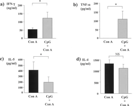
.Effect of CpG ODN on serum cytokine levels in Con A - induced hepatitis
Serum cytokine levels in mice 12 hours after Con A injection (0.25
mg/mouse)with or without prior injection of 50
J.igof CpG ODN are shown in Figure 3. Serum levels of IFN
-yand T
NF
-11'in mice treated with CpG ODN and Con A were significantly higher than those in mice treate d with Con A alone (Figures 3a and b,
ALT *
(lUlL)
*
* *
15000 i I
10000
5000
o
Control Con A* *
*
II
CpG
*
II
CpG non-CpG +
Con A + Con A
Fig. 1. Serum AL T levels in 5 groups of mice. Group 1 mice as controls were injected intradermally with PBS. Group 2 mice were injected intravenously with Can A. Group 3 mice were injected intradermally with CpG ODN. Group 4 mice were injected intradermally with CpG ODN 48 hours before Can A injection.
Group 5 mice were injected intradermally with non-CpG ODN 48 hours before Can A injection. Data are shown as means±SD for 5 groups of 8 mice each.
Representative results of 3 experiments are shown. • P < 0.05.
ROLE OF CpG OD IN CONCANAVALIN A-INDUCED HEPATITIS IN MICE 45
ALT (lUlL) 15000
10000
5000
o
0.5 Jl g*
5 Jl g CpG
+ Con A
*
50 Jl g
Fig. 2. Serum AL T levels in mice injected with different amounts of CpG ODN before Can A injection. Mice were injected intradermally with 0.5, 5 or 50 flg of CpG ODN 12 hours before Con A injection. Data are shown as means±SD in 4 mice in each group. Representative results of 3 experiments are shown. * P
<
0.05.a) I(ng/ml) FN-y 200
*
b) TNF-a 200*
(pg/ml)
150 150
100 100
50 50
Con A CpG Con A CpG
+ +
Con A Con A
*
NSC)
IL-S 600 d) I.L-4 1500 r---l(pg/ml) 500 (pg/ml)
400 1000
300
200 500
100
Con A CpG Con A CpG
+ +
Con A Con A
Fig. 3. Serum levels of IFN-y (a), TNF-11' (b), IL-5 (c) and IL-4 (d) in mice 12 hours after Can A injection with or without prior injection of CpG ODN. Representa- tive results of 3 experiments are shown. • P <0.05.
46 K. ABE et al.
(a)
(b)
Fig. 4. Liver histology in (a) a mOllse treated with Con A alone and (b) a mOllse treated with CpG ODN and Con A (hematoxylin and eosin stain; original magnification 100 x)
respectively). On the other ha nd, serum IL- 5 level in mice treated with CpG ODN and Con A was significantly lower than tha t in mice treated with Con A a lone (Figure 3c). Serum IL-4 level in mice trea ted with CpG ODN and Con A was also lower than that in mice treated with Co n A alone (Figure 3d), but the difference was not significant.
Live!' histology zn Con A-induced hejJatitis
In liver histology, mi ce treated with CpG ODN and Con A sho wed more exten
-sive midzo nal necrosis than did mice treated with Con A alone (Figure 4). In mic e trea ted with CpG ODN a lone, hepa tocyte necrosis was not observed (data not shown).
DISCUSSION
Con A is a lectin that stimul ates lymphocytes or monocytes to secre te various cytokines in vitro, and it is known to induce liver injur y mediated by cell ular immunity when inj ected int o mi ce ') Therefore, Con A- induced hepatiti s is used as an experimental mode l of immune- medi ated liver injury. On the other hand, CpG DNA de rived from bacteria ca n potently stimulate a Thl immune response
7).Although the actions of CpG DNA resemble those of LPS, CpG DNA stimul ates
di stinct toll- like receptors as well as various cell types
'5 ).In this stud y, we showe d
ROLE OF CpG ODN IN CONCANAVALIN A-INDUCED HEPATITIS IN MICE 47
that Con A-induced hepatitis was aggravated by the injection of CpG ODN, with increases in serum levels of Th1-type cytokines, IFN-y and
TNF-aand a decrease in serum IL-5. In addition, prior injection of CpG ODN significantly increased serum AL T levels in mice treated with Con A in a dose-dependent manner. Thus, CpG DNA is thought to be one of the factors aggravating immune-mediated liver injury via augmentation of a Th1 immune response.
Several cytokines, including
TNF-a,IFN-y and IL-4, have been shown to be involved in the pathogenesis of Con A -induced hepatitis, because neutralization of these cytokines conferred protection against Con A-induced hepatitis
2 - 4).Tagawa et al. suggested that the induction of Fas antigen expression on hepatocytes after treatment with Con A is mediated by IFN-y, and that this elevated expression of Fas antigen promotes apoptosis of hepatocytes
5).In our model, serum IFN-y level was increased in mice treated with CpG ODN and Con A compared with that in mice treated with Con A alone. Since CpG DNA stimulates dendritic cells to produce IL- 12, which promotes the production of IFN-y by T cells and natural killer cells
l!),it is possible that exacerbation of Con A-induced hepatitis by CpG DNA is mediated by enhanced production of IFN-y. Similar to IFN-y, the enhanced levels of TNF-
a
were thought to be involved in the exacerbation of Con A-induced hepatitis by CpG DNA.
TNF-ais known to be released from macrophages or T cells stimulat- ed by CpG DNA as well as Con AI6,17). Heikenwalder et al. reported that livers of repeated CpG-ODN -treated mice showed multifocal infiltrates of Band T cells and marked Kupffer cell hyperplasia expanded the sinusoidsI8). These cells are thought to produce Th1-type cytokines in our model. Apoptotic mechanisms are likely to be involved in liver injury related to
TNF-a.In this study, 12 hours after Con A challenge, TNF
-awas not detected in sera of mice treated with Con A alone because it has been reported that plasma level of TNF
-apeaked at 2 hours17).
Louis et al. reported that IL-5-dependent activation of eosinophils and Fas/Fas
ligand interaction constitute the two major effecter pathways of liver injury in Con
A-induced hepatitisI9). They showed that natural killer T cells are a major source
of IL-5, which causes eosinophil-mediated liver damage. However, our study
showed increases in serum Th1-type cytokines and a decrease in serum IL-5 in mice
treated with CpG ODN and Con A compared with those in mice treated with Con A
alone, suggesting that CpG DNA-enhanced production of Th1-type cytokines may
suppress the production of IL-5. Since Satoh et al. reported that the infiltrating
cells in the liver of Con A-induced hepatitis mice were predominantly T
lymphocytes
20),Con A-induced hepatitis may be aggravated by T cells activated by
enhanced production of Th1-type cytokines by CpG DNA even though the produc-
tion of IL-5 was reduced in our models. Serum levels of IL-4 were also reduced in
mice treated with CpG ODN and Con A compared with those in mice treated with
Con A alone, but the difference was not significant. These results suggest that
production of IL-4 by various cells may be more resistant to the suppression of Th1-
type cytokines than that of IL-5 in our models, but further experiments are neces-
48 K. ABE et al.
sary to clarify this phenomenon.
ACKNOWLEDGEMENT
This work was supported in a part by a grant from the Intractable Hepatitis Research Committee of the Health and Welfare Ministry of Japan.
REFERENCES
1. Tiegs G, Hentschel J, Wendel A. A T cell-dependent experimental liver injury in mice inducible by concanavalin A. J Clin Invest, 90: 196-203, 1992.
2. Mizuhara H, O'Neill E, Seki N, Ogawa T, Kusunoki C, Otsuka K, Satoh S, Niwa M, Senoh H, Fujiwara H. T cell activation-associated hepatic injury: mediation by tumor ne- crosis factors and protection by interleukin 6. J Exp Med, 179: 1529-1537, 1994.
3. Toyabe S, Seki S, Iiai T, Takeda K, Shirai K, Watanabe H, Hiraide H, Uchiyama M, Abo T. Requirement of IL-4 and liver NK1+ T cells for concanavalin A-induced hepatic injury in mice. J Immunol, 159: 1537-1542, 1997.
4. Kusters S, Gantner F, KunstIe G, Tiegs G. Interferon gamma plays a critical role in T cell-dependent liver injury in mice initiated by concanavalin A. Gastroenterology, 111 : 462-471, 1996.
5. Tagawa Y, Sekikawa K, Iwakura Y. Suppression of concanavalin A-induced hepatitis in IFN-gamma(-/-) mice, but not in TNF-alpha(-/-) mice: role for IFN-gamma in activating apoptosis of hepatocytes. J Immunol, 159: 1418-1428, 1997.
6. Tokunaga T, Yamamoto H, Shimada S, Abe H, Fukuda T, Fujisawa Y, Furutani Y, Yano 0, Kataoka T, Sudo T, et al. Antitumor activity of deoxyribonucleic acid fraction from Mycobacterium bovis BCG. 1. Isolation, physicochemical characterization, and antitumor activity. J Nat! Cancer Inst, 72: 955-962, 1984.
7. Krieg AM, Yi AK, Matson S, Waldschmidt TJ, Bishop GA, Teasdale R, Koretzky GA, Klinman DM. CpG motifs in bacterial DNA trigger direct B-cell activation. Nature, 374: 546-549, 1995.
8. Sato Y, Roman M, Tighe H, Lee D, Corr M, Nguyen MD, Silverman GJ, Lotz M, Carson DA, Raz E. Immunostimulatory DNA sequences necessary for effective intradermal gene immunization. Science, 273: 352-354, 1996.
9. Brazolot Millan CL, Weeratna R, Krieg AM, Siegrist CA, Davis HL. CpG DNA can induce strong Th1 humoral and cell-mediated immune responses against hepatitis B surface antigen in young mice. Proc Nat! Acad Sci USA, 95: 15553-15558, 1998.
10. Tokunaga T, Yamamoto T, Yamamoto S. How BCG led to the discovery of immunos- timulatory DNA. Jpn J Infect Dis, 52: l-ll, 1999.
11. Jakob T, Walker PS, Krieg AM, von Stebut E, Udey MC, Vogel JC. Bacterial DNA and CpG-containing oligodeoxynucleotides activate cutaneous dendritic cells and induce IL- 12 production: implications for the augmentation of Th1 responses. Int Arch Allergy Immunol, 118: 457-461, 1999.
ROLE OF CpG ODN IN CONCANAVALIN A-INDUCED HEPATITIS IN MICE 49 12. Miyata M, Hiroko K, Tomomi S, Yukio S, Reiji K. Unmethylated oligo-DNA containing CpG motifs aggravates collagen-induced arthritis in mice. Arthritis Rheum, 43: 2578- 2582, 2000.
13. Sparwasser T, Miethke T, Lipford G, Borschert K, Hacker H, Heeg K, Wagner H.
Bacterial DNA causes septic shock. Nature, 386: 336-337, 1997.
14. Frances R, Benlloch S, Zapater P, Gonzalez 1M, Lozano B, Munoz C, Pascual S, Casellas lA, Uceda F, Palazon 1M, Carnicer F, Perez-Mateo M, Such J. A sequential study of serum bacterial DNA in patients with advanced cirrhosis and ascites. Hepatology, 39:
484-491, 2004.
15. Hemmi H, Takeuchi 0, Kawai T, Kaisho T, Sato S, Sanjo H, Matsumoto M, Hoshino K, Wagner H, Takeda K, Akira S. A Toll-like receptor recognizes bacterial DNA.
Nature, 408: 740-745, 2000.
16. Shoda LK, Kegerreis KA, Suarez CE, Mwangi W, Knowles DP, Brown WC. Immunos·
timulatory CpG-modified plasmid DNA enhances IL-12, TNF-alpha, and NO production by bovine macrophages. 1 Leukoc Bioi, 70: 103-112, 200l.
17. Gantner F, Leist M, Lohse AW, Germann PG, Tiegs G. Concanavalin A-induced T -cell- mediated hepatic injury in mice: the role of tumor necrosis factor. Hepatology, 21: 190- 198, 1995.
18. Heikenwalder M, Polymenidou M, lunt T, Sigurdson C, Wagner H, Akira S, Zinkernagel R, Aguzzi A. Lymphoid follicle destruction and immunosuppression after repeated CpG oligodeoxynucleotide administration. Nat Med, 10: 187-92, 2004.
19. Louis H, Le Moine A, Flamand V, Nagy N, Quertinmont E, Paulart F, Abramowicz D, Le Moine 0, Goldman M, Deviere 1. Critical role of interleukin 5 and eosinophils in con·
canavalin A-induced hepatitis in mice. Gastroenterology, 122: 2001-2010, 2002.
20. Satoh M, Kobayashi K, Ishii M, Igarashi T, Toyota T. Midzonal necrosis of the liver after concanavalin A-injection. Tohoku 1 Exp Med, 180: 139-152, 1996.