九州大学学術情報リポジトリ
Kyushu University Institutional Repository
ヒト型ハイブリドーマのモノクローナル抗体生産性 増強に関する研究
菅原, 卓也
九州大学農学研究科食糧化学工学専攻
https://doi.org/10.11501/3065533
出版情報:Kyushu University, 1992, 博士(農学), 課程博士 バージョン:
権利関係:
CHAPTER Vlli PURIFICATION OF IMMUNOGLOBULIN PROSUCTION STIMULATING FACTOR-lip
1. Introduction
IPSF-II a derived from Namalwa lysate was purified and identified as glyceraldehyde-3-phosphate dehydrogenase (GPD). which is a key enzyme in the glycolytic pathway as mentioned previously. There
were some evidences that Namalwa lysate contained another IPSF active substance. The IPSF was named IPSF-IIP and purified from supernatant of 50 °/o saturated ammonium sulfate solution of Namalwa cell lysate. It was supposed that IPSF-IIP was different from IPSF-Ila
the reason that there was a fraction which had IPSF activity in spite of lack of GPD (36 KD peptide).
In this chapter, IPSF-IIP was purified from Namalwa cell lysate and estimated its molecular size.
2. Materials and methods
2-1. Cells and cell culture
Namalwa cells derived from human Burkitt's lymphoma were cultured to extract IPSF-IIP in ERDF medium (Kyokuto Pharmacy Industrial Co.) supplemented with 2 °/o fetal bovine serum (FBS) and ITES as mentioned previously (Sugahara et al., 1991 a). To obtain a
large number of the cells, a high density continuous culture system SHC-1 (Shimadzu) was employed. A human-human hybridoma cell line, HB4C5 was used to assay of IPSF activity during the purification of IPSF-IIP. HB4C5 producing human lung cancer specific monoclonal
antibody (MAb) of IgM type, was fusion product of human lymphocytes and a human fusion partner, NAT-30 (Murakami et al., 1985).
2-2. Assay ofiPSF activity
HB4C5 and other hybridoma lines were inoculated at 1x10⁵ cells/ml in 96 well culture plate containing 200 111 of ITES-ERDF medium supplemented with IPSF-II�. After a 6 hour cultivation, the
amount of secreted immunoglobulin was measured by ELISA as mentioned previously (Sugahara et al., 1991 a).
2-3. Purification of IPSF-11� from Namalwa cell lysate All the procedures are described in results section.
3. Results
3-1. Hydrophobic interaction chromatography of Namalwa cell lysate
Namalwa cells washed with ice-cold phosphate buffered saline (PBS) were ultrasonically homogenized in 10 mM sodium phosphate buffer, pH 7.4 (NaPB) at 4 °C. The cell lysate was fractionized by salting out using 50 °/o saturated ammonium sulfate at 4 °C. The supernatant was further purified by hydrophobic interaction column chromatography. A hydrophobic interaction column (BUTYL TOYOPEARL 650M, Tosoh, 22 mm I.D. x 25 em) was equilibrated with 10 mM NaPB containing 2 M ammonium sulfate. The supernatant was applied to the column and non-adsorbed materials were removed with 10 mM NaPB containing 2 M ammonium sulfate. The absorbed substances were then eluted stepwise with 10 mM NaPB containing ammonium sulfate at the concentration of 1.1, 0.9, 0. 7, 0.5 and 0 M.
The eluted fractions at each concentration of ammonium sulfate were
dialyzed to 10 mM N aPB and each dialyzed fraction was assessed for the IPSF activity.
IPSF activities were detected in the fractions eluted at the concentration of ammonium sulfate 1.1 M and 0. 7 M (Fig. VIII-1).
According to the SDS-PAGE analysis, IPSF activity of 1. 1 M fraction was derived from IPSF-Ila, but that of 0. 7 M fraction derived from another IPSF, namely, IPSF-II�.
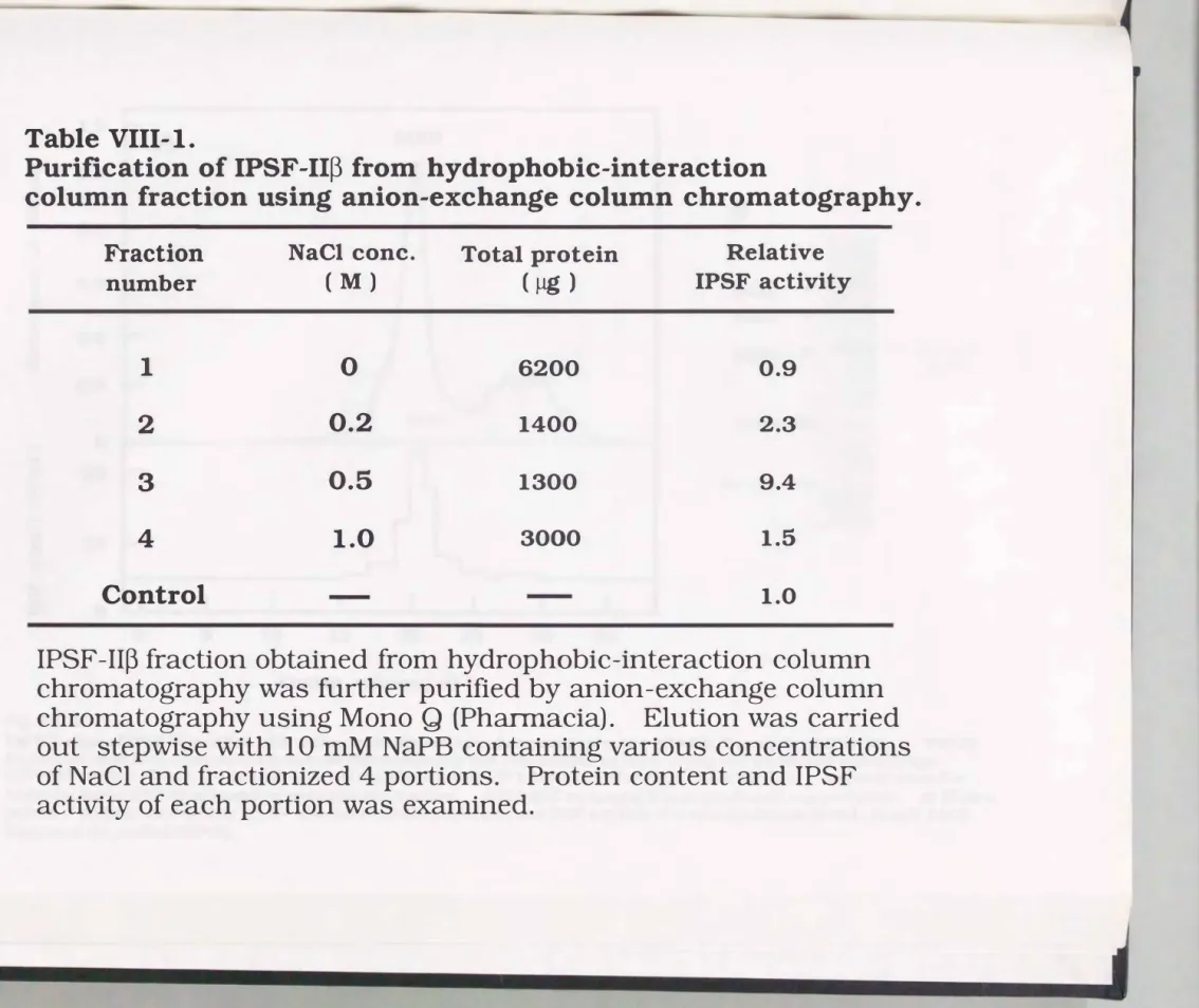
3-2. Anion-exchange chromatography of IPSF-11� fraction obtained from hydrophobic interaction column
IPSF-II� fraction was further purified by anion-exchange column
chromatography (Mono Q, Pharmacia). Following the equilibration with 10 mM NaPB (pH 7.4), IPSF-1I� fraction obtained by hydrophobic interaction column chromatography was applied to the anion-exchange column. After washing off the non-adsorbed materials with 10 mM NaPB, stepwise elution was carried out with NaPB containing 0.2, 0.5 and 1.0 M NaCI. As shown in Table VIII-1, IPSF-II� was collected in the fraction eluted at 0.5 M NaCl.
3-3. Gel ffitration of IPSF-11� fraction and molecular weight analysis by SDS-PAGE
For further purification and estimation of molecular size of IPSF
II�. the 0.5 M NaCl fraction concentrated by freeze-drying was gel filtrated with a TSKgel G3000SW column (Tosoh, 7.5 mm I.D. x 60 em) equilibrated with 100 mM NaPB (pH 7.4). One major peak was detected by measuring absorbance at 280 nm, and this fraction possessed IPSF activity (Fig. VIII-2A). From the standard curve of molecular sizes, the molecular size of IPSF-II� was calculated as 46 KD.
The subunit structure of IPSF-II� was analyzed by SDS-PAGE (Fig. VIII-
2B). The IPSF-II� still gave a single band after the treatment of 2-
mercaptoethanol on the SDS-PAGE electrophorogram, indicating that the protein was composed of a single peptide.
-....]
�
1.2 2.0
�
-a u d
d 1.0 0
0 CJ
ct) � �
0.8 �
� aS 1.0 �
� '3 en
CJ 0.6
d e
� aS
�
-::s0 0.4 d
ell 0
�
0�
0.2
�
0...
.E. 'CD 20
CJ d
0 10
CJ
=s � 0
20 40 10 20 30 40 50 60 70 80 90
Elution Volume (ml)
Fig. Vlll-1.
Hydrophobic interaction column chromatography of Namalwa cell lysate fractionized by ammonium sulfate precipitation. A supernatant obtained by salting out using 50°/o saturated ammonium sulfate was applied to the column and non-adsorbed materials were removed with 10 mM NaPB containing 2 M
ammonium sulfate. The absorbed substances were then eluted stepwise with 10 mM NaPB containing ammonium sulfate at the concentration of 1.1, 0.9, 0.7, 0.5 and 0 M. The eluted fractions at each
concentration of ammonium sulfate were dialyzed to 10 mM NaPB and IPSF activity of each dialyzed fraction was assayed with HB4C5 cells. The upper part shows the elution pattern of protein measured by the lN absorbance at 280 nm (solid line) and the stepwise gradient pattern of ammonium sulfate concentration (dotted line). The lower part shows the IPSF activity by the elevated IgM production of HB4C5.
'-1 ()1
Table VIII-I.
Purification of IPSF-II� from hydrophobic-interaction
column fraction using anion-exchange column chromatography.
Fraction number
1 2 3 4 Control
NaCl cone.
( M)
0 0.2 0.5 1.0
Total protein ( �)
6200
1400
1300 3000
Relative
IPSF
activity
0.9
2.3
9.4 1.5
1.0
IPSF-II� fraction obtained from hydrophobic-interaction column chromatography was further purified by anion-exchange column chromatography using Mono Q (Ph
arrnacia). Elution was carried out stepwise with 10 mM NaPB containing various concentrations of NaCl and fractionized 4 portions. Protein content and IPSF
activity of each portion was examined.
"' 0)
1.2 .
A
46KDa 1.0 I
0 =
I I \ I B
CX) � 0.8
� cd
Q) 0.6
(,)
r I \ I
94KD __.= cd
67KD __.
-e 0 0.4
f/)
I J \ I
43KD __.,
.� .___ IPSF -II ��
(46 KD)0.2 0
I __)
�'-./ "'- I
30KD __.-
a 20... I IL. I 20.1KD __.
"Q.()
-
= (,)= 10
0 (,)
::e "Q.() 0
�
0 5 10 15 20 25 30 35
Elution volume ( ml )
Fig. VIII-2.
Gel filtration of IPSF-np purified with anion-exchange column chromatography and SDS-PAGE analysis of IPSF-11�. IPSF-1I�
fraction obtained from anion-exchange column chromatography was concentrated by freeze-drying and gel filtrated with a TSKgel G3000SW column ( Tosoh, 7.5 mm I. D. x 60 em) equilibrated with 100 mM sodium phosphate buffer (pH 7.4). An arrow shows the
molecular size ofiPSF-II� estimated by molecular size markers. SDS-PAGE containing 2-mercaptoethanol was performed. A:. Elution pattern of proteins measured by the UV absorbance at 280 run (upper) and IPSF activities of obtained fractions (lower) .. B: SDS-PAGE analysis of the purified IPSF-II�.
4. Discussion
We previously reported that immunoglobulin production stimulating factor-II a (IPSF-II a) was purified from extract of Namalwa cells and had a molecular size of 112 KD (Sugahara et al.. 1991 a).
The IPSF-Ila was composed of two 36 KD and a 40 KD subunits. The 36 KD subunit which exclusively retained IPSF activity was identified by amino acid sequence analysis of its N-terminus as glyceraldehyde -3- phosphate dehydrogenase (GPD). a key enzyme in the glycolytic pathway (Sugahara et al., 1991b). We have found another IPSF. IPSF
IIP. in the Namalwa cells. As the result of purification . IPSF-IIP was estimated as a monomeric polypeptide of 46 KD by gel filtration and SDS-PAGE analyses.
5. Conclusion
IPSF-11� was purified from Namalwa cell lysate fractionized by salting out. The supernatant obtained by 50 °/o saturated ammonium sulfate solution of Namalwa lysate was applied to hydrophobic interaction column. The adsorbed materials were eluted stepwise.
stepwise elution could separate IPSF-II a and -II�, because IPSF-II a was eluted at higher concentration of ammonium sulfate.
IPSF- II� was estimated as a single peptide having 46 KD of molecular size by gel filtration and SDS-PAGE analyses.
CHAPTER
IXIDENTIFICATION OF IPSF-11�
1. Introduction
IPSF active subunit of IPSF-Ila (36 KD peptide) was estimated as glyceraldehyde-3-phosphate dehydrogenase (GPD) by amino acid sequence analysis of the 36 KD peptide. As well as IPSF-II a, IPSF-
liP
was purified from Namalwa cell lysate and estimated as a single peptide which possessed 46 KD of molecular size. Amino acid sequence of the 46 KD peptide was examined in this chapter.
2. Materials and methods
2-1. Analysis of amino acid sequence of IPSF-
liP
IPSF-II� fragments obtained by digestion with lysylendopeptidase were fractionized with reverse-phase column chromatography. The fractionized peptides were analyzed their amino acid sequences with a 4 77 A protein sequencer and 120A analyzer (Applied Biosystems).
2-2. Assay of enolase activity
Enolase derived from rabbit muscle was purchased from Boehringer Mannheim (Germany). It was confirmed by SDS-PAGE analysis that this enzyme preparation did not contain glyceraldehyde-3- phosphate dehydrogenase identified as IPSF-II a. Enolase activity was quantitatively monitored spectrophotometrically by measuring the absorbance at 240 nm derived from the reaction product, phosphoenolpyruvate at 25 oc (Kornblatt et al., 1989). The reaction mixture was composed of 1 mM magnesium acetate, 250 mM KCl, 0.1
mM EDTA, 1 mM 2-phosphoglycerate and enolase or IPSF-IIP in 50 mM imidazole buffer (pH 7 .1).
3. Results
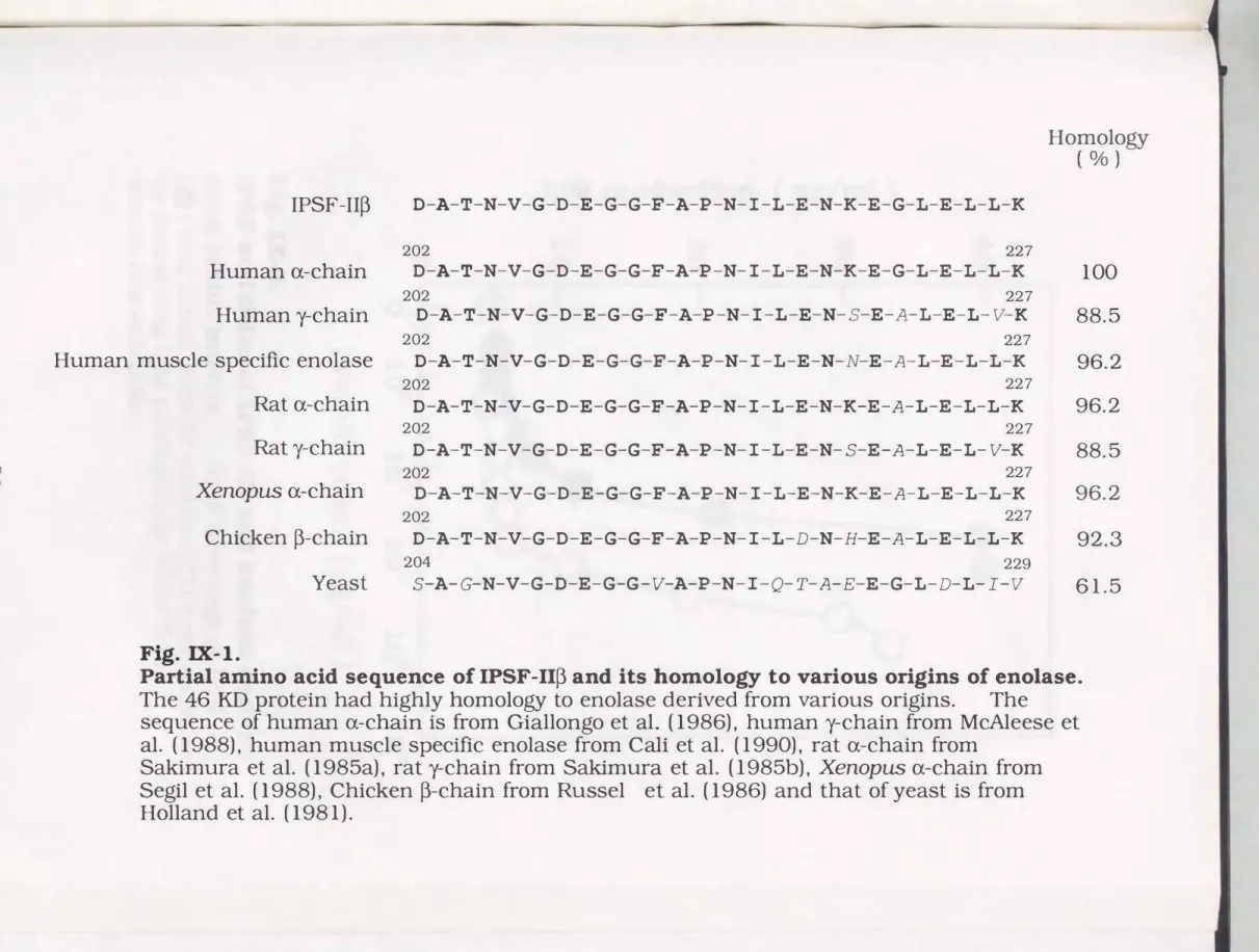
3-1. Partial amino acid sequence of IPSF-IIP
Partial amino acid sequence of a fragment of IPSF-IIP was shown in Fig. IX-1. The sequence was highly homologous to enolase (EC 4.2.1.11) of various origins. Enolase is a one of the enzymes in the glycolytic pathway which catalyzes conversion of 2-phospho-D-glycerate to phosphoenolpyruvate. With regard to the amino acid sequence, IPSF-np was completely homologous to human enolase a-chain.
3-2. IPSF activities of IPSF-IIP and enolase
Enolase derived from rabbit muscle stimulated IgM production of HB4C5 (Fig. IX-2). The amount of IgM produced in the medium was maximized at the concentration of 35 ng/ml by the addition of 700
�g/ml of enolase for 6 h. Whereas IPSF-IIP was needed only 60 �g/ml to accomplish the maximum stimulation of IgM production of the hybridoma line. It is noteworthy that IPSF-IIP has 12-fold higher activity than the rabbit muscle enolase.
3-3. Enolase activity of IPSF-IIp
Specific enzymic activity of IPSF-IIP was 1. 7-fold stronger than that of rabbit muscle enolase of which enzymic activity was 30 unit/ml (Fig. IX-3).
00
...
IPSF-I Ip 0-A-T-N-V-G-0-E-G-G-F-A-P-N-I-L-E-N-K-E-G-L-E-L-L-K
202 227
Homology
( o/o )
Human a-chain o-A-T-N-V-G-0-E-G-G-F-A-P-N-I-L-E-N-K-E-G-L-E-L-L-K 100
202 227
Human y-chain 0-A-T-N-V-G-0-E-G-G-F-A-P-N-I-L-E-N-5-E-A-L-E-L-V-K 88.5
202 227
Human muscle specific enolase o-A-T-N-V-G-0-E-G-G-F-A-P-N-I-L-E-N-N-E-A-L-E-L-L-K 96.2
202 227
Rat a-chain D-A-T-N-V-G-0-E-G-G-F-A-P-N-I-L-E-N-K-E-A-L-E-L-L-K 96.2
202 227
Rat�chain o-A-T-N-V-G-0-E-G-G-F-A-P-N-I-L-E-N-5-E-A-L-E-L-V-K 88.5
202 227
Xenopus a-chain 0-A-T-N-V-G-0-E-G-G-F-A-P-N-I-L-E-N-K-E-A-L-E-L-L-K 96.2
202 227
Chickenp-chain o-A-T-N-V-G-0-E-G-G-F-A-P-N-I-L-D-N-H-E-A-L-E-L-L-K 92.3
Fig.
IX-1.204 229
Yeast 5-A-G-N-V-G-o-E-G-G-V-A-P-N-I-Q-T-A-E-E-G-L-D-L-r-v 61.5
Partial amino acid sequence of IPSF-IIP and its homology to various origins of enolase.
The 46 KD protein had highly homology to enolase derived from various origins. The sequence of human a-chain is from Giallongo et al. (1986), human y-chain from McAleese et al. (1988), human muscle specific enolase from Cali et al. (1990), rat a-chain from
Sakimura et al. (1985a), rat y-chain from Sakimura et al. (1985b), Xenopus a-chain from Segil et al. (1988), Chicken P-chain from Russel et al. (1986) and that of yeast is from Holland et al. (1981).
40
30
20
10
•
0
Fig. IX-2.
Protein cone. ( �/ml )
IPSF activities of IPSF-11� and enolase derived from rabbit muscle. IPSF activities of IPSF-II�
(e) and rabbit muscle enolase ( 0) were assessed by measuring IgM produced by HB4C5 in
serum-free condition.
0.5
a 0.4
= 0 �
�
�0.3
as GJ
(.); 0.2
� 0
�
f/l0.1
o ��--��--��----�---��
0 2 4 6 8 10
Time (min.)
Fig. IX-3.
Enolase activities of IPSF-11� and enolase derived from rabbit muscle. Reaction mixture was composed of 1 mM magnesium acetate, 250 mM KCl, 0.1 mM
EDTA, 1 mM 2-phosphoglycerate and 0.5 mg/ml of enolase or IPSF-11� in 50 mM imidazole buffer (pH 7.1).
The enzymic activity was monitored
spectrophotometrically by increasing absorption at 240
nm parallel to the concentration of the reaction product,
phosphoenolpyruvate at 25 °C. e; IPSF-11�, Q; Rabbit
muscle enolase.
4. Discussion
The partial amino acid sequence for 26 residues of the 46 KD protein as IPSF-II� completely coincided with human enolase a-chain.
Furthermore, the sequence of IPSF-II� showed high homology with those of enolase originated from various origins.
Enolase derived from rabbit muscle had IPSF activity, but the specific activity was much lower than that of IPSF-11�. The reason for
the lower IPSF activity of the rabbit muscle enolase was supposed that it was derived from different species, and moreover, from muscle that mainly contains �-chain of enolase. The enzymic activity of IPSF-11�
was also observed, and the activity was 1. 7 -fold stronger than that of enolase.
Homology of amino acid sequences between GPD and enolase is low, and there are no specific consensus amino acid sequences between them. If there is a similarity of the modes of action of these IPSFs in spite of this low homology of primary structures, it is considered that IPSF activities of GPD and enolase are derived from secondary or tertiary structures of their peptides.
5. Conclusion
Amino acid sequence of
IPSF-IIP
was analyzed after enzymic digestion of 46 KD pepeide. The partial amino acid sequence ofIPSF
np
was completely homologous to human enolase a-chain.Enolase derived from rabbit muscle had
IPSF
activity, though the specidicIPSF
activity of the enzyme was much lower than that ofIPSF
up.
IPSF- liP
possessed enolase enzymic activity. The specific enzymic activity ofIPSF-np
was 1. 7 -fold higher than that of rabbit muscle enolase.CHAPTER X CHARACTERIZATION OF 1PSF-ll�
1. Introduction
IPSF-II� was purified and identified as human enolase a-chain on the basis of the partial amino acid sequence and enzymic activity.
Then in this chapter, we characterized IPSF-1I� by pH and heat stability, time course effects and effects on various hybridomas.
2. Materials and methods
2-1. Cells and cell culture
For investigation of the effect of IPSF-II� on various hybridomas,
seven human-human hybridoma lines and six mouse-mouse hybridoma lines were used. Among the human-human hybridomas EMK-F7, SU
I, FMK-H3 and KMK-41 were obtained by fusion of lymphocytes with a fusion partner, H0-32 3 (Ohashi et al., 1986). HFlOB4 was a hybridoma line made by fusing NAT-30 with lymphocytes as well as HB4C5. BD9-Dl2 was a fusion product of in vitro immunized human lymphocytes and a fusion partner, �H12 (Kawahara et al., 1992).
EMK-F7, SU-1 and BD9-Dl2 produced both IgG and IgM. FMK-H3, KMK-41 and HFlOB4 were producers of IgM. All mouse-mouse
hybridoma lines were established by fusion of splenocytes with a fusion partner, P3-X63-8AgU1. Among mouse-mouse hybridomas, CG53/2/4 and CGl/1 produced IgG, YM2/6 and YM4/9 produced IgM and IGELa2 and IGELb4 were producers of IgE, respectively. All of these human
human and mouse-mouse hybridoma lines were established in our laboratory. These hybridomas were maintained in 5 °/o FBS-ERDF medium.
3. Results
3-1. Heat and pH stability of IPSF activity of 1PSF-ll�
IPSF-1I� purified with the hydrophobic interaction column was incubated at various temperature for 30 minutes. After cooling, IPSF activities of the treated IPSF-II� were assayed. As shown in Fig. X-1, IPSF-1I� lost its IPSF activity at the temperature higher than 50 °C.
For analysis of pH stability, IPSF-II� was treated with buffer solutions of various pHs at 4 oc for 24 h. Though IPSF activity of IPSF
II� was maintained in such alkaline condition as pH 13, the activity was so unstable in acidic condition that it was completely lost its activity below pH 4 (Fig. X-2). This is contrastive to the fact that enolase activity of IPSF-II� was lost in alkaline condition, above pH 9.
3-2. Time-course of IPSF-11� activity
The time course of IPSF-1I� activity was examined on IgM production of HB4C5 in serum-free medium in the presence of 25
�g/ml of IPSF-II�. The IgM production of HB4C5 was stimulated no sooner than IPSF-1I� was added. After 72 h, the amount of IgM produced in the IPSF-II� supplemented medium reached 2 �g/ ml.
IPSF-II� enhanced IgM production more than 3-fold (Fig. X-3A), but did not affect cell growth of hybridoma (Fig. X-3B). The IgM productivity per 1 o5 cells per 6 h was stimulated more than 3-fold by IPSF-1I� (Fig. X-3C).
3-3. Effect of IPSF-11� on MAb productions with various hybridomas
The stimulating activity of IPSF-II� was examined on various hybridoma lines derived from both human and mouse cells. As shown in Table X-1, IPSF- II� highly enhanced IgM production of HB4C5 and
HF10B4 derived from NAT-30. EMK-F7, FMK-H3, KMK-41 and BD9- D 12 derived from either H0-323 or �H 12 were weakly stimulated their IgG and IgM production. SU-1 producing both IgG and IgM, was stimulated its IgM production as much as 3. 5-fold, but IgG production was not stimulated so much. IPSF- II� enhanced IgM production of mouse-mouse hybridoma lines 1.4-fold, but did not affect IgG production. Whereas IgE productions of mouse-mouse hybridoma lines were partially suppressed by IPSF-II�.
100
�
80
�
0'--'
:>-.
·�
� 60
·"
�
u as Cl.)
� 40
·"
�
as
,....
Cl.)
�
20
Fig. X-1.
0 20 40 60 80 100 Teiilperature ( °C)
Heat stability of IPSF-IIp. The IPSF activity
was measured by bioassay using IPSF-IIP samples
heated in varying temperature for 30 min.
�
80
�
0"--'
>-..
�
60
....
....
�
� (.)
as
u � 40
....
�
as
...
u
�
20
0
Fig. X-2.
2 4 6 8 10 12 14 pH
PH stability of IPSF-11�. IPSF-II� was treated with buffer solutions of various pHs at 4 oc for 24 h.
The IPSF-II� solutions were then dialyzed to 10 mM
NaPB (pH 7.4) to estimate their IPSF activities.
2
-
1
0
Fig. X-3.
B
D D
D 0
12 24 36 48 60 72 Time (hour)
0 6121824 36 48 60 Time (hour)
72
Time-course of IPSF activity on the IgM production of HB4C5 cells in serum-free medium. HB4C5 was incubated in
ITES-ERDF medium in the presence of 25 mg/ml of IPSF-II�.
Cell growth and IgM production were measured at various culture time. Panel A: Cell growth of HB4C5. Panel B: Accumulation of IgM in cultured medium. Panel C inserted in panel B: IgM productivity per 105 HB4C5 cells for 6 h.
0;
cultured inITES-ERDF medium,
D;
cultured in ITES-ERDF medium containing 25 J.tg/ml of IPSF-II�.Table X-1.
Effect of IPSF-ll� on MAb productions with various hybrodomas.
Hybridomas Parent cell I so type Ig cone. ( ng/ml) Relative activity None IPSF IPSF/None Human
EMK-F7 H0-323 IgG 7.4 8.5 1.1
IgM 9.0 10.9 1.1
FMK-H3 H0-323 IgM 2.4 4.8 2.0
KMK-41 H0-323 IgM 37.6 53.6 1.4
SU-1 H0-323 IgG 11.0 13.6 1.2
IgM 7.3 25.5 3.5
BD9-D12 A4Hl2 IgG 13.5 17.2 1.3
IgM 13.3 18.8 1.4
HB4C5 NAT-30 IgM 5.4 28.6 5.3
HF10B4 NAT-3 0 IgM 7.7 43.1 5.6
Mouse
CG53/2/4 P3-X63-8AgU1 IgG 131 108 0.8
CG1/1 P3-X63-8AgU1 IgG 23 23 1.0
YM2/6 P3-X63-8AgU1 IgM 549 762 1.4 YM4/9 P3-X63-8AgU 1 IgM 700 945 1.4
IGELa2 P3-X63-8AgU1 IgE 90 70 0.8
IGELb4 P3-X63-8AgU 1 IgE 120 97 0.8
All these hybridomas were inoculated in ITES-ERDF medium containing 20 Jlg/rnl of IPSF-II� at the density of 1x10 5cells/ml. After 6h, the amount of imn1unoglobulin was measured by ELISA.
4. Discussion
Alkaline-treated IPSF-IIP still showed IPSF activity, though it had lost the enzymic activity. This result suggested that the IPSF activity of enolase did not descend from its enzymic activity, as same as it had been confirmed that G PD served as IPSF-II a irrespective of its enzymic activity. It is evident that both enolase and GPD have another function as IPSF besides the enzymic function in the glycolytic pathway. But the fact that pyruvate kinase and phosphoglycerate kinase, the enzymes in the glycolytic pathway, did not have IPSF activities suggests that not all enzymes in the glycolytic pathway are IPSFs.
IPSF activity of IPSF-IIP was specific for IgM production of both human-human and mouse-mouse hybridoma lines. Especially, IPSF-IIP solely stimulated the IgM production of SU -1, a producer of both IgG and IgM. It is interesting that IgE production of mouse-mouse hybridoma lines were partially suppressed by IPSF-np. IgE production of lymphocytes derived from human lymphonodulus were also partially suppressed by IPSF-IIP.
We notice some similarities of the IPSF activities between IPSF-Ilcx and IPSF-IIP. when the modes of action of IPSF-IIP are investigated.
IPSF-II a and IPSF-IIP were not suppressed their IPSF activities by actinomycin D treatment of HB4C5 and enhanced immunoglobulin synthesis of in vitro translation system using rabbit reticulocyte lysate.
These IPSFs selectively stilnulated IgM production of hybridoma lines.
These facts disclose both IPSF-II a and IPSF-IIP enhance cellular protein productivity by stimulating translation process. Judging from all these results, there is a good possibility that the modes of action of IPSF-IIP are similar to that of IPSF-Ilcx.
GPD as IPSF-II a is a protein having a binding affinity with nucleic acids (Perucho et al., 1980), and is one of the three major RNA-binding proteins in rabbit reticulocyte lysate (Ryazanov, 1985). GPD binds to mono- and polyribosomes through RNA (Ryazanov et al., 1988). We suppose that these findings are concerned with IPSF activity of IPSF
Ila, because some DNA binding proteins such as histone H1, H2A and H2B and lactate dehydrogenase serve as IPSFs (as shown in CHAPTER XI). If IPSF-I1� acts on hybridon1a lines like IPSF-IIa, it is expected that IPSF-11� (i.e. enolase) may have a binding affinity with DNA or RNA.
In addition, both GPD and enolase are identified as heat-shock proteins of Xenopus laevis and yeast, respectively (Nickells et al., 1988 and lid a et al., 1985). Uncovering the mechanisms of the action of these IPSFs will contribute to the effective enhancement of cellular productivities of MAbs of hybridomas.
5. Conclusion
IPSF- II p was stable in alkaline condition, though lost its IPSF activity below pH 4. IPSF-np lost 70 °/o of its IPSF activity by heating at 50 °C for 30 min.
IPSF-IIP stimulated Immunoglobulin production of human-human
hybridoma HB4C5 cells as soon as it added to the medium and the effect was maintained for 3 days. Though HB4C5 cells were enhanced its immunoglobulin productivity, the cell proliferation rate was not affected.
IPSF-IIP enhanced IgM production both human-human and mouse
mouse hybridomas. Especially, IPSF-np stimulate only IgM production of SU-1 cells, a producer of both IgM and IgG. IgE production of mouse-mouse hybridomas were slightly suppressed by IPSF-np.
CHAPTER XI IPSF ACTIVITIES OF NUCLEIC ACID BINDING PROTEINS AND POLY-BASIC AMINO ACIDS
1. Introduction
IPSF-Ila was purified from Namalwa cell lysate using liquid chromatography and identified as glyceraldehyde-3-phosphate dehydrogenase (GPD; EC 1.2.1.12) on the basis of N-terminal amino acid sequence and enzymic activity as shown in CHAPTER V and VI.
GPD is not only a key enzyme in glycolytic pathway but also has various features except for its enzymic activity (Ryazanov, 1985, Ryazanov et al., 1988, Grosse et al., 1986, Lin et al., 1986, Launay et al., 1989, Lachaal et al., 1990 and Shin et al., 1973). One of the major features of GPD is the binding activity with nucleic acids, such as single-strand DNA and RNA (Perucho et al., 1977, Perucho et al., 1980).
We supposed that there were some relations between IPSF activity and nucleic acid binding activity. Some nucleic acid binding proteins are known. Histones (H1, H2A, H2B, H3 and H4), which are basic proteins, form complex with DNA in nucleus of eucaryotic cell.
Protamine, as well as histones molds complex with DNA in nucleus in spermatozoon of vertebrate. Lactate dehydrogenase was identified as single-strand DNA binding protein by Perucho et al. (1980).
In this chapter, IPSF activities of nucleic acid binding proteins, such as histones, protamine and lactate dehydrogenase were investigated. In conjunction with basic proteins, some poly-basic amino acids were assaied their IPSF activity.
2. Materials and methods
2-1. Materials
Histone H1, H2A, H2B, H3 and H4 derived from calf thymus and lactate dehydrogenase were purchased from Boehringer Mannheim.
Salmon roe and herring protamine sulfates were purchased from Wako pure chemical, ploy-L-lysine, poly-D-lysine and poly-L-arginine were purchased from SIGMA.
2-2. Assay of IPSF activity on hybridomas
IPSF activity was examined by measuring amounts of antibodies secreted by hybridomas in the medium by ELISA method as described previously (Yamada et al., 1989). HB4C5 cells (1 x 10⁵ cells/ml) suspended in ITES-ERDF medium supplemented with various concentrations of sample peptides were inoculated in 96 well plate for 6 h. After 6 hours, antibody concentration of cultured medium was determined.
3. Results
3-1. Effects of histones on IgM production of HB4C5 cells
Though histone H1, H2A and H2B showed maximum IPSF activity at 6 �g/ml, the range of optimal concentration was very narrow (Fig.
XI-1). H3 and H4 did not enhance IgM production of HB4C5 cells.
High concentrations of histones (above 20 �g/ml) had bad influences to the cells, so that the cells could not produce IgM.
3-2. Effects of protamines on IgM production of HB4C5 cells
As shown in Fig. XI-2, protamine derived from salmon roe or herring did not have IPSF activity.
3-3. Effect of lactate dehydrogenase on IgM production of HB4C5
cells
The addition of 800 �g/ml of lactate dehydrogenase enhanced IgM production about 5-fold as much as control. Lactate dehydrogenase had IPSF activity, nevertheless specific activity was much lower than that of GPD (Fig. XI -3).
3-4. Effects of poly-basic amino acids on IgM production of HB4C5
The addition of poly-L-lysine or poly-D-lysine resulted in 3- or 2. 5- fold accumulation of IgM in the cultured medium, respectively (Fig. XI- 4). Poly-L-arginine slightly enhanced IgM production of HB4C5. As same as histones, the range of optimal concentration of these poly
lysines was narrow.
60
I
0 ;Hl50 ... 6 ;H2A V; H2B
�
40
l-
D ;H3s • ;H4
...
'a1)
�
-
� 0 30
:p C) ::J
"0 0 I-.
20
I
e0.
c..o �
'a1)
c..o �
10
0
0 0.01 0.1 1 10 100 1000
Protein cone. (Jlg/ml)
Fig.
XI-1.IPSF activities of various his tones.
8
...'ol)
1=:
0 +:j 0 u �
"0 0
� 0..
:E 'ol)
-
30 �---.
25
20
15
®
10
5
0
0 0.01 0.1 1.0
0
; Salmon roue
; Herring1000 Protamine cone. ( �/rnl )
Fig. XI-2.
IPSF activities of protamines derived from salmon
roe and herring.
a
...'ol)
�
_...
� 0 :;j
u ;j '"d 0
� �
:;;s
'ol)�
60
50
40
30
20
10
0 0.1
Fig.
XI-3.1 10 100
LDH cone.
( �g/ml )
1000
IPSF activity of lactate dehydrogenase (LDH).
1""""1
...
s
'of)
�
.-
� 0
·� -+---l C)
� ;j 0 �
...
�
0
t..J
�
'of)
�
70
I
0; Poly-L-Lysine
60
1- /),. ; Poly-D-Lysine
I 0
; Poly-L-Arginine
50
40
30
20
10
0 1 1 ilL Ill I I I I I I I I I I
0
0.01 0.1Fig.
XI-4.1 10
Cone. ( J.tg/ml )
100 1000
IPSF activities of poly-L-Lys, poly-D-Lys and poly-L-Arg.
4. Discussion
Histone Hl, H2A and H2B had IPSF activities, but histone H3 and H4 did not enhanced IgM production. The former histones are
categorized into lysine-rich histones and the latter are arginine-rich histones. Protamine which is arginine-rich protein did not shown IPSF activity. From these results, it was assumed that lysine residues of these basic proteins participated in IPSF activities. IPSF activities of poly-lysine and poly-arginine were investigated to carry out further analysis of these experimental results. Though poly-L- and poly-D
lysine obviously acted as IPSF, poly-L-arginine showed only weak activity. These results suggest that lysine residues of proteins affect IgM production of hybridomas. The effect may be physical or chemical one, because poly-D-lysine has IPSF activity as well as poly-L-lysine.
5. Conclusion
Lysine-rich his tones, such as H l, H2A and H2B, showed IPSF activity, but the region of their optimal concentrations were very narrow.
Hist one H3 and H4 and protamines, that are arginine rich peptide, did not possess IPSF activity.
Poly-L-lysine and poly-D-lysine showed IPSF activities, but poly-L
arginine was not identified as IPSF because of the weakness of the activity. These results could be tied in with the results mentioned above.
CHAPTER Xll VffiUS CONTAINMENT CONTINUOUS CULTURE OF ANIMAL CELLS
1. Introduction
Many viruses enter a latent state, in which their genomes are present but inactive in the cell and no progeny are produced. Such animal cells may produce virus particles due to physiological alteration in the culture condition. It will cause a serious problem for safety issues in that the animal cell products produced by such cells are used for diagnosis and therapy. To cope with this problem, we examined the possibility of adapting a hollow fiber module, which had been proved to eliminate viruses, for high-density continuous culture of animal cells.
2. Materials and methods
2-1. The BMM hollow fiber
The benberg microporus membrane (BMM) hollow fiber (ASAHI CHEMICAL CO.) has a multi-layer wall structure and a mean pore size which can be precisely controlled throughout a range of 20 to 100 nanometers. The BMM hollow fiber having a mean pore size of 35 nanometers can reduce hepatitis C virus content to one part per 103.5 to 104 or less in a single filtration and to one part per 106 in double filtration (Yuasa et al., 1991). It can not only eliminate viruses but also permit a high protein recovery rate (Tsurumi et al., 1990). The BMM hollow fiber used here had 0.06 m2 filtration area and a mean pore size of 40 nanometer.
2-2. Cell culture system
High-density continuous culture system SHC-1 (Shimadzu) equipped with 500 ml culture vessel was used here. The flow sheet of SHC- 1 culture system installed the BMM hollow fiber was shown in Fig.
XII- 1. SHC- 1 has a 500 ml culture vessel. The cells and cultured medium are separated by crossflow filter and fresh medium is supplied to the culture vessel. Cell-free spent medium stocked in the tank was filtrated with this module continuously.
2-3. Cell and cell culture
Human-human hybridoma cell line HB4C5 cells producing anti human lung cancer specific antibody (IgM type) were cultured in ERDF medium supplemented with 10 �g/ml of insulin, 20 �g/ml of transferrin, 20 �M ethanolamine, 25 nM sodium selenite and 30 �g/ml of yolk lipoprotein (YLP) ( Murakami et al., 1985 and Murakami et al.,
1988).
....
__....
Crossflow Filterrt
ll �
/' "'\ /' "\
Jl
,,
� '�·�---
Fresh Medium Spent Medium
l J
"'
...v
...r 1
BMM Hollow Fiber
/
:
.
::
.1:-:
r h
-- -
a
"\
· y
A
.::
..,.
[h[L
Magnetic Stirrer
&
Heater
Virus-free Cultured Medium
Fig. XII-1.
_ .... ...
--
Flow sheet of SHC-1 high-density continuous
Gas Exhaust
� , C02, Air Inlet DO and pH Sensor Temp. and Level Sensor
culture system installed the
BMMhollow fiber filtration
system.
3. Results
3-1. Semi-continuous Filtration of spent medium from high-density continuous culture of HB4C5 cells
High-density culture was continued over 30 days as shown in Fig.
XII-2. Viable cell density exceeded lxl07 cells/ml in 2 days and was kept at a density of 2xl07 cells/ml for 20 days.
Over 30 days, 20 L of spent medium was obtained and filtrated continuously. Filtration of 20 L of spent medium required only one module. After the 20th day of culture, much of the scum of the cells was observed in the spent medium because of the worse cell conditions. Even with those conditions, this module showed good filtration capacity.
3-2. Protein recovery rate of the BMM hollow fiber
To examine the protein recovery rate of this fiber, immunoglobulin concentration of spent medium before and after filtration was measured by ELISA method. Adsorption of immunoglobulin to the fiber does not matter in practical use (Fig. XII -3).
� Q
� � ....
,..-4
....
.g
....>
,..-4
... a
,..-4 Ul
,..-4
� CJ
...., � ....
Ul
= � 'tS
,..-4 ,..-4
� CJ
,..-4 �
.g >
100
80 60
40
20 5x10⁷
1x10⁷ 7
5x10⁶ 6
5
4 1x10⁶
3 5x10⁵
2 1
0
0 5 10 15 20 25 30
Time (day)
Fig. XII-2.
High-density continuous culture of HB4C5 cells
with SHC-1 high-density continuous culture system installed the BMM hollow fiber module. Human-human hybridoma HB4C5 cells were cultured in ERDF medium supplemented with ITES and 30 J.ig/ml of YLP.
'Ql)
a
= 0 ....
...., CJ = 'tS
�
:e 'Ql) ...
�
�
0
�
�
...
s
�
'--"'
� 0
·� ..._;
(ij �
..._;
� Q) C,) �
0 C,)
�
'oJ)�
10
8 6
4 2
0 0
Fig. XII-3.
5 10 15
0 ; Before filtration
@;After filtration
20 25 30
Time (day)
IgM concentration of continuous cultured medium
before and after filtration using the BMM hollow fiber
4.Discussion
Containment of virus causes a serious problem for production of biologically active substances by animal cells and safe use of them.
Generally, even though animal cells which do not contain virus particles can be doubted the presence of virus gene in their genomes.
These viruses are inactive in the cell and no progeny are produced in normal cell culture conditions. But alteration of the cell culture conditions such as high-density culture may cause the production of virus particles. We can solve this problem if the virus can be confined in the culture vessel, even the cells produce virus particles due to the alteration of culture conditions.
So, the BMM hollow fiber module which can eliminate virus particles was adapted to high-density continuous culture system to filtrate spent medium. BMM hollow fiber module possessing 0.06 m2 filtration area was able to filtrate 20 L of spent medium over 30 days.
This result shows that this module has no problem in practical use. In addition to filtration capacity, this module permits a high protein recovery rate.
Application of this module to the production of animal cell products will be very useful to solve the problem caused by viruses.
5. Conclusion
The BMM hollow fiber possessing 0.06 m2 filtration area could filtrate spent medium up to 20 L. This result shows that this module has no problem in practical use for containment of virus into culture vessel.
This hollow fiber module exhibited high protein recovery rate so much so that the loss of products could be ignored.
CHAPTER Xlli LARGE-SCALE, HIGH-DENSITY FREEZING OF ANIMAL CELLS AND ITS APPLICATION TO HIGH-DENSITY CULTURE
1. Introduction
Animal cells are producing various type of biologically active substances, such as hormones, growth factors, interferrons, interleukins, cytokines, and immunoglobulins. Most of them are glycoproteins which should be produced using mammalian cells to obtain intact molecules. For mass production of such bioproducts, low-density large-size cultures or high-density small-size cultures have been studied. High-density culture systems equipped with perfusion systems, hollow fibers and various types of fixed beds have already been developed (Lim et al., 1980, Yoshioka et al., 1990 and Tolbert et al., 1983). Some of them are commercially available now.
In the large-scale cultures of animal cells, it often takes more than 10 days to obtain maximum cell densities and maximum production rates of bioproducts, because of their slow proliferation rates. To reduce the lag periods, inoculation of animal cells at higher cell densities could be effective. In the present paper, we studied on the large-scale high-density freezing of high-density cultured cells and application of the frozen cells to high-density cultures.
2. Materials and methods
2-1. Materials
ERDF medium was purchased from Kyokuto Pharmacy Industrial Co. (Tokyo, Japan). insulin from Novo (Denmark). transferrin from
Sigma (St. Louis, Mo), and fetal calf serum (FCS) from Hyclone Laboratories, Inc (Utha, USA).
2-2. Cell and cell culture
Human Burkitt's lymphoma cell line, Namalwa cells were used to examine the possibility of large-scale, high-density freezing of animal cells. Namalwa cells were maintained in ERDF medium supplemented with 9 �g/ml of insulin, 20 �g/ml of transferrin, 20 mM of ethanolamine, 25 nM of sodium selenite, and 2 °/o FCS (ITES-FCS
ERDF) at 37 °C under humidified atmosphere containing 5 °/o C02 I 95 0/o air. Cell number was counted with an electric cell counter and cell viability was determined by the dye exclusion test using 0.1 °/o trypan blue.
2-3. Freezing and thawing of cells
Freezing medium used here was ERDF medium supplemented with 20 °/o FCS and 10 °/o dimethyl sulphoxide (DMSO). The cells were resuspended in the freezing medium, and then frozen at -80 °C.
For high-density freezings, the cells suspended in the freezing medium were centrifuged and then resuspended in the freezing medium before freezing. For large-scale freezing, blood transporting bags (250 ml vol., Kawasumi Chern., Tokyo, Japan) were used. Less than 25 ml of cell suspension was injected into the freezing bag and then frozen in the deep freezer. Thawing was underwent quickly in a 50 oc water bath, and then the cell suspension was centrifuged to remove the freezing medium. Cells were washed twice with ERDF medium to remove DMSO before inoculation into culture dishes or culture vessels.
2-4. High-density culture
Cells were cultured in a perfusion culture system, Shimadzu SHC-1 (Shimadzu, Kyoto, Japan), in 500 ml of ITES-FCS-ERDF medium at 37
°C. Through cultivation, DO level of medium was maintained at 4.0
ppm and pH at 7.0. To maintain the above culture conditions, 0-500 ml of medium was exchanged a day, depending on cell density. Cell suspension was taken periodically to determine cell density and cell viability.
3. Results
3-1. High-density culture of Namalwa cells
To start high-density culture of Namalwa cells at 4x10⁵ cellslml using 500 ml of culture volume, 2x 1 as cells were necessary. At least 200 ml of cultures were necessary to prepare such a large number of cells by conventional dish cultures, in which maximum cell densities were around 1 x 1 Q6 cells I ml. Usually it took a week to prepare the seeding cells by dish culture. When the cells were inoculated at 4x10⁵ cellslml, almost 10 days cultivation was necessary to exceed 107 cellslml as shown in Fig. XIII-1.
3-2. Inoculation of Namalwa cells from frozen bag
Mter 12 days-cultivation in the suspension culture system, 250 ml of the cell suspension with a viable cell density containing 2.0 x 10⁷ cellslml was withdrawn from the culture vessel. The cells were frozen at 1.2x10⁷ cellslml (25 ml I bag) and stored at -80 °C. Namalwa cells from frozen bag were then inoculated into the perfusion culture system at 5.0 x 10⁵ cellslml. As shown in Fig. XIII-2, the cells proliferated normally with viabilities higher than 90 °/o and the cell density was
reached 1x10⁷ cells/ml after 9 days. This indicates that the frozen high-density cultured cells can be applicable to start another high
density culture, and that high-density inoculation shorten the lag period greatly.
5x10⁷
5x10⁶
�
...::::l rJ)
(1)
�
0 ...
rJ) � (1) a ::::l
1x10⁶
u (1)
-(1)
�
...>
5x1o5
0 2 4 6
Fig.
XIII -1.8
Stock
10 12
Time (day)
14
High-density culture
ofNamalwa cells.
16
s
...;:::::::: rJ)
� v
b "(j)
� v
0
...
a) u
:a v ro
>
5x1o6
1x10⁶
5x10⁵
0 2 4 6 8 10 12 14 16 18 20 22
Time (day)
Fig. XIII-2
High-density culture of Namalwa cells from large-scale stock bag.
4. Discussion
Since animal cells are producing various biologically active substances, industrial-scale production of bioproducts using cultured cells is under investigation. To produce bioproducts massively, mass culture of animal cells is essential. In addition to that, improvement of productivity and deduction of production cost would be important. We have been studied on mass production of human MAb by serum-free high-density culture of human-human hybridomas. Use of serum-free medium enabled us to cut down not only culture cost but also purification cost of bioproducts. However, MAb productivity of human
human hybridomas in serum-free medium was often lower than that in serum-supplemented medium. The decrement of MAb productivity in serum-free media could be overcome by the addition of immunoglobulin production stimulating factors mentioned previous chapter.
High-density culture of hybridomas is also effective to reduce production cost and many hybridomas are high-density cultured in serum-free medium. However, proliferation rates of mammalian cells are much slower than bacteria and it takes 2 to 3 weeks to obtain high cell densities over 107 cells/ml. To skip the long lag periods for production of biomaterials, we tried here large-scale freezing of animal cells and high-density inoculation of the frozen cells.
Namalwa cells, producing immunoglobulin production stimulating factors, could be applied to large-scale freezing. The frozen cells in large-scale could be cultured in high-density continuous culture system. This technique could be applied to many human-human and mouse-mouse hybridomas applied to large-scale and high-density freezing (Ninomiya et al., 1991).
The usage of blood transporting bag as a freezing container increased freezing volume from below 2 ml of freezing tubes to 25 ml.
Namely, if large-scale freezing and high-density freezing are combined, cells can be frozen and stored at about 100 times higher cell number than the conventional cases. The number of cells containing in a bag (at least 3. 0 x 109 cells) is enough for inoculation of cells into a 500 ml culture vessel at 6.0 x 10⁶ cells/ml (10 times higher cell density than normal inoculations prepared by dish cultures). The inoculation at such a high cell density reduced the lag period previous to production phase greatly. Since a half volume of cell suspension could be withdrawn every 2 days, the high-density culture and freezing system is useful for preparation of seed cultures not only for itself but also for larger scales of high-density cultures.
All four human-human and mouse-mouse hybridomas were frozen by the large-scale high-density freezing method and stored for 140 days in the longest case, without detectable decrease in viability and proliferative activity. This suggest that the freezing methods is applicable to various types of animal cells.
5. Conclusion
Large-scale freezing technique could be applied to high density continuous culture of animal cells. This technique can reduce the lag period previous to production phase so that the production of bioproducts using animal can be performed efficiently.
CHAPTER XIV CONCLUSION
Monoclonal antibody (MAb) productivity of human-human hybridomas is very low. Hybridomas must be enhanced their productivities to obtain enough amount of MAbs, efficiently. To boost production of MAbs of hybridoma, immunoglobulin production stimulating factor (IPSF) was screened in various cell lines. Several non-protein IPSFs such as potassium or sodium phosphate (Sato et al., 1989) and phenyl compounds (Maeda et al., 1990) were found. Suger compounds, i. e. fructose and chitosan, were also identified as IPSF.
Cellular protein IPSF was first found in the 3 M KCl extracts of human lung adenocarcinoma PC-8 cells (Shinmoto et al., 1988). IPSF-I isolated from a human lymphoblastoid cell line H0-323 was a complex protein of 410 KD (Toyoda et al., 1990). Another IPSF, IPSF-11, was purified, characterized and identified from human Burkitt's lymphoma N amalwa cells in this thesis.
IPSF-11 was obtained by purification from Namalwa cell lysate. To pre-fractionize Namalwa cell lysate by salting out, ammonium sulfate was added to the cell lysate up to a point of 50 °/o saturation. The supernatant obtained after centrifugation was then purified by hydrophobic interaction column chromatography using BUTYL TOTOPEARL 650M gel. Since the IPSF activities were broadly distributed, active fractions were grouped into three portions (A, B and C). Fraction A was further purified by gel filtration. The IPSF named I PSF-Ila was identified as a 112 KD protein composed of a 40 KD and two 36 KD subunits by SDS-PAGE analysis. The 36 KD subunit exclusively showed IPSF activity. The 40 KD subunit protein did not take part in IPSF activity. Although the role of the 40 KD protein is still unknown, there is a possibility th at it may contribute to
stabilization or transportation of the 36 KD protein in vivo. Therefore, the amino acid sequence of the 36-KD protein was analyzed for the 20 amino acid residues from the N-terminus. Surprisingly, the amino acid sequence of the protein completely coincided with that of glyceraldehyde-3-phosphate dehydrogenase (GPD) from human liver, and was highly homologous with those of GPDs derived from various origins. GPD is a one of the key enzymes in the glycolytic pathway and found in exceedingly large quantities in many cells and tissues.
Though GPDs from human muscle, rabbit muscle and B.
stearothermophilus revealed IPSF activities, there were great differences betweeng their specific IPSF activities. However, the IPSF activity of GPD was not derived from its enzymic activity, because GPD which lost enzymic activity completely retained its IPSF activity.
Immunoglobulin production of the translation-suppressed hybridomas was still stimulated by GPD. The mRNA level for immunoglobulin in the cell was not increased by the addition of sufficient amounts of GPD.
These results indicate that GPD does not accelerate transcription of mRNA from DNA to enhance immunoglobulin production. The effect of GPD on in vitro translation system was therefore examined. The addition of GPD increased immunoglobulin production in the in vitro translation system using rabbit reticulocyte lysate 50 °/o more than in the control system. Furthermore, GPD stimulated translation activity of cell-free translation system made from HB4C5 cell lysate. These
results suggest that hybridomas are stimulated translation process of protein synthesis by GPD. But, it is unclear why endogenous GPD in the hybridoma does not act as IPSF. It is commonly assumed that GPD is an inactive or low-active form that is activated during incorporation into the hybridomas. Putative modes of action of IPSF-Ila are expected that GPD activates the resting ribosome at the beginning of
the translation process. This hypothesis is supported by the fact that IPSF activity was inhibited at the higher culture temperature at which the translation process is inactivated the initiation of translation process by a marked reduction in formation of the 40 S ribosomal subunit/Met-tRNAr complex (Mizuno, 1975).
Among IPSF activities obtained by the hydrophobic interaction column chromatography, IPSF eluted by ammonium sulfate concentration 1.0-0.8 M was identified as IPSF-IIa, as described above.
Another protein possessing IPSF activity was eluted by the ammonium sulfate concentration 0. 7 M. The IPSF active fraction was purified by serial use of anion-exchange column chromatography and gel filtration.
The IPSF activity was detected in the protein fraction of which the molecular size was estimated about 45 KD. The protein was analyzed by SDS-PAGE. The most abundant protein on the gel revealed a
molecular size of 46 KD. This 46 KD protein was extracted from the gel and measured for the IPSF activity. The protein also stimulated production of immunoglobulin by hybridomas and was named IPSF-II�.
The partial amino acid sequence of IPSF- II� completely coincided with that of human enolase a-chain. Enolase is an enzyme in the glycolytic pathway, as well as GPD. Rabbit muscle enolase, which mainly contains �-chain, enhanced the production of immunoglobulin by hybridomas as well as by IPSF-II�. if used at a higher concentration, suggesting that IPSF- II� is either the a-chain itself or an isozyme.
IPSF-II� lost most of its IPSF activity when treated above 50 oc for 30 min. Its activity was completely preserved in a strong alkaline solution, though unstable in an acidic environment. Since enzymic function of the enolase loses irreversibly in such a strong alkaline condition as pH 11-13, enzymic activity of enolase does not contribute to its IPSF activity, as well as IPSF-II a . There are also some