PHYSIOLOGICAL FUNCTION AND STRUCTURE OF
CADAVERINE TRANSPORTER CadB
WARAPORN SOKSAWATMAEKHIN
2006
GRADUATE SCHOOL OF
PHARMACEUTICAL SCIENCES
CHIBA UNIVERSITY
JAPAN
PHYSIOLOGICAL FUNCTION AND STRUCTURE OF
CADAVERINE TRANSPORTER CadB
WARAPORN SOKSAWATMAEKHIN
A DISSERTATION SUBMITTED IN PARTIAL FULFILLMENT OF
THE REQUIREMENTS OF THE DEGREE OF
DOCTOR OF PHILOSOPHY
(PHARMACEUTICAL SCIENCES)
DEPARTMENT OF CLINICAL BIOCHEMISTRY
GRADUATE SCHOOL OF PHARMACEUTICAL SCIENCES
CHIBA UNIVERSITY
JAPAN
CONTENTS
ABSTRACT……….1
INTRODUCTION………....3
CHAPTER I Physiological Function of Cadaverine Transporter CadB Introduction………...13
Results………14
Discussion……….30
CHAPTER II Identification of the Cadaverine Recognition Site on Cadaverine Transporter CadB Introduction………...34
Results………...35
Discussion………..51
CHAPTER III Materials and Methods………..53
ACKNOWLEDGEMENTS……….69
REFERENCES………70
LIST OF PUBLICATIONS……….84
ABSTRACT
The functions of the putative cadaverine transport protein CadB were studied in Escherichia coli. CadB had both cadaverine uptake activity, dependent on proton motive force, and cadaverine excretion activity, acting as a cadaverine-lysine antiporter. The Km values for uptake and excretion of cadaverine were 20.8 and 303 µM, respectively. Both the cadaverine uptake and cadaverine-lysine antiporter activities of CadB were functional in cells. Cell growth of a polyamine-requiring mutant was stimulated slightly at neutral pH by the cadaverine uptake activity and greatly at acidic pH by the cadaverine-lysine antiporter activity. At acidic pH, the operon containing cadB and cadA, encoding lysine decarboxylase, was induced in the presence of lysine. This caused neutralization of the extracellular medium and made possible to produce CO2 and cadaverine and aminopropylcadaverine instead of putrescine and spermidine. The induction of the cadBA operon also generated a proton motive force. When the cadBA operon was not induced, the expression of speF-potE operon, encoding inducible ornithine decarboxylase and a putrescine-ornithine antiporter, was increased. The results indicate that the cadBA operon plays important roles in cellular regulation at acidic pH.
Amino acid residues involved in the activities of cadaverine uptake and cadaverine-lysine antiporter were further identified by site-directed mutagenesis of the CadB protein. It was found that Tyr73, Tyr89, Tyr90, Glu204, Tyr235, Asp303, and Tyr423 were strongly involved in both uptake and excretion and that Tyr55, Glu76, Tyr246, Tyr310, Cys370, and Glu377 were moderately involved in both activities. Mutations of Trp43, Tyr57, Tyr107, Tyr366, and Tyr368 mainly affected uptake activity, and Trp41,
Tyr174, Asp185, and Glu408 had weak effects on uptake. The decrease in the activities of the mutants was correlated with the increase in the Km value. Mutation of Arg299 mainly affected excretion activity, suggesting that Arg299 is involved in the recognition of the carboxy group of lysine. These results indicate that amino acid residues involved in both activities, or solely in the excretion activity, are mainly located in the cytoplasmic loops and the cytoplasmic side of transmembrane segments, whereas residues involved in the uptake activity are located in the periplasmic loops and the transmembrane segments. The SH group of Cys370 seemed to be important for both activities, because the activities were inhibited by the existence of Cys125, Cys389, or Cys394 together with Cys370. The relative topology of twelve transmembrane segments was determined by inserting cysteine residues at various sites and measuring the degree of inhibition of transport through crosslinking with Cys370. The results suggest that a hydrophilic cavity is formed by the transmembrane segments II, III, IV, VI, VII, X, XI and XII.
INTRODUCTION
Polyamines (putrescine, spermidine and spermine) are necessary for cell proliferation and differentiation [1-5]. The mutant cells deficient in polyamine biosynthesis are isolated from Escherichia coli [6], Saccharomyces cerevisiae [7] and chinese hamster ovary cells [8]. These cells required polyamines for cell growth. Putrescine, cadaverine and spermidine are found in almost all living species, except two orders of Archaea, Methanobacteriales and Halobacteriales, whereas spermine is mainly found in eukaryotic cells [3,9]. In addition, extreme thermophile, Thermus thermophilus produces unique polyamines such as N4-aminopropylspermidine and N4-aminopropylspermine [10].
At physiological conditions, polyamines carry a positive charge on each nitrogen atom. Their positive charges enable polyamines to interact with DNA, RNA, nucleotide triphosphates and other acidic constituents within the cells [11]. Estimation of the distribution of polyamines in bovine lymphocytes, rat liver and E. coli from the binding constants of polyamines to macromolecules revealed that most polyamines exist as complex with RNA [12,13]. After that, there were several other reports about the effects of polyamines on protein synthesis. These included stimulation of the synthesis of some proteins [14-18], Ile-tRNA formation [19-21] and an increase in the fidelity of protein synthesis [22-24]. Polyamines also induce the in vivo assembly of 30S ribosomal subunits [25-27]. Thus they can influence various stages of protein synthesis.
In E. coli, the polyamines function as specific regulators that stimulate the protein synthesis at the translational level have been reported. Polyamines induce
structural changes of RNA at the Shine-Dalgarno (SD) sequence and the initiation codon AUG of mRNA, therefore facilitate formation of the initiation complex [28,29]. Polyamines also facilitate UUG codon-dependent initiation of mRNA translation [30] and stimulate the suppression of UAG amber termination codon by facilitating the binding of suppressor tRNA to ribosomes [31]. The genes whose expression is enhanced by polyamines at the level of translation are classified as part of the “Polyamine modulon” [32]. A periplasmic oligopeptide binding protein OppA [28] and several transcription factors Cya [30], RpoS [31], FecI and Fis [32], are assigned as Polyamine modulon thus far and are involved in the control of cell proliferation.
In mammalian cells, It is supposed that both of the stimulation of protein synthesis by polyamines [33,34] and the function of active eukaryotic translation initiation factor 5A (eIF-5A) are involved in the cell proliferation [35-38]. Active eIF-5A contains hypusine [N ε-(4-amino-2-hydroxybutyl)lysine] [39,40], which is derived from spermidine by conjugation of the aminobutyl moiety of spermidine to a specific lysine residue by deoxyhypusine synthase, followed by deoxyhypusine hydroxylase-mediated hydroxylation [41-43]. This post-translational modification converts an inactive eIF5A precursor into an active eIF5A (hypusine-containing form). Inhibitors of either deoxyhypusine synthase or deoxyhypusine hydroxylase exert anti-proliferative effects on various mammalian cells [44,45]. Depletion of polyamines by inhibitors of polyamine biosynthetic enzymes causes defect in maturation of eIF-5A followed by cell growth arrest [46]. It indicated that eIF-5A and its hypusine modification are critical for cell proliferation.
Each kind of polyamines can exert almost the same functions in different effective concentrations. The degree of effective concentration is in the order diamines
> triamines > tetraamines [47]. In their ability to stimulate protein synthesis, 1 mol of spermine is equivalent to 5 mols of spermidine and 100 mols of putrescine [34]. In addition to cell growth, it has been shown that polyamines can regulate the cell death process known as apoptosis [48]. High concentrations of polyamines cause the decrease of protein synthesis and ATP, damage to mitochondria, and leading to cell death [49-58]. Because polyamines are bivalent regulators of cellular function, promoting cell growth or cell death, therefore the regulation of intracellular polyamine concentration is sophisticated and thorough [3,59]. Intracellular polyamine concentration is regulated by biosynthesis, degradation and transport.
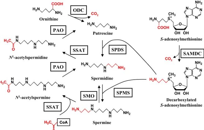
The polyamine metabolism pathway in E. coli and the genes involved in the pathway are shown in Fig. 1 and Table 1. Putrescine is synthesized either from decarboxylation of ornithine by ornithine decarboxylase or the combined action of decarboxylation of arginine by arginine decarboxylase and hydrolysis of agmatine by agmatine ureohydrolase [60]. Putrescine is converted into spermidine by sequential combination with decarboxylated S-adenosylmethionine derived from S-adenosylmethionine which is formed by S-adenosylmethionine decarboxylase, SAMDC [61,62]. The difference between polyamine biosynthesis in E. coli and mammalian cells is the presence in E. coli of both ornithine decarboxylase and arginine decarboxylase. In contrast to E. coli, mammalian cells do not have arginine decarboxylase and synthesize putrescine only by the ornithine decarboxylase pathway [60-62]. The polyamine biosynthesis and degradation pathway in mammalian cells are shown in Fig. 2.
Regulation of polyamine biosynthesis is complex and both ODC and SAMDC are rate-limiting enzymes in the pathway of polyamine biosynthesis. In
Fig.1. Polyamine metabolism and related genes in E. Coli. CAD, cadaverine; PUT,
putrescine; SPD, spermidine; SAM, S-adenosylmethionine; adiA, inducible arginine decarboxylase; speA, biosynthetic arginine decarboxylase; speC, constitutive ornithine decarboxylase; speF, inducible ornithine decarboxylase ; speD, S-adenosylmethionine decarboxylase; ldcC, constitutive lysine decarboxylase; cadA, inducible lysine decarboxylase; gsp, Glutathionylspermidine synthetase / amidase; speG, Spermidine acetyltransferase Orn speC speF PUT CAD Aminopropyl CAD SAM speD Decarboxylated SAM SPD Arg speA adiA AGM Lys ldcC cadA N1 N2 COOH N1COOH N2 N1 N2 N1 N2 speE gsp speG speB speE Glutathionyl-SPD Acetyl-SPD
Table 1 Polyamine-related genes in E. coli
Gene Length (kbp)
Enzyme Direction* Location(min) Ref.
ldcC speE speD speF speG speA speB speC gsp adiA cadA 2.1 1.7 3.5 0.6 3.1 2.2 1.4 3.4
Constitutive lysine decarboxylase
Spermidine synthase
S-Adenosylmethionine decarboxylase Inducible ornithine decarboxylase Spermidine acetyltransferase Biosynthetic arginine decarboxylase Agmatinase (agmatine ureohydrolase) Constitutive ornithine decarboxylase Glutathionylspermidine synthetase / amidase
Inducible arginine decarboxylase Inducible lysine decarboxylase
+
-
-
+-
-
-
0.5 2.9 15.5 35.7 66.4 66.9 67.6 93.5 [63] [64] [65] [66] [67] [68] [69] [70] +, Clockwise;-
, counterclockwise.Fig. 2. Polyamine metabolic pathway in mammalian cells. ODC, ornithine
decarboxylase; PAO, polyamine oxidase; SAMDC, S-adenosylmethionine decarboxylase; SMO, spermine oxidase; SPDS, spermidine synthase; SPMS, spermine synthase; SSAT, spermidine/spermine N1-acetyltransferase.
H2N NH2 COOH H2N NH2 H2N N H NH2 H N N H NH2 H2N N N N N NH2 O OH OH S+ CH3 H2N COOH N N N N NH2 O OH OH S+ CH3 H2N CO2 CO2 Ornithine Putrescine Spermidine Spermine S-adenosylmethionine Decarboxylated S-adenosylmethionine N1-acetylspermine N1-acetylspermidine H N N H NH2 H3C O H N N H NH2 H N H3C O H3C O CoA H3C O CoA ODC SAMDC SSAT PAO SMO SSAT PAO SPDS SPMS
E. coli, ODC is regulated both post-translationally, by covalent modification [71] and/or interaction with inhibitor proteins [72,73], and transcriptionally [73,74]. Antizyme is a non-competitive protein inhibitor of ODC, the synthesis of which is induced by polyamine [75]. The inhibition of ODC by antizyme is reversible and active ODC can be recovered from inactive ODC-antizyme complexes under certain conditions [72,76]. In mammals, there is evidence involving antizyme in ODC degradation [77-80] and polyamine transport for down regulation of the intracellular polyamine level [81-83]. Production of antizyme is induced by polyamines through translational frameshifting [84]. Antizyme is encoded by two overlapping open reading frames (ORF1 and ORF2), and translational frameshifting forward one base just before the stop codon of ORF1 is required for the synthesis of full length (i.e. active form) of antizyme. Antizyme can bind to ODC leading to degradation of ODC by the 26S proteasome [85,86]. The degradation of ODC by proteasome is ATP-dependent, but ubiquitine- independent [87,88].
In E. coli, there are two parallel pathways in spermidine degradation. Spermidine is converted to either glutathionylspermidine by glutathionyl spermidine synthase [89] or acetylspermidine by spermidine acetyltransferase [90]. On the other hand, in mammalian cells the first step of polyamine degradation is N1-acetylation of spermidine and spermine by spermidine/spermine N1-acetyltransferase, SSAT, to form N1-acetylspermidine and N1-acetylspermine using acetyl-CoA [91,92]. These N1-acetylpolyamines are then oxidized by FAD-dependent polyamine oxidase to form putrescine and spermidine, respectively [93]. The oxidation of acetylated polyamines produce 3-acetamidopropanal and H2O2, which are both toxic to cells [94,95]. Again, spermine is then directly oxidized by spermine oxidase to produce
spermidine and 3-aminopropanal [96-99]. The 3-aminopropanal is spontaneously converted to acrolein, which is a highly reactive compound that can bind to lysine and histidine residues and interferes the function of proteins [100,101]. Acrolein and spermine oxidase are significantly increased in the plasma of chronic renal failure [102] and stroke patients [103], so that they can use as markers for the diagnosis of these diseases.
Although the polyamine metabolic pathways are well sophisticated, the transport of polyamines plays crucial roles in regulation of cellular polyamine level which have not been clearly known. In the case of polyamine-deficient mutant, polyamine uptake increases. Then accumulation of polyamines in cells stimulate cell growth [104,105]. Moreover, excretion of polyamines from cells is vital process. Polyamine transport of the cell appears to be regulated by the growth status of that cell, which is inactive in the growing cell and active in the resting cells [106,107].
To gain understanding of the polyamine transport systems, the genes encoding polyamine transporter in E. coli have been identified [104], and some of the properties of these transporters have been studied at a biochemical, molecular and structural level. Although, bacteria differs from fungi and mammals, the study of polyamine transport systems in bacteria could provide some information that may help to understand the polyamine transport systems in other cellular systems.
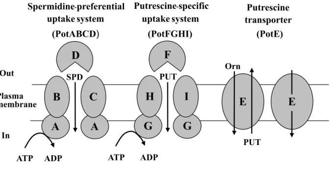
In E. coli, the rate of polyamine uptake is in the order: putrescine > spermidine > spermine [108]. Three polyamine transport systems were identified and characterized in E. coli (Fig. 3) [109]. The uptake of polyamine is mainly catalysed by two polyamine-uptake systems. Both of the systems are ATP binding cassette (ABC) transporters. One is a putrescine-specific system consisting of four proteins,
Fig. 3. Polyamine transport system in E. coli. PUT, putrescine; SPD, spermidine; Orn, ornithine
E
E
Orn PUTG
H
G
I
F
PUT ATP ADP SPD ADP ATPA
C
A
B
D
Out Plasma membrane In uptake system (PotABCD) uptake system (PotFGHI) Putrescine transporter (PotE) Spermidine-preferential Putrescine-specificPotF (the substrate-binding protein), PotG (an ATPase), PotH and PotI (channel-forming proteins) [110]. The second is spermidine-preferential uptake system, consisting of PotA (an ATPase), PotB and PotC (channel-forming proteins), and PotD (a substrate-binging protein) [111]. The structure of substrate binding proteins PotD and PotF were solved by X-ray crystallography [112-114] and polyamine binding sites were characterized by site-directed mutagenesis of the genes [115,116]. The third transport system is catalysed by PotE. PotE is a member of amino acid-polyamine-organocation (APC) transporter family, mediated both uptake and excretion of putrescine [65,117]. The uptake of putrescine was dependent on membrane potential, whereas the excretion was catalyzed by the putrescine/ornithine exchange reaction [118,119]. Like PotE, a cadaverine-lysine antiporter CadB exists in E. coli and others [120-122].
The E. coli cadBA operon and the adjacent cadC gene encode lysine decarboxylase (cadA), a putative lysine-cadaverine antiporter (cadB), and CadC, a positive regulator of cadBA expression [120,123]. Expression of the cadBA operon is induced by acidic pH and lysine [124]. Production of cadaverine, its excretion and the consumption of a proton during the decarboxylation reaction contribute to an increase in the pH of the media and to cell growth. It is also proposed that the cadBA operon functions as a supplier of carbon dioxide [125].
To understand further the role of the products of the cadBA operon, it is important to elucidate the structure and function of the cadaverine transport protein CadB, which has not been studied thus far. In this study, the properties of cadaverine transport by CadB and its physiological functions in cells, particularly at acidic pH were investigated. Furthermore, cadaverine recognition site on CadB based on the
CHAPTER I
Physiological Functions of Cadaverine Transporter CadB
I-1 Introduction
The Escherichia coli cadBA operon and the adjacent cadC gene encode lysine decarboxylase (cadA), a putative cadaverine-lysine antiporter (cadB), and CadC, a positive regulator of cadBA expression [120,123]. Expression of the cadBA operon is induced by acidic pH and lysine [124]. It is thought that production and excretion of cadaverine and the consumption of a proton during the decarboxylation reaction contribute to an increase in pH of the extracellular medium and to cell growth. It is also proposed that the cadBA operon functions as a supplier of carbon dioxide [125].
Several secondary transporters which catalyze antiport of a weak acid and its product such as malate and lactate, coupled with decarboxylation, have been reported as secondary proton motive force generation systems [126,127]. The basis for energy coupling was first characterized in Oxalobacter by Anantharam et al [128]. Furthermore, one antiport system for a basic amino acid and its amine product (histidine/histamine), coupled with decarboxylation, has been reported to generate a proton motive force in Lactobacillus buchneri [129]. Thus, it is conceivable that the cadaverine-lysine antiport system, coupled with decarboxylation, also functions as a proton motive force generation system in E. coli.
To understand the role of the products of the cadBA operon, it is important to elucidate the function of the cadaverine transport protein CadB, which has not yet been studied in detail. The structure and function of PotE has been recently delineated. PotE, which functions as a putrescine-ornithine antiporter and a putrescine transporter, is encoded by the pPT71 (speF-potE) operon consisting of speF, a gene for inducible ornithine decarboxylase, and potE [65,117-119]. Since the amino acid sequences of
PotE and CadB show high similarity, CadB was hypothesized to be a cadaverine-lysine antiporter. In this study, the properties of cadaverine transport by CadB and its physiological functions in cells, particularly at acidic pH, were investigated.
I-2 Results
Uptake and Excretion of Cadaverine by CadB
Chromosomal location and schematics of the cadC gene, cadBA operon and speF-potE operon are shown in Fig. 4A. A positive or negative regulator gene for the speF-potE operon is not located close to the operon, and so far unidentified. Homology between the amino acid sequences of CadB and PotE was compared, and high sequence similarity was observed (30.7% overall identity) especially in the NH2-terminal region (Fig. 4B), suggesting that CadB has structure and function similar to PotE and that it may be involved in both uptake and excretion of cadaverine.
The cadB gene was cloned into the vector pMW119 under the control of lacUV promoter. pMW119 and pMWcadB were introduced into MA261cadC::Km, which is deficient in positive regulator CadC, and into JM109. Uptake and excretion of cadaverine by CadB were measured using intact cells of MA261cadC::Km/pMW119 or pMWcadB, and lysine-loaded inside-out membrane vesicles prepared from JM109/pMW119 or pMWcadB, respectively. The results are shown in Fig. 5. Uptake of cadaverine by CadB was observed at neutral pH and found to be immediately inhibited by CCCP (carbonyl cyanide m-chlorophenylhydrazone), a proton uncoupler, suggesting that uptake is dependent on the proton motive force (Fig. 5A). Putrescine and lysine inhibited cadaverine uptake by CadB about 20% at 25-times higher concentration (250 µM) of substrate (10 µM). Cadaverine uptake was inhibited by ornithine and agmatine with a lesser extent compared to that observed in putrescine and lysine, and arginine had no effect on the uptake (data not shown). The results suggest
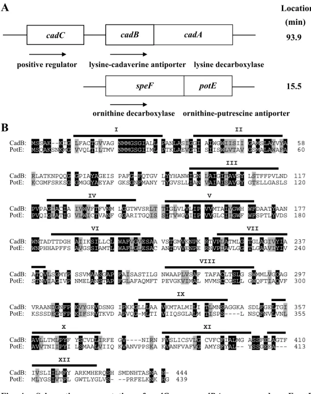
A
Location (min)93.9
positive regulator lysine-cadaverine antiporter lysine decarboxylase
15.5
ornithine decarboxylase ornithine-putrescine antiporter
B
I IICadB: MSSAK--KIG LFACTGVVAG NMMGSGIALL PANLASIGGI AIWGWIISII GAMSLAYVYA 58
PotE: MSQAKSNKMG VVQLTILTMV NMMGSGIIML PTKLAEVGTI SIISWLVTAV GSMALAWAFA 60 III
CadB: RLATKNPQQG GPIAYAGEIS PAFG-FQTGV LYYHANWIGN LAIGITAVSY LSTFFPVLND 117
PotE: KCGMFSRKSG GMGGYAEYAF GKSGNFMANY TYGVSLLIAN VAIAISAVGY GTELLGASLS 120 IV V
CadB: PVPAGIACIA IVWVFTFVNM LGGTWVSRLT TIGLVLVLIP VVMTAIVGWH WFDAATYAAN 177
PotE: PVQIGLATIG VLWICTVANF GGARITGQIS SITVWGVIIP VVGLCIIGWF WFSPTLYVDS 180
VI VII
CadB: WNTADTTDGH AIIKSILLCL WAFVGVESAA VSTGMVKNPK RTVPLATMLG TGLAGIVYIA 237
PotE: WNPHHAPFFS AVGSSIAMTL WAFLGLESAC ANTDVVENPE RNVPIAVLGG TLGAAVIYIV 240 VIII
CadB: ATQVLSGMYP SSVMAASGAP FAISASTILG NWAAPLVSAF TAFACLTSLG SWMMLVGQAG 297
PotE: STNVIAGIVP NMELANSTAP FGLAFAQMFT PEVGKVIMAL MVMSCCGSLL GWQFTIAQVF 300 IX
CadB: VRAANDGNFP KVYGEVDSNG IPKKGLLLAA VKMTALMILI TLMNSAGGKA SDLFGELTGI 357
PotE: KSSSDEGYFP KIFSRVTKVD APVQG-MLTI VIIQSGLALM TISPS----L NSQFNVLVNL 355 X XI
CadB: AVLLTMLPYF YSCVDLIRFE GV----NIRN FVSLICSVLG CVFCFIALMG ASSFELAGTF 410
PotE: AVVTNIIPYI LSMAALVIIQ KVANVPPSKA KVANFVAFVG AMYSFYAL-- YSSGEEA--- 413 XII
CadB: IVSLIILMFY ARKMHERQSH SMDNHTASNA H- 444
PotE: MLYGSIVTFL GWTLYGLVS- --PRFELKNK HG 439
Fig. 4. Schematic representation of cadC gene, cadBA operon and speF-potE operon (A), and alignment of the amino acid sequence of CadB and PotE (B). A.
Chromosomal location and encoding proteins of cadC gene, cadBA operon and speF-potE operon are shown. B. Sequence similarities of CadB and PotE were analyzed according to the method of Needleman and Wunsch [130] using a DNASIS program (Hitachi Software Engineering Co. Ltd.). The identical amino acid residues are shown by black boxes with white lettering, and the similar residues by gray boxes.
speF potE
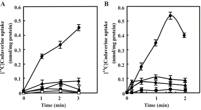
Fig. 5. Cadaverine uptake (A) and excretion (B) by CadB. A. Cadaverine uptake
activity was measured using intact cells of MA261cadC::Km containing pMW119 or pMWcadB with 10 µM [14C]cadaverine as a substrate as described in “Experimental procedures”. Where
indicated, 40 µM CCCP, 250 µM putrescine or 250 µM lysine was added to the reaction mixture. , MA261cadC::Km/pMWcadB; , MA261cadC::Km/pMWcadB + CCCP; U, MA261cadC::Km/pMWcadB + putrescine; , MA261cadC::Km/pMWcadB + lysine; z, MA261cadC::Km/pMW119. Km and Vmax values of cadaverine uptake by intact cells were measured by changing the concentration of substrates from 10 to 200 µM and shown in the figure. B. Cadaverine excretion activity was measured by cadaverine uptake by lysine-loaded inside-out membrane vesicles of JM109 containing pMW119 or pMWcadB with 100 µM [14C]cadaverine as a substrate as described in “Materials and Methods”. Where indicated, 40
µM CCCP was added to the reaction mixture. , JM109/pMWcadB; , JM109/pMWcadB + CCCP; z, JM109/pMW119; {, JM109/pMW119 + CCCP. Km and Vmax values of cadaverine uptake by lysine-loaded inside-out membrane vesicles were measured by changing the concentration of substrates from 100 µM to 2000 µM. Values are mean ± S. D. of triplicate
6 4 2 0 90 30 60 0 [ 14 C] Cadaverine uptak e (nmol/mg pr otein) Time (sec) 90 30 60 0 Time (sec) 0.05 0.15 0.10 [ 14 C] Cadaverine uptak e (nmol/mg pr otein) 0 0.20 10 8 Uptake Km : 20.8 µM Vmax : 11.8 nmol/mg Excretion Km: 390 µM Vmax : 0.683 nmol/mg A B
that CadB has a high specificity for cadaverine as the uptake substrate. Cadaverine uptake activity was low at acidic pH (data not shown). The Km value for cadaverine was calculated as 20.8 µM from Lineweaver-Burk plots. Excretion was measured by cadaverine uptake into lysine-loaded inside-out membrane vesicles. Compared to the vesicles from JM109/pMW119, the inside-out membrane vesicles prepared from JM109/pMWcadB exhibited a high activity which was not inhibited by CCCP, indicating that cadaverine is excreted by CadB through cadaverine-lysine antiporter activity (Fig. 5B). When cadaverine uptake was measured using unloaded inside-out membrane vesicles of JM109/pMWcadB, the activity was found to be 5% less than that measured with lysine-loaded inside-out membrane vesicles. Cadaverine uptake activity with inside-out membrane vesicles prepared at pH 7.5 rather than at pH 6.6 was low (data not shown). The Km value for cadaverine was calculated as 390 µM from Lineweaver-Burk plots.
Function and Properties of CadB at Neutral pH
CadB catalyzes the uptake of cadaverine at neutral pH. Polyamines [putrescine (1, 4-diaminobutane), spermidine and spermine] are necessary for normal cell growth [1,3] and putrescine and spermidine are usually presented in E. coli. We therefore examined whether cadaverine (1, 5-diaminopentane), taken up by CadB, contributes to cell growth in a polyamine-deficient mutant MA261 similar to effects of exogenous polyamines in this mutant. The E. coli polyamine-deficient mutant MA261 is deficient in the speB gene encoding agmatine ureohydrolase, and the speC gene encoding ornithine decarboxylase [131]. Thus, MA261 cells can not synthesize putrescine, although putrescine transported into cells can be converted to spermidine. When MA261 cells were cultured in the presence of cadaverine, cell growth was stimulated compared to cells cultured in the absence of cadaverine, but the effect of cadaverine on cell growth was smaller than that of putrescine (Table 2). The
Table 2 Cell growth of E. coli MA261 and MA261cadC::Km at neutral pH
Cells Addition Generation time Growth stimulation
(h) (-fold) MA261 None 5.25 ± 0.59 1.0 Putrescine (10 µg/ml) 1.68 ± 0.09 3.1 Putrescine (30 µg/ml) 1.68 ± 0.07 3.1 Cadaverine (10 µg/ml) 3.11 ± 0.23 1.7 Cadaverine (30 µg/ml) 3.03 ± 0.15 1.7 MA261cadC::Km None 4.92 ± 0.71 1.0 Putrescine (10 µg/ml) 1.88 ± 0.03 2.6 Putrescine (30 µg/ml) 1.71 ± 0.02 2.9 Cadaverine (10 µg/ml) 3.98 ± 0.33 1.2 Cadaverine (30 µg/ml) 3.90 ± 0.33 1.3
Cells were grown in medium A in the absence and presence of cadaverine or putrescine. Cell growth was followed by measuring A540. Values are mean ± S. D. of four determinations.
stimulatory effect of cadaverine on the growth of MA261cadC::Km cells, deficient in the cadC gene, a positive regulator of the cadBA operon, became smaller (Table 2). The difference of the stimulation effect of cell growth between putrescine and cadaverine became greater in MA261cadC::Km than in MA261. Cadaverine uptake activity of MA261cadC::Km cells was also low compared to MA261 cells (data not shown). Then, the polyamine content was measured under these conditions (Table 3). Cadaverine and aminopropylcadaverine accumulated in the polyamine-requiring mutant MA261, but there was less accumulation in the CadC-deficient mutant (Table 3). Aminopropylcadaverine can be synthesized from cadaverine by spermidine synthase, like spermidine from putrescine, and it also stimulates cell growth like spermidine [132]. Levels of putrescine and spermidine were decreased slightly after exposure to cadaverine. The results indicate that cadaverine transported by CadB stimulates cell growth of polyamine-deficient mutant of E. coli by itself or after conversion to aminopropylcadaverine, although the expression of CadB mRNA was low at neutral pH (see Fig. 10). When a strain MA261cadB::Km was used instead of MA261cadC::Km, essentially the same results were obtained (data not shown).
The characteristics of cadaverine uptake by CadB were examined using right side-out membrane vesicles of JM109/pUCcadB (Fig. 6). Right side-out membrane vesicles of JM109/pUCcadB showed high cadaverine uptake activity compared to the vesicles of JM109/pUC119 (vector) when the vesicles were energized by potassium ascorbate-phenazine methosulfate [133]. Uptake of cadaverine by CadB was greatly inhibited by CCCP, which dissipates the proton motive force, valinomycin, which dissipates the membrane potential, and nigericin which dissipates the transmembrane pH gradient (Fig. 6A), suggesting that cadaverine uptake was dependent on a proton motive force composed of both ∆pH and ∆Ψ. This was confirmed by measurement of cadaverine uptake by generating artificial ion gradients. When a proton motive force composed of both ∆pH and ∆Ψ was generated, cadaverine uptake was observed.
Table 3 Polyamine content in E. coli MA261 and MA261cadC::Km cultured at neutral pH
Cells Addition Polyamine (nmol/mg protein)
Putrescine Cadaverine Spermidine Aminopropyl-
cadaverine MA261 None 0.08 ± 0.02 0.63 ± 0.09 5.22 ± 0.57 4.02 ± 0.46 Putrescine (10 µg/ml) 19.8 ± 2.34 0.37 ± 0.05 15.5 ± 1.83 ND Cadaverine (10 µg/ml) ND 8.55 ± 0.97 3.58 ± 0.53 16.4 ± 1.77 MA261cadC::Km None 0.13 ± 0.03 0.03 ± 0.01 6.45 ± 0.74 4.61 ± 0.52 Putrescine (10 µg/ml) 24.9 ± 3,35 ND 14.8 ± 1.64 ND Cadaverine (10 µg/ml) ND 1.03 ± 0.18 4.78 ± 0.62 8.43 ± 0.95
Cells were cultured for 11 h in the presence and absence of putrescine or cadaverine and polyamine content was measured as described in "Materials and Methods". Values are mean ± S. D. of triplicate determinations. ND, not detected.
Fig. 6. Effect of ionophores (A) and artificially imposed ion gradients (B) on cadaverine uptake by right side-out membrane vesicles of JM109/pUCcadB. A.
Effect of various ionophores on cadaverine uptake was measured in the presence of the electron donor system constituting of potassium-ascorbate-phenazine methosulphate. Right side-out membrane vesicles were prepared from JM109/pUCcadB (z, , S, ) or from JM109/pUC119 ( ). Cadaverine uptake activity was measured in the absence (z, ) and presence of 10 µM nigericine (), 8 µM valinomycin (S), or 40 µM CCCP (). B. Cadaverine uptake by right side-out membrane vesicles prepared from JM109/pUCcadB was measured under artificial ion gradients as described in “Materials and Methods”. z, ∆p; S, ∆Ψ; , ∆pH; , none. Values are mean ± S. D. of triplicate determinations.
A
0 0.1 0.2 0.3 0.4 0.5 0.6 0 1 2 3 Time (min)B
[ 14 C] Cadave rine upta k e (nmol/m g p ro te in ) 0 0.1 0.2 0.3 0.4 0.5 0.6 [ 14 C] Cadave rine upta k e (nmol/mg pr otein) 0 1 2 Time (min)However, cadaverine uptake was greatly diminished when only ∆pH or ∆Ψ was generated (Fig. 6B). The pH inside cells was slightly acidified during cadaverine uptake (data not shown). The result suggests that cadaverine may be transported with protons, although the stoichiometry of proton and cadaverine transport is not known.
Function and Properties of CadB at Acidic pH
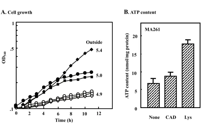
The growth of MA261 and MA261cadC::Km cells was compared at pH 5.2 in the presence and absence of lysine or cadaverine (Fig. 7A) in a synthetic medium. When MA261 cells were cultured at an initial acidic pH (5.2) in the presence of 5 mM lysine, stimulation of cell growth and neutralization of the medium were observed compared to cells cultured in the absence of lysine. Under this condition, expression of CadB mRNA greatly increased (see Fig. 10). Cadaverine did not support growth of MA261 cells at pH 5.2. Growth of MA261cadC::Km cells was not affected by lysine or cadaverine and neutralization of medium did not occur (Fig. 7A). Similar results were obtained with MA261cadB::Km (data not shown). When MA261 cells were cultured in the presence of lysine, the ATP content of the cells also increased (Fig. 7B). The contents of polyamines, cadaverine and aminopropylcadaverine in cells and medium were also measured (Table 4). When MA261 was cultured in the presence of lysine or cadaverine, an increase in cadaverine and aminopropylcadaverine was observed although cadaverine content was much higher with cells cultured in the presence of lysine than in the presence of cadaverine. In addition, the cadaverine content in the medium greatly increased when MA261 cells were cultured with lysine. Almost all lysine added to medium was converted to cadaverine. As a control, the cadaverine content in MA261cadC::Km cells and in medium was measured after culturing with lysine. The increase in cadaverine in these cells was small, and no cadaverine was excreted into the medium (Table 4). The results indicate that the cadBA operon was induced under acidic conditions in the presence of lysine, promoting decarboxylation of
Fig. 7. Cell growth and ATP content in cells cultured at acidic conditions. A.
MA261 and MA261cadC::Km cells were cultured in medium A, pH 5.2, containing 62 mM KH2PO4 instead of 40 mM K2HPO4 plus 22 mM KH2PO4 in the presence and absence of lysine or cadaverine. Cell growth was followed by measuring A540. After 11 h, pH of medium was measured and shown in the figure. MA261 (, , z) and MA261cadC::Km ( , , {) were cultured in the absence (, ) and presence of 5 mM lysine (, ) or cavaderine (z, {). Each point represents the mean of duplicate measurements. B. MA261 cells were cultured at acidic pH as described above in the absence and presence of 5 mM cadaverine or lysine for 11 h, and ATP content of cells was measured as described in “Materials and Methods”.CAD, cadaverine. Values are mean ± S. D. of triplicate determinations.
Outside 5.4
5.0
4.9
A
. Cell growthB
. ATP contentMA261 0 15 5 10 20 .1 .5 1 OD 540 A T P content (nmol/mg pr otein) Lys 0 2 4 6 8 10 12
Table 4 Polyamine content in E. coli cells and medium cultured at acidic pH
Strain Addition Polyamine (nmol/mg protein)
Putrescine Cadaverine Spermidine Aminopropyl-
cadaverine
Cells nmol/mg protein
MA261 None 1.82 ± 0.32 4.19 ± 0.55 16.1 ± 1.78 ND Lysine (5 mM) ND 192 ± 20.5 0.29 ± 0.05 5.05 ± 0.64 Cadaverine (5 mM) 0.11 ± 0.04 63.7 ± 7.54 3.60 ± 0.39 6.03 ± 0.74 MA261cadC::Km None 1.83 ± 0.23 23.2 ± 2.56 10.9 ± 1.23 ND Lysine (5 mM) 0.83 ± 0.09 44.1 ± 5.23 11.2 ± 1.33 ND Cadaverine (5 mM) ND 76.3 ± 8.21 6.10 ± 0.45 4.90 ± 0.43 Medium µM MA261 None ND 30.6 ± 4.5 ND ND Lysine (5 mM) ND 4660 ± 533 ND ND Cadaverine (5 mM) ND 4750 ± 567 ND ND MA261cadC::Km None ND ND ND ND Lysine (5 mM) ND ND ND ND Cadaverine (5 mM) ND 4890 ± 440 ND ND
Cells were cultured in medium A, pH 5.2, containing 62 mM KH2PO4 instead of 40 mM K2HPO4 plus 22 mM KH2PO4 for 11 h in the presence and absence of lysine or cadaverine, and polyamine content was measured as described in "Materials and Methods". Values are mean ± S. D. of triplicate determinations. ND, not detected.
lysine to cadaverine by lysine decarboxylase encoded by cadA and excretion of cadaverine by the cadaverine-lysine antiport activity of CadB, effects that support cell growth. The results also suggest that cadaverine and aminopropylcadaverine are used instead of putrescine and spermidine as polyamines for cell growth, because putrescine and spermidine contents decreased greatly in cells cultured in the presence of lysine.
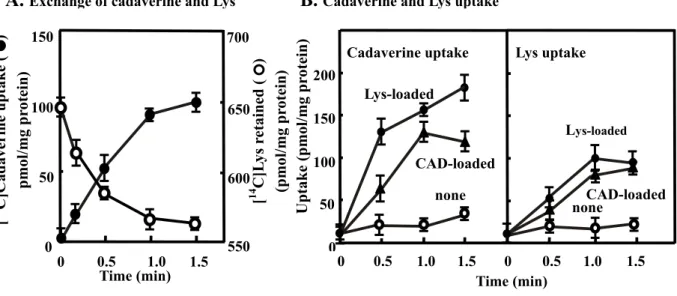
The characteristics of the cadaverine-lysine antiport activity of CadB were studied (Fig. 8). Inside-out membrane vesicles of JM109/pMWcadB were loaded with [14C]lysine or unlabeled lysine, and the stoichiometry of exchange of lysine with cadaverime was measured by adding unlabeled cadaverine or [14C]cadaverine, respectively. The amount of lysine retained in the membrane vesicles decreased as cadaverine uptake increased. The exchange ratio of lysine with cadaverine was nearly 1:1 (Fig. 8A). To examine whether CadB has not only cadaverine-lysine antiporter activity but also cadaverine-cadaverine and lysine-lysine exchange activity, lysine-loaded and cadaverine-loaded inside-out membrane vesicles were prepared, and cadaverine or lysine uptake activity was measured. As shown in Fig. 8B, significant cadaverine or lysine uptake activity was observed in vesicles loaded with cadaverine or lysine, indicating that CadB has cadaverine-lysine, cadaverine-cadaverine and lysine-lysine antiporter activities. [14C]Cadaverine uptake by lysine-loaded inside-out membrane vesicles was inhibited strongly by 10-fold excess of cadaverine and lysine (1 mM) of substrate (100 µM), about 20% by putrescine, and only slightly (about 5%) by ornithine, agmatine and arginine (data not shown). This suggests that the antiporter activity of CadB has a high substrate specificity for cadaverine and lysine.
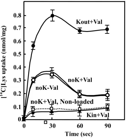
Whether electrogenic exchange of lysine with cadaverine takes place was next tested [129,134]. To examine this possibility, right side-out membrane vesicles of JM109/pUCcadB were loaded with cadaverine together with either N-methylglucamine (NMG) salts or potassium salts. Addition of NMG salts or potassium salts together with valinomycin generates a membrane potential whose polarity was either interior
positive (potassium outside, NMG inside) or interior negative (NMG outside, potassium inside). As shown in Fig. 9, when a membrane potential was not generated ({, U), significant cadaverine-lysine exchange activity was observed. When an interior positive membrane potential was generated (z), cadaverine-lysine exchange activity was greatly stimulated, whereas it was inhibited when an interior negative membrane potential was generated (). Accordingly, the process of cadaverine excretion with lysine uptake catalyzed by CadB will result in the generation of an interior negative membrane potential. When right-side-out membrane vesicles were prepared without cadaverine, no significant lysine uptake was observed ( ), indicating that this process is dependent on the presence of cadaverine as a counter substrate.
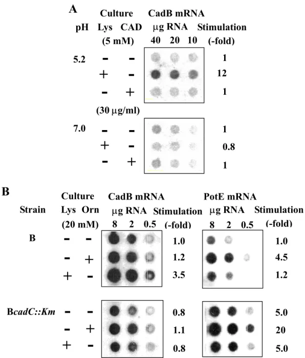
Expression of cadBA and speF-potE Operons
It has been reported that expression of the cadBA operon is induced by lysine at acidic pH [124]. Expression of the cadBA operon was compared at neutral and acidic pH using MA261 cells cultured in synthetic medium in the presence and absence of lysine or cadaverine (Fig. 10A). Expression of the cadBA operon was greatly stimulated only when cells were cultured at acidic pH in the presence of lysine. Expression of the cadBA operon was low at neutral pH even in polyamine-requiring mutant, MA261. Expression of the cadBA and speF-potE operons was then compared in E. coli B and BcadC::Km cultured at acidic pH in rich medium. In E. coli B the degree of induction of CadB mRNA by lysine was 3.5-fold. Using BcadC::Km cells, in which cadC, a positive regulator of cadBA expression is disrupted, there was no induction of CadB mRNA by lysine. Similarly, PotE mRNA could be induced 4.5-fold by ornithine. Although the precise comparison of the degree of expression of two operons is difficult by the dot blot analysis, the level of PotE mRNA in the presence of ornithine seems to be about 25% of the level of CadB mRNA in the presence lysine. At acidic pH, the level of CadB mRNA may be equal to or greater than that of PotE mRNA
Fig. 8. Exchange of cadaverine (CAD) with lysine (Lys) (A), and cadaverine and lysine uptake (B) by inside-out membrane vesicles. A. To measure the exchange
ratio of lysine with cadaverine, inside-out membrane vesicles of JM109/pMWcadB prepared in the absence of amino acid were preloaded with 1 mM [14C]lysine ({) or unlabeled lysine (z) for 1 h. Exchange activity was then measured by diluting the membrane vesicles by 1:10 and by adding 100 µM cadaverine ({) or [14C] cadaverine (z), respectively. B. Inside-out membrane vesicles of JM109/pMWcadB were prepared in the absence ({) and presence of 2.5 mM lysine (z) or cadaverine (S). [14C]Cadaverine (left) or [14C]lysine (right) uptake activity was measured as described in "Materials and Methods". CAD, cadaverine. Values are mean ± S. D. of triplicate determinations.
B. Cadaverine and Lys uptake
A. Exchange of cadaverine and Lys
Cadaverine uptake 0 0.5 1.0 1.5 noneCAD-loaded Lys-loaded 0 50 100 150 200 Uptake (pmol/mg pr otein) none CAD-loaded Lys-loaded 0 0.5 1.0 1.5 Lys uptake 550 600 650 700 0 50 100 150 0 0.5 1.0 1.5 [ 14 C]L ys r etained ( ) (pmol/mg pr otein)
Time (min) Time (min)
[ 14 C]Cadaverine uptake ( ) pmol/mg pr ot ein)
Fig. 9. Electrogenic exchange of lysine with cadaverine in right side-out membrane vesicles of JM109/pUCcadB. Right side-out membrane vesicles were
loaded with 1 mM cadaverine (z, {, U, ) or without cadaverine ( ) plus 100 mM phosphate as the NMG salt (z, {, U, ) or the potassium salt () and diluted 10-fold by the reaction mixture containing 100 µM [14C] lysine along with 100 mM sulfate plus 100 mM phosphate as the potassium salt (z) or the NMG salt ({, U, , ) in the presence (z, U, , ) and absence ({) of 1 µM valinomycin. Values are mean ± S. D. of triplicate determinations.
Kout+Val
noK-Val
noK+Val
Kin+Val
0
0.1
0.2
0.3
0.4
0.5
0.6
0.7
0.8
0
30
60
90
Time (sec)
[
14C]L
ys u
p
take
(nmol/m
g)
noK+Val, Non-loaded
Fig. 10. Expression of CadB and PotE mRNAs. A. Expression of CadB mRNA in
MA261 cultured at acidic and neutral pH in the presence and absence of lysine or cadaverine. MA261 cells were cultured at pH 5.2 or pH 7.0 as described in the legends of Fig. 8, and dot blot analysis of CadB mRNA was performed as described in "Experimental procedures". B. Comparison of expression of CadB and PotE mRNAs in E. coli B and BcadC::Km cultured at acidic pH in the presence and absence of ornithine or lysine. Culture of E. coli B and BcadC::Km, and dot blot analysis of CadB and PotE
A
CadB mRNA pH Lys CAD (5 mM) µg RNA Stimulation (-fold) 5.2 7.0 12 1 1 1 1 0.8--
-+
+
-
-
-
-
+
+
(30 µg/ml) 10 40 20 CultureB
Culture PotE mRNA-
-
-
-
+
+
--
-+
+
CadB mRNA µg RNA 3.5 1.0 0.8 0.8 1.1 1.2 Stimulation (-fold) 0.5 8 2 µg RNA Stimulation (-fold) 1.2 1.0 5.0 5.0 20 4.5 0.5 8 2 Lys Orn Strain B BcadC::Km (20 mM)regardless of the presence of lysine or ornithine, unless cadC was disrupted. When the cadC gene was disrupted, the level of PotE mRNA was increased by 5-fold. The results suggest that PotE and ornithine decarboxylase have functions that can compensate for the loss of CadB and lysine decarboxylase.
I-3 Discussion
In this study, CadB has not only cadaverine uptake activity but also cadaverine-lysine antiport activity has been shown. These data were obtained using E. coli MA261 intact cells, which is deficient in polyamine biosynthesis and right side-out and inside-out membrane vesicles prepared from E. coli JM109 cells. It was because preparation of right-side-out and inside-out membrane vesicles of MA261 strain was difficult and more active membrane vesicles could be made from E. coli JM109 cells. At neutral pH, CadB is involved in cadaverine uptake consuming a proton motive force as an energy source (Fig. 11). Cadaverine and aminopropylcadaverine can presumably substitute for the intracellular functions of putrescine and spermidine if these two polyamines are not available. However, expression of the cadBA operon was low at neutral pH. Consequently, the physiological effect of CadB at neutral pH is thought to be small and the effect would appear only in the specific polyamine-deficient strain. At acidic pH, CadB functions as a cadaverine-lysine antiporter (Fig. 11). The process of cadaverine excretion and lysine uptake catalyzed by CadB generates a membrane potential and decarboxylation of lysine by lysine decarboxylase generates a pH gradient through consumption of a cytoplasmic proton. This proton motive metabolic cycle causes neutralization of acidic conditions and an increase in the level of ATP in cells (Figs. 7 and 11). CO2 produced by lysine decarboxylase may also contribute to the various metabolic pathways including nucleotide biosynthesis. Although the surrounding concentration of protons is much higher at acidic pH, CadB functions as a
Fig. 11. Physiological functions of CadB in E. coli. CadA, lysine decarboxylase;
CadB, cadaverine transporter. At neutral conditions, CadB functions as cadaverine and proton symporter and cell growth was stimulated by cadaverine. At acidic conditions, CadB functions as electrogenic cadaverine-lysine antiporter and pH in the medium is increased by cadaverine. CadA (lysine decarboxylase) generates a pH gradient by consumption of a cytoplasmic proton. This process causes the increase in the level of ATP in cells. Cadaverine Out In Neutral conditions CadB CadB
Lys (in excess) Acidic conditions Out In Cadaverine + CO2 CadA Lys Cadaverine (CH2)4 HCCOO -+NH3 +NH3 +NH 3 +NH3 (CH2)5 Plasma membrane H+ H+
cadaverine/lysine antiporter which benefits bacterial cell growth. Thus, the cadBA operon plays important roles in cell growth at acidic pH. In this respect, CadB may contribute more than PotE because the cadBA operon seems to be expressed more than the speF-potE operon. This seems rational because lysine is more common than ornithine in nature. However, in a cadC-disrupted mutant, expression of the speF-potE operon is enhanced. These results suggest that the speF-potE operon replaces the function of the cadBA operon at acidic pH or cadC is a negative regulator of the speF-potE operon. As for the enhancement of expression of the speF-potE operon, we have previously reported that RNase III increases the translational efficiency of iODC-PotE mRNA by cutting the 5'-untranslated region of the mRNA [135]. However, it remains to be clarified whether RNase III is activated at acidic pH or not.
With regard to uptake activity, PotE may be more efficient than CadB, because the Km value of PotE for putrescine is 1.8 µM [118], whereas that of CadB for cadaverine is 20.8 µM. Polyamines (putrescine, spermidine and spermine) are known to stimulate protein synthesis [28,30,31,119]. The effective concentrations required for stimulation of protein synthesis are in the order putrescine > spermidine > spermine [14]. Cadaverine and aminopropylcadaverine also stimulate protein synthesis, and their effective concentrations are similar to those of putrescine and spermidine, respectively [47]. Thus, the slower growth of cells with cadaverine than with putrescine in the polyamine-requiring mutant MA261 (Table 2) was probably due to less accumulation of cadaverine than putrescine in the cells (Table 3). At acidic pH, cadaverine and aminopropylcadaverine accumulate in cells through the function of CadB and lysine decarboxylase, but the synthesis of putrescine and spermidine was inhibited to maintain an optimal concentration of "polyamines" including cadaverine and aminopropylcadaverine.
Polyamine content in cells was regulated by polyamine biosynthesis, degradation and transport. Although polyamine biosynthesis and degradation are well
characterized, properties of polyamine transport are not. In E. coli, it was reported that there are two ATP-dependent polyamine uptake systems in addition to PotE and CadB in E. coli [136], but only a PotE (or CadB)-like protein exists as a polyamine transport system in Rickettsia prowazekii [137]. Similarly, only PotE (or CadB)-like proteins have been found thus far as polyamine transport systems in Saccharomyces cerevisiae [138,139]. Thus, the secondary polyamine uptake systems like PotE or CadB may be the major polyamine uptake systems in certain cell types.
CHAPTER II
Identification of the Cadaverine Recognition Site
on Cadaverine Transporter CadB
II-1 Introduction
In polyamine transport, the properties of three polyamine transport systems were characterized in Escherichia coli [115,119,140]. They include spermidine-preferential and putrescine-specific uptake systems, which belong to the family of ATP-binding cassette transporters, and a protein, PotE, involved in the excretion of putrescine by a putrescine-ornithine antiporter activity. Furthermore, it has been reported that cadaverine and aminopropylcadaverine function as compensatory polyamines for cell growth [132], and both PotE and CadB, a cadaverine-lysine antiporter, are strongly involved in cell growth at acidic pH [105,124]. Like speF-potE operon [65], cadB is one component of the cadBA operon, in which cadA encodes lysine decarboxylase [120,123]. The speF-potE and cadBA operons both contribute to an increase in pH of the extracellular medium through excretion of putrescine and cadaverine, the consumption of a proton, and a supply of carbon dioxide during the decarboxylation reaction [105,125] so that expression of these two operons is important for cell growth at acidic pH. Other antiport systems for basic amino acids and their amine products (histidine/histamine and arginine/agmatine), coupled with decarboxylation, have been reported to generate a proton motive force in Lactobacillus buchneri and E. coli [129,141]. Although the function of CadB has been recently studied in detail [105], the structure of CadB is not well understood. When homology between the amino acid sequences of CadB and PotE was compared, high sequence
similarity was observed (30.7% overall identity) [105]. Amino acid residues involved in putrescine uptake and excretion by PotE have been previously identified [119]. In this study, cadaverine recognition site on CadB based on the information concerning putrescine recognition by PotE was identified.
II-2 Results
Identification of Amino Acid Residues Involved in Uptake and Excretion of Cadaverine
It is known that polyamines are recognized by proteins through their interaction with acidic and aromatic amino acid residues [115,116,119,142]. There are 9 aspartic acid and 7 glutamic acid residues in CadB. These residues were individually mutated to asparagine and glutamine, respectively, using site-directed mutagenesis of the cadB gene. Cadaverine uptake activity was measured using E. coli JM109 transformed with pMW encoding wild type or mutated CadB. Cadaverine excretion (i. e. cadaverine-lysine antiporter activity) was measured by [14C]cadaverine uptake using lysine-loaded inside-out membrane vesicles prepared from E. coli JM109 transformed with pMW encoding wild type or mutated CadB. As shown in Fig. 12, both uptake and excretion were greatly decreased with the mutants E204Q and D303N and moderately with E76Q and E377Q, and only uptake was moderately decreased with D185N and E408Q. There are 11 tryptophan and 13 tyrosine residues in CadB. These residues were mutated to leucine and the activities were measured. Both uptake and excretion were greatly decreased with the mutants Y73L, Y89L, Y90L, Y235L, and Y423L and moderately with Y55L, Y246L and Y310L, and only uptake was greatly decreased with
Fig. 12. Effect of Asp, Glu, Trp and Tyr mutations on the cadaverine uptake and excretion activities of CadB. Assays for cadaverine uptake and excretion were
performed under standard conditions. 100% activity of cadaverine uptake and excretion was 3.85 and 0.14 nmol/min/mg protein, respectively. Values are the mean ± S. D. of three samples. Activities of E. coli having pMW119 vector instead of pMWcadB or pMW mutated cadB were shown in the column labeled None. Amino acid residues indicated by white letters are involved in both activities, and residues in rectangles are involved in the uptake of cadaverine only.
II III IV V VI VII VIII X XII
Uptake Excretion C adB (W ild) No n e W41L W43L Y55L Y57L Y73L E76Q Y89L W94L Y107L W130L D 117N W142L W166L W168L D170N Y174L W178L D182N D185N W198L E204Q Y235L Y246L W269L W289L D303N 310LY E312Q D314N D349N E353Q D372N E377 429QE 436ND Q Y 423L Y 368L Y 366L E 408Q 50 100 0 Y 90L Relative activity (%)
W43L, Y57L, Y107L, Y366L, and Y368L and moderately with W41L and Y174L (Fig. 12).
In the previous studies of PotE, It was found that cysteine residues were involved in the recognition of putrescine by PotE [119]. There are 8 cysteine residues in CadB. Each cysteine was mutated to serine, and both uptake and excretion activities were measured. As shown in Fig. 13, both uptake and excretion activities were decreased with the mutant C370S, and then with C397S, but the effect of mutation was small with the mutants C12S, C125S, C196S, C282S, C389S and C394S. To confirm that Cys370 is important for CadB activities, all 8 cysteine residues were replaced by serine and both uptake and excretion were measured. As shown in Fig. 14, the activities of the ∆C mutant were decreased greatly, and they were effectively restored only with the mutant ∆C S370C, which has one cysteine residue at position 370, but not with the mutants ∆C S12C, S125C, S196C, S282C, S389C, S394C and S397C. The results confirmed that the SH group of Cys370 is involved in both uptake and excretion activities.
The most dramatic effects on both uptake and excretion of cadaverine were seen with the CadB mutants E204Q, D303N, Y73L, Y89L, Y90L, Y235L, and Y423L (Fig. 12). To confirm the importance of these amino acid residues, Glu204 was replaced by Asp and Asp303 by Glu. As shown in Fig. 15A, replacement of Glu204 and Asp303 by Asp and Glu did not restore the activities of CadB, indicating that Glu204 and Asp303 are critical for both activities. Similarly, Tyr73, Tyr89, Tyr90, Tyr235 and Tyr423 were replaced by Phe and Trp. Replacement of Tyr73, Tyr89 and Tyr235 by Phe and Trp did not restore the activities of CadB. Replacement of Tyr90 by Phe, but not by Trp, and replacement of Tyr423 by Phe and Trp slightly restored the activities of CadB. These results also
Fig. 13. Necessity of Cys370 residue for the uptake and excretion activities of CadB.
Assays were performed as described in the legend of Fig. 12. Values are the means ± S. D. of three samples. Amino acid residue (Cys370) indicated by white letter is involved in both activities.
0
50
100
150
CadB(W)
C12S
C125S
C196S
C282S
C389S
C394S
C397S
Relative activity (%
)
Uptake ExcretionC370S
Fig. 14. Necessity of Cys370 residue for the uptake and excretion activities of CadB.
Assays were performed as described in the legend of Fig. 12. Values are the means ± S. D. of three samples. Amino acid residue (Cys370) indicated by white letter is involved in both activities.
0
50
100
150
CadB
(Wi
ld
)
∆
C
∆
C
S12C
Relative activity (%
)
Uptake Excretion∆
C
S282C
∆
C
S389C
∆
C
S394C
∆
C
S397C
∆
C
S125C
∆
C
S196C
∆
C
S370C
Fig. 15. Cadaverine uptake and excretion activities of Glu204, Asp303, Tyr73, Tyr89, Tyr90, Tyr235 and Tyr423 mutants of CadB (A) and their expression (B). A. Assays
were performed as described in the legend of Fig. 12. Amino acid residues shown in the horizontal axis is the replaced amino acid residue. Values are the mean ± S. D. of three samples. B. Western blot analysis was performed using 100 µg of protein of the inside-out membrane vesicles.
L F W
Y73
Q D
E204
N E L F W L F W L F W L F W
D303 Y89 Y90 Y235 Y423
Uptake Excretion 0 50 100 Rela tive acti vi ty (% )
A.Cadaverineuptakeandexcretion activities
None Wild D303 N Y73L Y89L Y90L E204 Q Y235 L Y423 L
B. Expression of CadB mutants
indicate that these five tyrosine residues are critical for both activities of CadB. Expression of mutated CadB proteins was examined by Western blot analysis of inside-out membrane vesicles. Comparable amounts of mutated CadB proteins were expressed on the membranes, indicating that the mutations do not affect expression of CadB (Fig. 15B).
The Km values for uptake and excretion of cadaverine by mutants were then determined by Lineweaver-Burk plots. As shown in Table 5, the Km value for uptake of cadaverine by wild-type CadB was 20.8 µM. In all mutants, the Km value for cadaverine was increased, and this increase was parallel with the decrease in cadaverine uptake activity shown in Figs. 12 and 13. These results indicate that all amino acid residues related to the decrease in cadaverine uptake are involved in the recognition of cadaverine. The Km value for excretion of cadaverine by CadB was 390 µM (Table 5). In all CadB mutants, the Km value for cadaverine was increased, in parallel with the decrease in cadaverine excretion activity shown in Figs. 12 and 13. The change in the Km value for uptake of cadaverine was much greater than the change in the Km for excretion of cadaverine for any given mutant. This may be due to the existence of the limited amount of lysine in the inside-out membrane vesicles, which is the essential component for cadaverine-lysine antiporter activity.
In the case of putrescine-ornithine antiporter activity of PotE, Lys301 in C5 (cytoplasmic loop 5) is involved in recognition of the carboxy group of ornithine [119]. Thus, it was determined whether a basic amino acid(s) in C5 is involved in recognition of the carboxy group of lysine. As shown in Fig. 16, excretion but not uptake of cadaverine was markedly reduced in R299A, whereas both uptake and excretion activities were reduced to a similar, small extent in K308A, K320A, K321A and K329A
Table 5 The Km value of mutated CadB for cadaverine uptake and excretion Uptakea Excretiona Mutant Km (µM)b Degree of increase (-fold) Km (mM)b Degree of increase (-fold) Wild Y55L Y73L E76Q Y89L Y90L E204Q Y235L Y246L D303N Y310L C370A E377Q Y423L W41L W43L Y57L Y107L Y174L D185N Y366L Y368L E408Q 20.8 68.0 193 89.4 223 155 867 490 75.2 547 81.1 55.5 78.3 111 35.2 111 127 143 50.0 86.2 323 111 67.1 ± ± ± ± ± ± ± ± ± ± ± ± ± ± ± ± ± ± ± ± ± ± ± 0.46 8.23 19.2 3.06 27.9 13.5 70.7 109 6.93 3.92 10.4 0.97 3.61 9.26 1.02 2.16 9.45 11.2 4.29 6.10 35.3 9.26 4.45 3.27 9.28 4.30 10.7 7.45 41.7 23.6 3.62 26.3 3.90 2.67 3.76 5.34 1.69 5.34 6.11 6.88 2.40 4.14 15.5 5.34 3.23 0.39 1.02 3.86 1.65 2.05 2.37 2.67 2.82 1.30 2.96 1.10 0.99 0.72 2.61 ± ± ± ± ± ± ± ± ± ± ± ± ± ± – – – – – – – – – 0.01 0.06 0.06 0.14 0.13 0.27 0.38 0.26 0.05 0.42 0.10 0.18 0.08 0.94 2.62 9.90 4.23 5.26 6.08 6.85 7.23 3.33 8.97 2.82 2.54 1.85 6.69
a Assays were performed as described in the legend of Fig. 12.
b These values were calculated from the Lineweaver-Burk plots. Values are mean + S.D. of three samples.
Fig. 16. Effect of Arg and Lys mutations on the cadaverine uptake and excretion activities of CadB. Assays were performed as described in the legend of Fig. 12.
Values are the mean ± S. D. of three samples. The amino acid residues in rectangles is involved in lysine recognition.
0
50
100
150
Relative activity (%
)
CadB
(W
)
R299A
K308A
K320A
K321A
K329A
Uptake Excretion
K63A
R59A
mutants. As a control, basic amino acids in other cytoplasmic loops were also examined. As shown in Fig. 16, the uptake and excretion activities by basic amino acid mutants in other cytoplasmic loops ( R59A, K63A, R375A and R382A) did not decrease significantly. The results strongly suggest that Arg299 is involved in the recognition of lysine.
Location of Amino Acid Residues Involved in the Uptake and Excretion
Judging from the high sequence similarity between PotE and CadB (30.7% overall similarity) [105], CadB has twelve helices like PotE, as shown in Fig. 17. Amino acids important for both uptake and excretion were located in the transmembrane helices II, III, VI, VII, X, and XII or in the cytoplasmic region between transmembrane helices II and III (C2), VI and VII (C4), VIII and IX (C5), and X and XI (C6). Amino acid residues in the transmembrane helices are also located in the cytoplasmic side rather than the periplasmic side. In contrast to PotE, the Km value for cadaverine uptake by CadB is high (20.8 µM). Since numberous amino acid residues are involved in uptake of cadaverine, the participation of any individual residues in recognition of cadaverine may be weak. Those amino acids are mainly located in the transmembrane helices and the periplasmic loops and may be slightly separated from substrate binding site. However, it is expected that those amino acid residues are located close to each other in the tertiary-structure of CadB.
Transmembrane Helix Packing of CadB
It has been reported that a hydrophilic cavity of lactose permease having twelve transmembrane helices is formed between eight transmembrane helices: I, II, IV, V, VII,
![Fig. 5. Cadaverine uptake (A) and excretion (B) by CadB. A. Cadaverine uptake activity was measured using intact cells of MA261cadC::Km containing pMW119 or pMWcadB with 10 µM [ 14 C]cadaverine as a substrate as described in “Experimental procedures”](https://thumb-ap.123doks.com/thumbv2/123deta/8491145.921709/19.892.144.787.169.564/cadaverine-excretion-cadaverine-containing-cadaverine-substrate-experimental-procedures.webp)