Acta Med. Nagasaki 40: 30-34
Lung Xenotransplantation in the Rat
Takeshi NAGAYASU
First Department of Surgery, Nagasaki University School of Medicine, 1-7-1 Sakamoto, Nagasaki 852, Japan.
The aim of this study was to investigate the functional and morphological aspects of orthotopic lung xenograft rejection in a concordant hamster-to-rat donor-recipient species combination. All transplanted lungs had infiltrates during the 3 postoperative days. Complement-dependent cytotoxi- city assays revealed that anti-hamster lymphocytotoxic antibody titer increased to 5.2±1.1 (p < 0.05 vs. 1 day) 3 days after transplantation and reached 9.8±0.5 (p < 0.05 vs. 1, 3 and 5 days) 7 days after transplantation. Titers were not elevated 1 day after transplantation. The CD4+/CD8+ ratio in peripheral blood lymphocytes increased significantly at 3 days (p < 0.05, versus untransplanted) . At 5 th day, the ratio was lower than at 3 rd day (p < 0.05). There was no signifi- cant difference in B-cell population between rats before grafting and each group. Histologically, xenotrasplanted grafts were characterized by perivascular cellular infiltrates and edema 3 days after transplantation. The inflammatory infiltrate formed a dense cuff around the venules and arteri- oles. At 5 th day, arteriolar lumenal narrowing was observ- ed, and air-space containing many alveolar macrophages and lymphocytes. Immunohistochemical analysis of these grafts showed strong IgM and C3 deposits in the vascular endothelium, without any igG deposits. No IgM, C3 or IgG deposits were observed in normal hamster lung. This study indicates that both the cellular and humoral immune system mediate primary acute rejection in the hamster-to-rat orthotopic lung xenograft model.
Introduction
The development of immunosuppressive agents and surgical techniques has increased the number of patients eligible for organ transplantation. The shortage of donor organs, however, has become a serious problem. Xeno- transplantation, such as the baboon-to-human liver xenograft, recently reported by Starzl', may provide a solution to the shortage of donor organs. As with other organ xenografts2, lung transplantation may soon be performed using xenogenic organs.
In a well-established model of hamster-to-rat heteroto- pic cardiac xenotransplantation, rejection of a concordant xenograft was found to be mediated mainly by humoral immunity". The mechanism underlying lung xenograft rejection may differ, however, because the lung contains a considerable amount of lymphoid tissue, in contrast to
most other organs'. Tavakoli' has reported that in a guinea pig-to-rat discordant lung xenograft model, rejection occurred by a very rapid, predominantly humoral mecha- nism. However, there are no published papers on animal models of concordant lung xenograft. We have established a concordant hamster-to-rat orthotopic lung xenograft model to ivestigate the mechanism of lung xenograft rejection.
The aim of the present study was to investigate the functional and morphological aspects of orthotopic lung xenograft rejection in a concordant hamster donor to rat recipient model.
Materials and Methods Animals.
Female Golden Syrian hamsters weighing 100-170 g and Flscher (F344/L uvr ~) rats weigh 1,4 200-300 g were used as donors (SLC Inc, Japan), and inbred male Lewis (LEW/
Crj) rats weighing 200-300g were used as recipients (Charles River Inc, Japan).
Surgical procedure.
All animals were anesthetized with an intraperitoneal administration of 30 mg/kg of pentobarbital (Nembutal injection (R)), intubated, and ventilated at a tidal volume of 10 ml/kg and respiratory rate of 90 breaths/min using a respirator (Shinano-SN, Japan). Orthotopic lung trans- plantation was performed using a cuff technique, as previously described'. The pulmonary artery and pulmo- nary vein were anastomosed by the cuff technique and then the left main bronchus was anastomosed with a 9-0 polypropylene continuous suture for the cartilaginous ring and an interrupted suture for the membranous wall. All animals received humane care in compliance with the
"Guid
e for the Care and Use of Laboratory Animals of Nagasaki University".
Serial Chest Roentgenograms.
Chest roentgenograms were taken every day after
transplantation.
Experimental models
Rats were divided into four groups : group 1 (n = 4), animals were sacrificed 1 day after transplantation ; group 2 (n = 6), animals were sacrificed 3 days after transplantation ; group 3 (n = 7), animals were sacrificed 5 days after transplantation ; group 4 (n = 4), animals were sacrificed 7 days after transplantation.
Complement-dependent cytotoxicity (CDC) assay.
The concentration of lymphocytotoxic antibodies in the serum of xenografted rats was determined using a CDC assay'. After incubating the recipient's serum and comple- menting with hamster spleen cells (5x10⁶/ml) for 45 min, the percentage of cells staining with trypan blue was calculated. A score of 50% cell death was considered positive. Guinea pig serum diluted 1 : 4 served as the source of complement.
Peripheral lymphocyte subpopulations
Peripheral lymphocyte subpopulations were analyzed with monoclonal antibodies (MAbs ; SEROTEC LTD, Oxford). MAbs used in this study included phycoerythrin (PE)-conjugated anti-CD4 MAb (W3/25), fluorescein isothiocyanate (FITC)-conjugated anti-CD8 MAb (OX-8), PE-conjugated anti-B cell (RLN-9D3) and FITC- conjugated i anti-CD3 /rr. (Ir4\). T i race immunofl ri
mhesurfuore -
scence of individual cells was determined using a whole- blood labeling technique. Briefly, whole blood (100 #1) was incubated for 60 min on ice in the dark with 10 #1 of FITC and PE conjugated MAbs. Erythrocytes were subsequently lysed by a 10 min incubation with FACS Lysing Solution (Becton Dickinson). Cells were suspended in 1.0 ml of 0.5%
paraformaldehyde in phosphate buffered saline (0.5%
PFA-PBS, PH 7.4). Counts of positively stained cells were computed as a percent of total lymphocytes by CONSORT 30 software on the fluorescence activated cell sorter (Becton Dickinson). Controls consisted of allografted (F344 to LEW) and untransplanted animals.
Histopathlogy.
At autopsy, the lung and heart were removed en bloc, and 4% paraformaldehyde in phosphate buffered saline (4% PFA-PBS, PH 7.4) was injected into the trachea until the alveolar spaces were fully expanded. Slices of approxi- mately 0.5 cm thickness were cut from the lungs and stored at -80°C for immunohistochemical studies or fixed in 4%
PFA-PBS overnight for hematoxylin-eosin staining.
Immunohistochemistry.
The presence of rat complement deposits was determined directly using FITC-conjugated anti-human C3 MAb. The presence of rat IgM and IgG deposits on the vascular endothelium and mononuclear subsets of perivascular infiltrative cells in the xenografts were determined by means of the labeled streptavidin-biotin (LSAB) tech- nique'. Five #m serial cryostat sections were cut with a cryostat microtome, air dried for 60 min, and incubated in normal goat serum to inhibit nonspecific binding. Mouse affinity-purified MAb to tat IgM (heavy chain) and rabbit F (ab')2 anti rat IgG (whole molecule) were added at room temperature as primary antibodies. Following three washes in 0.05 M Tris-HC1 buffer, PH 7.2-7.6, sections were incubated for 10 min with diluted biotinylated antibody solution. Following three washes in 0.05 M Tris-HC1 buffer, pH 7.2-7.6, sections were incubated for 10 min with streptavidin alkaline phosphatase reagent (Dako Co, Ca).
Alkaline phosphatase was revealed by staining with the fast red substrate system (Dako Co, Ca). Sections were lightly counterstained with hematoxylin. Sections incu- bated with 0.05 M Tris-HC1 buffer, pH 7.2-7.6, instead of primary antibody served as negative controls.
Statistical analysis.
Groups were compared by the Mann-Whitney U test to determine the level of significance of any difference. Any p value of less than 0.05 was considered to indicate statisti- cal significance.
Results
In group 1 grafts, lung edema was observed, which was regarded as a reimplantation response of the transplanted lung. In group 2, 3 and 4, all transplanted lungs showed infiltrates during the 3 postoperative days. No evidence of significant change was observed in the native right lung of any of the rats.
Antibody titers are shown in Fig 1. Complement- dependent cytotoxicity assays revealed that anti-hamster lymphocytotoxic antibody titers increased to 5.2 ± 1.1
(loge) 3 days after transplantation (p <0.05 vs. 1 day), 7.0±0.8 (loge) 5 days after transplantation (p < 0.05 vs. 1 and 3 days) and reached 9.8 ± 0.5 (loge) 7 days after transplantation (p < 0.05 vs. 1, 3 and 5 days). Titers were not elevated 1 day after transplantation.
The ratio of CD4+/CD8+ in peripheral blood lympho- cytes increased significantly 3 days after transplantation
(p <0.05 vs. preoperative, Fig 2). Five days after trans- plantation, the ratio was decreased less than that at 3 rd day (p<0.01). Furthermore, there was a highly signifi- cant depression when compared with the control allotrans-
Fig. 1. Anti-hamster lymphocytotoxic antibody titer of lung xenograft recipients, measured by complement-
dependent cytotoxicity assay. Data given are means
±SD. (*P < 0.05 ; vs. group 1. * *P < 0.05 ; vs. groups 1 and 2. * * * P < 0.05 ; vs. groups 1, 2 and 3.)
Fig. 2. The CD4+/CD8+ ratio in peripheral blood lympho- cytes was significantly greater 3 rd day after trans-
plantation than it was preoperatively (* P < 0.05).
Five days after transplantation, the ratio was de-
creased less than that at 3 days (**P < 0.01). Further-
more, there was a highly significant depression when
compared with the control allotransplanted animals
5 days after transplantation after transplantation
(***P <0.05). Data given are means±SD.
planted animals 5 days after transplantation. (p <0.05).
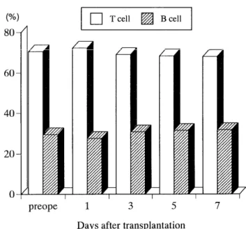
In peripheral T, B cell populations, there was no signifi- cant difference between rats before grafting and each group (Fig 3).
Histologically, perivascular edema was present due to reperfusion injury in group 1 grafts, but there was no evidence of perivascular mononuclear infiltration, intraal- veolar hemorrhage, or parenchymal necrosis. Group 2 grafts were characterized by perivascular cellular infil- trates and edema. The inflammatory infiltrate formed a
dense cuff around the venules and arterioles (Fig 4A).
Cytologically, the cells consisted of small round lympho- cytes, monocytes and occasional neutrophils. Neutrophils were also detected in the vacular endothelium. Mild lymphocytic bronchiolitis and moderate alveolar macro- phage accumulation was observed 3 days after trans- platation. Group 3 grafts showed obvious perivascular and interstitial infiltrates of mononuclear cells. The diameter
of arteriolar lumen was smaller and the airspase contained prominent alveolar macrophages and lymphocytes (Fig 4B). In addition, moderate peribronchiolar inflammatory cell infiltration, intraalveolar hemorrhage and hyaline membranes were observed. In group 4 rats, these signs of injury were more dramatic and were accompanied by parenchymal necrosis showed strong IgM and C3 deposits in the vascular endothelium (Fig 5A, B), but no IgG
deposits in group 2 grafts. No IgM, C3 or IgG deposits were observed in normal hamster lung.
Fig. 3. There was no significant difference in T or B cell populations between rats before grafting and each group.
Fig. 4. (A) Group 3 grafts were characterized by perivascular
cellular infiltrates. The inflammatory infiltrate formed a dense cuff around the small venules. The
cells consisted of small round lymphocytes, mono-
cytes with occasional neutrophils (H & E ; X480).
(B) Group 4 grafts showed an obvious perivascular
and interstitial infiltrates of mononuclear cells. The arteriolar lumen was narrow and the airspaces
contained prominent alveolar macrophages and
lymphocytes. Moderate peribronchiolar inflamma-
tory cell infiltration was present (H & E ; X300).
Discussion
Previous research"" has indicated that antibody- mediated immunity plays an important role in graft destruction in the hamster-to-rat cardiac xenograft model and that it occurs within 3 days in untreated recipients.
Liver xenografts, however, are more resistant to antibody- mediated injury and are rejected in about 7 days by a combination of humoral and cellular immunity"".
In the present study, we demonstrated that rejction of a hamster-to-rat lung xenograf t began within 3 days after transplantation. Although histologic examination of transplanted lungs revealed perivascular cellular infiltra- tion, the infiltrating cells differed from those observed in allograft models""', consisting not only lymphoid cells, but also of macrophages/monocytes. The perivascular infiltrates were associated with adherence of neutrophils to the vascular endothelium. In addition, recipient serum contained a moderate titer of cytotoxic antibodies as detected by a complement-dependent cytotoxicity assay
Fig. 5. (A) Immunohistochemical analysis of grafted lung in group 3 rats showed heavy IgM deposits in the vascu-
lar endothelium (X 300).
(B) C3 deposits were also observed in the vascular endothelium (X480).
and definite evidence of 1g1VI and C3 deposits were oberved in the vascular endothelium 3 days after transplantation.
Beyond 3 days, perivascular and intraalveolar cellular infiltration and edema progressed, followed by hemo- rrhaging and destruction of the pulmonary parenchyma.
This progression suggests that antibodies bound to the vascular walls, leading to infiltration of macrophages/
monocytes into the xenograft and macrophages/mono- cytes-mediated endothelial cell damage. Fryer et al." have reported that antidonor antibodies and macrophages
might interact through antibody-dependent cell-mediated cytotoxicity mechanisms to promote xenograft rejection.
Our results agree with these findings.
Intraalveolar hemorrhage and increased number of intraalveolar macrophages indicate damage on the alveo- lar capillary endothelium". Although airway damage was very infrequent 3 days after transplantation, mild lym- phocytic bronchiolitis and moderate alveolar macrophage accumulation was observed occasionally. Five days after transplantation, lymphocytic bronchitis associated with peribronchiolar inflammatory cell infiltration was ob- served, and airspaces contained many alveolar macro- phages and lymphocytes. Although the role of alveolar macrophages in our model is not clear, it may be a re- sponse to xenogeneic MHC molecules.
The result of our study also indicated that cellular immunity played an important role in concordant lung
xenograft rejection. The analysis of the peripheral lympho-
cyte subpopulations suggested that CD4;_ T cell popula-
tions were elevated early to activate other lymphocytes,
macrophages/monocytes and T cell dependent antibody production, and that CD8- T cell populations then in-
creased to complete the process of cellular rejection. But
the B-cell population was not significantly increased in the peripheral lymphocytes during rejection. Thus, cell- mediated immunity also might play a role in lung
xenograft rejection, as it does in allograft rejection". If
humoral xenograft rejection can be conquered in concor-
dant lung xenotransplantation, the T cell mediated re-
sponse may be as great a problem as in allotransplan- tation.
In conclusion, our experimental results suggest that both the cellular and humoral immune system mediate primary
acute rejection in the hamster-to-rat orthotopic lung xenograft model. Studies are underway to find a way to prevent lung xenograft rejection using immunosuppressive
agents, and to thereby prolong xenograft survival.
Acknowledgement
I would like to express deep appreciation to Emeritus Professor Masao Tomita, the First Department of Surgery, Nagasaki University School of Medicine and other staffs of the Department for their cooperation.
References
1) Starzl TE, rung J, Tzakis A, et al.: Baboon-to-human liver transplan- tation. Lancet 341: 65, 1993
2) Van Den Bogaerde J, Aspinall R, Wang MW, et al.: Induction of long-term survival of hamster heart xenografts in rats. Trans-
plantation 52 : 15, 1991
3) Bailey LL, Nehlsen-Cannarella SL, Concepcion W, Jolley WB.: Baboon to human cardiac xenotransplantation in a neonate. JAMA 254 : 3321,
1985
4) Monden M, Valdivia LA, Gotoh M, et al.: A crucial effect of sple- nectomy on prologing cardiac xenograft survival in combination with
cyclosporine. Surgery 105: 535, .1989
5) Prop J, Marck KW.: Lung transplantation in the rat. CRC Handbook of Microsurgery II : 493, 1983
6) Tavakoli R, Devaux JY, Nonnenmacher L, et al.: Discordant lung xenograft rejection in the rat. Transplantation 53 : 235, 1992 7) Mizuta T, Kawaguchi A, Nakahara K, Kawashima Y.: Simplified rat
lung transplantation using a cuff technique. J Thorac Cardiovasc Surg
97 : 578, 1989
8) Obata Y, Stockert E, O'Donnell PV, Okubo S, Snyder HW, Old LJ.: A new cell surface antigen of mouse leukemia defined by naturally
occurring antibody and its relationship to murine leukemia virus. J Exp
Med 13 : 747, 1976
9) Cordell JL, Falini B, Erber WN, et al.: Immunoenzymatic labeling of monoclonal antibodies using immune complexes of alkaline phos-
phatase and monoclonal anti-alkaline phosphatase conjugates in
immunohistochemistry. J Histochem Cytochem 32 : 219, 1984 10) Murase N, Starzl TE, Demetris AJ, et al.: Hamster-to-rat heart and
liver xenotransplantation with FK506 plus antiproliferative drugs .
Transplantation 55 : 701, 1993
11) Valdivia LA, Fung JJ, Demetris AJ, Starzl TE.: Differential survival of hamster-to-rat liver and cardiac xenografts under FK-506 immuno-
suppression. Transplant Proc 23 : 3269, 1991
12) Yousem SA, Berry GJ, Brunt EM, et al.: A workig formulation for the standardization of nomenclature in the diagnosis of heart and lung
rejection : lung rejection study group. J Heart Transplantation 9 : 593,
1990
13) Prop J, Wildevuur CRII, Nieuwenhuis P.: Lung allograft rejection in the rat. II. Specific immunological properties of lung grafts. Trans-
plantation 40 : 126, 1985
14) Fryer JP, Leventhal JR, Dalmasso AP, et al.: Beyond hyperacute rejection. Transplantation 59 : 171, 1995
15) Valdivia LA, Monden M, Gotoh M, et al.: Evidence that deoxys- pergualin prevents sensitization and first-set cardiac xenograft
rejection in rats by suppression of antibody formation. Transplanta-
tion 50 : 132, 1990
16) Tullius SG, Sablinski T, Heemann UW.: Immunobiology of heart rejection in experimental models. In : Immunology of heart and lung
transplantation. Rose ML & Yacoub MII, Great Britain, 42, 1993