• 検索結果がありません。

H e m o r r h a g i c C e r e b r a l I n f a r c t i o n P r o c e e d i n g s o f t h e 4 t h a n n u a l m e e t i n g o f T h e J a p a n e s e I s c h e m i c C e r e b r o v a s c u l a r D i s e a s e C o n f e r e n c e , N a g a s a k i , M a r c

N/A
N/A
Protected

Academic year: 2021

シェア "H e m o r r h a g i c C e r e b r a l I n f a r c t i o n P r o c e e d i n g s o f t h e 4 t h a n n u a l m e e t i n g o f T h e J a p a n e s e I s c h e m i c C e r e b r o v a s c u l a r D i s e a s e C o n f e r e n c e , N a g a s a k i , M a r c"

Copied!
49
0
0

読み込み中.... (全文を見る)

全文

(1)

Acta Med. Nagasaki. 22 : 1-49

Hemorrhagic Cerebral Infarction

Proceedings of the 4th annual meeting of

The Japanese Ischemic Cerebrovascular

Disease Conference, Nagasaki, March 18, 1977

CONTENTS

Part 1 : Pathology of hemorrhagic cerebral infarction Pathogenesis of cerebral infarction : a review

Hidekatsu Matsumura*, Emanuel R. Ross ... 3 A clinicopathologic study of hemorrhagic infarction of the brain

Masakuni Kameyama* ... 9 Part 2 : Experimental hemorrhagic cerebral infarction

Experimental hemorrhagic cerebral infarction. Its clinical and pathological aspects compared with those of pale infarct

Yoshinari Kamijo*, Julio H. Garcia, Jonathan A. Cooper ... 13 Acute brain swelling and hemorrhagic infarction by recanalized middle cerebral

artery occlusion. An experimental study

Mamoru Taneda**, Toru Hayakawa ...17 Experimental middle cerebral artery occlusion in monkeys

Schinichi Yoshida* * , Richard A. R. Fraser, Russel H. Patterson Jr . ... 18 Experimental blood-brain barrier (BBB) change and diapedetic bleedings following

temporary ischemia of relatively short duration in Mongolian Gerbils

Umeo Ito**, Kikuo Ono, Yutaka Inaba ...19 Experimental hemorrhagic cerebral infarction in the dog

Shobu Shibata**, Akio Yasunaga, Hirohisa Ono, Kazuo Mori ...20 Experimental middle cerebral artery occlusion and extra-intracranial arterial shunt

Akira Nishimoto**, Kazushi Kinugasa, Yuji Yamamoto, Hiroyuki Fujisawa,

Yasunori Yagyu, Takashi Ohmoto ...21 Pharmacological study for regional ischemic infarction of the anterior thalamus in the dog

Jiro Suzuki**, Takashi Yoshimoto, Tetsuya Sakamoto ...22

1

(2)

Part 3 : Clinical aspects of hemorrhagic cerebral infarction

Clinical aspects of internal carotid artery embolism with blood-stained cerebrospinal fluid

Tadayoshi Irino* ...23 Hemorrhagic cerebral infarction

Goro Araki* ...26 Hemorrhagic cerebral infarction ; its neuroradiological findings and pathogenesis

Kazuo Uemura*, Toshio Okudera, Kiyoshi Ishi, Hitoshi Fukasawa ... 29 part 4 : Hemorrhagic cerebral infarction as a neurosurgical complication

Pathogenesis of hemorrhagic cerebral infarction. A study with computed tomography, rCBF and fluorescein cortical angiography

Takeshi Kawase*, Masahiro Mizukami, Hiroshi Kin, Michiharu Nishijima, Goro Araki ...32

Three cases of postoperative hemorrhagic infarction of the brain

Hirohisa Ono**, Kazuo Mori ...34

Part 5 : Special lecture

Microneurosurgical Anastomosis ; A biochemical basis for improvement George M. Austin ...35

* invited speaker

** discusser

CHAIRMEN

Part 1 : Junichi Kawafuchi, Department of Neurosurgery, School of Medicine, Gumma University

Part 2: Hajime Handa, Department of Neurosurgery, Kyoto University Medical School Part 3: Takenori Yamaguchi, Department of Internal Medicine, School of Medicine,

Kyushu University

Part 4: Jiro Suzuki, Division of Neurosurgery, Institute of Brain diseases, School of Medicine, Tohoku University

Part 5 : Keiji Sano, Department of Neurosurgery, University of Tokyo, School of Medicine edited by

Hirohisa Ono, Department of Neurosurgery, Nagasaki University School of

Medicine

(3)

Pathogenesis of cerebral infarction: A. review

Hidekatsu Matsumura and Emanuel R. Ross

Division of Neuropathology, Department of Pathology

Loyola University Medical Center, Illinois, USA

Cerebrovascular disease and its sequelae, represents the commonest disorder of the nervous system. Within the U. S. , it has been estimated that slightly over 200,000 deaths per year occur as a result of cerebrovascular disease. The morbidity of this type of disease appears to be 10 times greater, with over 2 million persons suffering its ravage.

In considering all type of cerebrovascular disease, Kurtzkel5> estimated that 62 will be thromboembolic disease, 16% hemorrhage, 12% SAH and 10% other or illdefined cerebrovascular disorders. Older studies tend to indicate that cerebral hemorrhage is commoner than infarction. In more recent reviews by Kurtzke,15) Kreuger, Gordon and others, the incidence of cerebral hemorrhage has decreased, whereas the incidence of cerebral trombosis has increased. This is not only true in the United States, but also in Japan, where until recently cerebral hemorrhage made up the major portion of cerebro- vascular deaths. It now appears that thromboembolic disease is more common than cerebral hemorrahge. This has been confirmed by Katsuki14) where the proportion of hemorrhage to thrombosis is similar to that of Western countries. This is also supported by the reports of Otsu20) and Matsumoto1). It should be noted that a recent paper by Fukasawa9) indicates that in Northern Japan the incidence of cerebral hemorrhage still remains greater than thromboembelic disease.

Infarction results, when the blood supply to a region of the brain falls below a critical value. As Adams') pointed out this may be independent of arteriosclerosis. This may be due to a thromboembolic event, increased intracranial pressure or systemic hypotension.

The resulting necrosis may be ischemic, hemorrhagic or mixed. Thrombotic events generally give rise to pale or ischemic infarct whereas hemorrhagic infarcts are most commonly the result of an embolic process. It is the hemorrhagic infarction and its pathogentic mechanism that I would like to review at this time.

The macroscopic appearance of a hemorrhagic infarction is readily distinguished

from the pale or ischemic lesion, in that the affected gyri appear swollen, flattened with

multiple isolated or even confluent hemorrhages. The tissue is soft, and because of the

(4)

swelling the sulci in the affected area are generally obliterated. In contrast the ischemic or pale infarct is usually soft, pale, tan-yellow and is generally totally devoid of hemorrhagic discoloration.

The cut surface of the hemorrhagic infarct reveals hemorrhages which may be mild to severe and varies from area to area. The affected zone is sharply demarcated and generally limited to the cortical zone. The cortico-white junction is obliterated. Deeper white matter lesions are rarely hemorrhagic, but remain pale (Fig. 1). Involvement of the white matter by pale grey-white sharply demarcated lesions, appear to occur more commonly in hypertensive patients. Basal ganglia when affected, may likewise be hemor- rhagic. The basis for this will be discussed shortly.

Microscopically, ischemic neuronal degeneration is first to occur (Fig. 2). Rapid disappearance of oligodendroglia with swelling and eventual disappearance of astrocytes follows. Microglia being most resistent persists the longest. Myelin changes are characterized by a loss of stainability, followed by degenerative changes. Axonal degeneration follows shortly after the myelin changes. Most resistant to the affects of ischemia are the blood vessels themselves. These are the last structures to undergo degeneration. One further comment, in early lesions perivascular polymorphonuclear leucocytic aggregates are found.

These may be so prominent that an erroneous diagnosis of meningoencephalitis may be made by the unwary.

Despite intensive investigation and increasing knowledge, the question as to why one develops a hemorrhagic rather than ischemic infarct or vice versa is not fully understood.

Globus maintained that the ischemic and hemorrhagic infarcts were different stages of the same process. The escape of blood from a ruptured vessel within the area of ischemic necrosis transforms this to a hemorrhagic infarct. This view was later supported by Bohne.

In 1932, Westphal expressed the view that hemorrhagic infarction resulted from temporary arterial occlusion brought on by prolonged angiospasm.

As a result of his studies of human autopsy material, Fazio" stated that the hemorrhagic infarct was unrelated to massive hemorrhage and the result of a localized vasodilatation resulting in stasis and diapedesis. The vasodilatation was possibly due to a vaso - vascular reflex from some remote stimulus or possibly due to a local secretion of a vasodilating substance produced by a previous circulatory arrest.

Meyer and Denny-Brown'8 subsequently reported experimental data revealing that occlusion of large caliber vessels led to contraction of distal segments having a diameter of 50-25011. Shortly thereafter, as a result of a reduction in pressure, the contraction was

replaced by mild dilation of .some 10%.

An important factor in the development of an infarct is the status of the collateral

circulation. This is demonstrated by those cases where occlusion of one or even both carotid

arteries fail to give rise to any type of infarction. Vander Ecken and Adams24) demonstrated

the existence of a collateral circulation between the major vessels of the cerebrum by means

of the meningeal cortical vessels. Faris and co-workers6 considered the collateral circulation

as essential to the production of a hemorrhagic infarct. Hypertension was also considered

(5)

as a significant factor contributing to the development of hemorrhagic infarction. Both contributing factors were demonstrated in dogs who underwent both middle cerebral artery clipping and in whom surgical coarctation was performed. In the non-hypertensive controls, ischemic encephalomalacia was noted, whereas in the clipped middle cerebral artery - hypertensive dogs the lesions were hemorrhagic infarcts. The hypertension, with its increased intravascular pressure opened existing anastomosis between the major vessels thus confirming Vander Ecken and Adams /143 earlier work. It should be noted that in these experiments hemorrhages may have occurred as a result of fibrinoid degeneration of the arterioles resulting from hypertension.

Penry and Netsky21) observed that because of the existence of an adequate collateral circulation, embolic occlusion of a single leptomeningeal vessel failed to give rise to an infarct. Only when a cortical penetrating branch of the artery was occluded did a grey matter infarct arise. This extended only to the depth of sufficiently large anastomotic channel. Hain and co-workers occluding the middle cerebral artery of dogs produced both pale and hemorrhagic lesions. Clipping of the middle cerebral artery distal to the perforating ganglionic branch leads to ischemic necrosis, on the other hand clipping done so as to leave some perforating branches in front, as well as behind the clips, leads to hemorrhagic infarction. When the middle cerebral artery of monkeys was occluded pale infarction always occurred leading these investigators to conclude that the different response to middle cerebral artery clipping was due to a differentiation in collateral circulation distal to the clipping. This was confirmed by the observations of other investigators. Fazio'), Meyer and Denny-Brown") and others, Hain et a110), felt that the escape of blood in the hemorrhagic infarcts was due to diapedesis.

Fisher and Adams") in 1951, stated that of the 66 cases of hemorrhagic infarction all but three were due to embolic disease. Hence, the statement made earlier in this paper that essentially all hemorrhagic infarcts are embolic in origin.

In a subsequent paper, Adams') proposed that an embolic process also can give rise to mixed infarcts. The embolus, either by dilation or fragmentation is propagated peripherally restoring the circulation to the damage vessels, thus permitting free passage of blood through the already damaged vessel walls. Some vessels remaining plugged contiune to show ischemic necrosis.

Cobb and Hubbard') state that hemorrhage into the parenchyma arise from veins and capillaries secondary to venous stasis, with no evidence of bleeding from the arterial side. Cammermeyer in studying cases of arterial occlusion, venous thrombosis, and herniation estimated to be 1-12 days, also noted hemorrhages along veins and capillaries and rarely about arteries. The veins were distended, with some showing wide rupture of the wall. Cammermeyer believes the rupture to be due in part to the loss of support by the surrounding parenchyma. This loss is secondary to the necrotizing process of the infarct and partially due to the high venous back flow pressure which develops.

Recently we observed a biopsy specimen of a hemorrhagic infarct of a parietal lobe.

Our preliminary findings reveal attenuation of some endothelial cells within the capillary. The

(6)

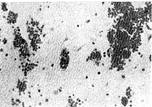
endothelial surface shows prominent undulations with many pinocytotic vesicle formation (Fig. 3-A). Occasionally discontinuities in the endothelial wall of the capillary were noted which varied in size. Plasma and fibrinous material were seen in these areas of discontinuity as well as in the enlarged pericapillary spaces (Fig. 3-B). These findings have been reported as the morphological basis of increased permeability of blood vessels and the break down of the blood brain barrier in various pathological conditions.

REFERENCES

1. Adams, JH, Scot Med J 12 : 339, 1967.

2. Adams, RD et al, Annual Rev Med 4 : 213, 1953.

3. Baker, AB et al, Neurology 13 : 445, 1963.

4. Cammermeyer, J, Acta Psychiat Neurol Scan 28 : 9, 1953.

5. Cobb, S et al, Am J Med Sci 178 : 693, 1929.

6. Faris, AA et al, Arch Neurol 9 : 468, 1963.

7. Fazio, C, J Neuropath Exp Neurol 8 :43, 1949.

8. Fisher, M et al, j Neuropath Exp Neurol 10 : 92, 1951.

9. Fukasawa, J, Tohoku J Exp Med 117 :357, 1975.

10. Hain, RF et al, j Neuropath Exp Neurol 11 : 34, 1952.

11. Hardin, C et al, Arch Neurol 9 : 473, 1963.

12. Hicks, SP et al, A. M. S. Arch Path 52 : 403, 1951.

13. Hirano, A, In Pathology of Cerebral Microcirculation (Ed. J Cervos-Navarro) 203p, 1974.

14. Katsuki, S et al, In Transactions of the Fifth Princeton Conference 99p, 1966.

15. Kurtzke, JF, Epidermology of Cerebrovascular Disease, Springer-Verlag Berlin 4 7,

60, 69p, 1969.

16. Little, JR et al, Stroke 7 : 35, 1976.

17. Matsumoto, N et al, Stroke 4 : 20, 1973.

18. Meyer, JS et al, Neurology 7 : 447, 1957.

19. Meyer, JS et al, A. M. A. Arch Neurol & Psychiat 72 :296, 1954.

20. Otsu, S, Jap. Circulat. J. 33 : 1459, 1969.

21. Penry, JK et al, Arch Neurol 3 : 1960.

22. Stehbens, WE, Pathology of the Cerebral Blood Vessels, C. V. Mosby Co. 13 1 p, 1972.

23. Sundt, TM et al, J Neurosurg 31 : 311, 1969.

24. Vander Ecken, HM et al, J Neuropath Exp Neurol 12 : 132, 1953.

25. Zilch, KJ, In Pathology of the Nervous System (J. Minckler), 1522p, 1971.

(7)

Fig. 1 . Punctate and confluent hemorrhages are present thrughout most of the left cerebral cortex. A localized ischemic infarct is noted in the

paramedial convexity. The subcortical white matter is pale, softened

and collapsed.

Fig. 2 . Typical ischemic neurons (center) scattered focal hemorrhages with

few scattered reactive microglia.

(8)

Fig. 3-A

Fig. 3-B

Fig. 3 A. Surface infolding of the capillary endothelial wall with arrows indicating an increased number of pinocytotic vesicles. X 12000.

Fig. 3 B. A discontinuity in the capillary endothelial wall is present between the arrows. An arrow head indicates the attenuation of the endothelial wall.

Plasma and fibrinous matpria arp seen in the pericapillary space, X 1600Q,

(9)

A clinicopathologic study of hemorrhagic infarction of the brain

Masakuni Kameyama

Department of Geriatric Medicine,

Faculty of Medicine, Kyoto University,

Kyoto, Japan.

If blood reflows into the area of encephalomalacia, red infarction may occur, which results in marked brain swelling and severely interferes the prognosis of the patients. Consequently, the pathogenetic mechanism of cerebral hemorrhagic infarction is an important problem.

The purpose of this study is to clarify the following two points from a clinicopathologic point of view :

1. frequency and distribution of hemorrhagic infarction in the brain, and 2. clinical conditions significantly differing between the pale and red infarct.

MATERIALS AND METHODS

For this investigation, 223 subjects aged sixty year or more and having cerebral infarction were selected at random from the routine autopsy series at the Yokufukai Geriatric Hospital. All of the brains were intensively studied with neuropathologic techniques. Red or hemorrhagic infarction signifies that hemorrhage is apparent to the naked eye in the distribution of an occluded vessel, while pale softening indicates an ischemic area, even though microscopic examination of the infarcted tissue may show diapedesis of red cells around the smaller vessels.

Hypertension was defined as repeatedly measured systolic blood pressure of more than 160 mmHg or diastolic pressure of more than 90 mmHg.

If two or more arteries at the base of the brain showed narrowing of more than 50 percent of the lumen, the grade of cerebral atherosclerosis was classified as severe.

RESULTS

1 . Distribution and frequency of hemorrhagic infarcts in the brains.

Of all subjects with cerebral infarct examined, frequency and distribution of the

hemorrhagic infarct were as follows (Table 1) :

(10)

Table 1 . Frequency of pale and red infarcts

Infarcts

arterial supply and location Total

pale red

Internal Carotid 3 (50%) 3 (50%) 6

Middle Cerebral localized { massive 28 40 (52%) (47%) 32 37 (48%) (53%) 60 77

Anterior Cerebral 16 (53%) 14 (47%) 30

Posterior Cerebral 3 (27%) 8 (73%) 11

Basilar (massive) 3 (100%) 0 3

Internal Capsule and Basal Ganglia 18 (64%) 10(36%) 28

Cerebellum 4 (50%) 4(50 °) 8

Hemorrhagic infarction was observed in 50% of the cases with intrenal carotid occlusion ; 53% of massive and 48% of partial softenings in the area of the middle cerebral ; 47% of the anterior cerebral and 73% of the posterior cerebral arteries. In basilar thrombosis, all of the intarctions were ischemic. In the internal capsule and basal ganglia, 36%, and in the cerebellum, 50% of the cases showed red infarcts.

Among the cerebral regions, areas of the posterior cerebral arteries were most vulnerable to the hemorrhagic infarction.

2. Frequency of arterial occlusion in cases with cerebral infarction at autopsy.

Complete occlusion of the internal carotid or middle cerebral arteries was observed in 23 (46.2%) of 49 cases with compatible infarctions; in the anterior cerebral, in all 8 cases ; and in the posterior cerebral in 3 (60%) of 5 cases. In total, frequency of arterial occlusions was 55% of 62 cases examined. A tendency was found that in cases with complete arterial occlusion, onset. of stroke was slower and the blood pressure at the stroke was lower than in the cases without occlusion.

3. Difference of clinicopathologic features between red and pale infarctions.

Cerebral infarctions were divided according to the location of necrosis into the cortico-subcortical and the penetrating artery group.

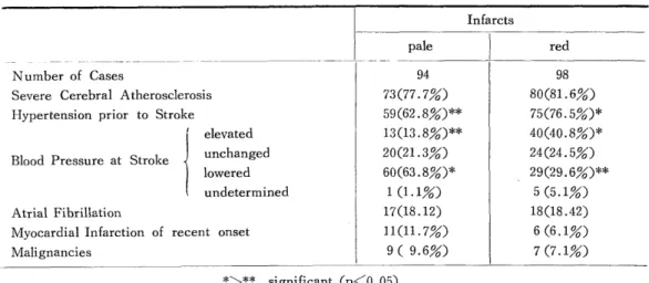
In the cortico-subcortical group (Table 2), presense of arterial hypertension prior to stroke was significantly more frequent in the hemorrhagic than in the ischemic infarcts.

Blood pressure elevation at stroke was a prominent feature in cases with red softening, while in pale infarctions blood pressure tended to be unchanged or lower. Frequency of atrial fibrillation, myocardial infarction of recent onset or malignancies of various kinds

did not differ between the pale and red infarctions. Severe cerebral atherosclerosis was

found in approximately equal frequency between the pale and red infarctions. However, in

the penetrating artery group (Table 3), a significant defference was noted only in the

frequency of severe atherosclerosis, being more frequent in the hemorrhagic infarctions.

(11)

Table 2 . Infarcts in the cortico-subcortical group

Infarcts

pale red

Number of Cases 94 98

Severe Cerebral Atherosclerosis 73(77.7%) 80(81.6%)

Hypertension prior to Stroke 59(62.8%)** 75(75.5%)*

elevated 13(13.8%)** 40(40.8%)*

Blood Pressure at Stroke unchanged 20(21.3%) 24(24.5%)

lowered 60(63.8%)* 29(29.6%)**

undetermined 1 (1.1%) 5 (5.1%)

Atrial Fibrillation 17(18.12) 18(18.42)

Myocardial Infarction of recent onset 11(11.7%) 6 (6.1%)

Malignancies 9 ( 9.6%) 7 (7.1%)

*>** significant (p<0.05)

Table 3 . Infarcts in the penetrating arteries group

Infarcts

pale I red

Number of Cases 21 10

Severe Cerebral Atherosclerosis 20(95.2%)* 7 (70%)**

Hypertension prior to Stroke 12(57.1%) 6 (60%)

elevated 3 (14.3%) 3 (30%)

unchanged 11(52.4%) 0 (40%)

Blood Pressure at Stroke l owered 7 (33.3%) 3 (30%)

undetermined 0 0

Atrial Fibrillation 3 (14.3%) 2 (20%)

Myocardial Infarction of renent onset 3 (14.3%) 2 (20%)

Malignancies 2 (9.5%) 1 (10%)

*~** p<0 .05

DISCUSSION

Pathogenetic mechanisms of hemorrhagic infarction have been repeatedly reported

by many authors'-'), in which recanalization of the thrombotic arteries or development of

callateral circulation into the infarcted area was considered most important. Faris, et a15),

stressed the importance of hypertension. I pointed out a significance of hypertention prior

to and at stroke in the pathogenesis of hemorrhagic infarction from a clinicopathologic

standpoint. Hypertension seems to be the significant factor in two ways') ; firstly, by

increasing the changes in the vessel wall ; and secondly, by increasing the available

intravascular pressure, thus rendering the collateral circulation functional. On the other

hand, hypertension may promote the bleeding of diapedes type in the necrotic vessels and

fragmentation of the blood clot. Atherosclerotic changes do not seem to have an importnat

(12)

role in the production of hemorrhagic infarction. The role of a fibrinolytic action in the local or general circulation remains to be clarified.

SUMMARY

Of 223 cases with cerebral infarction, red and pale softenings were observed in approximately equal frequency in the areas perfused by the internal carotid and middle cerebral as well as by the anterior cerebral arteries. In the posterior cerebral artery region hemorrhagic infarction was more preponderant. In the penetrating arterial system, pale infarcts were more common. Arterial occlusion was found at autopsy in about half of the cases with infarcts. The most signiticant difference, between the red and pale infarctions, was presence of hypertension prior to and at stroke in the hemorrhagic cases. Frequency of severe atherosclerosis, atrial fibrillation and myocardial infarction of recent onset or malignancies were not significantly different between the red and pale infarcts.

The pathogenetic mechanism of arterial hypertension in the hemorrhagic infarction was discussed.

REFERENCES

1. Hain, R. F., et al.: J. Neuropath. Exp. Neurol. 11 : 34, 1952.

2. Fazio, C. et al.. Ibid., 8 : 43, 1949.

3. Fisher, C. M., et al.: Ibid., 10 : 92, 1951.

4. Globus, J. H., et al.: Ibid., 12 :107, 1953.

5. Faris, A. A., et al : Arch. Neurol., 9 : 468, 1963.

(13)

Experimental hemorrhagic cerebral infarction

Its clinical and pathological aspects

compared with those of pale infarct

Yoshinari Kamijyo, * Julio H. Garcia**

and Jonathan A. Cooper**

* Department of Neurosurgery, Tenri Hospital, Nara, Japan.

** Department of Pathology , University of Maryland, U.S.A.

Using a minimally traumatic method for occluding the middle rerebral artery (MCA) the authors have evaluated in two groups of adult cats, with permanent and temporary clipping ; (1) the occurrence, type and severity of the resulting motor deficit, (2) extravascular leakage of a protein-bound tracer, (3) patterns of microvascular filling, (4) degree and timing of tissue swelling, and (5) leakage of red blood cells into the area of the brain supplied by the occluded artery.

MATERIALS AND METHODS

Transorbital occlusion of the MCA was performed on 34 adult cats in the manner described by the authors". In 18 cats the arterial clip remained in place until sacrifice (1 hour to 8 days post-occlusion), and in 16 cats the clip was removed either 1, 6 or 24 hours after occluding the vessel. Animals in the latter group were killed between one to eight days after the initial occlusion. Two to three hours prior to sacrifice, 10 ml of an Evans blue solution (1%) was injected via a forelimb vein. At the time of the sacrifice, approximately 100 ml of 1 : 2 dilution of carbon black in normal saline was infused through the inferior vena cava. The brains were fixed by immersion in a buffered solution of formaldehyde for a period of two weeks. The capillary filling pattern was evaluated after freezing fragments of brain and cutting slices, 200-microns thick. Other portions of the cerebral tissues were processed and embedded in paraffin for routine histologic examination.

RESULTS

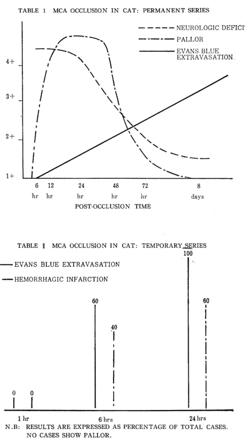
Table I displays the outcome of the experiments in animals in which permanent

occlusion of the artery results in observable ischemic lesions. The neurologic deficit was

(14)

characteristic and similar in all animals in whom it developed, in both the temporary and permanent groups. This consisted of hemiparesis, more severe in the forelimb, circular gait, and a late-appearing sign of forelimb flexion and hindlimb extension when the animal was lifted by the neck. This sign persisted long after the hemiparesis and circular gait disappeared, as they did in all cases within 2-3 days post-occlusion, regardless of the size of the lesion. The hemiparesis became maximal at approximately 24 hours.

Animals without neurologic deficit had no demonstrable lesions in the brain. It was noted that in the temporary group there was no evidence of neurologic deterioration after removal of the clip.

Evans blue extravasation was negligible when the MCA occlusion lasted one hour, but the extravasation occurred in animals in whom the occlusion was of six hours' duration or longer. The most extensive staining by the dye was seen in animals with arterial occlusions lasting 8 days. Reperfusion into the area of ischemia made the extravasation of the dye occur much earlier and more densely than in the permanently occluded animals.

The size of the lesions, however, was much smaller than in the latter.

Table II presents the outcome of the temporary occlusions. No instances of grossly visible pallor were found in this group, an observation which indicates patency of the capillary network within the territory of the temporarily occluded artery. It is to be noted that, as pallor was seen only in the animals with permanent occlusion, hemorrhagic softening developed only in the animals with temporary occlusion, increasing in frequency with the duration of the occlusion. Thus, 60% of the animals with temporary occlusions lasting 24 hours showed evidence of hemorrhagic infarction.

Table III demonstrates the distribution of the lesions in the two groups. Infarctions in the hemispheric cortex were far more common in animals with permanent occlusion, but in neither group were as common as lesions in the basal ganglia. Hemorrhagic infarction was also much more common in the basal ganglia than in the cortex.

In the group of cats having permanent MCA occlusion, the resulting lesions were all in the gray matter with the exception of one. In the animals with temporary occlusion, however, six of the ten cats developing infarctions showed them either predominantly or selectively in the white matter.

Microscopically, it was possible to follow the progressive derangement of the cortical

microangioarchitecture. Two to three hours after occlusion of the MCA, it was only the

deepest stratum of the cortex which showed non-filling by carbon black. After six hours

all strata were involved. By three days, some dilated vasculature appeard in the area of

pallor. Occlusions for 8 days resulted in complete replacement of the normal vascular

structure by dilated, tortuous channels and the infarcted tissue was completely replaced

by macrophages. In animals with the temporary occlusion it was possible to show patency

of all capillary networks within the areas of Evans blue staining. In the areas showing

hemorrhagic infarction there were severe stasis and congestion of the capillaries accompanied

by extravasation of red blood cells and lack of penetration of carbon black, except in some

of the long perforating vessels.

(15)

TABLE 1 MCA OCCLUSION IN CAT: PERMANENT SERIES

POST-OCCLUSION TIME

TABLE 9 MCA OCCLUSION IN CAT: TEMPORARY„,5ERIES

EVANS BLUE EXTRAVASATION

HEMORRHAGIC INFARCTION

N.B: RESULTS ARE EXPRESSED AS PERCENTAGE OF TOTAL CASES.

NO CASES SHOW PALLOR.

(16)

TABLE M MCA OCCLUSION IN CAT: COMPARISON OF TEMPORARY VS.

PERMANENT MCA CLIPPING (OCCLUSION OF 6 HOURS OR LONGER)

LOCATION OF LESIONS

BASAL GANGLIA CORTEX

TEMPORARY GROUP EVANS BLUE 58% 25%

HEMORRHAGIC INFARCTION 41% 25%

PERMANENT GROUP EVANS BLUE 92% 83%

HEMORRHAGIC INFARCTION NONE NONE

N.B.: RESULTS ARE EXPRESSED AS PERCENTAGE OF TOTAL CASES.

COMMENT

Ischemic neurological deficit produced by MCA occlusion in cat appears to correspond with the human stroke known as RIND. This would be based upon the anatomical peculiarity of the feline brain : (1) a location of the cortical motor area which sits over the border zone territory between ACA and MCA, and (2) the effective leptomeningeal anastomoses'). Although reperfusion of blood through ischemic areas results in hemorrhagic infarction or increased permeability to the protein tracer predominantly in the subcortical white matter, the size of the lesions is much smaller than that of permanent ischemia.

We suggest that reperfusion of ischemic brain responsible for RIND, prior to 24 hours after the arterial occlusion, may result in significant protection of the corresponding tissues and that hemorrhagic infarction proper may not be harmful.

REFERENCE

1) Kamijyo, Y. et, al Stroke 6: 361, 1975

(17)

Acute brain swelling and hemorrhagic infarction by recanalized MCA (middle cerebral artery) occlusion

-An experimental study-

Mamoru Taneda, Toru Hayakawa

* Department of Neurosurgery , Hanwa Hospital

** Department of Neurosurgery , Osaka University

Osaka, Japan

Rapid neurological deterioration after recanalization of the occluded major cerebral artery are frequently observed clinically. To elucidate its mechanism, the changes of superficial and deep cerebral microvasculatures in experimental recanalized cerebral infarction were studied by observation of cortical surface and its fluorescein angiogram through a cranial window and by post-mortem microangiograms. The MCAs of adult cats were occluded temporarily or permanently via a transorbital approach and focal cerebral ischemia was produced.

Rapid deterioration or acute death frequently occured after restoration of MCA flow.

Severe brain swelling with tentorial herniation was found in almost all of them but massive hemorrhage only in some. In the observation of cortical surface through the cranial window, the ischemic area decreased in size or completely disappeared and reactive hyperemia with perivascular hemorrhage frequently appeared immediately after restoration of flow. Fluorescein in this reflowed portion extravasated remarkablly. Thereafter, development of brain swelling was apparently accelerated. On microangiograms, the vessels in deep structures in the temporarily occluded cases were usually well visualized including fine arterioles even with only few exceptions.

The data obtained indicate that the reactive hyperemia might play the most important

role in causes of acute fatal outcome following recanalization of major cerebral artery,

because it has an intimate relationslhip with acute brain swelling. Homorrhagic infarction

itself may play a secondary or less important role as the direct cause of fatal outcome.

(18)

Experimental MCA occlusion in monkeys

Shinichi Yoshida, * Richard A. R. Fraser ,

** Russel H Patterson Jr . **

* Department of Neurosurgery

, University of Tokyo hospital, Tokyo, Japan

** Division of Neurosurgery, The New York Hospital-Cornell Medical Center, USA.

Following the introduction of microsurgical technique, embolectomy and thrombectomy for acute MCA occlusion have been increasingly popular without firm experimental supports . In order to find the appropriate indications for such direct vascular surgeries , the origin of the middle cerebral artery (MCA) was clipped temporarily via a transorbital route for 1 to 6 hours of duration. Clinical deficits and extents of infarction were compared with those of the group of permanent clipping. Three weeks later the surviving animals underwent a 2nd surgical procedure in which MCA on the other side was clipped for the same duration. At the termination of experiments retrograde transaortic perfusion was carried out with a mixture of carbon black and formalin . The final data were collected from 52 monkeys.

Results

1. Temporary occlusions for 1 to 3 hours produced no or mild clincal deficits and path- ological changes which were significantly less extensive than those produced by permanent occlusions.

2. Temporary occlusions for 6 hours produced clinical deficits and pathological changes close to those of permanent occlusion.

3. Macroscopic hemorrhage in the brain occurred in 1/3 of animals following release of MCA occlusion in 6 hours and resulted in a high mortality.

Clinical implications

1. Six hours following the onset, MCA embolectomy or thrombectomy may not only be ineffective but also dangerous because of the possibility of producing macroscopic hemorrhage in the brain leading to a high mortality.

2. Useful adjunctives should be searched to elongate the short golden period in which surgical restoration of cerebral blood flow is meaningful .

3. Treatment for ischemic brain edema should be studied .

(19)

Experimental blood-brain barrier (BBB) change and diapedetic bleedings following temporary ischemia of

relatively short duration in Mongolian Gerbils

Umeo Ito, Kikuo Ono, and Yutaka Inaba

Department of Neurosurgery, Tokyo Medical and

Dental University, Tokyo, Japan

The present study aims to elucidate a mechanism, especially of relation to BBB change, of the diapedetic bleedings which occur during recirculation following temporary ischemia of less than 6 hrs. 1. Comparative studies on diapedetic bleedings and BBB change for Evans blue (EB) : The left carotid artery of the ischemia-sensitive Gerbils was clipped for 30 mins, 1,3 and 6 hrs. Then animal brain was recirculated for 30 secs to 20 hrs. Evans blue was injected intravenously prior to releasing the clip. Animal brain was fixed by perfusion, and examined for EB permeation and histology. Diapedetic bleedings occurred in 20-60% of animals during 1-20 hrs of recirculation. This was not parallel in incidence of animals positive for BBB change (Table). There was certain topographical dislocations between the areas of BBB damage and diapedetic bleedings.

Diapedetic bleedings seemed to occur frequently along the veins and capillaries which drain the blood to the cerebral base and internal cerebral vein.

2. Electronmicroscopic study using horseradish peroxidase (HRP) as a tracer : HRP was injected intravenously at the end of recirculation for 1 hr, following temporary ischemia of 30 mins, 1 and 6 hrs. The brain was perfused with paraformaldehyde-glutalardehyde fixative in Na-caccodylate buffer, and treated for peroxidase reaction according to Reese

& Karnovsky. HRP extravastion was demonstrated electronmicroscopically in all 3 animals of 6 hrs ischemia group. The endothelial cells were well torelated the ischemic insult.

Pinocytotic vesicles containing HRP in the endothelial cells are significantly increased in number. So far as we have examined, no evidence of extravasation of HRP and red blood corpscles through the intercellular tight junction of the endothelial cells was observed.

Conclusion : The diapedetic bleedings after temporary ischemia of relatively short duration

seem to be mainly caused by the secondary circulatory disturbances especially of veins due

to brain edema etc.

(20)

* * *

Duration of Duration of No. of animals No. of peri- No. of No. of animals

temporary recirculation with hemorrhage vascular confluent with BBB damage

ischemia bleedings foci bleedings foci for EB

30 secs 0 0 0 0

15 wins 0 0 0 0

30 mins 1 hr 0 0 0 0

5 hrs 0 0 0 0

20 hrs 0 0 0 3

30 secs 0 0 0 0

15 mins 0 0 0 0

1 hr 1 hr 1 5 0 0

3 hrs 0 0 0 0

5 hrs 1 5 5 1

20 hrs 1 11 0 5

30 secs 0 0 0 0

15 mins 0 0 0 0

30 mins 0 0 0 1

3 hrs 1 hr 3 21 3 3

2 hrs 2 14 1 4

3 hrs 2 11 4 5

20 hrs 3 18 11 5

30 secs 0 0 0 0

15 mins 0 0 0 4

6 hrs 30 mins 0 0 0 4

3 hrs 2 5 1 5

10 hrs 3 18 3 5

20 hrs 2 42 32 5

* 5 animals are included in each group

Experimental hemorrhagic cerebral infarction in the dog.

Shobu Shibata, Akio Yasunaga, Hirohisa Ono and Kazuo Mori

Department of Neurosurgery, Nagasaki Universiry

School of Medicine, Nagasaki, Japan

The middle cerebral artery (MCA) of dogs was occluded by a clip and the collateral blood supply was compromised by subjecting to hemorrhagic hypotension for 1 hour.

Following restoration of the systemic blood pressure by infusion of the shed blood, an area

(21)

of ischemia in the territory normally supplied by the clipped artery could be easily demarcated by fluorescein angiography (FAG) through the lingual artery and the femoral vein and by carbon perfusion (CP) through the heart.

The involved cerebral tissue showed marked hemorrhagic infarction 24 hours after restoration of the blood pressure which localized in the boundary zone between the deep cortical layer and the subcortical white matter.

These findings correlated well with extent of the leptomeningeal circulation and were interpreted as due to rough capillary networks in the region which anastomose with those of the deep cortical layer and the subcortical white matter.

Sizes of the hemorrhagic infarction, demonstrated by FAG and CP, were smaller in a group of dogs in which the MCA clip was removed and the involved area was reperfused, than those of a group with the artery remained clipped. Severity of hemorrhage, postulated from amount of extravasated carbon particles, however, was more extensive in the former group.

Experimental MCA occlusion and extra-intracranial arterial shunt.

Akira Nishimoto, Kazushi Kinugasa, Yuji Yamamoto,

Hiroyuki Fujisawa, Yasunori Yagyu and Takashi Ohmoto

Department of Neurological Surgery, Okayama

University Medical School. Okayama, Japan.

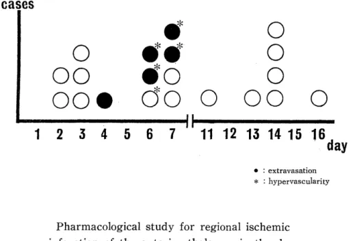
Occlusion of the middle cerebral artery (MCA) was experimentally produced by a transorbital route in 25 dogs. In 20 of these dogs, microangiography was performed with a 60% solution of barium sulfate (Micropaque) on the 2nd day to 16th day following the occlusion. Extravasation of Micropaque and hypervascularity were observed in 5 of 8 animals in which microangiography was done from 4 to 7days after the MCA occlusion.

However, other 12 animals demonstrated no extravasation microangiographically (Fig. 1).

In addition, extracranial (maxillary) artery-middle cerebral artery by-pass formations were performed 4 hrs. , 1 week and 3 weeks after the occlusion in 19 dogs. Histopathological study demonstrated that the size of infarct following a prompt by-pass at 4 hours after the occlusion was much smaller than that of control, but the size in late by-passes (1 week and 3 weeks) appeared to be almost the same as that of control. Some hemorrhagic parts were seen in the infarction area only in three of 7 cases of 1 week by-pass formation.

These results indicate that the vascular vulnerability to the hemorrhagic infarction

in the territory of occlusive vessel may be increased 1 week after the occlusion.

(22)

Fig. 1 MICROANGIOGRAPHICAL FINDINGS FOLLOWING M. C. A. OCCLUSION

Pharmacological study for regional ischemic infarction of the anterior thalamus in the dog

Jiro Suzuki. , Takashi Yoshimoto and Tetsuya Sakamoto

Division of Neurosurgery, Institute of Brain Diseases,

Tohoku University School of Medicine, Sendai, Japan.

Consequences of occluding a cerebral artery in experimental animals differed signi- ficantly in nature, extent and location of pathological changes of the brain. The authors developed a new experimental method in dogs which resulted in a high incidence (more than 70%) of localized ischemic infarction of the anterior thalamus as described elsewhere.

This report concerns with functional alteration of the ischemic anterior thalamus after perfusion of the tissue with pharmacological solutions.

Methods and results : Under Nembutal ansesthesia, the internal carotid, posterior communicating, anterior cerebral and middle cerebral artery of a dog were occluded tem- porarily through a temporal craniotomy.

Under electrical monitoring of the ischemic anterior thalamus and the brain cortex,

two types of experiments, 20% mannitol perfusion for short term and 7 days administration of

CDP-choline, were designed and changes in power spectrum of EEG were compared after

the administration with that of a control group of dogs without the treatment. The authors

confirmed significant effects of these pharmacological solutions on a functional improvement

of the ishemic tissue by noting a less extensive decrease in fast components of EEGs during

occlusion of the arteries and earlier recovery as well after release of the occlusion.

(23)

Clinical aspects of internal carotid artery embolism with blood-stained cerebrospinal fluid

Tadayoshi Irino

Division of Cerebrovascular Diseases, Hanwa Hospital,

Osaka, Japan

A large number of pathological and experimental studies have suggested that cerebral infarction in the acute stage often results in hemorrhagic infarction. ',I) However, there has been no sufficient clinical report regarding this pathological condition as yet. In this study, in order to clarify the clinical aspects of infarcted patients accompanying hemorrhagic infarction, acute stroke patients with internal carotid arterial embolism were observed clinically and comparisons were made between those with and without sanguinous or xanthochromic cerebrospinal fluid.

MATERIALS AND METHODS

Among the stroke patients who had an apoplectiform onset of symptoms and were diagnosed following both physical and angiographical findings within 24 hours after ictus,

18 patients had cerebral embolism caused by acute internal carotid artery occlusion and were given consecutive cerebrospinal fluid (CSF) examination at one to three-day intervals within a week after the onset. They were not treated with fibrinolytic agents or anticoag- ulants. Ten cases had colored CSF (sanguinous or xanthochromic) and the remaining eight cases did not. The former group was classified as Group I and the latter as Group II ; clinical observations for the two groups were compared below.

(1) Clinical findings

Clinical findings included the frequency of atrial fibrillation, hypertension (systolic pressure higher than 150 mmHg within a week) and outcomes estimated one month after onset.

(2) Follow-up cerebral angiographies

Follow- up cerebral angiographies were performed on the 18 patients to inspect

alterations of the occluded artery because some previous reports suggested that recirculation

of occluded arteries was closely related with the development of hemorrhagic infarction.1)

The occurence ratios of spontaneous recanalization were compared between those two

groups.

(24)

Table. Case Summary

Atrial Fibrillation I Recanalization Hypertension

Group I No.1 0 0

2 O O O

3 0

4 0 0

5 0 0

6* O

7

8* 0 0 0

9* O 0 0

10 0

Group II 11

12 0

13 0

14 0

15 0

16 0

17

18*

* fatal case within a month after onset

RESULTS

The Table summarized the results.

(1) Clinical findings i. Atrial fibrillation

Five cases of Group I (50%) and four of Group II (50%) showed atrial fibrillation on ECG.

ii. Outcomes within a month

All cases had poor prognosis, i. e . , the degree of himparesis did not improve enough to allow the patients to leave the bed without assistance. Three cases of Group I died within a month but one in Group II did.

iii. Hypertension

Five cases (50%) of Group I, but only one (13%) in Group II, were judged as hypertensive.

(2) Follow-up cerebral angiographies

Of the 18 cases, eight (44%) showed angiographical recanalization of the occluded

points within a few days ; all were in Group I. Group II had no recanalized cases.

(25)

DISCUSSION

According to the previous reports on animals, experimental hemorrhagic infarction can be observed either when the occluded cerebral arteries are recanalized after ligation2>

or when the animals with occluded cerebral arteries were made hypertensive." These reports explain well the present results of patients with recanalization and hypertension showing blood-stained CSF to indicate development of hemorrhagic infarction. This study suggests that recanalization of the occluded arteries is more closely related with the devel- opment of hemorrhagic infarction than hypertension as for the human beings.

On the other hand, pathological studies by Fisher suggested that hemorrhagic infarction could be observed in the autopsied brain where occluded cerebral artery had not been found pathologically." He said that hemorrhagic infarction could be caused by blood flow through recanalization, which, however, had not been confirmed angiographically during life. We have previously reported that angiographically demonstrated recirculation did not contribute to recovery of infarcted patients and that sanguinous CSF frequently occurred in recanalized cerebral infarction.5 6) In addition, we have shown that several pathological findings such as capillary blush or space taking signs were not infrequently observed on the postrecanalized angiograms, which suggested the development of hemorr- hagic infarction in recanalized embolism.')

CONCLUSION

The present findings summarized are :

(1) The presence of atrial fibrillation did not run parallel with the occurrence of colored CSF.

(2) Elevated blood pressure was observed in half of the patients with sanguinous CSF.

(3) Patients with colored CSF had poorer prognosis than those without CSF abnor- mality.

(4) Angiographically demonstrated recanalization was closely related with development of sanguinous CSF.

REFERENCES

1) Fisher M, Adams RD: Observations on brain embolism with special reference to the mechanism of hemorrhagic infarction. J Neuropath Exp Neurol 10 : 92-94, 1951

2) Harvey J, Rasmussen T: Occlusion of the middle cerebral artery. An experimental

study. Arch Neurol Psychiat 66 :20-29, 1951

3) Zulch KJ: Hemorrhage, thrombosis, embolism. In Minckler J (ed) : Pathology of the Nervous System. New York, McGraw-Hill, vol 2, 1971

4) Irino T, Taneda M, Minami T : Positive scans in angiographically proved cases of

(26)

recanalized cerebral infarction. Stroke 6 : 132-135, 1975

5) Irino T, Taneda M, Minami T et al : Angiographical extravasation of contrast medium in hemorrhagic infarction. Case report. Stroke 7 : 641-617, 1976

6) Irino T, Taneda M, Minami T : Sanguinous cerebrospinal fluid in recanalized cerebral infarction. Stroke, in press

7) Irino T, Taneda M, Minami T : Angiographic manifestations in postrecanalized cerebral infarction, Neurology, in press

Hemorrhagic cerebral infarction

Goro Araki

Institute of Brain and Blood Vessels Mihara Memorial Hospital, Gumma, Japan

INTRODUCTION

Although the pathogenesis of hemorrhagic infarction which may occur during the coruse of cerebral infarction is still not quite certain, moving embolus in an occlusive process has been recognized as a cause of the hemorrhagic infarction.') The author reports autopsied cases of cerebral infarction in the authors hospital which were analized clinico- pathologically and evaluates the possibility of making diagnosis of hemorrhagic infarction by computed tomography (CT) or angiographical study. By elucidation of several important clinical factors, the author discusses causes of the hemorrhagic infarction, such as moving embolus.

MATERIAL AND METHOD

1. Materials

Twenty-five autopsied cases of cerebral infarction including 15 cases of cerebral thrombosis and 10 cases of cerebral embolism are studied. The author found no hemorrhagic infarction in the autopsied cases of cerebral thrombosis while hemorrhagic infarctions were found in 7 cases out of 10 cases of cerebral embolism. The average age of the cases of cerebral embolism (3 males and 7 females) was 62.8.

2. Diagonstic criteria of cerebral embolism.')

The angiographical delineation of an abrupt round edged occlusion in a vessel leading

to an avascular region and of retrograde flow of the contrast material in collateral vessels

(27)

to the deprived area were taken as criteria of an embolic occlusion. Additional diagnostic sings of cerebral embolism, such as early filling of cerebral veins, early positive brain scanning and early appearance of low density in CT study were also used. The movement or disappearance of an occlusive process on subsequent angiographical studies was considered to be a more definite sign of an embolic phenomenon. Clinical examinations were per- formed carefully to evaluate whether or not the patients were suffered from auricular fibrillation.

RESULTS

1. Angiographic findings.2)

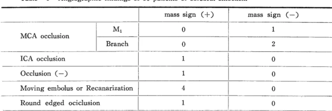
Carotid angiograms were performed on 10 patients of cerebral embolism, within 2 days after the onset in 7 cases, 7, 10, and 14 days after the onset in other cases. The angiographic findings of these 10 cases are summarised in Table 1. Three cases in this group of the patients had no mass sign angiographically. One of 2 cases of middle cerebral artery branch occlusion without mass sign died of a re-attack of cerebral embolism in the contralateral hemisphere. Another case, whose angiography was performed five hours after the attack, was found to have hemorrhagic infarction at necropsy, 2 days after the onset. The exact time of occurance of the hemorrhagic infarction was not determined in this case. Seven cases with angiographic mass signs consisted of 1 case of internal carotid occlusion, 1 case without apparent occlusion, 4 cases with moving embolus or with recanalization and 1 case with the abrupt round edged occlusion in the basilar artery. In 4 case with the moving embolus or with recanalization the time interval between the onset of the clinical symptoms and angiograpic examination was less than 24 hours in 1 case, 2 days in 2 cases, 7 days in 1 case and 21 days in 1 case.

2. CT study

CT studies were performed on 3 cases. A high density area observed in 2 cases enlarged gradually during the course up to death. The high density in the CT study of these 2 cases were found in accordance with the foci of hemorrhagic infarction at necropsy.

However, another case, which was found to have a middle cerebral artery branch occlusion

Table 1 Angiographic findings of 10 patients of cerebral embolism

mass sign (-}-) mass sign (-)

M1 0 1

MCA occlusion

Branch 0 2

ICA occlusion 1 I 0

Occlusion (-) l 1 0

Moving embolus or Recanarization 4 0

Round edged ociclusion 1 0

(28)

without angiographical mass sign, also had cerebral infarction of hemorrhagic type in autopsy. It was difficult to correlate angiographical findings of the vessel occlusion to presence of the high density area in CT study, because the moving embouls was seen in the subsequent angiograms in the cases of cerebral embolism.

3. Relationship between cerebral embolism and hemorrhagic infarction (Table 2).

Two of 4 cases with demonstrated moving embolus on angiograms showed hemorrhagic infarction pathologically, but the other two cases were found to have anemic infarction.

Therefore it was apparent that the movement or disappearnce of embolus did not always lead to hemorrhagic infarction. The case with the abrupt round edged occlusion of the basilar artery and the other case without apparent occlusion of the vessels were also found to have hemorrhagic infarction pathologically. No explanation could be made at this time with regard to the absence of the angiographical mass signs of recanalization in two cases of hemorrhagic infarction associated with middle cerebral artery occlusion. Two cases of moved embolus with a high density area in CT study were noted to have relatively favorable prognosis.

Table 2 CAG findings and types of cerebral infarction

Occlusion (+) Moving embolus Round edged Occlusion (-) Mass sign Mass sign

or occlusion Mass sign (-F) (+) (-)

Recanarization ICA MCA

M, 1 Branch

Hemorrhagic 2 1 1 1 1 1

infarction

Anemic infarction 2 0 0 0 0 1

Tota' 4 1 1 1 1 2

CONCLUSION

1. No obvious hemorrhagic infarction was found in autopsied cases of cerebral thrombosis while the hemorrhagic infarctions were apparent in 7 of 10 autopsied cases of cerebral embolism. Two of these 7 cases had moving embolus or recanalization angiogra- phically.

2. It was noteworthy that 2 autopsied cases of moving embolus or recanalization showed the anemic infarction. It appaered that the movement or disappearance of an occlusive process does not always lead to the hemorrhagic infarction.

3. CT study is a useful examination to diagnose presence of hemorrhagic infarction.

The author experienced 4 cases of hemorrhagic infarction (2 autopsied cases and 2 clinical cases) diagnosed by CT study.

4. Two patients with hemorrhagic infarction diagnosed clinically by CT study were

(29)

relatively benign in the clinical course.

The significance of the hemorrhagic infarction with regard to patients prognosis must be further investigated especially with CT and angiographical study.

REFERENCES

1) Arie Liebeskind et al : Stroke, 2 : 440, 1971.

2) Irino, T et al : Brain and Nerve, 27 : 1089, 1975.

Hemorrhagic cerebral infarction; its neuroradiological findings and pathogenesis

Kazuo Uemura*

Toshio Okudera*

Kiyoshi Ishii* and

Hitoshi Fukasawa**

Division of Radiology* and Pathology**

Research Institute of Brain and Blood Vessels, Akita, Japan

INTRODUCTION

Hemorrhagic infarction is thought to be an established entity in the neuropathology.1) However, it has not been fully exploited clinically because of its diagnostic difficulty. This report describes results of analysis of the hemorrhagic infarction cases, with findings of cerebral angiography, computed tomography and autopsy.

MATERIALS

During 7 year period from 1969 to 1976, autopsies were performed on a total of 62

cases with cerebral infarction. Hemorrhagic infarction was found in 13 cases. Cerebral

angiography was done on 12 cases of them (Group 1, Table 1). Group 2 (Table 2) consisted

of 32 cases with cerebral infarction studied by CT (computed touography) and cerebral

angiography. Of 21 cases showed revascularization of occluded arteries. We have had no

case who had suspected hemorrhagic infarction by CT and verified by autopsy.

Fig.  2  .  Typical  ischemic  neurons  (center)  scattered  focal  hemorrhages  with           few  scattered  reactive  microglia.
Table  3  .  Infarcts  in  the  penetrating  arteries  group
TABLE  1  MCA  OCCLUSION  IN  CAT:  PERMANENT  SERIES
Fig.  1  MICROANGIOGRAPHICAL  FINDINGS  FOLLOWING        M.  C.  A. OCCLUSION
+7

参照

関連したドキュメント

[r]

     ー コネクテッド・ドライブ・サービス      ー Apple CarPlay プレパレーション * 2 BMW サービス・インクルーシブ・プラス(

のようにすべきだと考えていますか。 やっと開通します。長野、太田地区方面  

Q is contained in the graph of a

※ MSCI/S&amp;P GICSとは、スタン ダード&プアーズとMSCI Inc.が共 同で作成した世界産業分類基準 (Global Industry Classification

[r]

[r]

[r]