ERM
(Physiological roles of ERM protein on
the kidney)
ERM
(Physiological roles of ERM protein on
the kidney)
2019
3
March 2019
Doctoral Program in Pharmacy
Graduate School of Pharmacy
Ritsumeikan University
1
5
ERM
15
19
24
ERM
29
33
38
43
45
46
55
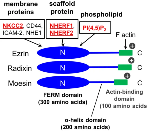
Ezrin, Radixin, Moesin ERM Actin
1) ERM N 300
200 α C
100
2,3) N Four-point-one, Ezrin, Radixin,
Moesin (FERM) 85%
FERM Na+, K+,
2Cl− cotransporter 2 (NKCC2) Na+,H+-exchanger 1 (NHE1)
CD44 ICAM 2
phosphatidylinositol 4,5-bisphosphate (PI(4,5)P2)
Na+,H+-exchanger regulatory factor
(NHERF)
Podocalyxin Na -dependent phosphate transporter 2A (Npt2a) Cystic fibrosis transmembrane conductance
regulator (CFTR) 3-5)
ERM C Actin
(Fig. 1) ERM N
C
(Fig. 2) 2)
ERM GTPase Rho GTPase
ERM Rho GDP/GTP Dbl Rho GTPase
GDI) FERM Rho GTPase Rho-GDI
Rho GTPase (Fig. 3)2,6) ERM
Rho GTPase Rho GTPase
ERM Ezrin Radixin Moesin
Ezrin Ezrin 5% Ezrin (Vil2kd/kd) H+,K+-ATPase 7) Radixin Radixin (Rdx−/−) multi-drug resistant protein 2 (MRP2)
8) Moesin Moesin (Msn−/y) T B 9) Rho GTPase 10) ERM Ezrin Radixin Moesin (TAL) (Fig. 4) Vil2kd/kd Npt2a
D Ca2+ Transient receptor potential vanilloid 6 (TRPV6)
12) ERM 100 Na+ 65% TAL 25-30% 5% (Fig. 5) Ezrin NHERF2 1 Podocalyxin 4 Ezrin Vil2kd/kd Ezrin TAL NKCC2 Moesin TAL NKCC2 ± LLC-PK1 Moesin cNKCC2 (NKCC1/NKCC2 ) 13) Moesin Moesin Msn−/y Moesin TAL NKCC2 TAL Moesin
8-11 Vil2kd/kd Msn−/y Vil2kd/kd
Ezrin 2 3
En2 (Homeobox protein engrailed-2) SA (splicing acceptor) IRES (Internal ribosome entry site) lacZ neo (neomycin-resistance gene) PA (polyadenyl site)
Ezrin 5% 7) Msn−/y X Moesin 3 3’ loxP/ -neo/loxP SA/PA Moesin 14) C57BL/6J Jcl ICR 12 3 mm 50 mM 95 10 1 M Tris-HCl (pH 8.0) 5 12,000 rpm 20 PCR PCR DNA
KOD FX (TOYOBO) Vil2kd/kd Msn−/y
primer Table 1 10 µL PCR
PCR Vil2kd/kd primer 380 bp Ezrin 290 bp Msn−/y primer
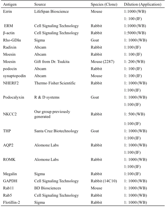
450 bp Moesin 280 bp (TERUMO) (PBS) 4% 0.1 M (pH 7.4) 1 4 (RM2245 Leica) 4 µm ( ) 0.01 M 121 20 ± PBS (Table 2) 4 0.1% Tween 20 PBS-T (Alexa Fluor 488 Alexa Fluor 594
Alexa Fluor 633 ) 1
PBS-T Mounting Medium (VECTASHIELD Vector Laboratories) (FV-1000D IX-81 Olympus)
(H.E.)
(Merck) 2% 2% 0.1 M (pH 7.4) 1 4 0.1 M 2% 0.1 M 4 70: 30 (Quetol 812 EM) 1 100% 60 48 ± 70 nm
2% 15 lead stain solution
(Sigma) 3
(JEM-1200EX JEOL)
Lysis buffer (150 mM NaCl 3 mM KCl 5 mM EDTA 3 mM EGTA 0.1% Triton X-100 60 mM HEPES)
(KINEMATICA)
4 800×g 5 min Total tissue lysate
20 mL PBS PBS
2 ×10⁶ /mL Dynabeads M-450 Tosylactivated (Veritas)
1 mg/mL Collagenase
70 µm (BD Biosciences) Dynabeads Magnetic particle concentrator (DYNAL BIOTECH)
± Pierce BCA Protein
Assay Kit (Thermo Scientific)
SDS (2% Sodium Dodecyl Sulfate (SDS) 2% 2-mercaptoethanol 20% glycerol 0.01% Bromophenol Blue (BPB) 50 mM Tris-HCl (pH 6.8)) 65 15 Laemmli
SDS-PAGE ( 8-12.5%)
Polyvinylidendifluoride (PVDF) 5% TBS-T
1 ± (Table 2) 4
1 Immobilon Western Chemiluminescent HRP Substrate (Millipore) LAS-4000mini (Fujifilm)
ImageJ
Adriamycin (ADR) 50 mg/kg body weight ADR (Wako Purechemical) 7
Lipopolysaccharide (LPS) 200 µg LPS (Escherichia coli O111:B4 strain Invivogen)
24
3,000×g 15 4 1,000×g 15
Rho
RhoA Rac1 Cdc42 G-LISA activation assay (Cytoskeleton) G-LISA Cell Lysis Buffer 4 3,000×g 15
Rho-GTP affinity G-LISA plate 30
RhoA Rac1 Cdc42 GTP
490 nm
27G (TERUMO) 4 1,000×g 15 3,000×g 15
DRI-CHEM 4000i (Fujifilm) Na+ K+ Cl−
Na+ K+
Cl− (ENa EK ECl) (FENa FEK FECl) ENa (µmol/100 g BW/day) = UNa (µM) ×V (L/day) ×100 / BW (g)
FENa (%) = (UNa (mM) / PNa (mM)) / (Ucr (mg/dL) / Pcr (mg/dL)) ×100
V: 1 BW: UNa: Na+ PNa: Na+ Ucr:
Pcr:
HCO3− pH (ABL555
Radiometer)
(glomerular filtration rate: GFR) GFR fluorescein isothiocyanate (FITC)-inulin
75 100 120 FITC-inulin
2- GFR (Graph Pad software GraphPad Prism)
TAL
HEPES (130 mM NaCl 2.5 mM NaH2PO4 4 mM KCl 1.2 mM MgSO4 6 mM
L-alanine 1 mM Na citrate 5.5 mM glucose 2 mM CaCl2 10 mM HEPES (pH 7.4))
1 mg/mL Collagenase
HEPES 37 30
HEPES 200 µm
HEPES
TAL 1.2 mg/mL Sulfo-NHS-SS-HEPES (L-alanine Na citrate ) 4 30
100 mM glycine
Sulfo-NHS-SS-HEPES Lysis buffer (150 mM NaCl 5 mM EDTA 1% Triton X-100 0.1% SDS 50 mM HEPES (pH 7.5) protease inhibitor cocktail (cOmplete Roche)) 200 µg
Streptavidin-coated agarose beads (Sigma) 4
wash buffer (150 mM NaCl 5 mM EDTA 0.1% Triton X-100 50 mM HEPES (pH 7.5)) 2 50 mM dithiothreitol (DTT) 10%
2-mercaptoethanol SDS 65 15 beads
NKCC2
TAL 37 0 30
60 NKCC2
PBS 50 mM Sodium 2 mercaptoethane sulfonate (MESNa) MESNa
25 mM iodoacetamide 4 15
500 µg Streptavidin-coated agarose
beads (Sigma) 4 50 mM DTT
10% 2-mercaptoethanol SDS ±
homogenization buffer (120 mM NaCl 20 mM KCl 1 mM EDTA 1 mM EGTA 10 mM Tris-HCl (pH 7.5) protease inhibitor cocktail)
20,000×g 4 20
500 µg 1 mL OptiPrep (Axis Shield) 8-34% (10 mL) OptiPrep
Gradient Master (SK-bio)
(P40ST ) 4 100,000×g 18
1 mL 2 acetone 30
4 10,000×g 15
100 µL SDS ±
fractionation buffer (10 mM KCl 1.5 mM MgCl2
1 mM EDTA 1 mM EGTA 250 mM sucrose 1 mM DTT 20 mM HEPES (pH 7.4) protease inhibitor cocktail) 27 G
10 20 4 10,000×g 5
4 100,000×g 1 fractionation buffer TN buffer (150 mM NaCl 5 mM EDTA 1% Triton X-100 1 mM DTT 25 mM Tris-HCl (pH 7.4) protease inhibitor cocktail)
500 µg 1 mL 2 OptiPrep (60%) OptiPrep 40% OptiPrep 35% 30% 25% 15% ± 40% ( ) 35% 30% 25% 15% 3 mL 2 mL 2 mL 2 mL 2 mL P40ST 4 170,000×g 15 1 mL 2 acetone 30 4 10,000×g 15 100 µL SDS ± NKCC2
TAL NKCC2 FluxOR Thalium detection kit (Thermo Fisher Scientific) TAL kit
FluxOR Dye loading buffer 37 1 Tl+ FluxOR 2.7 mM probenecid 100 µM ouabain FluxOR assay buffer (135 mM Na-gluconate 1 mM MgCl2
1 mM Na2SO4 1 mM CaCl2 15 mM HEPES (pH 7.4)) 2 100 µM
FluxOR assay buffer 30
37 100 µM
Tl+ uptake buffer (135 mM NaCl 5 mM Tl2SO4 100 µM Ouabain
20 mM HEPES (pH 7.4)) 37 5 Tl+
FluxOR assay buffer 2 Lysis buffer (SH-8100Lab Corona Electric)
Tl+ NKCC2
3% sucrose 7
RNA
RNeasy Mini Kit (QIAGEN) RNA RNeasy Mini Kit Buffer RLT
1% (v/v) 2-mercaptoethanol 600 µL 16,000×g
3 70% RNeasy
8,000×g 15
Buffer RW1 8,000×g 15 Buffer RPE 2 RNA RNase free NanoDrop 2000 (Thermo)
260 nm RNA
Omniscript Reverse Transcription Kit (QIAGEN)
RNA 20 µL 2 µg Gene Amp PCR system 9700 (Applied Biosystems) 37 1
Quantitative Real-time PCR
mRNA SYBR Premix Ex Taq (TaKaRa)
cDNA 20 Cyclooxygenase-2 (COX-2) Glyceraldehyde 3-phosphate dehydrogenase (GAPDH)
ABI PRISM7000 (Applied Biosystems) PCR
Ct GAPDH
2 Student T test Turkey’s post-hoc test one- way ANOVA p < 0.05
ERM
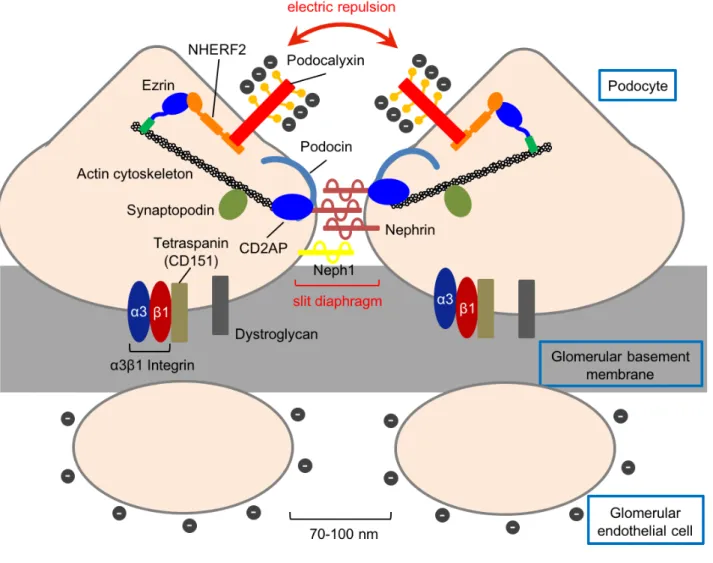
70-100 nm 20-50 15) 16) (Fig. 6) 80% 17)1 IV collagen Alport´s Alport´s 16) (slit diaphragm) Actin ± 18) ± ± 23 19) Adriamycin (ADR) in vivo 20) ± Nephrin 21,22)
NPHS2 Podocin 23)
24) C
CD2-associated protein (CD2AP)
CD2AP Nephrin
25) (Fig. 6) CD2AP Nephrin Actin
Nephrin 26) Nephrin IgG-like 1 Neph1 Nephrin 27) (Fig. 6) Integrin Tetraspanin Dystroglycan Integrin αβ α3β1 Integrin 28) Tetraspanin 1 CD151 α3β1 Integrin 29) Dystroglycan Actin 30) (Fig. 6) Podocalyxin Podocalyxin O- 1 31) Podocalyxin 24 32) Podocalyxin Podocalyxin C PDZ (PSD-95 / Dlg / ZO-1) ( DTHL) NHERF2 PDZ Podocalyxin NHERF2
Ezrin Actin (Fig. 6) Podocalyxin 4) Podocalyxin/NHERF2/Ezrin Podocalyxin Ezrin Ezrin Vil2kd/kd Podocalyxin ± Ezrin
ERM ERM Ezrin (Fig. 7) Ezrin CD34 Ezrin
(Fig. 8A) Radixin Moesin
(Fig. 7) Moesin CD34 (Fig. 8B)
ERM Ezrin
Vil2kd/kd Vil2kd/kd
Vil2kd/kd H.E.
Vil2kd/kd (Fig. 9A)
Vil2kd/kd (Fig. 9B) (µm) Vil2kd/kd (Fig. 9C) Vil2kd/kd Vil2kd/kd Vil2kd/kd SDS-PAGE CBB ± BSA Vil2kd/kd ± 60 kDa (Fig. 10)
Vil2kd/kd
Vil2kd/kd Ezrin
Ezrin NHERF2
Podocalyxin Ezrin
NHERF2 Podocalyxin (Fig. 11) Actin
Synaptopodin Podocin
(Fig. 11)
Vil2kd/kd Ezrin (Fig.
12) NHERF2 Podocalyxin Synaptopodin
Podocin (Fig. 12)
Ezrin NHERF2 Podocalyxin
Radixin Moesin Ezrin (Fig. 13) Vil2kd/kd Ezrin Vil2kd/kd Ezrin ± ERM Ezrin
Moesin (Fig. 14A) Vil2kd/kd
Ezrin Radixin
Moesin (Fig. 14A) Ezrin
Podocalyxin Rho-GDIα Vil2kd/kd
(Fig. 14) NHERF2
Vil2kd/kd
Vil2kd/kd Podocalyxin Ezrin Vil2kd/kd Ezrin ADR LPS Vil2kd/kd ADR 7 ADR ADR ADR ± (Fig. 15) LPS LPS 24 ± (Fig. 16) ADR ADR 5 33,34) LPS 24 ± 34) ADR LPS Vil2kd/kd ADR ± (Fig. 15) LPS ± (Fig. 16) Vil2kd/kd
Vil2kd/kd Rho GTPase GTPase Rho GTPase
Actin
Rho GTPase Actin
35) Rho GTPase RhoA
Rac1 Cdc42 RhoA Rac1
(lamellipodia) Cdc42 (filopodia)
36,37) Ezrin NHERF2
Podocalyxin Actin Rho GTPase
2) GST
Ezrin FERM Rho-GDI
6,38) Rho GTPase Rho-GDI
Rho
Vil2kd/kd Rho GTPase
Vil2kd/kd RhoA
Rac1
Cdc42 (Fig. 17A)
ADR LPS Rho GTPase
ADR RhoA Vil2kd/kd
(Fig. 17A) Rac1
Vil2kd/kd (Fig. 17A) Cdc42
ADR (Fig. 17A)
LPS RhoA ADR Vil2kd/kd
(Fig. 17B) Rac1 Vil2kd/kd
RhoA (Fig. 17B) Vil2kd/kd
Ezrin
±
Ezrin Moesin Ezrin Podocalyxin
Synaptopodin Podocin Table
3 ERM Moesin Radixin
Podocalyxin Vil2kd/kd Ezrin ERM Ezrin Ezrin Podocalyxin Ezrin NHERF2 Podocalyxin C 4) Vil2kd/kd Podocalyxin Podocalyxin 32) NHERF2 Podocalyxin/NHERF2/Ezrin Podocalyxin 39) Vil2kd/kd NHERF2 Ezrin NHERF2 NHERF2 Podocalyxin
Ezrin Ezrin Ezrin Podocalyxin Ezrin Vil2kd/kd Vil2kd/kd ± Podocalyxin 32) 40) Podocalyxin 31,32) Ezrin Podocalyxin Podocalyxin Ezrin Vil2kd/kd
Podocin CD2AP Neph1
26,41,42) α3 β1 Integrin
CD151
28,29,43) Vil2kd/kd
Ezrin
ADR LPS
Vil2kd/kd ADR
Vil2kd/kd
LPS
Podocalyxin/NHERF2/Ezrin Podocalyxin Actin
4,44) Ezrin
Vil2kd/kd ADR LPS
Ezrin Podocalyxin/NHERF2/Ezrin
Vil2kd/kd Rho GTPase
Ezrin Actin
Rho GTPase Actin
3) Rho GTPase RhoA Rac1 Cdc42
Actin 35,38) Rho GTPase GDP GTP Cdc42 Rac1 RhoA 45) Rac1 Rac1
EHT1846 Rho (ROCK) SAR407899 47) Rho GTPase Rac1 Vil2kd/kd Rac1 Ezrin RhoA 37) Vil2kd/kd RhoA Cdc42 45) Vil2kd/kd Cdc42 Vil2kd/kd Rac1 RhoA
Ezrin Rho GTPase Rho GDI FERM
Rho GDI Rho GTPase
6) Vil2kd/kd Rac1
Ezrin Rho GDI
Rac1 Rho GDIα
Rac1
48) Rho GDI Rac1
(Table 4)
Vil2kd/kd ADR LPS
RhoA (Table 4) RhoA Rac1
Rac1 RhoA
37,49) Vil2kd/kd Rac1
Vil2kd/kd Podocalyxin ADR LPS
±
Vil2kd/kd Rho GTPase
ERM
ERM Ezrin
Ezrin FERM NHERF1
50) ± LLC-PK1 NHERF1
Npt2a C PDZ Ezrin/NHERF1/Npt2a
Npt2a 50-52)
Vil2kd/kd Npt2a NHERF1
±
11) Vil2kd/kd Npt2a
Ca2+ 11)
Ezrin ERM 1 Moesin
Na+, K+, 2Cl− 2 (NKCC2) 13)
Moesin FERM NKCC2 C LLC-PK1
13) Msn−/y Moesin PI(4,5)P2 1 1 (S1PR1) T Moesin S1PR1 53) in vitro
Moesin F-actin PI(4,5)P2
54)
NKCC2 (SLC12A1) Cl− SLC (Solute carrier) 12 NKCC2 1,099
( 1,095 ) 120 kDa
12 N C
55) NKCC2
(Thick ascending limb: TAL)
Na+ K+ Cl− 1:1:2 (GFR) TAL NKCC2 TAL NKCC2 25-30% NKCC2 Na+ Cl− Na+,K+-ATPase ClC-Kb K+ ROMK (Fig. 18) NKCC2 I Bartter 56) NKCC
57) NKCC2 black people NKCC2 Na+ 58,59) NKCC2 NKCC2 NKCC2 NKCC2 NaCl (PTH) cAMP/PKA NKCC2 60,61) (ANP) (NO) cGMP 62,63) 2 (PDE2) cAMP NKCC2
64) (Fig. 19A) cAMP/PKA
NKCC2 N Ser126 C Ser874 NKCC2 65) cAMP/PKA NKCC2 VAMP2 66) NKCC2 67) Methyl-β-cyclodextrin NKCC2 68) NKCC2 (Fig. 19B) Moesin NKCC2
Msn−/y
NKCC2 NKCC2
Moesin
Moesin Moesin
(Fig. 20) Moesin
Megalin TAL Tamm Horsfall
protein (THP) Aquaporin 2 (AQP2) Msn−/y Moesin (Fig. 20) Msn−/y Msn−/y 14) Msn−/y H.E. Moesin Msn−/y (Fig. 21) Msn−/y Moesin Msn−/y Msn−/y Cl− (Table 5) Na+ Cl− (ENa ECl) (Table 5) Msn−/y Msn−/y (GFR)
GFR Na+ Cl− (FENa FECl) Msn−/y Cl− pH Msn−/y (Table 5) Msn−/y Cl− Msn−/y TAL NKCC2 Moesin TAL Msn−/y ± ± TAL THP Msn−/y TAL TAL NKCC2 ROMK AQP2 Msn−/y (Fig. 22A)
Msn−/y TAL NKCC2 ROMK
(Fig. 22B, C) NKCC2 Ares 68) TAL Msn−/y TAL Sulfo NHS SS biotin NKCC2 ± NKCC2 Msn−/y NKCC2 THP NKCC2 Msn−/y (Fig.
Msn−/y NKCC2
Msn−/y TAL Moesin
NKCC2
37 30 60 NKCC2 Sodium 2 mercaptoethane sulfonate MESNa NKCC2 4 30 60 NKCC2 37 NKCC2 (Fig. 24) Msn−/y Msn−/y NKCC2 (Fig. 24) Msn−/y NKCC2 Koulen 69) Msn−/y
postnuclear fraction Optiprep
NKCC2 Rab5 (
) Rab11 ( )
NKCC2 Rab11 Rab5
(Fig. 25A) Moesin 30-40% Rab5 (Fig. 25A, C) Msn−/y
Rab5 NKCC2
(Fig. 25B, C)
NKCC2 Msn−/y
Optiprep
Flotillin-2 NKCC2
(Fig. 26A) Msn−/y NKCC2
Msn−/y NKCC2 (Fig. 26B) Msn−/y NKCC2 Moesin NKCC2 NKCC2 Msn−/y TAL K+ Tl+ (FluxOR) NKCC2 Tl+ Tl+ (Fig. 27A, B) NKCC2 (100 µM) 75% Msn−/y NKCC2 Tl+ (Fig. 27C, D) Msn−/y NKCC2 NKCC2 NKCC2 Msn−/y 3% NKCC2 NKCC2 Msn−/y NKCC2 Msn−/y (Fig. 28)
Msn−/y COX-2 Msn−/y GFR (Table 5) TAL GFR - (TGF) TGF NKCC2 NaCl NaCl
Cyclooxygenase-2 (COX-2) Prostaglandin E2 (PGE2)
GFR 70,71) TAL Moesin Moesin NKCC2 (Fig. 29A) TGF COX-2 real-time PCR ± mRNA Msn−/y COX-2 mRNA
(Fig. 29B-D) PGE2 EIA Msn−/y
Moesin Moesin Moesin TAL Msn−/y TAL NKCC2 ROMK Msn−/y TAL NKCC2
Moesin TAL NKCC2 Msn−/y
Msn−/y 24 Na+ Cl− Msn−/y Cl− TAL Msn−/y NKCC2 Msn−/y K+ Tl+ Moesin NKCC2 Msn−/y TAL NKCC2 Msn−/y NKCC2
Msn−/y 37 NKCC2 NKCC2 NKCC2 Rab11 Msn−/y Rab5 NKCC2 T S1PR1 Moesin S1PR1 53) Moesin S1PR1 NKCC2 S1PR1 Msn−/y HeLa
in vitro Moesin PI(4,5)P2
F-actin
54) Moesin
Msn−/y NKCC2
Moesin FERM PI(4,5)P2
C Actin
Msn−/y NKCC2 NKCC2 TAL NKCC2 72,73) Ortiz NKCC2 Methyl-β-cyclodextrin NKCC2 68) Moesin PI(4,5)P2 2,3) Moesin PI(4,5)P2 NKCC2 Msn−/y NKCC2 NKCC2 V2 cAMP/PKA NKCC2 N Ser126 C Ser874 NKCC2 61) NKCC2 Msn−/y NKCC2 Moesin NKCC2 Carmosino LLC-PK1 Moesin cNKCC2
13) cNKCC2 NKCC1 N HA-tag C
apical sorting information (930-1,079)
NKCC2 NKCC1/NKCC2 Carmosino NKCC2 LLC-PK1 cNKCC2 TAL NKCC2 TAL NKCC2 Moesin NKCC2 Moesin NKCC2 Moesin Msn−/y - GFR Msn−/y GFR TGF NKCC2 NaCl COX-2 70)
NKCC2 NaCl COX-2 PGE2
GFR
70,71,74) Msn−/y COX-2 mRNA
PGE2
Msn−/y NKCC2
NaCl COX-2
PGE2 TGF GFR
Msn−/y NKCC2
NKCC2 Msn−/y
NKCC2 NaCl
1. Ezrin Ezrin
Podocalyxin Ezrin Vil2kd/kd
Vil2kd/kd
Podocalyxin
±
Vil2kd/kd
Rho GTPase Rac1 RhoA Ezrin 2. Moesin TAL TAL NKCC2 NKCC2 Msn−/y NKCC2 NaCl Na+ Cl− Cl− ERM Ezrin TAL NKCC2
ERM Actin Ezrin Radixin Moesin
Ezrin Rho GTPase Moesin
Ezrin Moesin Ezrin
Rho GTPase Ezrin
Radixin Moesin
Ezrin Radixin Moesin in vivo
1. Kawaguchi K, Hatano R, Matsubara M, Asano S. Internalization of NKCC2 is impaired in thick ascending limb of Henle in moesin knockout mice. Pflügers Archiv.
470(7), 1055-1068, 2018.
2. Hatano R, Takeda A, Abe Y, Kawaguchi K, Kazama I, Matsubara M, Asano S. Loss of ezrin expression reduced the susceptibility to the glomerular injury in mice. Sci.
1) Fehon RG, McClatchey AI, Bretscher A. Organizing the cell cortex: the role of ERM proteins. Nat. Rev. Ml. Cell Biol. 11(4), 276–287, 2010.
2) Kawaguchi K, Yoshida S, Hatano R, Asano S. Pathophysiological Roles of Ezrin/Radixin/Moesin Proteins. Biol. Pharm. Bull. 40(4), 381–390, 2017. 3) Tsukita S, Yonemura S. Cortical actin organization: lessons from ERM (ezrin/radixin/moesin) proteins. J. Biol. Chem. 274(49), 34507–34510, 1999.
4) Takeda T, McQuistan T, Orlando RA, Farquhar MG. Loss of glomerular foot processes is associated with uncoupling of podocalyxin from the actin cytoskeleton. J. Clin.
Invest. 108(2), 289–301, 2001.
5) Short DB, Trotter KW, Reczek D, Kreda SM, Bretscher A, Boucher RC, Stutts MJ, Milgram SL. An apical PDZ protein anchors the cystic fibrosis transmembrane conductance regulator to the cytoskeleton.J. Biol. Chem. 273(31), 19797–19801, 1998.
6) Takahashi K, Sasaki T, Mammoto A, Takaishi K, Kameyama T, Tsukita S, Takai Y. Direct interaction of the Rho GDP dissociation inhibitor with ezrin/radixin/moesin initiates the activation of the Rho small G protein. J. Biol. Chem. 272(37), 23371– 23375, 1997.
7) Tamura A, Kikuchi S, Hata M, Katsuno T, Matsui T, Hayashi H, Suzuki Y, Noda T, Tsukita S, Tsukita S. Achlorhydria by ezrin knockdown: defects in the
formation/expansion of apical canaliculi in gastric parietal cells. J. Cell. Biol. 169(1), 21–28, 2005.
8) Kikuchi S, Hata M, Fukumoto K, Yamane Y, Matsui T, Tamura A, Yonemura S, Yamagishi H, Keppler D, Tsukita S, Tsukita S. Radixin deficiency causes conjugated hyperbilirubinemia with loss of Mrp2 from bile canalicular membranes. Nat. Genet. 31(3), 320–325, 2002.
homeostasis. Int. Immunol. 24(11), 705–717, 2012.
10) Liu X, Yang T, Suzuki K, Tsukita S, Ishii M, Zhou S, Wang G, Cao L, Qian F, Taylor S, Oh MJ, Levitan I, Ye RD, Carnegie GK, Zhao Y, Malik AB, Xu J. Moesin and myosin phosphatase confine neutrophil orientation in a chemotactic gradient. J. Exp.
Med. 212(2), 267–280, 2015.
11) Hatano R, Fujii E, Segawa H, Mukaisho K, Matsubara M, Miyamoto K, Hattori T, Sugihara H, Asano S. Ezrin, a membrane cytoskeletal cross-linker, is essential for the regulation of phosphate and calcium homeostasis. Kidney Int. 83(1), 41–49, 2013. 12) Chen YX, Li Y, Wang WM, Zhang W, Chen XN, Xie YY, Lu J, Huang QH, Chen N. Phosphoproteomic study of human tubular epithelial cell in response to transforming growth factor-beta-1-induced epithelial-to-mesenchymal transition. Am. J. Nephrol. 31(1), 24–35, 2010.
13) Carmosino M, Rizzo F, Procino G, Zolla L, Timperio AM, Basco D, Barbieri C, Torretta S, Svelto M. Identification of moesin as NKCC2-interacting protein and analysis of its functional role in the NKCC2 apical trafficking. Biol. Cell. 104(11), 658–676, 2012.
14) Doi Y, Itoh M, Yonemura S, Ishihara S, Takano H, Noda T, Tsukita S. Normal development of mice and unimpaired cell adhesion/cell motility/actin-based
cytoskeleton without compensatory up-regulation of ezrin or radixin in moesin gene knockout. J. Biol. Chem. 274(4), 2315–2321, 1999.
15) Bulger RE, Eknoyan G, Purcell DJ 2nd, Dobyan DC. Endothelial characteristics of glomerular capillaries in normal, mercuric chloride-induced, and gentamicin-induced acute renal failure in the rat. J. Clin. Invest. 72(1), 128–141, 1983.
16) Patrakka J, Tryggvason K. Molecular make-up of the glomerular filtration barrier.
Biochem. Biophys. Res. Commun. 396(1), 164–169, 2010.
17) Tian X, Ishibe S Nephrol. Targeting the podocyte cytoskeleton: from pathogenesis to therapy in proteinuric kidney disease. Dial. Transplant. 31(10), 1577–1583, 2016. 18) Rodewald R, Karnovsky MJ. Porous substructure of the glomerular slit diaphragm in
the rat and mouse. J. Cell. Biol. 60(2), 423–433, 1974.
19) Endlich N, Simon O, Göpferich A, Wegner H, Moeller MJ, Rumpel E, Kotb AM, Endlich K. Two-photon microscopy reveals stationary podocytes in living zebrafish larvae. J. Am. Soc. Nephrol. 25(4), 681–686, 2014.
20) Hackl MJ, Burford JL, Villanueva K, Lam L, Suszták K, Schermer B, Benzing T, Peti-Peterdi J. Tracking the fate of glomerular epithelial cells in vivo using serial multiphoton imaging in new mouse models with fluorescent lineage tags. Nat. Medl.
19(12), 1661–1666, 2013.
21) Kestilä M, Lenkkeri U, Männikkö M, Lamerdin J, McCready P, Putaala H,
Ruotsalainen V, Morita T, Nissinen M, Herva R, Kashtan CE, Peltonen L, Holmberg C, Olsen A, Tryggvason K. Positionally cloned gene for a novel glomerular protein nephrin is mutated in congenital nephrotic syndrome. Mol. Cell 1(4), 575–582, 1998. 22) Ruotsalainen V, Ljungberg P, Wartiovaara J, Lenkkeri U, Kestilä M, Jalanko H, Holmberg C, Tryggvason K. Nephrin is specifically located at the slit diaphragm of glomerular podocytes. Proc. Natl. Acad. Sci. USA 96(14), 7962–7967, 1999.
23) Boute N, Gribouval O, Roselli S, Benessy F, Lee H, Fuchshuber A, Dahan K, Gubler MC, Niaudet P, Antignac C. NPHS2, encoding the glomerular protein podocin, is mutated in autosomal recessive steroid-resistant nephrotic syndrome. Nat. Genet. 24(4), 349–354, 2000.
24) Kawachi H, Koike H, Kurihara H, Sakai T, Shimizu F. Cloning of rat homologue of podocin: expression in proteinuric states and in developing glomeruli. J. Am. Soc.
Nephrol. 14(1), 46–56, 2003.
25) Schwarz K, Simons M, Reiser J, Saleem MA, Faul C, Kriz W, Shaw AS, Holzman LB, Mundel P. Podocin, a raft-associated component of the glomerular slit
diaphragm, interacts with CD2AP and nephrin. J. Clin. Invest. 108(11), 1621–1629, 2001.
286(5438), 312–315, 1999.
27) Donoviel DB, Freed DD, Vogel H, Potter DG, Hawkins E, Barrish JP, Mathur BN, Turner CA, Geske R, Montgomery CA, Starbuck M, Brandt M, Gupta A, Ramirez- Solis R, Zambrowicz BP, Powell DR. Proteinuria and perinatal lethality in mice lacking NEPH1, a novel protein with homology to NEPHRIN. Mol. Cell Biol. 21(14), 4829–4836, 2001.
28) Kreidberg JA, Donovan MJ, Goldstein SL, Rennke H, Shepherd K, Jones RC,
Jaenisch R. Alpha 3 beta 1 integrin has a crucial role in kidney and lung organogenesis.
Development. 112(11), 3537–3547, 1996.
29) Sachs N, Kreft M, van den Bergh Weerman MA, Beynon AJ, Peters TA, Weening JJ, Sonnenberg A. Kidney failure in mice lacking the tetraspanin CD151. J. Cell Biol. 175(1), 33–39, 2006.
30) Regele HM, Fillipovic E, Langer B, Poczewki H, Kraxberger I, Bittner RE,
Kerjaschki D. Glomerular expression of dystroglycans is reduced in minimal change nephrosis but not in focal segmental glomerulosclerosis. J. Am. Soc. Nephrol. 11(3), 403–412, 2000.
31) Nielsen JS McNagny KM. The role of podocalyxin in health and disease. J. Am. Soc.
Nephrol. 20(8), 1669–1676, 2009.
32) Doyonnas R, Kershaw DB, Duhme C, Merkens H, Chelliah S, Graf T, McNagny KM. Anuria, omphalocele, and perinatal lethality in mice lacking the CD34-related protein podocalyxin. J. Exp. Med. 194(1), 13–27, 2001.
33) Wang Y, Wang YP, Tay YC, Harris DC. Progressive adriamycin nephropathy in mice: sequence of histologic and immunohistochemical events. Kidney Int. 58(4), 1797– 1804, 2000.
34) Sun YB, Qu X, Zhang X, Caruana G, Bertram JF, Li J. Glomerular endothelial cell injury and damage precedes that of podocytes in adriamycin-induced nephropathy.
PLoS One. 8(1), e55027, 2013.
glomerular podocyte function. Can. J. Physiol. Pharmacol. 91(10), 773–782, 2013. 36) Blattner SM, Hodgin JB, Nishio M, Wylie SA, Saha J, Soofi AA, Vining C, Randolph A, Herbach N, Wanke R, Atkins KB, Gyung Kang H, Henger A, Brakebusch C, Holzman LB, Kretzler M. Divergent functions of the Rho GTPases Rac1 and Cdc42 in podocyte injury. Kidney Int. 84(5), 920–930, 2013.
37) Kistler AD, Altintas MM, Reiser J. Podocyte GTPases regulate kidney filter dynamics.
Kidney Int. 81(11), 1053–1055, 2012.
38) Hirao M, Sato N, Kondo T, Yonemura S, Monden M, Sasaki T, Takai Y, Tsukita S, Tsukita S. Regulation mechanism of ERM (ezrin/radixin/moesin) protein/plasma membrane association: possible involvement of phosphatidylinositol turnover and Rho-dependent signaling pathway. J. Cell Biol. 135(1), 37–51, 1996.
39) Cunningham R, Esmaili A, Brown E, Biswas RS, Murtazina R, Donowitz M, Dijkman HB, van der Vlag J, Hogema BM, De Jonge HR, Shenolikar S, Wade JB, Weinman EJ. Urine electrolyte, mineral, and protein excretion in NHERF-2 and NHERF-1 null mice. Am. J. Physiol. Renal Physiol. 294(4), F1001–F1007, 2008. 40) Kang HG, Lee M, Lee KB, Hughes M, Kwon BS, Lee S, McNagny KM, Ahn YH, Ko JM, Ha IS, Choi M, Cheong HI. Loss of podocalyxin causes a novel syndromic type of congenital nephrotic syndrome. Exp. Mol. Med. 49(12), e414, 2017.
41) Roselli S, Heidet L, Sich M, Henger A, Kretzler M, Gubler MC, Antignac C. Early glomerular filtration defect and severe renal disease in podocin-deficient mice. Mol.
Cell Biol. 24(2), 550–560, 2004.
42) Liu G, Kaw B, Kurfis J, Rahmanuddin S, Kanwar YS, Chugh SS. Neph1 and nephrin interaction in the slit diaphragm is an important determinant of glomerular
permeability. J. Clin. Invest. 112(2), 209–221, 2003.
43) Pozzi A, Jarad G, Moeckel GW, Coffa S, Zhang X, Gewin L, Eremina V, Hudson BG, Borza DB, Harris RC, Holzman LB, Phillips CL, Fassler R, Quaggin SE, Miner JH, Zent R. Beta1 integrin expression by podocytes is required to maintain glomerular
44) Orlando RA, Takeda T, Zak B, Schmieder S, Benoit VM, McQuistan T, Furthmayr H, Farquhar MG. The glomerular epithelial cell anti-adhesin podocalyxin associates with the actin cytoskeleton through interactions with ezrin. J. Am. Soc. Nephrol. 12(8), 1589–1598, 2001.
45) Scott RP, Hawley SP, Ruston J, Du J, Brakebusch C, Jones N, Pawson T. Podocyte- specific loss of Cdc42 leads to congenital nephropathy. J. Am. Soc. Nephrol. 23(7), 1149–1154, 2012.
46) Shibata S, Nagase M, Yoshida S, Kawarazaki W, Kurihara H, Tanaka H, Miyoshi J, Takai Y, Fujita T. Modification of mineralocorticoid receptor function by Rac1 GTPase: implication in proteinuric kidney disease. Nat. Med. 14(12), 1370–1376, 2008.
47) Babelova A, Jansen F, Sander K, Löhn M, Schäfer L, Fork C, Ruetten H, Plettenburg O, Stark H, Daniel C, Amann K, Pavenstädt H, Jung O, Brandes RP. Activation of Rac-1 and RhoA contributes to podocyte injury in chronic kidney disease. PLoS One.
8(11), e80328, 2013.
48) Sun Y, Guo C, Ma P, Lai Y, Yang F, Cai J, Cheng Z, Zhang K, Liu Z, Tian Y, Sheng Y, Tian R, Deng Y, Xiao G, Wu C. Kindlin-2 Association with Rho GDP-Dissociation Inhibitor α Suppresses Rac1 Activation and Podocyte Injury. J. Am. Soc. Nephrol.
28(12), 3545–3562, 2017.
49) Sander EE, ten Klooster JP, van Delft S, van der Kammen RA, Collard JG. Rac downregulates Rho activity: reciprocal balance between both GTPases determines cellular morphology and migratory behavior. J. Cell Biol. 147(5), 1009–1022, 1999. 50) Bretscher A, Edwards K, Fehon RG. ERM proteins and merlin: integrators at the cell cortex. Nat. Rev. Mol. Cell Biol. 3(8), 586–599, 2002.
51) Khadeer MA, Tang Z, Tenenhouse HS, Eiden MV, Murer H, Hernando N, Weinman EJ, Chellaiah MA, Gupta A. Na+-dependent phosphate transporters in the murine
osteoclast: cellular distribution and protein interactions. Am. J. Physiol. Cell Physiol. 284(6), C1633–1644, 2003.
52) Mahon MJ. Ezrin promotes functional expression and parathyroid hormone-mediated regulation of the sodium-phosphate cotransporter 2a in LLC-PK1 cells. Am. J. Physiol.
Renal Physiol. 294(3), F667–675, 2008.
53) Nomachi A, Yoshinaga M, Liu J, Kanchanawong P, Tohyama K, Thumkeo D, Watanabe T, Narumiya S, Hirata T. Moesin controls clathrin-mediated S1PR1 internalization in T cells. PLoS One. 8(12), e82590, 2013.
54) Barroso-González J, Machado JD, García-Expósito L, Valenzuela-Fernández A. Moesin regulates the trafficking of nascent clathrin-coated vesicles. J. Biol. Chem. 284(4), 2419–2434, 2009.
55) Ares GR, Caceres PS, Ortiz PA. Molecular regulation of NKCC2 in the thick ascending limb. Am. J. Physiol. Renal. Physiol. 301(6), F1143–F1159, 2011. 56) Simon DB, Karet FE, Hamdan JM, DiPietro A, Sanjad SA, Lifton RP. Bartter's syndrome, hypokalaemic alkalosis with hypercalciuria, is caused by mutations in the Na-K-2Cl cotransporter NKCC2. Nat. Genet. 13(2), 183–188, 1996.
57) Takahashi N, Chernavvsky DR, Gomez RA, Igarashi P, Gitelman HJ, Smithies O. Uncompensated polyuria in a mouse model of Bartter's syndrome. Proc. Natl. Acad.
Sci. USA. 97(10), 5434–5439, 2000.
58) Aviv A1, Hollenberg NK, Weder A. Urinary potassium excretion and sodium sensitivity in blacks. Hypertension, 43(4), 707–713, 2004.
59) Jung J, Basile DP, Pratt JH. Sodium reabsorption in the thick ascending limb in relation to blood pressure: a clinical perspective. Hypertension, 57(5), 873–879, 2011. 60) Molony DA, Reeves WB, Hebert SC, Andreoli TE. ADH increases apical Na+, K+,
2Cl- entry in mouse medullary thick ascending limbs of Henle. Am. J. Physiol. 252(1), F177–F187, 1987.
61) Dublineau I, Pradelles P, de Rouffignac C, Elalouf JM. Differential short-term desensitization to vasopressin, isoproterenol, glucagon, parathyroid hormone and calcitonin in the thick ascending limb of rat kidney. Pflügers Archiv. 420(1), 16–22,
62) Neant F, Bailly C. Luminal and intracellular cGMP inhibit the mTAL reabsorptive capacity through different pathways. Kidney Int. 44(4), 741–746, 1993.
63) Bailly C. Effect of luminal atrial natriuretic peptide on chloride reabsorption in mouse cortical thick ascending limb: inhibition by endothelin. J. Am. Soc. Nephrol. 11(10), 1791–1797, 2000.
64) Ares GR, Caceres P, Alvarez-Leefmans FJ, Ortiz PA. cGMP decreases surface
NKCC2 levels in the thick ascending limb: role of phosphodiesterase 2 (PDE2). Am.
J. Physiol. Renal. Physiol. 295(4), F877–F887, 2008.
65) Gunaratne R, Braucht DW, Rinschen MM, Chou CL, Hoffert JD, Pisitkun T, Knepper MA. Quantitative phosphoproteomic analysis reveals cAMP/vasopressin-dependent signaling pathways in native renal thick ascending limb cells. Proc. Natl. Acad. Sci.
USA. 107(35), 15653–15658, 2010.
66) Caceres PS, Mendez M, Ortiz PA. Vesicle-associated membrane protein 2 (VAMP2) but Not VAMP3 mediates cAMP-stimulated trafficking of the renal Na+-K+-2Cl- co-
transporter NKCC2 in thick ascending limbs. J. Biol. Chem. 289(34), 23951–23962, 2014.
67) Ares GR, Ortiz PA. Dynamin2, clathrin, and lipid rafts mediate endocytosis of the apical Na/K/2Cl cotransporter NKCC2 in thick ascending limbs. J. Biol. Chem. 287(45), 37824–37834, 2012.
68) Ares GR, Ortiz PA. Constitutive endocytosis and recycling of NKCC2 in rat thick ascending limbs. Am. J. Physiol. Renal. Physiol. 299(5), F1193–F1202, 2010.
69) Koulen P, Cai Y, Geng L, Maeda Y, Nishimura S, Witzgall R, Ehrlich BE, Somlo S. Polycystin-2 is an intracellular calcium release channel. Nat. Cell Biol. 4(3), 191–197 2002.
70) Schnermann J. Juxtaglomerular cell complex in the regulation of renal salt excretion.
Am. J. Physiol. 274(2), R263–279, 1998.
71) Deng A, Wead LM, Blantz RC. Temporal adaptation of tubuloglomerular feedback: effects of COX-2. Kidney Int. 66(6), 2348–2353, 2004.
72) Welker P, Böhlick A, Mutig K, Salanova M, Kahl T, Schlüter H, Blottner D, Ponce- Coria J, Gamba G, Bachmann S. Renal Na+-K+-Cl- cotransporter activity and
vasopressin-induced trafficking are lipid raft-dependent. Am. J. Physiol. Renal
Physiol. 295(3), F789–F802, 2008.
73) Yu MJ, Pisitkun T, Wang G, Aranda JF, Gonzales PA, Tchapyjnikov D, Shen RF, Alonso MA, Knepper MA. Large-scale quantitative LC-MS/MS analysis of
detergent-resistant membrane proteins from rat renal collecting duct. Am. J. Physiol.
Cell Physiol. 295(3), C661–C678, 2008.
74) Araujo M, Welch WJ. Cyclooxygenase 2 inhibition suppresses tubuloglomerular feedback: roles of thromboxane receptors and nitric oxide. Am. J. Physiol. Renal
Fig. 1 Schematic Figure of ERM Proteins.
The FERM domain and actin–binding domain are located at the amino– and carboxy– terminal of ERM proteins, respectively. The FERM domain consisted of about 300 amino acids binds to membrane proteins such as NKCC2, CD44, ICAM–2 and NHE1, and membrane phospholipid PI(4,5)P2.
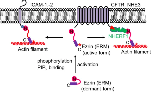
Fig. 2 ERM Proteins Function as Cross-Linkers between Membrane Proteins and Actin Filaments Directly or Indirectly via the Scaffold Proteins.
The interaction between the amino- and carboxy-terminal domains of ERM proteins makes them dormant. Phosphorylation of Thr567 in ezrin (Thr564 in radixin or Thr558 in moesin) by Rho kinase or protein kinase C, or PIP2 binding to the FERM domain, opens
up the dormant conformation into the active open conformation. (Left half) ERM proteins directly cross-link between ICAM-1 or -2 and actin filaments. (Right half) ERM proteins cross-link between the CFTR or NHE3 and actin filaments indirectly via the scaffold protein NHERF1.
Fig. 3 Regulation of Rho GTPases by Ezrin (ERM Proteins).
Rho GTPases in the GTP-bound form are active and interact with downstream effectors. They have an intrinsic GTPase activity and catalyze the conversion of bound GTP to GDP. The GDP-bound Rho GTPases are inactive and have greatly reduced affinity for downstream effectors. Rho-GEFs (such as Dbl) induce the exchange of bound GDP for GTP and activate Rho GTPases. On the other hand, Rho-GAPs inactivate Rho GTPases by increasing their intrinsic GTPase activity, resulting in accelerated hydrolysis of GTP to GDP. Rho-GDIs interact to stabilize the inactive GDP-bound Rho GTPases. Ezrin (or ERM proteins) promotes the dissociation of Rho-GDI from Rho GTPases. ERM proteins also function as a binding protein of GEF (Dbl) to activate Rho GTPases.
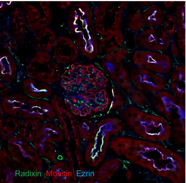
Fig. 4 Distributions of ERM Proteins in Mouse Kidney.
In glomeruli, ezrin was exclusively detected in podocytes; radixin and moesin were detected in endothelial cells. In addition, radixin and moesin co-localised with ezrin in the apical membranes of the proximal tubules (green: radixin, red: ezrin and blue: moesin).
Fig.5 Na+ Reabsorption at Different Parts of the Nephron.
In the renal tubule, 65%, 25–30%, 5% of Na+ filtered through the glomeruli are reabsorbed in proximal tubule, thick ascending limb (TAL), and distal tubule, respectively.
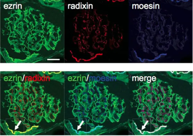
Fig. 7 Distributions of ERM Proteins in Mouse Glomeruli.
In WT mouse kidneys, glomerular localization of ERM proteins was investigated by immunofluorescence analysis. Ezrin, radixin and moesin showed different localization in glomeruli. Ezrin was exclusively detected in podocytes; radixin and moesin were detected in endothelial cells, but not podocytes. Radixin and moesin co-localized with ezrin in the apical membranes of the proximal tubules (Arrow).
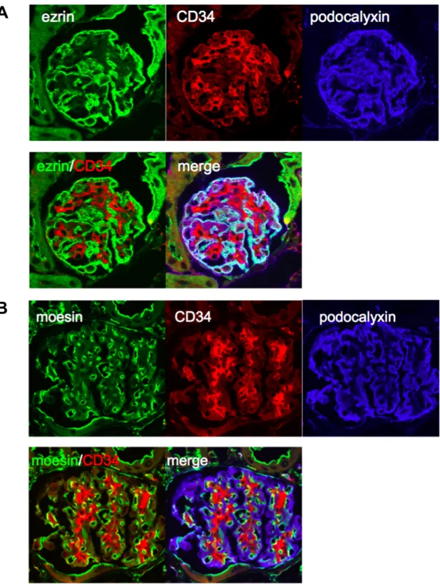
Fig. 8 Immunofluorescent Analysis for Ezrin and Moesin in Glomeruli.
Coimmunofluorescent analysis for ezrin (A) and moesin (B) with CD34, an endothelial cell marker, and podocalyxin, a podocyte marker were performed in WT mice kidney.
A
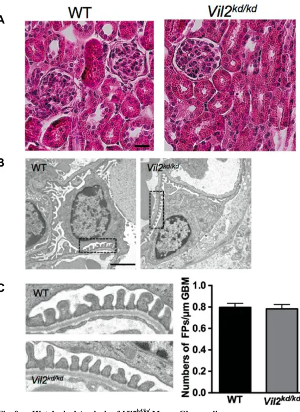
Fig. 9 Histological Analysis of Vil2kd/kd Mouse Glomeruli.
There were no morphological abnormalities in the Vil2kd/kd glomeruli observed by H.E.
staining (Scale bar: 25 µm) (A) or electron microscopic analysis (magnification × 3,610, scale bar: 2 µm) (B). Areas enclosed by dotted lines are magnified, and (magnification × 19,000) were shown in (C) and the number of foot processes/µm of glomerular basement
A
B
Fig. 10 Spot Urine was Separated by SDS-PAGE and Stained with CBB.
As a positive control, BSA (0.5, 1, 2.5 and 5 µg) was loaded. Apparent urinary albumin leakage was not observed in WT and Vil2kd/kd mouse urine.
Fig. 11 Immunofluorescence Analysis for Proteins Expressed in Glomerular Podocytes in WT Mice Glomeruli.
Immunolocalization of ezrin, other related proteins (NHERF2 and podocalyxin), and marker proteins (synaptopodin and podocin) was investigated in WT mouse glomeruli (Scale bar: 20 µm).
Fig. 12 Immunofluorescence Analysis for Proteins Expressed in Glomerular Podocytes in Vil2kd/kd Mice Glomeruli.
Immunolocalization of ezrin, other related proteins (NHERF2 and podocalyxin), and marker proteins (synaptopodin and podocin) was investigated in Vil2kd/kd mouse glomeruli.
Fig. 13 Immunofluorescent Analysis for ERM Proteins in WT and Vil2kd/kd Mice Glomeruli.
Localizations of radixin and moesin were also investigated in WT (A) and Vil2kd/kd (B)
mouse glomeruli. Ezrin, radixin, and moesin were coimmunostained with podocalyxin.
A
Fig. 14 Western Blot Analysis of Glomerular Proteins in Isolated Glomeruli from WT and Vil2kd/kd Mice.
Glomerular protein expression levels were investigated by immunoblotting. As a control, total kidney cortex lysate from WT mouse was used (A). Densitometric analysis was performed (n = 3–5, respectively) (B). *p < 0.05, vs. WT.
Fig. 15 Morphological Changes in Podocytes and Functional Changes in Adriamycin-induced Glomerulopathy.
Adriamycin (50 mg/kg) was administrated intravenously. After 7 days, spot urine was collected, separated by SDS-PAGE and stained with CBB. As a positive control, BSA (0.5 and 1 µg) was loaded. Urinary albumin excretion was increased in WT mice but not
Vil2kd/kd mice (WT: n = 7, Vil2kd/kd: n = 7) (A). Urinary albumin and creatinine concentrations were measured and ACR (mg albumin/mg creatinine) was calculated (B). Electron microscopic analysis was performed in these mice (magnification × 19,000, Scale bar: 1 µm). Number of foot processes/µm of GBM was measured. Foot process
B
Fig. 16 Morphological Changes in Podocytes and Functional Changes in LPS-induced Glomerulopathy.
LPS (200 µg) was administrated intraperitoneally. After 24 h, spot urine was collected, separated by SDS-PAGE, and stained with CBB (A). As a positive control, BSA (0.5 and 1 µg) was loaded. Urinary albumin and creatinine concentrations were measured and ACR (mg albumin/mg creatinine) was calculated (WT: n = 9, Vil2kd/kd: n = 9) (B). Electron microscopic analysis was performed in these mice (magnification × 19,000, Scale bar: 1 µm). Number of foot processes/µm of GBM was measured (C).
B
Fig. 17 Rho-GTPase Activity Assay Using Isolated Glomeruli.
Rho-GTPase activity—including RhoA, Rac1 and Cdc42—was measured by an ELISA-based Rho-G-LISA assay. Isolated glomeruli from both untreated and ADR-treated WT and Vil2kd/kd mice were used in this study (n = 5–11) (A). Isolated glomeruli from both
untreated and LPS-treated WT and Vil2kd/kd mice were used in this study (n = 5–8) (B).
A
Fig.18 Electrolytes Reabsorption in TAL.
In TAL, 25–30% of Na+ filtered through the glomeruli are reabsorbed. TAL plays an important role in the maintenance of salt and fluid homeostasis. NKCC2 plays an essential role in this process.
Fig.19 Schematic Figure on Exocytosis and Endocytosis of NKCC2.
The hormonal stimulation of the TAL recruits exocytosis of NKCC2. This process is promoted by phosphorylation of Ser126 and Ser874 of NKCC2 via the cAMP/PKA pathway. Vasopressin binds vasopressin V2 receptor (V2R) and increases intracellular cAMP (A).
Lipid raft localization of NKCC2 is important for the endocytosis of NKCC2 since this
A
Fig. 20 Immunofluorescent Analysis of WT and Msn−/y Mice.
Kidney tissue sections from WT and Msn−/y mice were co-immunostained with moesin,
and megalin (marker for proximal tubule) (A), or THP (Tamm Horsfall protein: marker for TAL) (B). White arrow indicates apical membrane of proximal tubule, in which positive immunostaining for moesin is observed (A). Yellow arrowhead indicates apical membrane of TAL, in which positive immunostaining for moesin is observed (B). Moesin
A
B
Fig. 21 Histological Analysis of WT and Msn−/y Mice.
Hematoxylin & Eosin staining was performed using the kidney tissue section from WT and Msn−/y mice. (Scale bar 25 µm)
Table 5 Biochemical Parameters of Plasma and Urine and GFR in WT and
Fig. 22 Expressions and Localizations of Moesin and Other Proteins (NKCC2, THP, and ROMK) in the Kidney.
Immunoblotting for moesin and other proteins expressed in TAL (NKCC2, THP) using mouse renal medulla lysate (A). Densitometric analysis was performed (n = 3, respectively) (A). Subcellular localizations of NKCC2, co-stained with THP (B) and ROMK (C) were examined in WT and Msn−/y mice by the immunofluorescent analysis.
A
B
Fig. 23 Surface Biotinylation Assay for the NKCC2.
Surface biotinylation assay was performed using medullary tubular suspensions from WT and Msn−/y mice. Surface expression levels of NKCC2, moesin, and THP were compared
with total expression levels. Densitometric analysis for surface expressions of NKCC2 and THP was performed as shown in graph (n = 11, respectively). * p < 0.05 vs. WT.
Fig. 24 Internalization Assay for the NKCC2.
Internalization assay was also performed using medullary tubular suspensions. Densitometric analysis for internalized NKCC2 was performed. Data are expressed as a percentage of basal surface expression of NKCC2. (n = 3, respectively). * p < 0.05 vs. WT (37 °C)
A
Fig. 25 Subcellular Fractionation of Renal Medulla Lysate by Density Gradient Centrifugation.
Subcellular fractionation of renal medulla lysate from WT (A) and Msn−/y (B) mice was performed by 8%–34% OptiPrep-based density gradient centrifugation method. The distribution of Rab5 (endosome) or Rab11 (recycling endosome) was examined. Densitometric analysis for moesin, NKCC2, Rab5, and Rab11 was performed. Data are expressed as a percentage of total protein expression (n = 3, respectively) (C). Open circle (○) represents protein expressions in WT kidney. Closed triangle (▲) represents protein
Fig. 26 Fractionation of Lipid Raft by Density Gradient Centrifugation.
Lipid raft fractions were separated by the 10–40% Optiprep-based density gradient centrifugation method in WT (A) and Msn−/y (B) mice. The expression of flotillin-2 as a marker of lipid raft was examined. The fractions containing flotillin-2 were shown by bold lines at the bottom of figures
A
Fig. 27 Analysis for NKCC2 Activity in Medullary Tubular Suspension.
Time-dependent Tl+ uptake (10 mM Tl+, 37 °C) was examined using medullary tubular suspension from WT mice in the presence or absence of Furosemide (100 µM) (n = 3, respectively) (A). Concentration-dependent Tl+ (2.5, 5, 10, 30, and 40 mM Tl+) uptake (37 °C, 5 min) were examined using medullary tubular suspension from WT mice in the presence or absence of Furosemide (100 µM) (n = 3, respectively) (B). Inhibition of tubular Tl+ uptake (10 mM Tl+, 37 °C, 5 min) by Furosemide (12.5, 25, 50, 100, and 200 µM Furosemide) (n = 3, respectively) (C). Furosemide sensitive-tubular Tl+ uptake activity (10 mM Tl+, 37 °C, 5 min) was compared between WT and Msn−/y mice (n = 4, respectively) (D). *p < 0.05 vs WT. **p < 0.01 vs. WT. Furosemide sensitive-tubular Tl+
A
B
Fig. 28 Analysis for Surface Expression of NKCC2 and THP in Water Loading Condition.
Surface expressions of NKCC2 and THP in the medullary tubules after water loading were examined by surface biotinylation and immunoblotting (A). Densitometric analysis was performed for the surface expression levels of NKCC2 in water loading condition (n
†
A
Fig. 29 Analysis for Moesin Expression in the Cortical TAL and Macula Densa Cells, and Real-time PCR and Western Blot Analysis for Renocortical COX-2 Expression.
Immunostaining for moesin, NKCC2, and THP was performed. In the cortical TAL, all of these proteins were expressed. In the macula densa cells (NKCC2+/ THP−: white arrow) of WT mouse, moesin was expressed at apical membrane and co-localized with NKCC2. G: glomeruli (A). Renocortical mRNA and protein expression of COX-2 was examined by real-time PCR (n = 9, respectively) (B) and Western Blot. (n = 11,
Fig. 30 Assay for Renocortical PGE2 Level.
Renocortical PGE2 level was examined by EIA assay (WT; n = 8, Msn-/y; n = 5),