Introduction
The common minke whale Balaenoptera acutor-ostrata is the smallest species of Balaenopteran whale, and is regarded as an important cetacean resource in Japan as well as in Northern Europe
such as Norway and Iceland. Common minke whales are currently classified into two subspe-cies: B. a. scammoni in the north Pacific and B. a. acutorostrata in the north Atlantic. In the South-ern Hemisphere, there is also a distinctive popula-tion of “dwarf” minke whales B. a. subsp. howev-er, the taxonomical classification of this popula-tion remains under debate(RICE, 1998).For man-aging whales appropriately, the population esti-mates of the subspecies or stocks levels are re-quired. Therefore, taxonomy is one of the most important concerns for the whale management. Genetic information has recently become a major key for classification in many animals. However, Société franco-japonaise dʼocéanographie, Tokyo
Presence of an interparietal bone and morphological variation in the
vertex of the skull in North Pacific common minke whale
Gen NAKAMURA1)*, Megumi TAKAHASHI1), Ayumi HIROSE1), Ryotaro HAYASHI1), Yoshihiro FUJISE2), Naoko MIYAKAWA1), Satoko INOUE1)and Hidehiro KATO1)
Abstract: The vertex of the skull is regarded as one of the most important categorical traits in
taxonomy in baleen whales. However the previously reported species-specific characteristics for the North Pacific common minke whale, were based on only a very limited number of specimens Therefore, we aimed to assess the efficacy of these morphological characteristics as categorical traits, based on specimens of North Pacific common minke whales collected from the JARPNII survey(n =106).The frontal border of the nasal bones, as well as the positional relationship of the end of premaxilla and the nasal bones, were varied widely by individual, indicating that these traits are not appropriate for species classification. However, the end of the maxilla was located posteriorly to the end of the premaxilla and the nasal bones in all whales surveyed. Therefore, we conclude that this characteristics can serve as universal species-specific morphological character-istics for the North Pacific common minke whales. Furthermore, we found an interparietal bone in all whales surveyed. This bone has been regarded as one of the specific character of “dwarf” minke and have not been reported from any common minke whales in the North Pacific.
Keywords : Common minke whales, Balaenoptera acutorostrata, skull morphology
1)Laboratory of Cetacean Biology, Tokyo University of Marine Science and Technology, 4-5-7 Konan, Minato-ku, Tokyo 108-8477, Japan
2)Institute of Cetacean Research, 4-5 Toyomi-cho, Chuo-ku, Tokyo 104-0055, Japan
*Corresponding author: Tel: +81-03-5463-0561 Fax: +81-03-5463-0561 E-mail: [email protected]
especially in cetaceans, even though their genetic differences were not clear, some morphological differences were clearly observed between stocks or populations in different oceans (MIYAZAKI, 1994). Morphological characteristics therefore still remain an important key for clas-sification in cetaceans.
The vertex of the skull is the highest part of the skull, and all dorsal skull bones aggregate at this point(Fig. 1).This shift in the bonesʼ position, generally called as “telescoping” is the result of adaptation to an aquatic lifestyle(MILLER, 1923), and can be used as an important categorical characteristic for taxonomy in baleen whales, especially Balaenoptera(OMURAet al., 1970; 1981; OMURA and KASUYA, 1976; WADA et al., 2003; MEIRELLESand FURTADO-NETO, 2004; MAGALHÃESet al., 2007).
Morphological comparison of the bones in the vertex of the common minke whale and the Antarctic minke whale B. bonaerensis have been performed by several authors(OMURA, 1975; ARNOLD et al., 1987; HORWOOD, 1989; ZERBINI and SIMÕES-LOPES, 2000).Schematic illustration of the vertex showing interspecific differences have de-scribed(HORWOOD, 1989).A convex frontal bor-der of the nasal bones(viewed dorsally), the posterior end of the nasal bones being located more anteriorly to the end of premaxilla, and the end of maxilla being located posterior to the nasal bones were all species-specific morphological characteristics for North Pacific common minke whales(OMURA, 1975).However, Omuraʼs study was based on only 6 whales, including juvenile animals. Therefore, there still remains the uncer-tainty that these results are due to individual variation or growth-dependent changes, and a greater number of individuals should be exam-ined to confirm the consistency of these differen-ces. Furthermore, based on photographs in OMURA(1957), ZERBINI and SIMÕES-LOPES(2000)
pointed out that the North Pacific minke whales also might possess an interparietal bone, which has not been reported previously in this species. Against this background, in the present study we focused on the form of the frontal border of the nasal bones, the positional relationships of the end of premaxilla, maxilla, and the nasal bones, and the existence/nonexistence of an interparie-tal bone, which have been held to be possible species-specific characteristics. We examined
in-Fig. 1. Dorsal view of the skull of common minke
whale and the vertex of the skull(surrounded by dotted line). F: Frontal, Ma: Maxilla, Na: Nasal, Oc: Occipital, Pa: Parietal, Pm: Premaxilla, T: Temporal.
dividual variation, sexual differences, and growth-dependent changes in these morphological char-acteristics to assess their efficacy as categorical traits for classifying whale species.
Materials and Methods
We used 106 North Pacific minke whales(64 males, body length: 1.68–8.05 m; 42 females, body length: 3.73–8.55 m)collected from water off Ayukawa, Miyagi pref. and Kushiro, Hokkaido pref. Japan, in 2009–2012 during the Japanese Whale Research Program under Special Permit in the Western North Pacific-Phase II(commonly known as JARPN II)survey(Fig. 2),conducted under the special permit from the government of Japan in accordance with Article VIII of the International Convention for the Regulation of Whaling(GOVERNMENT OF JAPAN, 2002). Body length was recorded as the length between the
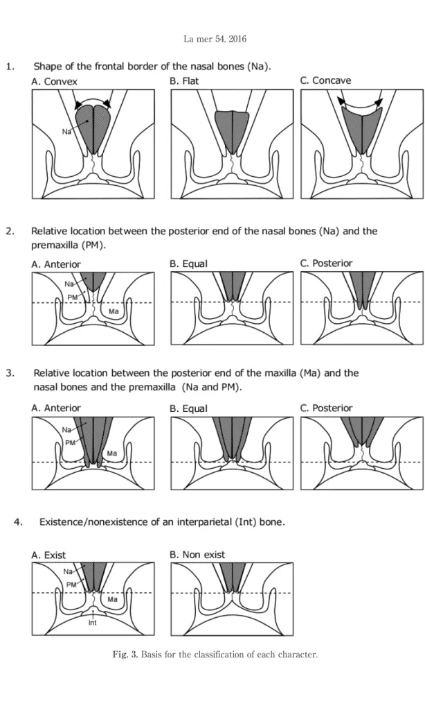
tip of rostrum and the notch of the fluke. After flensing, any flesh or connective tissue remaining on the skull was completely removed manually by knife and the skull was then ob-served. The below characteristics were chose for the present analysis in order to elucidate individ-ual variation, sex differences, and growth-depend-ent changes in morphology. Each characteristic was classified based on form or position(Fig. 3). To avoid the effect of growth related morphologi-cal changes, the fetus was used only for the observation of the existence/nonexistence of the interparietal bone. Animals with unclear morpho-logical characteristics, due to the incompleteness of removing connective tissues were removed from analysis.
1. Shape of the frontal border of the nasal bones(Fig. 3-1)
Animals were classified into 3 types based on form of the frontal border of the nasal bones: convex, flat, or concave.
2. Relative location between the posterior end of the nasal bones and the premaxilla(Fig. 3-2)
Animals were classified into 3 types based on whether the end of the nasal bones was posi-tioned anterior, equal, or posterior to the end of the premaxilla.
3. Relative location between the posterior end of the maxilla and the nasal bones and premaxilla(Fig. 3-3)
Animals were classified into 3 types based on whether the posterior end of the maxilla was positioned anterior, equal, or posterior to the end of the other two characteristics(the nasal bones and premaxilla).
4. Existence/nonexistence of an interparietal bone and any individual variation(Fig. 3-4) By removing any flesh or remaining connective tissue carefully, especially around the parietal
Fig. 2. Sampling area. Research was conducted in
coastal waters off Ayukawa, Miyagi prefecture and Kushiro, Hokkaido prefecture. The areas are 50 nautical miles from each port(illustrated by circle).
and occipital bones, the existence/nonexistence of an interparietal bone was examined.
Results
1. Shape of the frontal border of the nasal bones
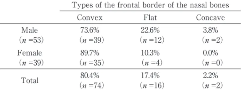
There was variation in shape of the frontal border of the nasal bones. The animals with convex type were dominant(80.4%, n =74),how-ever animals with flat(17.4%, n =16)or concave border(2.2%, n =2)were also observed(Table 1).Such flat or concave borders have not been reported previously. Therefore the shape of the frontal border of the nasal was not uniform and varied individually. Sex ratios within each border type were compared to clarify the presence or absence of sex differences. Proportional differen-ces between each types were not statistically
significant, indicating that there is no sexual differences in the shape of frontal border of the nasal bones(Chi-square test, df = 2, p > 0. 05) (Table 1).
Concave type was observed in only the class with body length > 7 m(n = 2). Although it appeared that animals with flat type nasal bones occurred relatively frequently in the >7 m class, no statistical difference was observed(Chi-square test, df =6, p >0.05)(Table 2)
2. Relative location between the posterior end of the nasal bones and the premaxilla The end of the nasal bones was positioned anterior(52.9%, n =54)or equal(41.2%, n =42)to the end of the premaxilla. On the other hand, we also observed a few animals with the end of the nasal bones positioned posterior to the premaxilla (5. 9%, n = 6), which has not been previously Table 1. Frequency and sex ratio of each types of the frontal border of
the nasal bones.
Types of the frontal border of the nasal bones
Convex Flat Concave
Male 73.6% 22.6% 3.8%
(n =53) (n =39) (n =12) (n =2)
Female 89.7% 10.3% 0.0%
(n =39) (n =35) (n =4) (n =0)
Total (n =74)80.4% (n =16)17.4% (n =2)2.2%
Table 2. Frontal border types of the nasal bones in each body length class. Body length class
Total <5.0m 5.1-6.0m 6.1-7.0m 7.1m < Convex (n =21)84.0% (n =25)92.6% (n =14)87.5% (n =14)58.3% (n =74)80.4% Flat (n =4)16.0% (n =2)7.4% (n =2)12.5% (n =8)33.3% (n =16)17.4% Concave (n =0)0.0% (n =0)0.0% (n =0)0.0% (n =2)8.3% (n =2)2.2% Total 25 27 16 24 92
reported(Table 3). Sex ratios were compared between position types to clarify the presence or absence of sex differences. Proportional differen-ces between each types were not statistically significant, indicating that there is no sexual differences in the relative position between the posterior end of the nasal bones and premaxilla (Chi-square test, df =2, p >0.05)(Table 3).
No clear trends were observed, and there were no statistical differences between each body length classes(Chi-square test, df =2, p >0.05) (Table 4)
3. Relative location between the posterior end of the maxilla and the nasal bones and premaxilla
The distance from the end of the maxilla and these other two characteristics varied by individ-ual. However, for all animals(n =106),the end of
the maxilla was posterior to the end of the pre-maxilla and the nasal bones.
4. Existence/nonexistence of an interparietal bone and any individual variation
In all animals(n =106),we found a bone that was separate from the occipital, frontal, and parie-tal bones. In the feparie-tal stage, this bone was not fused with other bones; therefore, we concluded it to be the interparietal bone(Fig. 4).The size and shape varied by individual, but no sex differences or growth-dependent change in shape were ob-served(Fig. 5).
Discussion
The vertex of the skull has come to be regard-ed as an important trait for categorizing baleen whales. OMURA(1975)previously described the frontal border of the nasal bone in North Pacific Table 3. Frequency and sex ratio of position of posterior end of nasal bones to that of the
premaxilla.
Position of posterior end of nasal bones to that of the premaxilla
Anterior Equal Posterior
Male 51.6% 43.6% 4.8%
(n =62) (n =32) (n =27) (n =3)
Female 55.0% 37.5% 7.5%
(n =40) (n =22) (n =15) (n =3)
Total (n =54)52.9% (n =42)41.2% (n =6)5.9%
Table 4. Frequency of the position of posterior end of nasal bones to that of the premaxilla. in each body length class.
Body length class
Total <5.0m 5.1-6.0m 6.1-7.0m 7.1m < Anterior (n =13)48.2% (n =12)48.0% (n =12)63.2% (n =17)54.8% (n =54)52.9% Equal (n =11)40.7% (n =13)52.0% (n =4)21.0% (n =14)45.2% (n =42)41.2% Posterior (n =3)11.1% (n =0)0.0% (n =3)15.8% (n =0)0.0% (n =6)5.9% Total 27 25 19 31 102
minke whales as protruding forward—that is, the border was convex. From the present study, it was clearly indicated that the animals with this convex type of the nasal bones were predominant (80. 4%); however, flat-or concave-type animals were also observed. This study furthermore re-vealed that the positional relationship between the end of the premaxilla and the nasal bones also varied. Because of such wide individual variation, we concluded that the shape of the frontal border of the nasal bones and the positional relationship between the end of the premaxilla and the nasal bone are not an appropriate categorical trait.
The end of the premaxilla and the nasal bones extend more posteriorly and are in line with the maxilla in the Antarctic minke whale, Brydeʼ s whale, and pygmy blue whale(OMURAet al., 1970; 1981; OMURA, 1975).The degree of the telescop-ing, posterior extension of the rostral bones, in common minke whales tend to be lower than that in other baleen whales. The present study found that the posterior end of the maxilla was located posterior to the end of the premaxilla and the nasal bones in all animals. This positional
relation-ship could therefore be a universal characteristic among North Pacific common minke whales.
Our study also found that all whales surveyed possessed the interparietal bone. The existence of this bone has been reported for dwarf minke whales and Antarctic minke whales, but not for the North Pacific common minke whales(OMURA, 1975; ARNOLDet al., 1987; ZERBINIand SIMÕES-LOPES, 2000).As indicated by ZERBINIand SIMÕES-LOPES (2000), we found the interparietal bone in OMURAʼs figures(1975).Therefore OMURA(1975) overlooked the existence of this bone, but present study declared that the North Pacific common minke whales also have interparital bone.
In conclusion, our study found that the position-al relationship between the posterior end of the maxilla and the nasal bones and premaxilla is a suitable characteristic for classification of North Pacific common minke whale. The study also established that the North Pacific common minke whale possesses the interparietal bone. It should be noted that the present study was based on only appearance, not quantitative measurements. As for the future study, measurement based
Fig. 4. Photograph(left)and illustration(right)of the vertex of skull of common minke whale. F: Frontal, Ip:
study such as using caliper or 3D scanner would be more effective to evaluate the morphological variation and/or species specific character.
In the North Pacific common minke whales, the existence of two stocks has been reported(KATO et al., 1992).In the present study, we used the samples collected from the coastal waters off
Ayukawa and Kushiro, where the two stocks is mixing(HATANAKA and MIYASHITA, 1997), how-ever we did not consider inter stock differences and therefore still remain the uncertainty that the shape and positional variation includes inter-stock differences. The utility and/or importance of vertex characteristics as categorical traits for
Fig. 5. Individual variation in size and shape of interparietal of common minke whale. Interparietals are marked
whale classification would be revealed more clearly by future studies using more specific measurements and analyses of size or proportion of these characteristics as well as considering stock information.
Acknowledgments
We thank the former leaders of the JARPN II Survey; Genta Yasunaga and Takeharu Bando of the Institute of Cetacean Research; Toshiya Kishiro and Hideyoshi Yoshida of the National Research Institute of Far Seas Fisheries. Repre-sentative Director Yoshiichi Shimomichi, former Exploration Chief Shigeo Tabata, Takahiro Hara and the staff of the Association for Community-Based Whaling. We also thank the members of the Laboratory of Cetacean Biology, Tokyo Uni-versity of Marine Science and Technology and the anonymous reviewers for quick and valuable review.
References
ARNOLD, P. W., H. MARSHand G. HEINSOHN(1987):The
occurrence of two forms of minke whales in east Australian waters with a description of external characters and skeleton of the diminutive or dwarf form. Sci. Rep. Whales Res. Inst.,38, 1-46.
GOVERNMENT OFJAPAN(2002):Research plan for
ceta-cean studies in the western North Pacific under Special Permit(JARPN II).Paper SC/54/O2. Pre-sented to the 54th IWC Scientific Committee
Meeting; May 2002(unpublished).46pp.
HATANAKA, H. and T. MIYASHITA(1997): On the
feeding migration of Okhotsk Sea. West Pacific stock of minke whales, estimates based on length composition data. Rep. Int. Whal. Commn., 47,
557-564.
HORWOOD, J. W.(1989):Biology and exploitation of the
minke whale. CRC Press, Florida, 248pp.
KATO, H., T. KISHIRO, Y. FUJISEand S. WADA(1992):
Morphology of minke whales in the Okhotsk Sea, Sea of Japan and off the East Coast of Japan, with respect to stock identification. Rep. Int. Whal.
Commn.,42, 437-442.
MAGALHÃES, F. A., M. M. SEVERO, C. H. TOSI, R. G. GARRI,
A. N. ZERBINI, S. CHELLAPPAand F. J. L. SILVA(2007):
Record of a dwarf minke whale(Balaenoptera acutorostrata)in northern Brazil. J. Mar. Biol. Ass. UK, 2, Biodiversity Records, 1, 1-2.
MEIRELLES, A. C. O. and M. A. A. FURTADO-NETO(2004):
Stranding of an Antarctic minke whale, Balaenop-tera bonaerensis, Burmeister, 1867, on the north-ern coast of South America. LAJAM, 3, 81–82. MILLER, G. S.(1923):The telescoping of the cetacean
skull. Smithson. misc. collect.,76, 1–71.
MIYAZAKI, N.(1994):Skull morphology of small
ceta-cean: A consideration of taxonomic problems in the Short-finned Pilot Whale, Globicephala macro-rhynchus, in Japanese waters. Honyurui Kagaku [Mammalian Science], 34, 31-42(in Japanese
with English abstract).
OMURA, H.(1957):Osteological study of the little piked
whale from the coast of Japan. Sci. Rep. Whales Res. Inst., 12, 1-21.
OMURA, H.(1975): Osteological study of the minke
whale from the Antarctic. Sci. Rep. Whales Res. Inst., 27, 1-36.
OMURA, H., T. ICHIHARAand T. KASUYA
(1970):Osteolo-gy of pygmy blue whale with additional informa-tion on external and other characteristics. Sci. Rep. Whales Res. Inst., 22, 1-27.
OMURA, H. and T. KASUYA(1976):Additional
informa-tion on skeleton of the minke whale from the Antarctic. Sci. Rep. Whales Res. Inst., 28, 57–68. OMURA, H., T. KASUYA, H. KATOand S. WADA(1981):
Osteological study of the Brydeʼs whale from the central South Pacific and eastern Indian Ocean. Sci. Rep. Whales Res. Inst.,33, 1-26.
RICE, D. W.(1998): Marine mammals of the world:
systematics and distribution. Special Publication Number 4 of the Society for Marine Mammalogy. Allen Press, Lawrence, Kansas, 231pp.
WADA, S., M. OISHIand T. K. YAMADA(2003):A newly
discovered species of living baleen whale. Nature,
426, 278-281.
ZERBINI, A. N. and P. C. SIMÕES-LOPES
(2000):Morphol-ogy of the skull and taxonomy of southern hemisphere minke whales. Paper SC/52/OS10
presented to the 52nd IWC Scientific Committee
Meeting; June 2000(unpublished).28pp.
Received: December 4, 2015 Accepted: February 1, 2015