Review J Tokyo Wom Med Univ 89(Extra1): E75-E99, 2019 Jul
Potential of Adjuvant-induced Lymphangiomas in Mice:
Its Advantages as an Animal Model to Study Lymphatic Endothelial Cells
Taichi Ezaki
Department of Anatomy and Developmental Biology, School of Medicine, Tokyo Women s Medical University, Tokyo, Japan (Accepted April 15, 2019)
Studies on the lymphatic system have made remarkable progress with the discovery of lymphatic endothelial cell-specific markers. To discover lymphatic endothelial cell-specific markers, we adopted a Freund s incomplete adjuvant (FIA)-induced lymphangioma model in rodents. The tumor was used as an antigen source of mouse lym-phatic endothelium to produce monoclonal antibodies. We obtained LA102, which recognizes lymlym-phatic endothe-lial cells, but not blood vessel endotheendothe-lial cells. We found LA102 to be a homolog of mouse CD90.2 (Thy-1.2). Using LA102 and other specific markers for microvessels, including lectins, we have developed 3D-imaging techniques to characterize lymphatic networks to differentiate from blood vessels. This model has also been adopted to in-vestigate the relationship between peritoneal mesothelium and lymphatic endothelium. At three days after FIA injection, simple squamous mesothelial cells became cuboidal and detached from each other to lose their polarity and formed multi layers. Various-sized fat droplets gradually fused with each other, and the fat-storing cells be-came large fat cells or formed large chimeric follicular structures. At four weeks or later, these cell masses formed tubular structures draining the fat out of the peritoneal cavity. Taking up fat (FIA) droplets, not only po-doplanin+
mesothelial cells, but also bone marrow-derived macrophages and some interstitial mesenchymal cells were involved in tumorigenicity. We suggest a sequential change from mesothelial to lymphatic endothelial cells via fat-storing lymphangioma cells after FIA stimulation. These phenomena seem to be a defense mechanism, where mesothelial-endothelial transformation might occur via fat incorporation to drain the extrinsic adjuvant oil out of the peritoneal cavity. The significance of FIA-induced lymphangiomas and mesothelial cell diversity might be important for the interrelationship between fat cells and lymphatic endothelial cells.
Key Words: adjuvant, fat, lymphangioma, lymphatic endothelial cell, mesothelial cell
Introduction
Animal tumor models have been used to
under-stand normal cellular life events, because tumors
may reflect mirror images of normal cells.
1There
are several tumor models to investigate various
phenomena, such as tumor immunity as specific
tar-gets for immune reactions,
2-6thymic nurse cell as a
thymic microenvironment in spontaneous
thymo-mas,
7-10and various tumor angiogenesis.
11-13In this
re-view, I introduce how an adjuvant-induced
lym-phangioma rodent model was adopted and
demon-strate its potential and use as various experimental
strategies.
Corresponding Author: Taichi Ezaki, Department of Anatomy and Developmental Biology, School of Medicine, Tokyo Women s Medical University, 8-1 Kawada-cho, Shinjuku-ku, Tokyo 162-8666, Japan. [email protected]
doi: 10.24488/jtwmu.89.Extra1_E75
Copyright Ⓒ 2019 Society of Tokyo Women s Medical University. This is an open access article distributed under the terms of Creative Commons Attribution License (CC BY), which permits unrestricted use, distribution, and reproduction in any medium, provided the original source is properly credited.
Figure 1 Monoclonal antibodies against rat vascular en-dothelial cells.
The specific reactivity of each monoclonal antibody to vari-ous rat microvasculature is expressed as the width range of bars. For example, B1 recognizes endothelial cells of arterioles and capillaries on the arterial side, but not of ve-nous side and veve-nous-type vessels.
As an Antigen Source of Lymphatic Endothelium
to Identify the Lymphatic Endothelial Cell
Specific Markers
1.Initial studies on vascular endothelial cells
Blood vascular and lymphatic systems play
es-sential roles in local tissue microcirculation.
Mi-crovessels have characteristic structures to exhibit
their special functions for local microcirculation.
14For example, arterioles are called as resistance
ves-sels because they constitute the principal
compo-nent of peripheral resistance to blood flow that
regulates the blood pressure. Capillaries, as
ex-change vessels, may exhibit marked differences in
their structures and permeability properties
de-pending on the functional sites in various organs.
Capillaries converge to form post-capillary venules
(PCVs) or venules of larger size, where cellular or
macromolecular passages can take place under
both physiological and pathological conditions.
15Therefore, we tried to develop strategies to
in-vestigate the morphological and functional
charac-teristics of each microvessel. Monoclonal antibodies
(mAbs) against rat vascular endothelial cells were
produced in mice to characterize and assess the
functional properties of the antigens recognized
(Figure 1).
16B1 recognizes rat arteriolar endothelial
cells and arterial-side capillaries, but not those of
venous-type vessels. Contrarily, B 110 recognizes
venous-side capillaries, venules including PCVs,
some sinuses and small veins, but not any
arterial-type vessels (Figure 2a). B8 recognizes endothelial
cells in splenic marginal sinuses and certain types
of capillaries. B43 recognizes lymph node venules
and hepatic sinusoids. Immunohistochemical
identi-fication of microvessels using a panel of these mAbs
will provide valuable information for studies on
lo-cal blood microcirculation.
Despite its critical role in tissue fluid homeostasis,
macromolecule or lipid absorption, and immune or
malignant cell migration, however, the lymphatic
system has not received considerable attention.
Lymphatic capillaries have highly permeable
struc-tures, and they readily capture fluids,
macromole-cules, particulate matters, and cells from the
con-nective tissue spaces. However, segmental and
functional studies on microvessel networks have
not always been successful due to the lacks of
reli-able techniques to identify these ramifications in
the local tissue (Ezaki & Kotani, in this volume
17). In
part this has been due to a lack of specific markers
for lymphatic endothelial cells. Therefore, we first
produced a mouse mAb, B27, against rat lymphatic
endothelium.
18B27 strongly recognizes endothelial
cells of most rat lymphatics, including lacteals,
lym-phatic capillaries in the diaphragm (Figure 2b) at
the absorption sites in the peritoneal cavity,
collect-ing lymphatics, and thoracic duct. However, B 27
also recognizes the endothelium of some other
types of blood vessels. Hence, double
immunostain-ing of B27 should be combined with type IV
colla-gen of basement membrane to discriminate
be-tween lymphatic capillaries and blood capillaries on
tissue sections (Figure 2c).
2.Requirement for more specific strategies to
identify lymphatic endothelial cells
To better understand the lymphatic functions, it
is important to discriminate lymphatic vessels from
blood vessels and characterize lymphatic
endothe-lial cells under both physiological and pathological
conditions.
19-22Furthermore,
various
vascular
lineage-specific genes have been identified in both
lymphatic and blood vascular endothelial cells in
cultures.
23Therefore, mAb availability would
con-tribute to immunohistochemical identification of
lymphatic vessels particularly in mice and
under-stand the molecular basis of their functions. In the
last two decades, several useful markers for
lym-phatic vessels have been reported. For example,
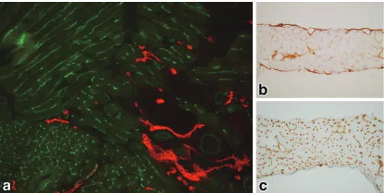
Figure 2 Specificity of B110 and B27 monoclonal antibodies to rat microvasculature. a: Reactivity of B110 to capillary networks continuing two accompanying arteriole (A) and venule (V) in the mesentery. Note that B110 recognizes capillaries on the venous side (*starting point and flow direction: arrows) and venule (V), but not any arterial-type vessels including arteriole (A) as shown in Figure 1.
b: B27 recognizes lymphatic endothelial cells (red) of the lymphatic sinuses (*: black car-bon ink has been absorbed by the lymphatic vessels after a peritoneal injection) underly-ing the peritoneal surface of the diaphragm. Note that peritoneal mesothelial cells are also recognized by B27 and took up some carbon particles (arrowheads), suggesting their close relationship. (from Ezaki et al., 1990)18
c: As B27 also recognizes the endothelium of some types of blood vessels, it is necessary to combine double immunostaining of B27 (red) with type IV collagen of the basement mem-brane (arrows) to discriminate lymphatic capillaries and blood capillaries in sections of sub-cutaneous tissue of the skin. Note that lymphatic capillary (LC) is strongly stained by B27 (red), whereas blood capillaries (BC) are lined with basement membranes (dark brown). (from Ezaki et al., 1990)18
LYVE-1 is a specific receptor of hyaluronic acid.
24Podoplanin is a marker for renal podocytes and is
co-localized on lymphatic vessels.
25,26Prox-1 is
an-other nuclear marker for developing lymphatic
en-dothelial cells.
27However, most of these markers
re-ported have not been originally discovered on
lym-phatic vessels ; their empirical relationship with
lymphatic vessels was realized accidentally.
20,24,25,27Furthermore, their immunohistochemical
distribu-tion is not strictly confined to lymphatic endothelial
cells, but shared with some blood vessel types.
20,28-30To discover novel lineage-specific markers of
lymphatic origin, we sought to produce another
mAb directed at mouse lymphatic endothelium.
However, there were several technical limitations.
Firstly, the source material of lymphatics as an
anti-gen for immunization is difficult to obtain. Secondly,
lymphatic-specific antigens seem to have weak
im-munogenicity compared with those of blood vessels,
against which several specific antibodies have been
raised, probably due to their relatively strong
im-munogenicity. Lastly, there are no good screening
procedures for selecting lymphatic-specific
antibod-ies. To overcome these limitations, we applied
sev-eral technical devices to obtain more confined
lym-phatic vessel-specific antibodies. We induced
be-nign lymphangiomas in mice by intraperitoneal
in-jection of Freund s incomplete adjuvant ( FIA ) .
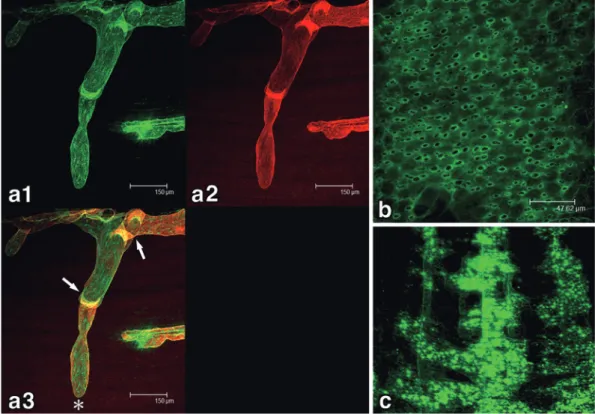
31Figure 3 Reactivity of LA102 and LA5 to mouse vascular endothelial cells.
a: Double immunofluorescent staining of a C57BL/6 mouse tongue tissue section with LA102 (red) and LA5 (green). The distribution of both lymphatic and blood vessels in the tongue muscle layers is apparent.
b: Immunoperoxydase staining (DAB reaction: brown) of lymphatic vessels in the dia-phragm are strongly stained with LA102.
c: Immunoperoxydase staining (DAB reaction: brown) of blood vessels in the diaphragm are stained with LA5.
This tumor model provided considerable amount of
relatively pure source material of lymphatic
endo-thelium.
32Furthermore, we employed a mild
en-zyme treatment followed by the neuraminidase
treatment
33to expose very minor hidden cell
sur-face antigens in lymphatic endothelial cells, and
used a rapid differential immunization protocol to
overcome the possible weak immunogenicity of
lymphatic endothelial cells.
34By trial and error, we
successfully produced a rat mAb, LA102 (or LEC
26), reacting predominantly with mouse lymphatics,
but not with any types of blood vessel. During the
immunization, we could also obtain another mAb,
LA5 (or BEC12), reacting selectively with most of
peripheral blood vessels, but not to any type of
lym-phatic vessel (Figure 3).
These novel antibodies would prove useful for
studies on mouse blood vascular and lymphatic
mi-crocirculation with several advantages.
34First, the
mAbs against vascular endothelial cells can be used
to characterize and assess the functional properties
of the antigens recognized. Second, confocal
laser-scanning microscopy can be used for 3D imaging of
the microvascular networks by infusing various
fluorescent lectins into circulation (described later).
Third, endothelial cell proliferation can be identified
by BrdU immunostaining of the tissue.
35Finally, the
functional aspects of microvessels can be further
in-vestigated by establishing some lymphatic
endothe-lial cell lines in vitro after separating lymphatic
en-dothelial cells from adjuvant-induced
lymphan-giomas. Several mouse lymphatic endothelial cell
lines have been successfully established using these
mAbs for positive separation using
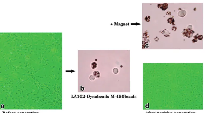
magnetic-activated cell sorting (MACS) (Figure 4). These cell
lines can be useful to characterize lymphatic
endo-thelial cells in vitro at the molecular level.
363.Mouse Thy-1 antigen as a specific marker
for lymphatic vessels
Interestingly, the antigen recognized by LA 102
exists on not only lymphatic endothelial cells, but
also some types of lymphoid cells, particularly the
T-cell lineage cells. While characterizing the
anti-gens recognized by this antibody, we found it to be
a homolog of CD90.2 1.2), but not CD90.1
(Thy-1.1). The following evidence supports this finding.
First, 94% of thymocytes and 51.2% of spleen cells
were positive for LA102. The LA102-positive spleen
cells were almost all positive for CD 3. Second, T
lymphocyte lineage-derived tumor cells were LA
Figure 4 Positive separation of lymphatic endothelial cells from adjuvant-induced lymph-angiomas by MACS using magnetic beads conjugated with LA102.
a: FIA-induced lymphangiomas of C57BL/6 mice were digested with collagenase D (Boer-hinger-Mannheim Biochemicals: 2 mg/mL in RPMI1640 medium with 1% fetal calf serum) for 30 min at 37 ℃ and single cell suspension was prepared. The cell mixture was cultivat-ed as the primary culture for several days to obtain single cell layers. Note that the shape of cell mixtures varies, indicating that they are heterogeneous cell populations before positive separation. b: The cells were treated with 0.05% trypsin, collagenase (200 U/mL), and 1 mM EDTA in PBS at 37 ℃ for 30 min to obtain as single-cell suspension. The cells were then incubated with LA102-conjugated Dynabeads M-450 at 4 ℃ for 30 min. c: The cell mixture was then positively separated for LA102-positive cells by MACS. Note that all separated cells bear magnetic beads. d: After positive separation, the cells appeared almost homogeneous and like a cobble stone in cultures.
102 positive, but those from B lymphocyte and
fi-broblast lineages were negative.
34Third, LA 102
only recognized its antigens on tissues from C 57
BL/6, BALB/c, and DBA/2 mice (all Thy-1.2
ground ) , but not from AKR mice ( Thy-1.1
back-ground) (Figure 5a&b). Forth, pre-treatment of
tis-sue sections with phosphatidylinositol-specific
phos-pholipase C from Bacillus cereus (PI-PLC) (P5542-5
UN; Sigma-Aldrich, St. Louis, MO, USA )
substan-tially diminished the reactivity of LA102 and Thy-1
antibodies against their original targets (
Fig-ure 5c-e). This indicates that the antigen molecule
recognized by LA102 is a glycoprotein anchored to
the plasma membrane through a
glycosylphosphati-dylinositol ( GPI ) tail covalently linked to the
carboxyl-terminal amino acid. The GPI-anchored
glycoproteins can be specifically cleaved off the cell
membrane by phospholipase C treatment.
37Finally,
the molecular size of LA102 is approximately 26-27
kDa, which is almost equivalent to that of CD 90,
with 25-29 kDa in its reduced form. We also found
that Thy-1.2 ( CD 90.2 ) is a homolog of LA 102 in
terms of molecular size and 45% mass-matching
ra-tio in a mass-spectrogram analysis between the two
molecules as well as their tissue co-localization
(un-published data). These data suggest that Thy-1 (CD
90) is another specific marker for lymphatic
endo-thelial cells in mice. Jurisic et al.
38also reported that
mouse lymphatic endothelial cells express high
lev-els of Thy-1 in situ and in vitro.
Thy-1 glycoprotein has been well characterized
in rodents and other species
39since it was first
de-scribed in 1964.
40Thy-1, the smallest member of the
immunoglobulin superfamily, is classified as a CD90
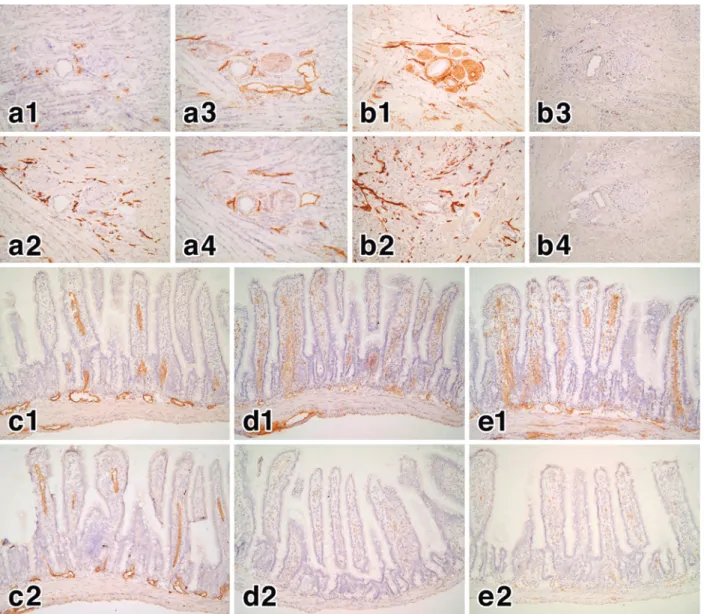
Figure 5 Effects of Thy-1 background and phospholipase C treatment on the antigens recognized by LA102 and other antibodies.
a: Cryosections of the tongue of C57BL/6 mice (Thy-1.2 type background) were stained by the immunoperoxidase reaction (DAB: brown) with either an anti-Thy1.1 antibody (a1), LYVE-1 (a2), an anti-Thy1.2 antibody (a3), or LA102 (a4). Note that no lymphatic vessel structure is recognized by the anti-Thy1.1 antibody, whereas the lymphatic vessels in the tongue are strongly stained by other antibodies including LA102. The positive stains in a1 are non-specific background stains (mainly of plasma cells, but not lymphatic vessels). b: Cryosections of the tongue of AKR mice (Thy-1.1 type background) were stained by the immunoperoxidase reaction (DAB: brown) with either an anti-Thy1.1 antibody (b1), LYVE-1 (b2), an anti-ThyLYVE-1.2 antibody (b3), or LALYVE-102 (b4). Note that no lymphatic vessel structure is recognized by both the anti-Thy1.2 antibody and LA102, whereas the lymphatic vessels in the tongue are strongly stained by the anti-Thy1.1 antibody and LYVE-1. The positive stains in b3 and b4 are non-specific background stains, but not any lymphatic vessel struc-tures.
c-e: Cryosections of the tongue of C57BL/6 mice (Thy-1.2 type background) were treated either without (c1, d1, e1) or with (c2, d2, e2) phospholipase C (0.2 U/mL: Sigma) in 10 mM Tris buffer with 0.05% BSA for 30 min at 37℃ , and then stained by the immunoperoxidase reaction (DAB: brown) with LYVE-1 (c1, c2), LA102 (d1, d2), or an anti-Thy1.2 antibody (e1, e2). Note that phospholipase C treatment completely diminished immunostaining of both LA102 (d2) and anti-Thy1.2 antibody (e2), but not that of LYVE-1 (c2), indicating that the antigens recognized by LA102 and anti-Thy1.2 antibody are both GPI-anchored glycopro-teins.
of size 25-29 kDa in its reduced form. Surprisingly,
CD 90 is differentially expressed and distributed
among species and tissues of the same species. In
human, CD90 is widely expressed on the surface of
neuronal and various stromal cells, but only on a
few blood leukocytes or lymphocytes.
41In rodent,
CD90 is present on various cells including
thymo-cytes, peripheral T lymphothymo-cytes, nerve cells,
fibro-blasts, myofibro-blasts, epidermal cells, and bone marrow
stem cells.
42As CD90 is highly expressed on rodent
thymocytes, it is regarded as a differentiation
marker for mouse T lymphocytes.
43,44Moreover, in
human and rat, CD90 is a marker for blood vessel
endothelial cells and fibroblasts.
45-47Indeed the
di-verse distribution of CD 90 indicates that it plays
some significant roles in different tissues and
spe-cies.
48However, its exact functions and natural
ligands are still unknown. Because it belongs to the
Ig superfamily, it is suggested that it has acts as a
cell adhesion molecule and mediates some
cell-to-cell interactions. CD90 has been reported to play
po-tential role in cell adhesion required for tumor
pro-gression and inflammation.
38The most obvious difficulty in assigning a single
function to the molecule is its unusual distribution.
39It is not only present on various cell types in a
cies, but also on different cell types in different
spe-cies. In mice, amino acid polymorphism exists in
thymocytes and brain Thy-1 with two allelic
forms-Thy-1.1 (CD90.1) and Thy-1.2 (CD90.2)―defined
us-ing alloantisera.
49The only difference in amino acid
in the extra membranous portions of Thy-1.1 and
Thy-1.2 in mouse brain is Arg and Gln that
inter-change at residue 89, respectively.
50As LA102 is a
homolog of Thy-1.2, further investigation on their
molecular characteristics is required.
4.Application to 3D-imaging of lymphatic
ves-sels by various imaging techniques
1)Basic research using various imaging
strate-gies
Although various novel lymphatic endothelial cell
(LEC) markers have been discovered and
informa-tion on lymphatic system has been accumulated,
the distribution of each marker antigen does not
necessarily coincide with each other. Therefore, it is
difficult to differentiate LECs from other cell
line-ages, such as blood endothelial cells (BECs) and
lym-phoid cells. Specific marker availability for LECs
en-ables the characterization of lymphatic function and
visualization of lymphatic vessels at the 3D level.
51Herein, I will focus on the diversity of LEC markers
by immunohistochemical characterization of
anti-gen distribution with mAbs and other useful
mark-ers. In studies on angiogenesis or vasculogenesis,
the special relationship between newly formed
ves-sels and existing vesves-sels should be analyzed by
3D-imaging. This is because there is a limitation to
un-derstanding the actual 3D distribution of vascular
networks on usual tissue sections (Figure 6a&b).
By the mid 1990s, several sophisticated techniques
have been developed and applied for 3D-imaging of
microvasculature, such as vascular casts observed
by the scanning electron microscopy ( SEM ) (
Fig-ure 6 c ) . In addition to specific mAbs, various
lectins
52that recognize specific sugar chain residues
can also be used to identify specific parts of BECs
(Figure 6d). We have also successfully applied
vari-ous lectins to identify tissue-specific vascular
endo-thelial cells in mouse
53or placental villous cells in
hu-man.
54Morikawa sought to define specific lectins
that individually bound to vascular endothelial cells
and LECs in mice.
55The results revealed that ConA
and MAL-I bound strongly to LECs than to BECs
and mesothelial cells. In contrast, LEL bound to
BECs and mesothelial cells, but not to LECs (
Ta-ble 1). Although the binding of these lectins to
endo-thelial cells is not strictly specific, the use of lectins
is very useful, because these labeled lectins can be
simply injected either intravenously or
subcutane-ously/intraperitoneally to vitally stained blood
ves-sels or lymphatic vesves-sels, respectively (Figure 7).
Furthermore, Isogai and his colleagues developed
the live imaging technique
56and studied
lymphangi-ogenesis in zebrafish.
57Using two-photon time-lapse
imaging, they demonstrated that the thoracic duct
endothelial cells are derived from primitive veins
56and provided insights into the origin of lymphatic
endothelial cells ; they suggested that the
mecha-nisms controlling endothelial cell differentiation
dif-fered in the head and trunk of zebrafish.
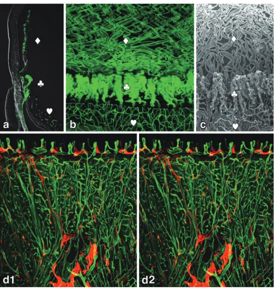
57There-Figure 6 3D-imaging of uveal vascular structures and a stereoview of microvasculature in the mouse tongue using lectin.
a: A sagittal section of a mouse eye ball. Blood vessels were stained by intravenous in-jection of FITC-conjugated tomato LEL lectin (Vector Laboratories, USA) to label all the blood vessels (green). b: A whole-mount preparation of the anterior pole of a mouse eyeball. Blood vessels were stained in the same way as 6a. Note that the 3D-images of the vascular distribution patterns are completely different in each part of the uvea. c: The same view of the uvea of vascular corrosion casts by the scanning electron microscopy (SEM). Courtesy Prof. K. Toida (Kawasaki Medical School, Kurashiki, Japan). Blood vessels in the iris ( ♢ ), ciliary body ( ♧ ), and retina ( ♡ ) in a-c.
d: Stereo-pair immunofluorescent micrographs of a mouse tongue stained with FITC-con-jugated tomato LEL lectin (Vector Laboratories, USA) (green) for blood vessels and LYVE-1 (red) for lymphatic vessels. The stereo-pairs of pictures, 6° tilted (dLYVE-1 and d2), were captured with the same magnification using a Leica TCL-SL confocal laser scanning micro-scope (Leica, Wetzlar, Germany). Capillary loops in the papillae and underlying lymphatics are clearly demonstrated.
fore, the spatial relationship between newly formed
and existing vessels can be easily analyzed by
3D-imaging in studies on angiogenesis during various
pathological conditions or vasculogenesis during
de-velopment.
2 ) Clinical diagnostic and therapeutic
applica-tions
Lymphangiography has been used for clinical
di-agnosis when lipiodol or indigo carmine are
subcu-taneously injected, or
99 mTc-labeled human albumin
is applied for scintigraphy of lymphatics. However,
Figure 7 Identification of lymphatic vessels by lectins.
a: Confocal double fluorescent images of a lymphatic capillary (*) in the thoracic side of the mouse diaphragm stained by intraperitoneal injections with FITC-conjugated MAL-1 (Vector Laboratories, USA) (aMAL-1: green) and LAMAL-102 (a2: red). The merged image (a3). The blind end (*) of the lymphatic capillary and subsequent collecting vessel portion with valves (arrows) are clearly seen.
b&c: Confocal fluorescent images of the macula cribriformis on the peritoneal surface of the mouse diaphragm (b) and lymphatic sinuses (c) immediately underneath the mesothe-lium of the diaphragm stained by the intraperitoneal injection of FITC-conjugated ConA (Vector Laboratories, USA) (green).
Table 1 Reactivity of various lectins to endothelial cells and mesothelial cells*.
Lectins Sugar binding to Lymphatic ECs
Blood vessels ECs
Mesothelial
cells Remarks
ConA Mannose ++ + (venules) + strong binding at stomata
LEL GlcNAc − ++ ++
MAL-II Sialic acid NT NT NT
UEA-I Fucose − − −
MAL-I ++ − + strong binding at stomata
RCA-I Galactose /GalNAc − ++ + strong binding at stomata
GSL-I B4 ++ ++ + blood vessel ECs stained with lectin i.p.
*Tested by S. Morikawa (unpublished data). All lectins were purchased from Vector Laboratories, USA. EC, endothelial cell; i.p., intraperitoneal injection; NT, not tested.
side effects of such tracer substances are a concern.
In addition to the radioactive substances, lipiodol of
iodine compounds derived from lipid-soluble
con-trast media might cause embolization or allergic
re-actions to iodine. Pigmented solutions such as
in-digo carmine or isosulfan blue remain for a long
pe-riod in the subcutis. Therefore, lymphangiography
using new fluorescent dyes, such as indocyanine
green (ICG), and magnetic resonance imaging (MRI)
are more often applied for lymphatic vessel
imag-ing. Indocyanine green was first introduced in
fluo-rescent lymphangiography to assess lymphatic
function in patients with edema.
58Since the proposal
of the sentinel node theory by Morton et al.,
59ICG
has become one of the most commonly used tracers
in clinical diagnosis and navigation surgery for
ma-lignant diseases.
60,61Advanced MRI for lymphatic
vessels
62and real-time imaging of the lymphatic
sys-tem using ultrasonography and Sonazoid
63are also
reported to visualize various lymphatic vessels.
Unveiling the Relationship Between Peritoneal
Mesothelium and Lymphatic Endothelium
The lymphatic system plays important roles in
draining of tissue fluid and transport of lipids,
mac-romolecules and immune cells. The actual
mecha-nisms underlying its key function, such as the
selec-tive uptake of materials, are yet to be established.
Both fluid drainage and cell trafficking in the body
cavity, particularly peritoneal cavity, are clinically
important for the pathogenesis of ascites,
inflamma-tion, and malignant cell metastasis. There are
sev-eral studies on lymph drainage from the peritoneal
cavity.
64-68The rate and pattern of absorption of
liq-uid, cells, and insoluble particulates differ with
ma-terials. Fritz and Waag
69have reported the
transdia-phragmatic lymphatic transport of intraperitoneally
administered particulate marker. Bettendorf
70,71re-ported the peritoneal resorption of latex particles
via diaphragmatic lymphatics. However, his studies
have mostly focused on the normal structural and
functional aspects of routes of lymph absorption of
various materials under physiological conditions.
Yuan et al.
72reported the lymphatic drainage of
in-flammatory cells from the peritoneal cavity in a
sheep peritonitis model. The obstruction of lymph
drainage or other lymphatic disorders under
patho-logical conditions may also be important issues in
clinical treatments for ascites.
In order to establish a tumor model for studies on
the lymphatic drainage from the peritoneal cavity,
we induced benign lymphangiomas in rats
32and
mice
73,74by intraperitoneal FIA injection according
to the method of Mancardi et al..
31This tumor model
could help unveil the relationship between
perito-neal mesothelium and lymphatic endothelium.
1 . General changes in the peritoneal cavity
surface after FIA injection
We first performed morphological and functional
characterization of FIA-induced lymphangiomas
with respect to their phenotypes and effect on
lymph drainage from the peritoneal cavity in rats.
32In approximately two months after the
intraperito-neal FIA injections ( 1 mL emulsion ) with a
two-week interval, tumors developed in the peritoneal
cavity, such as the diaphragm, omentum, and liver
surface. To distinguish lymphatics from blood
ves-sels, various mouse mAbs against rat
microvascula-ture (B27, B1, and B110) were used. We also used a
tomato lectin ( Lycopersicon esculentum lectin, LEL )
for blood vessel staining
35and 5 -nucleotidase
reac-tivity
75for lymphatic vessel staining. We found that
the tumors themselves lacked the typical lymphatic
phenotypes, but were partly positive for blood
ves-sel markers (B1). Type IV collagen was negative or
very weakly positive. The results suggest that
these tumors might share some characteristics of
both lymphatic capillaries ( 5 -nucleotidase : partly
strong-positive ) and arterial-side capillaries ( B 1 :
positive), although they showed a honey comb-like
or granulomatous morphology.
76However, the
draining capacity of lymphangioma via
diaphrag-matic lymphatics to parathymic lymph nodes was
remarkably reduced, indicating that the tumor lost
its absorptive function as lymphatics.
32We then investigated the phenotype of the tumor
in depth in mice.
73Interestingly, within three days
after a single intraperitoneal FIA injection, the
peri-toneal cavity surface started to change and formed
typical benign lymphangiomas (Figure 8a-d). One to
two weeks after FIA injection, we found that
peri-toneal mesothelial cells elongated (cuboidal) and lost
their polarity, and gradually formed thick stratified
cell masses all over the peritoneal membrane
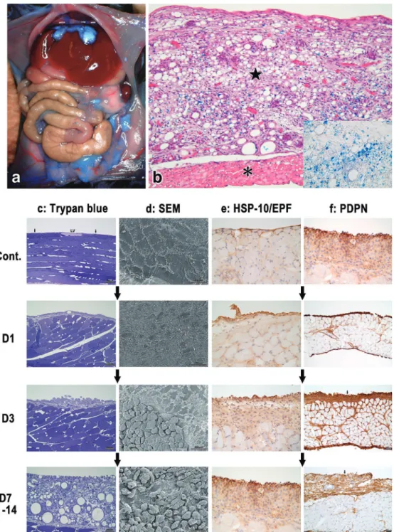
(Fig-ure 8c&d, 9a-e). By day 3 of FIA injection, we found
an increase in the early pregnant factor ( EPF or
HSP-10 )
77and podoplanin expression in peritoneal
mesothelial cells (Figure 8e&f). EPF has been
re-ported to be produced and secreted into blood by
some types of mineral-oil-induced tumor.
77,78Figure 8 Development of lymphangiomas induced by a peritoneal FIA injection in mice. a: A macroscopic view of peritoneal cavity in a mouse two weeks after a peritoneal FIA injection containing EM-blue (blue dye). Note that not only the surface of the liver, dia-phragm, and mesenteries including the omentum, but also the surface of abdominal walls colored blue, indicating FIA is taken up by lymphangiomas developed almost all over the peritoneal surface. b: Light microscopic view of the lymphangioma ( ★ ) developed on the surface of the diaphragm (*) four weeks after the FIA injection (Hematoxylin and Eosin staining). Note that FIA containing EM-blue dye is taken up by the tumor cells (inset: a higher magnification of the tumor without HE staining). c-f: Sequential changes of the abdominal surface before (Cont.) and after FIA injection (D1: day 1, D3: day 3, D7-14: day 7-14). The tumor development were observed by semithin epon sections stained with try-pan blue (c), scanning electron microscopy (d: SEM), cryosections stained by the immuno-peroxidase reaction (DAB: brown) against mouse HSP-10/EPF (R&D Systems, Inc.) (e), and also against mouse podoplanin (AngioBio Co., USA) (f: PDPN). Note that not only peritoneal mesothelial cells but also many peritoneal free cells (some of them bearing large vacuoles) are seen on the surface of the lymphangiomas (in c&d: D3, D7-14).
Figure 9 Ultramicroscopic changes of the lymphangiomas after FIA injection.
Transmission electron micrography (TEM) and scanning electron micrography (SEM) were performed to characterize the lymphangiomas at electron microscopic level. a: Normal sur-face of the peritoneal wall. The SEM image (inset) of the mesothelium. The mesothelial cells are simple squamous with short microvilli on their surface toward the peritoneal cavity. b: Three to five days after FIA injection. The SEM image (inset) of the mesothelium. Note that the mesothelial cells became taller and cuboidal, with increased microvilli all around the cell surface, indicating they have lost their polarity. Some cells take up FIA into their cytoplasm. c-d: One to two weeks after FIA injection. Mesothelial cells have loosened the cellular connections with each other, and then several peritoneal free cells attach and get further into the interstitial spaces underneath mesothelial cells (c). Some mesothelial cells formed multilayers with abundant and long microvilli on the bilateral sides of their surface. Several cells in the newly formed tumors have various sizes of fat (FIA) droplets (d). (from Ezaki and Desaki, 2012)73 e: At four weeks or later, the fat droplets fused with each other and increase in size in the cells. The fat-storing cells became larger in size, and then some adjacent cells gradually fused with each other to form cell clusters. f-g: At two to four months or later, the fat-storing cells fused with each other and gradually formed tubular structures like lymphatic vessels. (from Ezaki and Desaki, 2012)73
model, EPF might have been induced locally
(Fig-ure 8e), but its serum level was unaltered at least
until seven days (unpublished data). Contrarily, the
increase in local podoplanin expression was
remark-able and the whole lymphangiomas became
strongly positive (Figure 8f). However, the tumor
contained some LA5
+blood vessels, but not
LYVE-1
+or LA102
+cells. This suggests that the tumor
it-self is mesothelial in origin, rather than lymphatic
endothelial cells. By day 7-14, the tumors showed
heterogeneous expression of lymphatic endothelial
markers. At four weeks or later, they formed
typi-cal honeycomb-like lymphangiomas consisting of
various-sized fat-storing cells (Figure 8b, 9d-f). As
they developed, the fat-storing cells fused with each
other and gradually formed tubular structures like
lymphatic vessels ( Figure 9 e-g ) or large follicles.
Consequently, LYVE-1
+and LA102
+functional
lym-phatic vessel structures were detected at
approxi-mately four weeks after FIA injection as described
later. These sequential changes appear to be unique
if the phenomenon is regarded as a process in
lym-phangiogenesis. Shimizu et al. also found similar
vasculogenic patterns, where individual LYVE-1
+stromal cells gathering around the trabecular
arter-ies gradually joined and fused together, and finally
formed various tubular structures as the initial
lym-phatic in developing spleen at 18.5 days of the
em-bryonic age in mice (unpublished data).
2 . Morphological and functional phenotypes
of cells involved in FIA-induced lymphangiomas
Although the tumors were podoplanin
+, typical
lymphatic vessel-like structures expressing
LYVE-1 and LALYVE-102 only appeared at the later stages (four
weeks or later). Therefore, the initial
name“lym-phangioma”for this animal tumor model
31might not
be suitable. This idea prompted us to characterize
the phenotype of the adjuvant-induced tumor.
To clarify the involvement of bone
marrow-derived cells in tumor development, GFP
+bone
marrow cells (1.5×10
7cells/100
μL) from C57BL/6
(GFP-Tg) mice were intravenously injected to
le-thally irradiated (12 Gy) syngeneic mice. The
recipi-ent bone marrow chimeric mice were
intraperito-neally injected FIA (0.2 mL) two weeks after bone
marrow cell transplantation. Substantial number of
GFP
+cells accumulated in FIA-induced
lymphan-giomas from the beginning of tumor development
(Figure 10a&b, e-h ) . We confirmed that most of
these cells had some macrophage or myeloid cell
markers, such as CD 68 ( Figure 10 c ) , Gr-1 (
Fig-ure 10d), and CD11c (figFig-ure not shown). We also
found that some interstitial cells bearing fibroblast
marker (S100A4) were involved in this tumor
(fig-ure not shown). Therefore, we concluded that
lym-phangiomas of several cell origins including at least
mesothelium, macrophages, and fibroblasts were
formed as chimeric cell masses by fusing with each
other to dispose the extrinsic FIA. In other mineral
oil-induced tumors, besides granulomatous
re-sponses that might correspond to our tumor model,
plasmacytomas were quite common.
76,79Interest-ingly, there was a strain difference in the incidence
of plasmacytomas, but there were no reports
re-garding this in oil-induced granulomas.
80Further-more, the involvement of M1 (iNos
+) and M2 type
macrophages (arginase
+) was compared on days 3
and 120 after the FIA injection. (Figure 10e-h) On
day 3, both M 1 proinflammatory and M 2
anti-inflammatory macrophages increased in the
tu-mors, whereas only a few M 2 type macrophages
were found in the tumors at very late stage (day
120). However, further studies should be conducted
to clarify the biological significance of M 1 / M 2
macrophage balance
81,82in this tumor model.
Gene expression levels of podoplanin, CCL2,
Prox-1, TGF-β, VEGFc, and TNF-α in lymphangiomas
were examined using RT-PCR at various times
af-ter FIA injection (Figure 11). Considerable increase
in podoplanin and CCL2 expression was observed
by day 1, and an increase in TGF-β and VEGFc
from day 3. However, one of the lymphatic
endothe-lial markers, Prox-1, did not show any significant
change. A remarkable increase in podoplanin
ex-pression was also confirmed in tissue sections. As
podoplanin
+cells secrete CCL 2 to recruit CD 68
+cells in wound-healing lesions,
83,84the increase in
po-doplanin and CCL 2 expression in lymphangiomas
might support the massive accumulation of bone
marrow-derived macrophages ( CD 68
+cells ) from
the peritoneal cavity into tumors.
Furthermore, we investigated the expression of
various fat-storing-cell-related markers in
lymphan-giomas. For example, adipophilin is known to be
positive for fat-accumulating hepatocytes in
alco-holic cirrhotic fatty livers or lipid-storing CD 68
+macrophages. Both adipophilin
+and CD 36
+cells
might correspond to CD 68
+macrophages.
85,86We
found a considerable increase in cells positive for
adipophilin and CD36 (Figure 12a&b).
Fat-droplet-Figure 10 Involvement of bone marrow-derived intraperitoneal cells in FIA-induced lymphangiomas.
a: GFP+ (green) bone marrow-derived cells accumulated in lymphangiomas ( ☆ ) developed on the muscular layer of the diaphragm (*) seven days after FIA-injection. b: Confocal triple immunofluorescent image of the cellular masses in lymphangiomas of GFP+ bone marrow cell-reconstituted mice four weeks after the FIA injection. GFP (green), podoplanin (red), and CD68 (blue). Note that the cell masses are found to be chimeric cell mixtures (arrowheads) from different origins and many of them contain various sizes of fat droplets (*). Podoplanin+ cells (arrow) surround these cell mixtures. c: Immunoperoxidase staining of lymphangiomas developed two weeks after the FIA injection for CD68, a macrophage marker. d: Immunoperoxidase staining of lymphangiomas developed two weeks after the FIA injection for Gr-1, a myeloid differentiation antigen (Ly-6G/Ly-6C).
e-f: Confocal triple immunofluorescent image of the cellular masses in lymphangiomas of GFP+ bone marrow cell-reconstituted mice three days after FIA injection. Both M1 (red in e: iNos) and M2 (red in f: arginase) type macrophages (CD68: blue) were located in lymphan-giomas at a very early stage of tumor development. g-h: Confocal triple immunofluorescent image of the cellular masses in lymphangiomas of GFP+ bone marrow cell-reconstituted mice 120 days after the FIA injection. Note that there are some M1 type macrophages (red in g: iNos) are still present in lymphangiomas, but a very few M2 type macrophages (red in h: arginase). ☆ : lymphangioma, *: muscular layer of the diaphragm in c-h.
Figure 11 Expression level of lymphatic and inflammation related factors in the dia-phragm using the RT-PCR assay.
A quantitative analysis was performed using the RT-PCR to determine the expression level of podoplanin (PDPN), CCL2, Prox-1, TGF-β, VEGFc, and TNF-α in the diaphragm at various times (control, and days 1, 3, and 14) after FIA injection. The results were normal-ized using β-actin as the internal control. To calculate the expression level of genes, the delta-delta comparative threshold method was used. Note that remarkable increases in PDPN and CCL2 expression were seen as early as day 1, and an increase in TGF-β and VEGFc from day 3. However, one of the lymphatic endothelial markers, Prox-1, did not show any significant change (*p < 0.01). (from Ezaki et al., 2018)74
storing cell accumulation in tumor might be similar
to the pathogenesis of atherosclerotic plaques in the
arterial wall where lipid droplet-containing
macro-phages also accumulate.
87,88Interestingly, both these
phenomena are closely related to lipid or oil that
triggers macrophage infiltration under the
endothe-lium or mesotheendothe-lium in atherosclerosis or
FIA-induced lymphangiomas, respectively. However,
adiponectin (Figure 12c), one of the major
adipoki-nes, production was not very significant.
The massive uptake of fat (FIA) into
lymphan-giomas prompted us to investigate fat metabolism
in relation to fat droplets in the tumor. Fatty
acid-binding proteins (FABPs) are known to be involved
in promoting cellular uptake, and transporting and
targeting fatty acids to specific metabolic
path-ways.
89,90Therefore, we investigated the possible
in-volvement of FABPs in FIA-induced tumor
devel-opment using FABP-KO mice and specific
antibod-ies against mouse FABP1, FABP3, FABP4, FABP5,
and FABP 7.
91In normal C 57 BL / 6 mice, a single
peritoneal
FIA
injection
induced
typical
honeycomb-like
lymphangiomas
consisting
of
various-sized ring-like fat-storing cells. These cells
were strongly positive for podoplanin, F4/80, and
FABP5 and FABP7. Contrarily, significant positive
staining for FABP4 was not observed (Figure 12d).
In atherosclerosis, FABP4 (aP2 or adipocyte FABP)
expression by macrophages promoted
atherogene-sis,
89whereas FABP 5 ( Figure 12 e ) , rather than
FABP4, appeared to be more responsible in our
tu-mor model, and the follicular structures mainly
con-sisted of FABP7
+cells ( Figure 12 f ) . On the
con-trary, in FABP3-, FABP5-, FABP7-KO mice (data
not shown), there was no significant difference in
the incidence of tumors when compared with that
Figure 12 Expression of various fat-related markers in lymphangiomas.
a: Immunoperoxidase staining of lymphangiomas for adipophilin (PROGEN Biotechnik GmbH, Germany), a macrophage marker relating to lipid droplet-associated proteins. b: Immunoperoxidase staining of lymphangiomas for CD36 (Cascade Bioscience, MS, USA), a macrophage marker also relating to lipid droplet-associated proteins. c: Immunoperoxi-dase staining of lymphangiomas for adiponectin (R&D Systems, Inc., USA), an adipokine. d-f: Immunoperoxidase staining of lymphangiomas for FABP4 (d), FABP5 (e), and FABP7 (f). DAB+ cells in FABP4 staining (d) are non-specific background stains. All anti-FABP an-tibodies were gifted by Prof. Owada Y. (Tohoku University, Sendai, Japan). All cryosections of the lymphangiomas ( ★ ) developed two to four weeks after FIA injection were colored by DBA reaction in a-f.
in normal B 6 mice. However, tumors developed
more vigorously in FABP7-KO mice than in other
KO strains. Among FABP-KO mice, relatively
smaller fat-storing cells were seen in FABP 3-KO
mice, whereas the follicle-type structures were
more commonly found in FABP5-KO mice. In
con-trast, the tumors in FABP7-KO mice showed less
fat-storing cells with more inflammatory and
fi-brous components. The results suggest that a close
relationship between the extrinsic FIA metabolism
and fat-storing cell mass is involved in this tumor
model, because the tumor cells contain FIA in their
cytoplasm as various-sized lipid droplets.
74However,
the actual role and biological significance of each
FABP in the tumorigenesis remain to be further
clarified.
During tumor development, the tumor contained
some LA 5
+blood vessels, but not LYVE-1
+or
LA102
+cells until about four weeks after FIA
injec-tion. This was confirmed by other vascular
mark-ers, such as blood vessels with CD31 (Figure 13a)
and lymphatic vessels with Prox-1 and VEGFR-3
( figure not shown ) . Only the typical lymphatic
vessel-like structures expressed LYVE-1 and LA
102 at the late stages ( four weeks or later )
(Fig-ure 13c-f). This corresponds to the time when the
tubular structures are found after fusion of various
cell masses (Figure 9e&f). Furthermore,
vasohibin-2 (VASHvasohibin-2), a tumor angiogenesis-promoting factor,
was detected in the tumor (Figure 13b). It was
ex-pressed preferentially in mononuclear cells derived
from the bone marrow and promoted angiogenesis
in the mouse.
92It is also known that VASH-2 is
re-quired for epithelial-mesenchymal transition of
ovarian cancer cells via regulation of TGF-β
signal-ing.
93As the tumors develop, the fate of extrinsic
FIA might be a problem. In the tumor, we found
ab-normal draining routes from the peritoneal cavity
at very late stages. Once the oil droplets are stored
in cells, they are gradually secreted out of these
cells into, for example, follicular structures (
Fig-ure 14a&b), or through the remodeled lymphatic
vessel-like structures (Figure 9g, 13c&d). They
fi-nally reached either the draining lymph nodes of
peritoneal cavity (Figure 14e) or abnormal
collat-eral routes penetrating abdominal wall to dispose
FIA to the skin subcutis (Figure 14f&g). The
sig-nificance of these complementary transformations
might involve tissue remodeling after acute or
chronic inflammation and lymphangiogenesis to
drain undesirable fat droplets in the peritoneal
cav-ity.
3 . Relationship between peritoneal
mesothe-lium and lymphatic endothemesothe-lium
The actual mechanisms underlying
tumorigenic-ity of this tumor model is unclear. In the 1960 s,
there were several reports on the development of
variety of tumors after the intraperitoneal injection
of some mineral oils or adjuvants.
79,80Furthermore,
in 1985, Leak et al.
76reported that peritoneal
meso-thelium developed granulomatous tumors in
re-sponse to mineral oil or pristane. Quinn
78reported
that EPF (or HSP-10 ) is involved in the initiation
and maintenance of various peritoneal responses to
mineral oil. We also found some mesothelial and
in-terstitial cells including fat-stored cells expressed
EPF. During the first one or two weeks, a
remark-able increase in EPF expression was observed in
the tumors (Figure 8e). However, the biological role
of EPF in tumor development after FIA injection
remains unclear. As EPF is produced during
vari-ous responses under both physiological and
patho-logical conditions,
77there might also be some other
factors involved in this tumor model; this should be
further investigated.
The fact that peritoneal mesothelial cells became
tall in height (cuboidal) and lost their polarity, and
gradually formed thick stratified cell masses all
over the peritoneal membranes one to two weeks
after FIA injection may suggest the potential of the
mesothelial cells to transform into benign tumors
forming cell masses with other type of cells (
Fig-ure 10b, 14b). The increase in podoplanin and its
gene expression in FIA-induced tumors (
Fig-ure 8f&11) might also be related to the potential
di-versity of mesothelial cells and possible
transforma-tion into lymphatic endothelial cells. As podoplanin
correlates with ezrin redistribution to membrane
projections and cytoskeleton and cell motility
regu-lation,
94podoplanin might play a role in inducing
these sequential changes in tumor progression by
linking mesothelial and lymphatic endothelial
func-tions. Leak et al.
76also found that an intraperitoneal
injection of pristane, a mineral oil, induced
squamous mesothelial cells to become cuboidal and
lose their polarity. They reported that pristane
in-duced fibrin networks, as in inflammation, involving
extracellular matrices, such as fibronectin, resulting
in huge granuloma-like structures attracting
perito-neal free cells. Simultaneously, vigorous
angiogene-sis and lymphangiogeneangiogene-sis occurred, probably due
to platelet activation upon engagement by
podo-planin.
94,95These results indicate that FIA-induced tumors
are heterogeneous cell population forming chimeric
cellular masses resulting from mutual cell fusion
af-ter fat droplet uptake. In addition to podoplanin
+mesothelial cells, bone marrow-derived
macro-phages (CD68
+), and some interstitial mesenchymal
cells are also involved in tumorigenicity in this
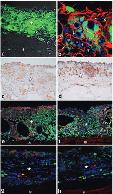
tu-Figure 13 Development of functional vascular structures in lymphangiomas.
Functional vascular structures were detected 28 days after FIA injection in C57BL/6 fe-male mice. Ten-micron thick cryosections were made and immunostained with CD31 (a), vasohibin-2 (VASH2), a tumor angiogenesis promoting factor (b).
In c-f, blood vessels were labed by intravenous injection with FITC-conjugated tomato lec-tin (LEL), the mice were then perfusion-fixed with 4% PFA for 10 min, washed and frozen in OCT compound. Ten-micron thick cryosections were made and immunostained. Triple fluorescent staining with LA102 (c), LYVE-1 (d), and tomato LEL lectin (e). The merged im-age (f). All blood vessels that have blood supply are strongly stained by the FITC-LEL (e). Note that LYVE-1+ lymphatic vessels extend from the borders between the diaphragm (*) and lymphangioma ( ☆ ) and formed various shaped lymphatic vessels, while LA102+ ves-sels mainly exist in lymphangiomas (c, f). Depending on their distribution sites, the positiv-ity of LYVE-1 and LA102 does not necessarily correspond with each other.
Figure 14 Drainage of FIA out of peritoneal cavity via the newly formed functional lym-phatics.
a: Transmission electron micrograph of a typical fat-storing follicular structure (*) on day 28 of FIA injection. b: A confocal triple immunofluorescent image of follicular structure similar to that shown in (a) on day 28 of FIA injection. Chimeric large cell masses contain-ing GFP+ bone marrow-derived cells (green), podoplanin+ mesothelial cells (red), and CD68+ macrophages (blue). Note that various sizes of fat droplets were incorporated and some were secreted inside the follicle (*).
c: The two main routes of lymphatic pathways from abdominal cavity in the normal con-trol animal. Carbon dyes injected intraperitoneally were absorbed from the diaphragm and transported into the thoracic cavity by lymphatic vessels. A main route is parasternal or anterior route (arrow heads), reaching the regional parathymic lymph nodes (*). The other is the dorsal or posterior route (double arrowheads), reaching the posterior medias-tinal lymph nodes (**). Di: diaphragm, Ht: heart, Lu: lung, St: sterna, Th: thymus. d: FIA (with EM blue) reached the parathymic lymph nodes (*) and the posterior mediastinal lymph nodes (**) within seven days after intraperitoneal injection. e: FIA (with EM blue) reached parathymic or posterior mediastinal lymph nodes more than four months after in-traperitoneal injection. Note that these regional lymph nodes swelled and fused together at the late stages of tumor development.
f: Formation of abnormal intermuscular draining routes from the peritoneal cavity (on the left) towards the subcutis in the abdominal skin (on the right). g: Higher magnification view of the abnormal intermuscular draining routes of FIA. Note that various-sized fat droplets or even whole fat-storing cells are transported in the tubular structures as shown in Fig-ure 9f&g.
Figure 15 Hypothesis: mesothelial-endothelial transformation.
The possible processes and mechanisms involved in the development of FIA-induced lymphangiomas. Sequential changes from mesothelial cells to lymphatic endothelial cells via various fat-storing lymphangioma cells are suggested. These phenomena may be inter-preted as one of the biological defense mechanisms to drain extrinsic adjuvant oil out of the peritoneal cavity.
mor model. We found the podoplanin
+cells induced
a chemokine CCL2 to recruit CD68
+bone
marrow-derived cells which accumulated from the
perito-neal cavity.
74Both M1 and M2 type macrophages
might be involved in the early stage of tumor
devel-opment. Macrophages are responsible for adipose
tissue remodeling
96and various pathological
condi-tions, such as atherosclerosis.
87,88It has also been
re-ported that the vast majority of macrophages
infil-trating the obese organ are arranged around dead
adipocytes, forming characteristic crown-like
struc-tures (CLS).
97The large follicular structures
consist-ing of podoplanin
+mesothelial cells and CD 68
+macrophages in our tumor model (Figure 14a&b)
may be very close or almost equivalent to CLS.
Fur-thermore, Detry et. al.
98also suggested the
possibil-ity that FIA-induced lymphangiomas formed
tubu-lar structures, like lymphatic vessels.
The results demonstrated in this study suggest
sequential changes from mesothelial cells to
lym-phatic endothelial cells via fat-storing
lymphan-gioma cells after FIA stimulations. These
phenom-ena may be interpreted as one of the biological
de-fense mechanisms to drain extrinsic adjuvant oil
out of the peritoneal cavity. We have suggested the
possibility of sequential transformation (Figure 15)
from peritoneal mesothelial cells to functional
lym-phatic vessels via fat-storing tumor cells after FIA
stimulation.
74,99Besides sharing the same
mesenchy-mal origin, both lymphatic endothelial and
mesothe-lial cells have several similarities in their function
and structure (as shown in Figure 2b). Moreover,
the two cell types might have some ability to
trans-form into various interstitial cells under
pathologi-cal conditions or in vitro. However, in vivo, there
has been no clear evidence to show the relationship
between the two cell types. The biological
signifi-cance of FIA-induced lymphangiomas and the
di-versity of mesothelial cells might be important with
respect to the relationship between fat cells and
lymphatic endothelial cells.
Finally, during our analyses of tumor
develop-ment in the peritoneal cavity, we observed biased
site difference in tumor occurrence. The tumors
easily developed almost everywhere, such as the
surface of the diaphragm, omentum, liver, spleen,
and abdominal wall, except the ovary.
99The ovarian
surface (or germinal) epithelial cells never formed
any tumor mass (n ≧ 59 females), although they
be-came strongly positive for podoplanin similar to
other peritoneal mesothelial cells forming
lymphan-giomas at different sites in the peritoneal cavity
(un-published data). These results suggest that the
peri-toneal mesothelial cells might have diversity in
their phenotypic transformation depending on the
site of peritoneal cavity. This might be due to the
fact that the majority of ovarian tumors derive
from the surface (or germinal) epithelial cells and
the heterogeneity of the mesothelium in response
to FIA.
4.Prospective for the clinical application of
this tumor model
The mechanism for lymphangiogenesis is of
ma-jor interest in both basic and clinical lymphology. It
is clear that FIA (or mineral oil) is involved in a high
degree of interrelationship between mesothelium
and endothelium and is a factor that bring these
two structures closer. This process has recently
gained considerable attention, and studies to
under-stand this process are important for explaining the
mechanism of lymphangiogenesis.
100If the shift
be-tween mesothelial and lymphatic endothelial cells
can be controlled, it might be possible to develop
treatment for chronic lymphedema and even
perito-neal sclerosis in patients on peritoperito-neal dialysis. The
hypothesis (Figure 15) might give some hints for
the possible application of peritoneal dialysis or the
treatment of chronic edemas.
Conclusions
The one of the important missions of lymphatic
ves-sels is to absorb fat as well as macromolecules and
lymphoid cells and transport them into the
sys-temic blood circulation. Once the lymphatic vessel
function is disturbed under various abnormal
condi-tions, some biological defense mechanisms recover
the draining capacity by involving various cell
types, such as mesothelial cells and fat droplet
con-taining macrophages. Therefore, adjuvant-induced
lymphangiomas would provide us useful
experi-mental animal models to clarify some of the
unan-swered aspects
17in the lymphatic system under
both normal and pathological conditions.
Acknowledgements
I received cooperation and support from several indi-viduals over 20 years while compiling this work. I thank my collaborators, Prof. Kenjiro Matsuno (Dokkyo Medi-cal University, Tochigi, Japan), Dr. Kazuhiko Kuwahara (Aichi Cancer Center Research Institute, Aichi, Japan), Dr. Junzo Desaki ( Ehime University, Ehime, Japan ) , Prof. Kouji Matsushima (Tokyo University, Tokyo, pan), Prof. Yuji Owada (Tohoku University, Sendai, Ja-pan), Prof. Nobuko Tokuda (Dokkyo Medical University, Tochigi, Japan ) , and Prof. Kazunori Toida ( Kawasaki Medical School, Okayama, Japan). In particular, I would like to express my sincere gratitude to my colleagues, the late Dr. Shunichi Morikawa, Dr. Kazuhiko Shimizu, Dr. Shuji Kitahara, Dr. Ayako Sedohara, Dr. Sachiko Kikuta, Dr. Masae Morishima, the late Mrs. Yasuko Yamazaki, Mrs. Hiromi Sagawa, Mrs. Kae Motomaru, Ms. Kazuko Nakada, and Ms. Iori Sato.
Some parts in this work were supported by Grant-in-Aid for Scientific Research (A)(2) in 2002-2003 (Grant Number: 14207001) and (B) in 2007-2009 (Grant Number: 19390053) from the Japan Society for the Promotion of Science (JSPS).
Conflicts of Interest: There are no conflicts of
inter-est to declare.
References
1.Rosai J, Levine GD: Tumors of the thymus. In Atlas of tumor pathology, 2nd series, fasc.13, pp34―161, Armed Forces Institute of Pathology, Washington,
D.C. (1976)
2.Ezaki T, Marbrook J: The haemolytic plaque reduc-tion technique to measure cytotoxicity with hybri-doma cells as a target. J Immunol Methods 54: 281― 290, 1982
3.Ezaki T, Skinner MA, Marbrook J: Spontaneous cy-totoxic T cells in murine spleen-cell cultures. I. Some characteristics of effector and precursor cells. Immunology 50: 343―349, 1983
4.Ezaki T, Skinner MA, Marbrook J: Spontaneous cy-totoxic T cells in murine spleen-cell cultures. II. Dis-tinguishing between spontaneous cytotoxic T cells and NK cells according to kinetics and target selec-tivity. Immunology 50: 351―357, 1983
5.Ezaki T, Marbrook J: The use of hybridoma cells as a murine model to study tumor immunity. Int J Cancer 35: 107―111, 1985
6.Skinner MA, Thompson K, Ezaki T et al: Effects of in vivo modulation of splenic natural killer cell ac-tivity on the growth of spleen-seeking tumour vari-ants. Br J Cancer 55: 259―263, 1987
7.Ezaki T, Kawatsu R, Matsuno K et al: Characteriza-tion of intrathymic and extrathymic T cell develop-ment in spontaneous thymoma Buffalo/ Mna rats. Thymus 16: 67―87, 1990
8.Ezaki T, Matsuno K, Kotani M: Unique thymic mi-croenvironment for T-cell development in sponta-neous thymoma BUF/Mna rats. In Lymphatic tis-sues and in vivo immune responses ( Imhof BA, Berrih-Aknin S, Ezine S eds), pp105 ― 109, Marcel Dekker, New York (1991)
9.Ezaki T, Matsuno K, Kotani M: Thymic nurse cells (TNC) in spontaneous thymoma BUF/Mna rats as a model to study their roles in T-cell development. Immunology 73: 151―158, 1991
10.Ezaki T, Uehara Y: Thymic nurse cells forming a dynamic microenvironment in spontaneous thy-moma BUF/Mna rats. Arch Histol Cytol 60: 39―51, 1997
11.Morikawa S, Baluk P, Kaidoh T et al: Abnormalities in pericytes on blood vessels and endothelial sprouts in tumors. Am J Pathol 160: 985―1000, 2002 12.Baluk P, Morikawa S, Haskell A et al:
Abnormali-ties of basement membrane on blood vessels and endothelial sprouts in tumors. Am J Pathol 163 : 1801―1815, 2003
13.Kitahara S, Suzuki Y, Morishima M et al: Vasohibin-2 modulates tumor onset in the gastrointestinal tract by normalizing tumor angiogenesis. Mol Can-cer 13: 99, 2014
14.Ezaki T: How can we detect lymphatic vessels ? Japanese Journal of Lymphology 35: 105―107, 2012 (in Japanese)
15.Kotani M, Nawa Y, Fujii H et al: Post-capillary ve-nules as the pathway for migrating B lymphocytes. Cell Tissue Res 152: 299―303, 1974
16.Ezaki T, Matsuno K, Fujii H et al: Characterization of rat vascular endothelial cell antigens by monoclo-nal antibodies. Acta Anatomica Nipponica 64: 390, 1989
17.Ezaki T, Kotani M: Mysteries in lymph, lymphatics, and lymphoid tissues. J Tokyo Wom Med Univ 89 (Extra 1): E4―E65, 2019
18.Ezaki T, Matsuno K, Fujii H et al: A new approach for identification of rat lymphatic capillaries using a monoclonal antibody. Arch Histol Cytol 53 (Suppl): 77―86, 1990
19.Swartz MA, Skobe M : Lymphatic function, lym-phangiogenesis, and cancer metastasis. Microsc Res Tech 55: 92―99, 2001
20.Scavelli C, Weber E, Agliano M et al: Lymphatics at the crossroads of angiogenesis and lymphangio-genesis. J Anat 204: 433―449, 2004
21.Baluk P, Tammela T, Ator E et al: Pathogenesis of persistent lymphatic vessel hyperplasia in chronic airway inflammation. J Clin Invest 115 : 247 ― 257, 2005
22.Tammela T, Petrova TV, Alitalo K: Molecular lym-phangiogenesis: new players. Trends Cell Biol 15: 434―441, 2005
23.Hirakawa S, Hong YK, Harvey N et al: Identifica-tion of vascular lineage-specific genes by transcrip-tional profiling of isolated blood vascular and lym-phatic endothelial cells. Am J Pathol 162: 575―586, 2003
24.Banerji S, Ni J, Wang SX et al: LYVE-1, a new homologue of the CD44 glycoprotein, is a lymph-specific receptor for hyaluronan. J Cell Biol 144 : 789―801, 1999
25.Breiteneder-Geleff S, Soleiman A, Kowalski H et al: Angiosarcomas express mixed endothelial pheno-types of blood and lymphatic capillaries: podoplanin as a specific marker for lymphatic endothelium. Am J Pathol 154: 385―394, 1999
26.Schacht V, Dadras SS, Johnson LA et al: Up-regulation of the lymphatic marker podoplanin, a mucin-type transmembrane glycoprotein, in human squamous cell carcinomas and germ cell tumors. Am J Pathol 166: 913―921, 2005
27.Wigle JT, Oliver G: Prox1 function is required for the development of the murine lymphatic system. Cell 98: 769―778, 1999
28.Partanen TA, Alitalo K, Miettinen M: Lack of lym-phatic vascular specificity of vascular endothelial growth factor receptor 3 in 185 vascular tumors. Cancer 86: 2406―2412, 1999
29.Mouta Carreira C, Nasser SM, di Tomaso E et al: LYVE-1 is not restricted to the lymph vessels: ex-pression in normal liver blood sinusoids and down-regulation in human liver cancer and cirrhosis. Can-cer Res 61: 8079―8084, 2001
30.Jackson DG, Prevo R, Clasper S et al: LYVE-1, the lymphatic system and tumor lymphangiogenesis. Trends Immunol 22: 317―321, 2001
31.Mancardi S, Stanta G, Dusetti N et al: Lymphatic endothelial tumors induced by intraperitoneal in-jection of incomplete Freund s adjuvant. Exp Cell Res 246: 368―375, 1999
32.Ezaki T, Kuwahara K, Morikawa S et al: Charac-terization of adjuvant-induced rat lymphangiomas as a model to study the lymph drainage from ab-dominal cavity. Japanese Journal of Lymphology 27: 1―10, 2004
33.Toshimori K, Araki S, Oura C: Masking of sperm maturation antigen by sialic acid in the epididymis of the mouse. An immunohistochemical study.