Title
Decrease of β-cells and Increase of α-cells in a Diabetic Patient
with Mitochondrial DNA 3243 (A→G) Mutation( 本文(Fulltext)
)
Author(s)
KOJIMA, Toshihiro; YAMAMOTO, Mayumi; FURUHASHI,
Naoki; SARUI, Hiroshi; TAKATSU, Hisato; TAKEDA,
Noriyuki; ISHIZUKA, Tatsuo; YAMADA, Koji; YASUDA,
Keigo
Citation
[Internal medicine] vol.[42] no.[12] p.[1193]-[1196]
Issue Date
2003-12-01
Rights
The Japanese Society of Internal Medicine (日本内科学会)
Version
出版社版 (publisher version) postprint
URL
http://hdl.handle.net/20.500.12099/27314
D
ecrease
of p-cells
and Increase
of a-cells
in a Diabetic
Patient
with Mitochondrial
DNA 3243 (A-*G)
Mutation
Toshihiro KOJIMA, Mayumi YAMAMOTO*,Naoki FURUHASHI**, Hiroshi SARUI***,Hisato TAKATSU****, Noriyuki TAKEDA*, Tatsuo ISHIZUKA*****, Koji Yamada****** and Keigo Yasuda*
Abstract
We report here a case of a 53-year-old womanwith
mitochondrial 3243 (A-*G) mutation diabetes. Diabetes mellitus was diagnosed at 39 years of age. At 50 years of age, cardiomyopathy and hypothyroidism were noted. At 53 years of age, the patient was admitted to our hospital for treatment of necrotic ulceration of toes. However, she died of massive heart failure. Pathological examination at autopsy showed a decrease in size, number, and acido-philicity of islets in the pancreas. Immunohistochemical stain revealed a decrease in the p-cell population to 20.5± 9.2% and a relative increase in a-cell population to 48.6±
ll.5%.
(Internal Medicine 42: 1193-1196, 2003)
K
ey words: diabetes mellitus, islet, insulin, glucagon,
mito-chondrial DNA 3243 (A-*G) mutation
Introduction
Mutation of mitochondrial DNA 3243 (A->*G) was first
reported by van den Ouweland et al in 1992 (1). Kadowaki
et al reported that the incidence of the mutation is 1% of dia-betic patients in Japan (2). Gerbitz et al estimated that 1.5% of diabetic patients have the mutation (3). Diabetes with the mitochondrial DNA 3243 (A-*G) mutation has several char-acteristics: progressively decreasing insulin secretion, mater-nalinheritance, frequent occurrence of deafness,
cardiomyo-pathy, disorder of the cardiac electrical pathway, muscle
atrophy, and rarely, obesity. At onset, many patients can re-gain sufficient insulin secretion by treatment with sulfo-nylureas, but most cases require insulin therapy to correct the
rapidly diminishing insulin secretion. As there are few re-ports of pathological analysis of the pancreas (4-7), the asso-ciation of diminishing insulin secretion with diabetes onset has not been fully clarified. Here, a deceased patient with diabetes with the mitochondrial DNA 3243 (A-*G) mutation was autopsied, and the a- and P-cells in the pancreas were
examined.
Case Report
A 53-year-old womanwith leg edema was admitted to our hospital complaining of ulceration of toes. She had been deaf in the right ear from birth and suffered sensory hearing loss in the left ear from 30 years of age. She had heart failure at 42 and cerebral infarction at 48. Her father had hypertension and acute myocardial infarction, but there was no family his-tory of diabetes mellitus, including her mother and grand-mother, or hearing loss. Although urinary sugar was first noted at 34 years of age during pregnancy, she did not con-sultmedical services. With a visual disturbance at 39 years of age, she consulted her family practitioner and was
diag-nosed with diabetes mellitus. She was medicated with sulfonylurea. However, the patient complained of thirst, and
her blood glucose control was poor. Although insulin
injec-tion therapy was started at 44 years of age, her HbAlc level
was over 8%. She was introduced to our hospital at 49 years
of age. Because of her short height, hearing loss, and
pro-gressive neuropathy, she was suspected to have diabetes
mel-litus related to a mitochondrial DNA mutation. The
mito-F
rom Department of Internal Medicine, Gifu Central Hospital, Gifu, *Department of Endocrinology, Diabetes, and Rheumatology, Gifu University School of Medicine, Gifu, **Department of Internal Medicine, Kanayama Municipal Hospital, Kanayama, ***Department of Internal Medicine, Murakami Memorial Hospital, Asahi University, Gifu, ****The First Department of Internal Medicine, Gifu Municipal Hospital, Gifu, *****Department of General Medicine, Gifu University School of Medicine, Gifu and ******Department of Internal Medicine, Toyokawa Municipal Hospital, Toyokawa
Received for publication May 6, 2003; Accepted for publication August 29, 2003
R
eprint requests should be addressed to Dr. Mayumi Yamamoto, Department of Endocrinology, Diabetes, and Rheumatology, Gifu University School of Medicine, 40 Tsukasa, Gifu 500-8705
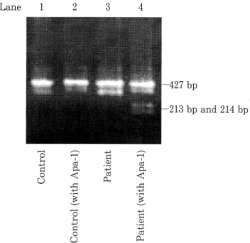
KOJIMA et al Lane 1 -427 bp -213 bp and 214 bp O O (3$< 2 o O OhCD
Figure 1. Mitochondrial mutation 3243 from adenine to
gua-nine was detected in white blood cells. Total DNA was isolated
from blood. A 427 bp fragment encompassing the tRNA Leu
(UUR) mutation site located at nucleotide 3243 was amplified
by PCR with forward primer, 5 -AAGGTTCGTTTGTTCAA
CGA (from 3029 to 3048) and reverse primer 5 -AGCGAAG
GGTTGTAGTAGCC (from 3437 to 3456). The fragment was
digested with Apa-1 at 37°C overnight, followed by
electropho-resis on a 1% agarose gel. Bands were visualized by ethidium
bromide. The mitochontrial DNA of the patient (Lane 4) was
partially separated to 214 bp and 213 bp with Apa-1 treatment.
chondrial 3243 mutation (A-*G) was detected in white blood cell analysis (Fig. 1). Anti-GAD antibody was negative. Suggested by the genetic information, dilated
cardiomyo-pathy and hypothyroidism were found at 50 years of age.
SerumlevelsofTSH, FT3, and FT4 were 5.64 juU/ml, 2.65
pg/ml, and 1.07 ng/dl, respectively, although
anti-thyro-globlin antibody, anti-TPO antibody, and TSH receptor
anti-body were negative.
Dilated
cardiomyopathy
was treated
with 120 mg of ubidecarenone, 40 mg of furosemide, and 2.5
mg of enalapril maleate per day. Hypothyroidism was
con-trolled by 75 jug of levothyroxine sodium per day. Protein
in-take was restricted to 40 g per day to protect renal function,
and calcium polystyrene sulfonate was used to avoid
hyper-kalemia. Serum levels of lactic acid and pyruvic acid were
increased to 31.8 (normal range: 3.3-14.9) mg/dl and 1.70
(normal range: 0.30-0.94) mg/dl, respectively. The patient
had noticed edema in the legs and face related to heart failure
since age 51. She was admitted to hospital several times for
treatmentof diabetic foot ulcers. At age 53 she was admitted
complaining of necrotic ulceration of the left first and second
toes. Physical examination on admission showed 140.0 cm
height, 32.0 kg body weight, 16.3 kg/m2body mass index, 72
beats/minheart rate, 127/85 mmHg blood pressure, and 36.3
°C body temperature. Consciousness was clear and bilateral
deafnesswas obvious. The cervical superficial lymph node
was not palpable, the thyroid gland was palpable and elastic hard, a systolic murmurof the heart was audible, respiratory soundwasclear, abdominal wall was soft and flat, bilateral dorsal pedis arteries were not palpable, patellar and Achilles tendonreflexes were diminished, tactile sensation and sense of temperature were diminished, and vibration sensations measured by C64 were bilaterally decreased. Diabetic preproliferative retinopathy and macular edema were
ob-served and treated with photocoagulation. There were
ulcerations on the left first and second toes. Biochemical
analysis revealed elevation of BUN (41.4 mg/dl) and
creatinine (1.5 mg/dl), and anemia as 9.7 g/dl of hemoglobin
concentration. Cardiomegaly was diagnosed by a 60.6%
chest thorax ratio upon chest X-ray. Ultrasound cardiograph
Table 1. Laboratory Data on Admission
Na:133mEq/Z K: 5.4mEq/1 BUN: 41.4mg/dl Cre:1.5mg/dl TP:6.9g/dl Alb: 4.0g/dl
AST: 24 IU/Z ALT: 16 IU/Z
CRP: 1.40 mg/dl ESR: 69 mm/h HbAlc:7.3% WBC:5,700/jlQRBC:309x107^1 Ht:29.5%Pit:27.7x10⁴/jli1 TSH:5.64mU/mlFT3:2.65pg/ml Anti-GADAb:negative Anti-TPOantibody:negative Chest X-ray:CTR60.6% UCG:EF0.30,severe MRandTR,Dd39mm,Ds31mm UrinaryExam.: pH6.0,protein (+),sugar (-),OB(-),keton body(-) U-protein:360mg/dayU-Alb: 153.0 mg/dayCcr:15.0ml/min,
U-C-peptide: 4.0 jug/day
Glucagon-inducedincremental C-peptideat6min:0.3 ng/ml(1.5-1.2) Cl:95mEq/Z
UA:8.1mg/dlT.bil:0.5mg/dlLDH:579IU/ZFPG:71mg/dlHb:9.7g/dlFT4:1.07ng/dlAnti-thyroglobulinantibody:negativeTSHreceptorantibody:negativeECG:STdepressioninI,II,aVF,V4-V6
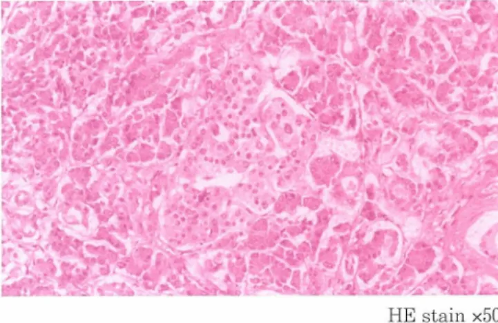
HE stain x50
Figure 2. Hematoxylin-eosin staining of pancreas. The islet is
acidophilic. Magnification is x50. 100 £ I £ 50 a-cell 0-cell
Figure 4. The percentage of insulin-positive and glucagon-positive cells was 20.5±9.2% and 48.6±9.2%, respectively,
in twenty-one islets. There is a decrease in p-cell
popula-tion and an increase in a-cell population.
demonstrated heart failure with 0.30 of ejection fraction and severe mitral and tricuspid regurgitation. ST depression in I, II, aVF, and V4-V6 induction was demonstrated by electro-cardiography. Although urinary protein excretion was only
360 mg/day, the creatinine clearance level was decreased to 15.0 ml/min. Insulin secretion had diminished because uri-nary C-peptide was decreased to 4.0 ug/day and glucagon-induced incremental C-peptide at 6 minutes was 0.3 ng/ml
(Table 1). The patient died in hospital at age 53 of severe heart failure despite intensive therapy, in April 1999.
Patho-physiological examination of the pancreas at autopsy re-vealed decreased size, number, and acidophilicity of islets (Fig. 2). Alpha- and P-cells were identified by immunohisto-chemical staining with insulin and glucagon (Fig. 3), which
showed a decrease in p-cell population (20.5±9.2%) and an increase in a-cell population (48.6±1 1.5%) (Fig. 4). Fibrosis
and ragged fiber were observed in myocardial muscle and
also in muscles of iliacus, psoas, and tongue. The kidney
ex-hibited
diabetic nephropathy. Discussion
The patient had the mitochondrial mutation 3243 (A-Kj), detected by peripheral white blood cell analysis, with diabe-tes mellitus, sensory deafness, cardiomyopathy, and hypo-thyroidism. Diabetes mellitus with a mitochondrial DNA mutation is known to result in a rapid and severe decrease in insulin secretion, which was the main cause of the onset of diabetes in this case. Only a few reports have investigated the pathological features of the pancreas in diabetic patients with a mitochondrial DNA mutation (A^G) (4-7). The four cases to date all reported a large decrease in the pancreatic P-cell population, as seen in this patient. Although an in-crease in the population of a-cells was observed in this case
immunohistochemical stain (anti-glucagon) xlOO (anti-insulin) xlOO
Figure 3. Immunohistochemical staining of representative islets. Magnification is xlOO. The same islet stained for glucagon (left)
and insulin (right).
KOJIMA et al
as well as in the case reported by Togashi et al (7),
Kobayashi et al (5) found a decrease in the number of a-cells. Miyazawa et al (4) and Kuzuya (6) did not mention any pathological alterations of the a-cells. However, the dra-matic decrease in the p-cell population together with the in-crease in a-cell population in this case may suggest a model of pancreatic pathological changes in typical type 2 diabetes. The greater reduction in P-cell population together with the increase in a-cells should decrease insulin secretion and dis-turb glucose metabolism more profoundly. Most
immuno-cytochemical studies report a smaller reduction in P-cell population (30-50%) in typical type 2 diabetes and only a slight increase in the a-cell population (8-10). Kobayashi et al suggests that mitochondrial DNA mutation 3243 may reduce mitochondrial enzymes by cytochrome c oxidase ac-tivity and increased succinate dehydrogenase, which
acceler-ates damage to P-cells through toxic hydroxyl radical generation by the electron transport system (5). Although we
did not observe superoxide generation or apoptosis of islet cells, the dramatic reduction in the population of P-cells and the increase in the population of a-cells in the islets of this patient suggests the pathogenesis of type 2 diabetes.
References
1) van den Ouweland JM, Lemkes HH, Ruitenbeek W, et al. Mutation in
mitochondrial tRNA(Leu)(UUR) gene in a large pedigree with mater-nally transmitted type II diabetes mellitus and deafness. Nat Genet 1:
368-371, 1992.
2) Kadowaki T, Kadowaki H, Mori Y, et al. A subtype of diabetes melli-tus associated with a mutation of mitochondrial DNA. N Engl J Med
330: 962-968, 1994.
3) Gerbitz KD, van den Ouweland JM, Maassen JA, Jaksch M.
Mito-chondrial diabetes mellitus: a review. Biochim Biophys Acta 1271:
253-260, 1995.
4) Miyazawa Y, Kikuchi T, Suzuki H, et al. An autopsy case of MELAS syndrome associated with diabetes mellitus; A study of mitochondrial DNA mutation in various tissues. Diabetes Journal 22: 164-167, 1994
(in Japanese).
5) Kobayashi T, Nakanishi K, Nakase H, et al. In situ characterization of islets in diabetes with a mitochondrial DNA mutation at nucleotide
po-s
ition 3243. Diabetes 46: 1567-1571, 1997.
6) Kuzuya N. Morphological and histological in situ studies on tissues from patients with mitochondrial diabetes mellitus (MDM). Nippon Rinsho 56 (Suppl): 547-553, 1998 (in Japanese).
7) Togashi M, Yamada H, Iwasaki N, et al. Selective loss of pancreatic P-cells in a diabetic patient with a mitochondrial 3243 mutation. J Jpn Diabet Soc 43: 455-458, 2000 (in Japanese).
8) Rahier J, Goebbels RM, Henquin JC. Cellular composition of the human diabetic pancreas. Diabetologia 24: 366-371, 1983.
9) Clark A, Wells CA, Buley ID, et al. Islet amyloid increased A-cells, re-duced B-cells and exocrine fibrosis: Quantitative changes in the pan-creas in type 2 diabetes. Diabetes Res 9: 151-159, 1988.
1
0) Kloppel G. Islet Histopathology in Diabetes Mellitus. in: Pancreatic Pathology. Kloppel G, Heitz PU, Eds. Churchill Livingston, New York, 1984: 154-192.