*1熊本大学医学部附属病院放射線科
*2日本医科大学医学部放射線科
*3慶應義塾大学医学部放射線科
*4群馬大学医学部核医学科
*5三重大学医学部放射線科
*6半田市立半田病院放射線科
*7金沢大学医学部核医学科
*8京都府立医科大学医学部放射線科 受付:13 年 8 月 1 日
最終稿受付:13 年 11 月 1 日
別刷請求先:熊本市本荘 1–1–1 (0 860–8556) 熊本大学医学部附属病院放射線科
冨 口 静 二 E-mail: [email protected]
I. は じ め に
201Tl 心筋 SPECT 像の画質を劣化させる主な要
因としては,体内での吸収 (減弱), 散乱線,低空
《原 著》
多施設共同研究による
201Tl 心筋 SPECT における 吸収散乱補正の臨床的有用性の検討
冨口 静二*1 汲田伸一郎*2 橋本 順*3 井上登美夫*4 野村 新之*5 江本 順一*6 中嶋 憲一*7 西村 恒彦*8
要旨 201Tl 心筋 SPECT における吸収散乱補正の臨床的有用性を検討する目的で,多施設共同研究に
より散乱線補正を加えた transmission 法による吸収補正を施行した.
対象は,冠動脈に有意な狭窄を認めない正常 26 例および冠動脈病変を認める虚血性心疾患 38 例 (うち心筋梗塞 26 例) である.
正常例では,吸収の影響により,男性では下後壁の集積低下が女性より高度で,女性では心基部前壁 の集積が男性より低かった.吸収散乱補正により 201Tl 心筋分布の性差は消失し,男性および女性とも に分布は均一となった.また冠動脈病変診断能に関しては,右冠動脈領域では,sensitivity および specificity ともに改善し,specificity の改善は有意であった.左回旋枝領域では sensitivity の改善を認め た.左前下行枝領域では sensitivity の向上は認めたものの,specificity は低下した.しかし,この specificity の低下は統計学的には有意ではなかった.
結論として 201Tl 心筋 SPECT における吸収散乱補正は,正常例では性差なく均一な局所分布が得ら れ,冠動脈病変診断能に関しては左前下行枝領域で specificity が低下するものの,従来診断能の低かっ た右冠動脈や左回旋枝領域で診断能の改善を認め,臨床的に有用と思われた.
(核医学 39: 37–46, 2002)
間分解能による部分容積効果があげられる.この ような要因は,画質の劣化とともに再構成像の アーチファクトの原因にもなっている.吸収は最 も重要な要因で,吸収によるアーチファクトとし ては,男性では横隔膜による下壁のカウント低 下,女性では乳房による前壁のカウント低下が 起こり,同部位の偽陽性所見の原因になってい る1〜3).したがって吸収補正の目的は,吸収によ るアーチファクトを改善し specificity を向上させ ることである.吸収補正の方法としては様々なも のがあるが,胸郭の場合,空気から骨まで様々な 減弱係数を持った組織が不均等に分布しており,
また個体差も大きいので,その補正のためには transmission computed tomography (TCT) を施行 し,個人およびスライス毎の減弱補正係数マップ
核 医 学
を作る必要がある.吸収補正はこのように TCT を通常の SPECT 検査に加えることで可能となっ
た1〜10).しかし,正確な減弱補正係数を算出する
ためには,TCT のデータに散乱線補正を加える必 要がある.本研究の目的は虚血性心疾患を対象と して,負荷 201Tl 心筋 SPECT において,病変診断 能を吸収散乱補正の有無で比較し,吸収散乱補正 の病変診断能における臨床的有用性および問題点 を明らかにすることである.なお,本稿は日本核 医学会 SPECT 定量ワーキンググループの全国 6 施設の参加のもとに実施した成績をまとめたもの である (Table 1).
II. 対 象
本研究の参加について文書による同意が得られ た 64 例,冠動脈に有意狭窄を認めない健康成人 26 例および責任冠動脈に有意狭窄を認める虚血 性心疾患 38 例 (心筋梗塞 26 例,狭心症 12 例) で ある.
1) 選択基準
以下の基準を満たす者を本試験の対象とする.
1. 年齢:特に制限しない.
2. 正常群:冠動脈に有意狭窄を認めない.
3. 患者群:心筋虚血による臨床症状を呈し た患者
a) 責任冠動脈に閉塞または 75% 以上の狭 窄を認めること
b) PTCR, PTCA, CABG が初回検査時は未 施行であること
2) 除外条件
次の条件の少なくとも一つに該当する者は,本 試験の対象としない.
1. 妊婦または妊娠している可能性のある婦 人ならびに授乳中の婦人
2. 重篤な合併症を有する患者 3. 冠攣縮を有する患者
4. その他,担当医が対象として不適当と判 断した患者
III. 方 法 検査方法:
負荷心筋シンチにおいては,原則として負荷は 運動負荷とし,心電図,血圧をモニターし,最大 負荷時に 201Tl 111 MBq 静注し,負荷を静注後 1 分継続した.負荷時 SPECT は静注後 5–10 分後よ り開始し,遅延像は 3–4 時間後に撮像した.
装置および撮像法:
使用装置は東芝社製 GCA7200 または Picker 社 製 prism 3000 を使用し,TCT および emission computed tomography (ECT) は同時収集法で施行 し,TEW 法またはこれに準ずる方法で散乱線補 正を施行した.以下に詳細な撮像条件を示す.
a. 東芝社製 GCA7200
TCT 線源には Sheet line source を用い TCT 核種 には 99mTc 740–1110 MBq (20–30 mCi) を使用し
た. コリメータは低エネルギー汎用コリメータを
使用した.TCT データは一検出器に Sheet line source を装着し,対向する検出器で収集し,ECT データは両検出器で収集した.エネルギーウィン ドウは 201Tl についてはメインピーク 70 keV,
ウィンドウ 20%, 99mTc についてはメインピーク
140 keV, ウィンドウ 20% とし,それぞれのメイ
ンピークに対し 7% のサブウィンドウをメイン ピークの両側に設定し, 6 ウィンドウでデータ収 集を行った. 収集条件は Step and shoot mode によ り 128×128 マトリックス,360 度 60 方向で,一 方向 20–30 秒で収集した.TCT データでは,プ ロジェクションデータを Butterworth フィルター (order 8.0, cutoff 0.14) 処理,および TEW 法によ る散乱線補正後に,ブランクデータで補正し吸収
Table 1 Multi-center trial sites 施設名 所属科名 担当者名 熊本大学附属病院 放射線科 冨口 静二 慶應義塾大学病院 放射線科 橋本 順 日本医科大学病院 放射線科 汲田伸一郎 群馬大学附属病院 核医学科 井上登美夫 三重大学附属病院 放射線科 野村 新之 半田市立半田病院 放射線科 江本 順一
多施設共同研究による Tl 心筋 SPECT における吸収散乱補正の臨床的有用性の検討 39
補正マップを作成した.再構成には filtered back projection 法で,Ramp filter を用いた.ECT デー タについては,Sheet line source を装着している検 出器のデータを Sheet line source 像をサブトラク ションすることにより,2 検出器分のプロジェク ションデータを作成し,TCT と同様の方法で再構 成像を作成した.作成した再構成像は散乱線補正 像と吸収散乱線補正像で,吸収補正には TCT 像 より作成した吸収補正マップを用い Chang の逐次 近似法により逐次近似回数 1 回で再構成した.
本法は,正常 9 例,心筋梗塞 20 例,狭心症 5 例の 34 例に施行した.
b. Picker 社製 prism 3000
TCT 線源には line source を用い TCT 核種には
99mTc 555 MBq (15 mCi) を使用した.コリメータ は心臓用のファンビームコリメータを使用した.
TCT データは line source に対向する検出器で収集 し,ECT データは 3 検出器で収集した.エネル ギーウィンドウは 201Tl についてはメインピーク 74 keV, ウィンドウ 35%, 99mTc についてはメイ ンピーク 140 keV,ウィンドウ 15% とし,201Tl のメインピークに対しサブウィンドウを 53 keV を中心に 7% のウィンドウと 92 keV 中心で 4%
ウィンドウでメインピークの両側に設定し,4 ウィンドウでデータ収集を行った.収集条件は continuous mode により 64×64 マトリックス,360
度 120 方向で,一方向 10 秒で収集した.再構成 は TCT データおよび ECT データともに逐次近似 法を用い,TCT データには Maximum-likelihood expectation-maximization (ML-EM) を用いて近似 回数 20 回で再構成し,ECT データは ordered sub- set expectation-maximization (OSEM) 法を用いて逐 次回数 4 回で,散乱線および吸収補正を行った.
作成した再構成像は散乱線補正なし像と吸収散乱 線補正像である.
本法は,正常 17 例,心筋梗塞 6 例,狭心症 7 例の 30 例に施行した.
評価方法:
多施設による本臨床試験の結果を統一的な基準 で客観的に評価するために判定委員 4 名にて視覚 評価をした.視覚評価は 4 名の合意により行い,
短軸像で心基部より,中央部,心尖寄りの 3 スラ イスを選択し,それぞれを 6 セグメントにわけ,
局所集積を評価した (Fig. 1).心尖部 (セグメント 19) に関しては,正中垂直長軸断像にて評価し た.局所集積はスコアにより評価し,スコアは正 常 0,軽度低下 1,中等度低下 2,高度低下 3,
欠損 4 とした.冠動脈病変診断においては,異常 値は,各セグメントの正常例での平均スコアおよ び標準偏差を算出し,平均スコアより 2SD 以下 のスコアを異常スコアとして判定した.なお,心 尖部よりの短軸像における側壁部 (セグメント 14, 15) は前下行枝または回旋枝の支配領域である可 能性があるため,評価より除外した.
検討項目:
1) 正常例における吸収散乱補正による局所の
201Tl 分布に及ぼす影響を視覚的評価により検討
した.
2) 負荷心筋シンチにおいて,虚血部位の診断 能を吸収散乱補正による向上を認めるかどうか視 覚的評価法を用い検討する.
統 計:
正常例の 201Tl 局所分布の検討には,各セグメ ントのスコアを用い,仮説値を 0 として一標本符 号検定を施行した.201Tl 局所分布の性差につい Fig. 1 Segments evaluated by visual assessment. Each
short axial view of basal, middle and apical region was divided into 6 segments. Apical region (segment 19) was evaluated on vertical long axial view.
核 医 学
Fig. 2 Mean scores in each segment on noncorrected stress images in normal male and female. Higher mean score was observed in anterobasal region in female and inferoposterior region in male.
Fig. 3 Mean scores in each segment on scatter and attenuation corrected stress images in normal male and female. Lower mean scores were ob- tained with scatter and attenuation correction than those without correction. However, higher mean scores were observed in segment 19.
ては,正常男性および女性の各セグメントにおけ るスコアを Mann-Whitney の U 検定を用い検討し た.また,sensitivity および specificity の吸収散乱 補正前後での有意差検定には,カイ 2 乗検定 (MacNemar 検定) を用いた.有意差はすべて p<
0.05 とした.
IV. 結 果
正常例の 201Tl 局所分布の検討では,女性では 吸収散乱補正しない場合に心基部前壁 (セグメン ト 1), 心室中隔 (セグメント 5), 後下壁 (セグメ ント 4, 10) でスコアが大きく,同部のカウント低 下の傾向を認めるも,各セグメントのスコアに統 Table 2 Comparison of diagnostic performance between SAC and NC images by visual analysis
LAD RCA LCX
NC SAC NC SAC NC SAC
Sensitivity 22/29 (76%) 23/29 (79%) 17/24 (71%) 19/24 (79%) 15/25 (60%) 19/25 (76%) Specificity 32/35 (91%) 27/35 (77%) 28/40 (70%)* 34/40 (85%)* 37/39 (95%) 37/39 (95%) Accuracy 54/64 (84%) 50/64 (78%) 45/64 (70%)* 53/64 (83%)* 52/64 (81%) 56/64 (88%) SAC: attenuation and scatter corrected, NC: uncorrected, LAD: left anterior descending artery, RCA: right coronary artery, LCX: left circumflex artery. *: statistically significant (p<0.05).
Table 3 Comparison of diagnostic performance between SAC and NC images by visual analysis (prism 3000)
LAD RCA LCX
NC SAC NC SAC NC SAC
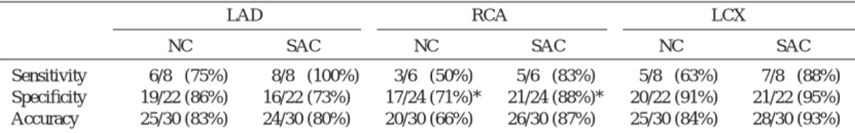
Sensitivity 6/8 (75%) 8/8 (100%) 3/6 (50%) 5/6 (83%) 5/8 (63%) 7/8 (88%) Specificity 19/22 (86%) 16/22 (73%) 17/24 (71%)* 21/24 (88%)* 20/22 (91%) 21/22 (95%) Accuracy 25/30 (83%) 24/30 (80%) 20/30 (66%) 26/30 (87%) 25/30 (84%) 28/30 (93%) SAC: attenuation and scatter corrected, NC: uncorrected, LAD: left anterior descending artery, RCA: right coronary artery, LCX: left circumflex artery. *: statistically significant (p<0.05).
多施設共同研究による Tl 心筋 SPECT における吸収散乱補正の臨床的有用性の検討 41
計学的には有意差は認めなかった (Fig. 2).吸収 散乱補正した場合においては,すべてのセグメン トで有意差は認めなかった (Fig. 3).男性では吸 収補正しない場合には,心室中隔 (セグメント 5) および後下壁 (セグメント 4, 10) でスコアが有意 に大きく,201Tl カウントの同部での低下が認め られた (Fig. 2).しかし,吸収散乱補正した場合 には,心尖部 (セグメント 19) での 201Tl カウント の低下を除き,すべてのセグメントでスコアに有 意差を認めず,201Tl の局所分布は均一化した (Fig. 3).
次に,男性と女性の各セグメントのスコアを比 較検討した.吸収散乱補正しない場合には,心尖 部 (セグメント 19) で,女性の方がスコアは有意 に大きく,また後壁 (セグメント 4) では,男性の 方がスコアは有意に大きかった (Fig. 2).しか し,吸収散乱補正した場合には,心尖部 (セグメ ント 19) でカウントの低下を認めるも,その他の セグメントでは男女間に有意差は認めず,吸収散 乱補正により 201Tl の局所分布における性差は改 善された (Fig. 3).
冠動脈病変診断能に関しては,右冠動脈 (RCA) 領域では,sensitivity および specificity ともに改善 し (吸収散乱補正なし;sensitivity 71%, specificity 79%, 吸収散乱補正あり; sensitivity 79%, specificity 85%), 特に specificity は有意に改善した.そのほ かには明らかな有意差は認めなかったが,左回旋 枝 (LCX) 領域では sensitivity の改善を認めた (吸 収散乱補正なし;sensitivity 60%, specificity 95%, 吸収散乱補正あり;sensitivity 76%, specificity 95%).左前下行枝 (LAD) 領域でも sensitivity の改
Table 4 Comparison of diagnostic performance between SAC and NC images by visual analysis (GCA-7200)
LAD RCA LCX
NC SAC NC SAC NC SAC
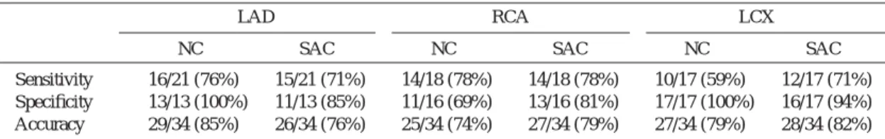
Sensitivity 16/21 (76%) 15/21 (71%) 14/18 (78%) 14/18 (78%) 10/17 (59%) 12/17 (71%) Specificity 13/13 (100%) 11/13 (85%) 11/16 (69%) 13/16 (81%) 17/17 (100%) 16/17 (94%) Accuracy 29/34 (85%) 26/34 (76%) 25/34 (74%) 27/34 (79%) 27/34 (79%) 28/34 (82%) SAC: attenuation and scatter corrected, NC: uncorrected, LAD: left anterior descending artery, RCA: right coronary artery, LCX: left circumflex artery.
善を認めたが,specificity は低下した (吸収散乱補 正なし;sensitivity 76%, specificity 91%, 吸収散乱 補正あり; sensitivity 79%, specificity 77%) (Table 2).
A
B
Fig. 4 Short axial stress and delayed images in a 32- year-old male without CAD. Reduced tracer accumulation was observed in the posterior and posteroseptal wall on both noncorrected images (A). Improvement in tracer distribution was obtained with scatter and attenuation correction on both images (B).
核 医 学
GCA7200 および Prism3000 で施行した例につ いての結果を Table 3 および Table 4 に示す.い ずれにおいても結果は全体の結果と同様の傾向 で,両機種間で診断能に差はないと思われた.
V. 症 例 呈 示
1. 32 歳,男性,正常例 (Fig. 4)
吸収散乱補正なしの短軸像にて,左室後壁およ び心室中隔に集積低下を認める (Fig. 4A).吸収散 乱補正により,同部の集積低下は改善し,左室心 筋の 201Tl 分布は均一となった (Fig. 4B).
2. 27 歳,女性,正常例 (Fig. 5)
吸収散乱補正なしの短軸像にて,左室後壁およ び心室中隔の集積低下に加え,心基部前壁にも集 積低下を認める (Fig. 5A).吸収散乱補正により,
同部の集積低下は改善し,左室心筋の 201Tl 分布 は均一となった (Fig. 5B).
3. 63 歳,女性,3 枝病変 (Fig. 6)
症例は労作性狭心症で,LAD および RCA の末 A
B
Fig. 5 Short axial stress and delayed images in a 27- year-old female without CAD. Reduced tracer accumulation was observed in the anterobasal and posterior wall on both noncorrected images (A).
Improvement in tracer distribution was obtained with scatter and attenuation correction on both images (B).
A
B
Fig. 6 Short axial stress and delayed images in a 63- year-old female with CAD. Reduced tracer accumulation was observed in the anterobasal and posterolateral wall on noncorrected stress image (A). Reverse redistribution was observed in the anterobasal wall and redistribution in the pos- terolateral wall on noncorrected delayed image (A). Reduced tracer accumulation on the stress images, reverse redistribution and redistribu- tion on the delayed image were more clearly demonstrated with the scatter and attenuation correction (B).
多施設共同研究による Tl 心筋 SPECT における吸収散乱補正の臨床的有用性の検討 43
梢に 99%, LCX の中枢に 99% の狭窄を持つ例で ある.吸収散乱補正なしの短軸像にて,左室後壁 および心室中隔の集積低下に加え,後側壁にも集 積低下を認め,遅延像にて後側壁に再分布を認め る (Fig. 6A).吸収散乱補正により,左室後壁およ び心室中隔の集積低下は改善し,後側壁の集積低
下はより鮮明となり,再分布の評価も補正なしに 比べ容易に評価できる (Fig. 6B).
4. 72 歳,男性,正常例 (Fig. 7)
吸収散乱補正なしの負荷直後短軸像にて,左室 後壁および心室中隔の集積が低下しており,遅延 像にて同部に再分布が認められるので,右冠動脈 領域の虚血が疑われる (Fig. 7A).吸収散乱補正に より,負荷直後短軸像および遅延像ともに左室心
筋の 201Tl 分布は均一となり,吸収散乱補正なし
の短軸像で認めた右冠動脈領域の虚血は偽陽性所 見と判断できる (Fig. 7B).しかし,吸収散乱補正 により心尖部前壁に集積低下部が出現している.
VI. 考 察
201Tl 心筋 SPECT において,201Tl の正確な心筋 内分布を画像化するためには,吸収および散乱線 補正が必要である.しかし,現在までの報告で は,TCT 法による吸収補正のみが施行されたもの
が多い5,6,11〜13).また,吸収補正のみでは,心臓
周囲の 201Tl 集積部からの散乱線の影響で,下壁
のカウントが増加することが指摘されており,吸 収補正とともに散乱線補正の必要性が強調されて
いる5,12).橋本ら14) は,TCT 法による吸収補正に
加え,TEW 法による散乱線補正を施行すること で,吸収補正による下壁のカウント増加が補正で きたと報告している.しかし,吸収および散乱線 補正の多数例での臨床的検討は少ないので,今回 多施設による共同研究を施行した.
散乱線補正に関しては,様々な方法が報告され ているが,TCT 法による吸収補正と組み合わせる ことを考慮すると,TEW 法9,14) および transmis- sion-dependent convolution subtraction (TDCS)
法15,16)が有力な方法と考えられる.今回の検討
では,比較的簡便な方法であり,また,多施設で 使用できるため散乱線補正法については,TEW 法およびこれに準ずる方法を選択した.
正常例での検討では,吸収散乱線補正しない場 合には,女性に比べ男性の方に,おもに下後壁で 吸収の影響を強く認めた.Ficaro ら10) の負荷
99mTc-MIBI SPECT を用いた検討でも,下後壁で
A
B
Fig. 7 Short axial stress and delayed images in a 72- year-old male without CAD. Reduced tracer accumulation was observed in the inferoposterior wall on the noncorrected stress images (A).
Redistribution in the inferoposterior wall was suspected on the noncorrected delayed image (A).
In this case, ischemia was suspected in the RCA territory. With scatter and attenuation correction, tracer accumulation in the inferoposterior wall was normalized on both stress and delayed images (B). However, reduced tracer accumulation was observed in the anterolateral wall on the stress and delayed images with scatter and attenuation cor- rection (B).
核 医 学
は男性の方に集積率が低く,下壁の局所集積は平 均で女性 84% に対し男性では 75%,後壁では女 性 72% に対し男性 65% と報告されており,201Tl でも同様の傾向であった.しかし,吸収散乱補正 によりこのような性差は消失し,性別の影響のな い画像で診断できる点は吸収散乱補正の臨床的有 用性の一つである.
吸収補正の冠動脈病変診断能における有用性に 関してはいくつかの報告がある6,10,12,17).99mTc- MIBI 心筋 SPECT における冠動脈病変診断能につ き,標準 bull’s eye map により冠動脈の有意な狭 窄を 70% 以上とした場合に,吸収補正により全 冠動脈支配領域で sensitivity は向上し,specificity は LAD 領域では変わらないが,RCA および LCX 領域で向上したと報告されている10).吸収補正に より specificity のみではなく,sensitivity の向上も 得られているが,これは吸収補正像では,正常で は心筋全体のトレーサ分布が均一で,局所集積の 変動が小さいために,異常灌流領域の検出が容易 になるためと考えられる10).Gallowitsch ら12) は,201Tl 心筋 SPECT においても,sensitivity お よび specificity が吸収補正により向上すると報告 している.また,LAD および RCA 領域の灌流 異常の severity および extent については,吸収補 正像の方が正確に評価できたと報告している.
一方,吸収補正により LAD および RCA 領域の sensitivity は向上し,RCA 領域の specificity は向 上するものの LAD 領域の specificity は低下する
結果19) や,視覚評価において,吸収により出現す
る欠損像は改善するものの冠動脈病変診断能には 改善を認めなかったとの報告もある 17,18).今回の 検討では,冠動脈病変診断能において吸収散乱補 正により右冠動脈領域の specificity 以外には,統 計学的に有意な改善を認めなかった.しかし,
LAD 領域の specificity の低下を除き,sensitivity および specificity は向上しており,臨床的に有用 と思われた.LAD 領域の specificity についても,
吸収補正のみでは 88% から 65% と 23% も低下
した19) が,今回の検討では 91% から 77% と,低
下の程度は 14% と軽度であった.Fakhri ら20)
は,シミュレーションにより,吸収補正は画像の 均一性を向上させ,散乱線補正はコントラストを 向上させると報告している.症例 3 において,後 側壁の虚血が吸収散乱補正により明瞭となったこ とには,散乱線補正によるコントラストの向上が 寄与しているものと考えられる.また,今回の診 断能の向上も,散乱線補正を加えた効果としてコ ントラストが向上したためと考えられ,吸収補正 に散乱線補正を加えた場合の臨床的有用性を示唆 する結果と思われた.しかし,吸収散乱補正を 行っても,依然,前壁の specificity が低い問題点 がある.これには,読影上の問題と,物理的要因 が関与している.読影上の問題としては,相対的 前壁の集積低下を異常と読影したことが一因と なっている.正常例での吸収補正による前壁の集 積低下の程度を認識することで,この問題はある 程度改善するものと考える.物理的要因として は,周囲組織よりの散乱線の補正が十分ではな かったこと,ガンマカメラの空間分解能が低いこ との 2 点が挙げられる.Links ら21) は,動きの補 正のみでは,sensitivity/specificity (%) は,LAD の 領域で 64/71%, RCA の領域で 71/81%, LCX の領 域で 32/94% であったのに対し,動き,吸収およ び深さ依存のボケの補正 (空間分解能補正) を施行 した場合には,順に 77/93%, 74/81%, 50/97% と 診断能が向上し,有意な改善は,LAD および RCA 領域の specificity の向上と報告している.こ の結果をみると有意ではないものの全領域で sen- sitivity および specificity がともに改善している.
Links ら20) によれば,深さ依存のボケの補正によ
り,空間分解能は 30% ほど改善し,コントラス トも向上すると報告している.今後正確な散乱補 正法に加え空間分解能を改善すればさらなる診断 能の向上が期待できると思われる.
今回の評価では 2 機種を使用したので, 施設間 での装置の違いや, 画質の違いを考慮し, 視覚的 評価で検討した. その結果, 2 機種間で診断能に差 はないものと思われた. しかし, 今後は多数例によ る定量的評価など, より客観的評価法で, 吸収散乱 補正の有用性を検討する必要があると思われた.
多施設共同研究による Tl 心筋 SPECT における吸収散乱補正の臨床的有用性の検討 45
VII. 結 語
201Tl 心筋 SPECT における吸収散乱補正は,正
常例では性差なく均一な局所分布が得られ,冠動 脈病変診断能に関しては左前下行枝領域で spec- ificity が低下するものの,従来診断能の低かった 右冠動脈や左回旋枝領域で診断能の改善を認め,
臨床的に有用と思われた.
謝辞:今回,技術協力いただいた東芝メディカル および島津製作所に感謝いたします.
文 献
1) Eisner RL, Tamas MJ, Cloninger K, et al: Normal SPECT Thallium-201 bull’s-eye display: gender differences. J Nucl Med 1988; 29: 1901–1909.
2) Manglos SH, Thomas FD, Gange GM, Hallwig BJ:
Phantom study of breast tissue attenuation in myo- cardial imaging. J Nucl Med 1993; 34: 992–996.
3) King MA, Tsui BMW, Pan TS: Attenuation compensation for cardiac single-photon emission computed tomographic imaging: Part 1. Impact of attenuation and methods of estimating attenuation maps. J Nucl Cardiol 1995; 2: 513–524.
4) Tsui BMW, Gullbert GT, Edgerton ER, et al:
Correction of nonuniform attenuation in cardiac SPECT imaging. J Nucl Med 1989; 30: 497–507.
5) 冨口静二,大山洋一,吉良朋広,吉良光子,中島 留美,辻 明徳,他: 201Tl 心筋 SPECT における トランスミッション・エミッション同時収集法の 評価.核医学 1996; 33: 1027–1035.
6) Ficaro EP, Fessler JA, Ackermann RJ, Rogers WL, Corbett JR, Schwaiger M: Simultaneous trans- mission-emission Thallium-201 cardiac SPECT:
effect of attenuation correction on myocardial tracer distribution. J Nucl Med 1995; 36: 921–931.
7) Chang W, Loncaric S, Huang G, Sanpitak P:
Asymmetric fan transmission CT on SPECT systems.
Phys Med Biol 1995; 40: 913–928.
8) Tan P, Bailey DL, Meikle SR, Eberl S, Fulton RR, Hutton BF: A scanning line source for simultaneous emission and transmission measurements in SPECT. J Nucl Med 1994; 34: 1752–1760.
9) Ichihara T, Motomura N, Ogawa K, Hasegawa H, Hashimoto J, Kubo A: Evaluation of SPECT quan- tification of simultaneous emission and transmis- sion imaging of the brain using a multidetector SPECT system with the TEW scatter compensation method and fan-beam collimation. Eur J Nucl Med
1996; 23: 1292–1299.
10) Ficaro EP, Fessler JA, Shreve PD, Kritzman JN, Rose PA, Corbett JR: Simultaneous transmission/emis- sion myocardial perfusion tomography. Diagnostic accuracy of attenuation-corrected 99mTc-sestamibi single-photon emission computed tomography.
Circulation 1996; 93: 463–473.
11) Prvulovich EM, Lonn AH, Bomanji JB, Jarritt PH, Ell PJ: Effect of attenuation correction on myocardial thallium-201 distribution in patients with a low likelihood of coronary artery disease. Eur J Nucl Med 1997; 24: 266–275.
12) Gallowitsch HJ, Sykora J, Mikosch P, et al:
Attenuation-corrected thallium-201 single-photon emission tomography using a gadolinium-153 moving line source: clinical value and the impact of attenu- ation correction on the extent and severity of perfu- sion abnormalities. Eur J Nucl Med 1998; 25: 220–
228.
13) Chouraqui P, Livischitz S, Sharir T, et al: Evaluation of an attenuation correction method for thallium-201 myocardial perfusion tomographic imaging of patients with low likelihood of coronary artery disease. J Nucl Cardiol 1998; 5: 369–377.
14) Hashimoto J, Ogawa K, Kubo A, et al: Application of transmission scan-based attenuation compensation to scatter-corrected thallium-201 myocardial single- photon emission tomographic images. Eur J Nucl Med 1998; 25: 120–127.
15) Meikle SR, Hutton BF, Bailey DL: A transmission- dependent method for scatter correction in SPECT. J Nucl Med 1994; 35: 360–367.
16) Hutton BF, Osiecki A, Meikle SR: Transmission- based scatter correction of 180 degrees myocardial single-photon emission tomographic studies. Eur J Nucl Med 1996; 23: 1300–1308.
17) Bestetti A, Tarola GL, Lomuscio A, et al: A comparison between attenuation-corrected and -uncorrected transmission-emission SPECT images obtained with Tl-201 in CAD patients. Giornal Italiano di Cardiologia 1999; 29: 411–417.
18) Lee DS, So Y, Cheon GJ, et al: Limited incremental diagnostic values of attenuation-noncorrected gating and ungated attenuation correction to rest/stress myocardial perfusion SPECT in patients with an intermediate likelihood of coronary artery disease. J Nucl Med 2000; 41: 852–859.
19) 冨口静二: 臨床編/心臓,吸収・散乱補正,
SPECT 機能画像 (西村恒彦編),メジカルビュー 社,東京,1998: 105–111.
20) Fakhri GE, Buvat I, Benali H, Todd-Polropek A, Paola RD: Relative impact of scatter, collimator response, attenuation, and finite spatial resolution
核 医 学 corrections in cardiac SPECT. J Nucl Med 2000; 41:
1400–1408.
21) Links JM, Becker LC, Rigo P, et al: Combined
Summary
Multi-center Study for the Evaluation of Clinical Usefulness of Attenuation and Scatter Correction on
201Tl Myocardial SPECT
Seiji T
OMIGUCHI*
1, Shin-ichiro K
UMITA*
2, Jun H
ASHIMOTO*
3, Tomio I
NOUE*
4, Yoshiyuki N
OMURA*
5, Junichi E
MOTO*
6, Kenichi N
AKAJIMA*
7and Tunehiko N
ISHIMURA*
8*1 Department of Radiology, Kumamoto University Hospital
*2 Department of Radiology, Nippon Medical School
*3 Department of Radiology, School of Medicine, Keio University
*4 Department of Nuclear Medicine, Gunma University School of Medicine
*5 Department of Radiology, Mie University School of Medicine
*6 Department of Radiology, Handa Municipal Hospital
*7 Department of Nuclear Medicine, Kanazawa University School of Medicine
*8 Department of Radiology, Kyoto Prefectural University of Medicine
corrections for attenuation, depth-dependent blur, and motion in cardiac SPECT: a multicenter trials. J Nucl Cardiol 2000; 7: 414–425.
The aim of this study was to evaluate the clinical usefulness of attenuation and scatter correction (AC, SC) on a 201Tl myocardial single-photon emission computed tomography (201Tl SPECT) as a multi-cen- ter trial.
With a dual-detecter and a triple-detector SPECT systems with a 99mTc transmission source, simulta- neous transmission/emission tomography (TCT/ECT) was performed on 38 patients with angiographically coronary heart disease (CHD) and 26 patients without evidence of CHD. Stress and delayed attenuation and scatter corrected images (SAC) and uncorrected im- ages (NC) were reconstructed.
On NC images of normal cases, influence of attenu- ation was greater in male than female. In comparison of 201Tl distribution between male and female, sig- nificant decrease in 201Tl activity was observed in the inferoposterior wall in male and that was observed in the anterobasal wall of the left myocardium in female.
Such a difference in 201Tl distribution between male and female disappeared on SAC images. On the diag- nostic performance for the identification of CHD, SAC images demonstrated improved specificity and
accuracy values in the right coronary arterial territory (RCA) with visual analysis statistically. Sensitivity value in the RCA was also improved, but it was not statistically significant. Sensitivity value in the left circumflex arterial territory (LCX) increased without decrease in specificity value on SAC images. In the left anterior descending arterial territory (LAD), sensi- tivity value increased on SAC images. Although specificity value decreased on SAC images in LAD territory, it was not statistically significant.
The difference in 201Tl distribution between male and female is improved in normal cases by attenuation and scatter correction on 201Tl myocardial SPECT.
Diagnostic performance of CHD is also improved by attenuation and scatter correction, especially in territo- ries of which specificity in assessing the absence of disease have been suboptimal. In conclusion, attenua- tion and scatter correction on 201Tl myocardial SPECT is considered to be clinically useful.
Key words: 201Tl myocardial SPECT, TCT, Scat- ter correction, Attenuation correction, Multi-center trial.