Development of New Fluorogenic OligoDNA Probes Utilizing Silylated Fluorescent Material
THESIS
Submitted to Gunma University for the Degree of
Doctor of Philosophy in Engineering
2015
Student ID: 12802171
Jakir Ahmed Chowdhury
Division of Molecular Science
Graduate School of Science and Technology Gunma University
Japan
I
Chapter 1
General Introduction 1
1.1 Fundamentals of nucleic acid 2
1.2 Hybridization probe 4
1.3 Principle of fluorescence generation 8
1.4 Fluorescence signal transduction 9
1.5 Fluorescence resonance energy transfer 10
1.6 Fluorescence nucleic acid hybridization probes 10 1.6.1 Adjacent probes or Light CyclerTM hybridization probe 11
1.6.2 TaqMan○RProbe or 5’-Nuclease probes 13
1.6.3 Strand-displacement probes or Yin-Yang probe 14
1.6.4 Molecular beacon probes 15
1.7 Fundamentals of molecular beacon and its spectroscopic principles 17
1.7.1 Loop sequence 17
1.7.2 Stem sequence 17
1.7.3 Fluorophore 18
1.7.4 Quencher 23
1.8 Applications of molecular beacon probes 24
1.8.1 Biosensor and biochip development 24
1.8.2 Genetic analysis 25
1.8.3 Real-time monitoring of DNA/RNA amplification during PCR 26
1.8.4 In-vivo imaging of cancer cells 26
1.8.5 In-vivo RNA detection 28
1.9 Problems of molecular beacon probes and recent solution 29
1.9.1 Signal-to-noise (S/N) ratio 29
1.9.2 Loop and stem nucleotides 32
1.9.3 Complexity of living environments 34
References
II
Polyamine-connected Deoxyuridine and Silylated Pyrene and Its Fluorescence Properties
2.1 Abstract 46
2.2 Introduction 46
2.3 Results and Discussion 52
2.3.1 Synthesis of [1-(Pyrenyl)dimethylsilylmethyl]-(2-cyanoethyl)
-N,N-diisopropylphosphoramidite 52
2.3.2 Synthesis of 5´-O-(4,4´-Dimethoxytrityl)-5-[N-[2-N,N-bis(2-trifluoro- acetamidethyl) amino]ethyl]carbamoylmethyl-2´-deoxyuridine-3ˊ-
O-yl(2-cyanoethyl)-N,N-diisopropylphosphoramidite 54 2.3.3 Synthesis of modified oligoDNA possessing polyamine-connected
deoxyuridine and silylated pyrene 56
2.3.4 Duplex forming ability of new MB probes 61
2.3.5 Fluorescence properties of new molecular beacon probes 62 2.3.6 The effect of mismatch in the target DNA to the fluorescence signal
of the probe 67
2.3.7 Thermal stability of new molecular beacon probes 68
2.4 Conclusion 69
2.5 Experimental 70
2.5.1 General Information 70
2.5.2 Synthesis of 1-(Chloromethyldimethylsilyl) pyrene 71 2.5.3 Synthesis of 1-(Acetoxymethyldimethylsilyl) pyrene 72 2.5.4 Synthesis of 1-(Hydroxymethyldimethylsilyl) pyrene 73 2.5.5 Synthesis of [1-(Pyrenyl)dimethylsilylmethyl]-(2-cyanoethyl)-
N,N-diisopropylphosphoramidite 74
2.5.6 Synthesis of 3´, 5´-O-Diacetyl-5-methoxycarbonylmethyl-2´
-bromo-2´-deoxyuridine 75
2.5.7 Synthesis of 3´, 5´-O-Diacetyl-5-methoxycarbonylmethyl-2´
-deoxyuridine 77
2.5.8 Synthesis of 5-[N-[N,N-Bis(2-trifluoroacetamidethyl)amino]ethyl]
-carbamoylmethyl-2´-deoxyuridine 77
III
2.5.10 Synthesis of 5´-O-(4,4´-Dimethoxytrityl)-5-[N-[2-N,N-bis(2-
trifluoroacetamid-ethyl)amino]ethyl]carbamoylmethyl-2´-deoxyuridine -3ˊ-O-yl(2-cyanoethyl)-N,N-diisopropylphosphoramidite 80
2.5.11 Oligonucleotides Synthesis 81
2.5.12 Melting Temperature Analysis 81
2.5.13 Fluorescence Measurements 82
References
Chapter 3
Synthesis and Fluorescence Properties of Pseudo-Dumbbell Type of Molecular Beacon Probes bearing a Modified Deoxyuridine and a Silylated Pyrene as a Fluorophore
3.1 Abstract 87
3.2 Introduction 87
3.3 Results and Discussion 92
3.3.1 Synthesis of [1-(Pyrenyl)dimethylsilylmethyl]-(2-cyanoethyl)
-N,N-diisopropylphosphoramidite 92
3.3.2 Synthesis of 5-methoxycarbamoylmethyl-3ˊ, 5ˊ-di-O-acetyl-2ˊ-
bromodeoxyuridine 92
3.3.3 Synthesis of 1-(2-Aminoethylamino) anthraquinone 93 3.3.4 Synthesis of 5-hydroxycarbamoyl methyl-5ˊ-O-DMTr
-2ˊ-deoxyuridine 93
3.3.5 Introduction of anthraquinone moiety to the C-5 position
of deoxyuridine 94
3.3.6 Synthesis of modified oligoDNA possessing C-5 modified
deoxyuridine and silylated pyrene 96
3.3.7 Duplex forming ability of new MB probes 107
3.3.8 Fluorescence properties of the probes 109
3.3.9 The effect of mismatch in the target DNA to the fluorescence
signal of the probes 115
3.3.10 The effect of deoxyinosine to the fluorescence signal of Probe 4 118 3.3.11 Hybridization abilities of molecular beacon type probe 119
IV
3.5.1 General Information 123
3.5.2 Synthesis of 1-(2-Aminoethylamino)anthraquinone 123 3.5.3 Synthesis of 5-Methoxycarbonylmethyl-2ˊ-deoxyuridine (17) 124 3.5.4 Synthesis of 5-Methoxycarbonyl methyl-5ˊ-O-DMTr-2ˊ-deoxyuridine 125 3.5.5 Synthesis of 5-Carboxy methyl-5ˊ-O-DMTr-2ˊ-deoxyuridine 126 3.5.6 Synthesis of 5´-O-(4,4´-dimethoxytrityl)-5-[N-[2-[N-(anthraquinon
-1-yl)amino]ethyl]carbamoylmethyl]-2´-deoxyuridine 127 3.5.7 Synthesis of 5´-O-(4,4´-dimethoxytrityl)-5-[N-[2-[N-(anthraquinon-1
-yl)amino]ethyl]carbamoylmethyl]-2´-deoxyuridine-3ˊ-O-yl(2
-cyanoethyl)-N,N-diisopropylphosphoramidite 128
3.5.8 Oligonucleotides Synthesis 129
2.5.12 Melting Temperature Analysis 129
2.5.13 Fluorescence Measurements 130
References
1
Chapter 1
General Introduction
2
1.1 Fundamentals of Nucleic Acid
Deoxyribonucleic acid, DNA is one of the most important molecules for every living life on earth. DNA was first identified and isolated as a distinct acidic substance stored inside nuclei in 1869 by Friedrich Miescher [1]. Thereafter, it took another 70 years to demonstrate [2]
that this DNA molecule carried genetic information. Another important step in the DNA study was made in 1953 by Watson and Crick [3]. They proposed a three-dimensional, right handed double helical B-DNA model of the DNA structure. A further advanced experimental studies revealed that DNA has other double-helical forms such as A-DNA and also many other three-dimensional firms, including triple [4] and quadruple helices [5-10].
On the other hand, ribonucleic acid, RNA which is closely related to DNA and also biologically very important molecule only forms A-type double helical structure.
The primary structures of DNA and RNA consist of phosphodiester-linked nucleotide units that contain a 2ˊ-deoxy–D-ribose or D-ribose sugar ring in DNA and RNA respectively and an aromatic nucleobases (Figure 1.1). The major bases are monocyclic pyrimidines or bicyclic purines. The major purines found in both DNA and RNA are adenine (A) and guanine (G). The major pyrimidines are cytosine (C), thymine (T) and uracil (U) in case of RNA. The resulting polynucleotide chain has a consistent 5ˊ→3ˊ polarity with both a negatively charged sugar-phosphate backbone and an array relatively hydrophobic nucleobases, amphiphilic features which ultimately drive the assembly and maintenance of secondary and tertiary nucleic acid structures.
In nature, DNA does not usually exist as a single molecule, but instead as a pair of molecules that are held tightly together [3, 11]. These two long strands entwine like vines, in the shape
3
of a double helix. The nucleotide repeats contain both the segment of the backbone of the molecule, which holds the chain together, and a nucleobase, which interacts with the other DNA strand in the helix. A nucleobase linked to a sugar is called a nucleoside and a base linked to a sugar and one or more phosphate groups is called a nucleotide. A polymer comprising multiple linked nucleotides (as in DNA) is called a polynucleotide. The DNA double helix is stabilized primarily by two forces: hydrogen bonds between nucleobases in horizontal direction and base-stacking interactions among aromatic nucleobases in perpendicular direction [3].
O O P O
OR O- O
O P O
O O- O
O O P O O- O
O O P O O- RO
5'
NH N N
O
NH2 N
G
N NH2
O N C
N N N
N
NH2 A OH
OH
HO
OH NH O
O N U
3'
O O P O
OR O- O
O P O
O O- O
O O P O O- O
O O P O O- RO
5'
NH N N
O
NH2 N
G
N NH2
O N C
N N N
N
NH2 A
NH O
O N T
3'
RNA DNA
Figure 1.1: The primary structures of DNA and RNA
4
1.2 Hybridization Probe
A hybridization probe is a fragment of DNA or RNA of variable length (usually 15-100 bases long) which is used in DNA or RNA samples to detect the presence of specific sequences that are complementary to the sequence in the probe. The probe thereby hybridizes to single-stranded nucleic acid (DNA or RNA) whose base sequence allows probe-target base pairing due to complementarity between the probe and target. The probe-target base pairing usually occurs by its hydrogen-bonding capability where the probe itself hybridize to the target sequence. Protein probes interact with their specific target through a blend of forces such as hydrophobic, ionic and hydrogen bonding at only a few specific sites. Whereas, nucleic acid probes interact with their complements through hydrogen-bonding (Figure1.2), at tens, hundreds or thousands of sites, depending on the length of the hybrid [12]
Figure 1.2: Sequence specificity of nucleic acid
5
The probe is tagged or labeled with a molecular marker of either radioactive or non- radioactive fluorescent molecules. The commonly used radioactive markers are 32P [13-15]
and non-radioactive marker digoxigenin [16-17]. The probes whose fluorescence signal changes upon hybridization with a target sequence is referred as fluorogenic hybridization probe [18]. In an electrochemical DNA hybridization detector, a short ssODN (a single- stranded DNA probe with 15–20 nucleotides long) is usually immobilized on a transducer (electrode) to create a DNA recognition element. The probe-modified electrode is then immersed into a solution of target DNA. When the target DNA contains a sequence that exactly matches that of the probe DNA, a hybrid duplex is formed at the electrode surface.
In the absence of a complementarity between the probe and target, no duplex is formed.
Hybrid formation is then translated into an electrical, analytically useful signal. Partial complementary binding of these DNAs may result, however, in a weak interaction that is more difficult to recognize.
Utilizing fluorescent dyes, meanwhile, several types of fluorogenic hybridization probes have been developed until date [19-26]. In general, we can divide them in two groups: dual- labeled and single-labeled probes.
The dual labeled probe utilizes an alteration of dye-dye interactions. The probe alone gives quenching effect by the close contact of the dye molecules. On the other hand, upon hybridization with a target sequence, the dyes are spatially separated which results in a significant fluorescence increase. These include either simplest linear probe or stem-loop structures probe (Figure 1.3). The fluorescence quenching in stem less dual labelled single stranded probe occurs by random probe coiling (Figure 1.4). A measurable change in
6
fluorescent intensity is accompanied by forming a duplex with the target. The energy transfer from the donor to the acceptor dye is the major mechanism of signal generation of
Figure 1.3: Schematic diagram of linear and stem-loop probe [cited from ref. 27]
a b
Figure 1.4: Dual labelled single stranded linear probe. (a) In absence of target, fluorescence quenching occur through random coiling of the probe (b) In presence of target, probe hybridize with its target and give an intense fluorescence signal. [cited from ref. 28]
7
this type of probe [19]. The donor dye is fluorescent, while the acceptor dye can be fluorescent [19] or non-fluorescent [20-21]. On the other hand, stem-loop type dual labeled probe has a sequence recognition region located in the loop and the stem has fluorescent dye and non-fluorescent dye which ensures quenching property by making a close contact of the dyes [29].
The single labeled probe utilizes the alteration of fluorescence efficiency of a single dye that takes place due to the hybridization event. This probe exploits the ability of the dyes to enhance fluorescence upon intercalation into double-stranded DNA. One example of such probe is hybeacon probes [22]. They are single-labeled oligonucleotides that increase their fluorescence upon hybridization. The inherent quenching ability of deoxyguanosine nucleotides has also been used to design single labeled probe formats (Figure 1.5) [23-26].
Figure 1.5: Schematic operation of singly labeled DNA-hairpins: The fluorophore is attached to the 5ˊ-end of the oligonucleotide and quenched by guanosine residues in the complementary stem via photoinduced intramolecular electron transfer. (cited from ref. 26)
8
1.3 Principles of Fluorescence Generation [30]
In the last few years, there have been a tremendous growth in utilization of fluorescence in the field of biological sciences [31]. Fluorescence is a kind of luminescence which is resulted from the emission of light from a substance. This phenomenon is extensively used in biotechnology, medical diagnostic, DNA sequencing and other disciplines. The pattern of fluorescence emission is varied depending on the types of hybridization probes. It needs to have at least one fluorophore in the probes which has the capability to absorb energy from light and by transferring this energy internally emit this energy as light of a characteristic wavelength. A fluorophore is usually raised from its ground state to higher vibrational level of an excited singlet state by absorbing energy. Then, it loses some of its energy as heat and come back once again to a relatively stable lowest vibrational level of excited singlet state.
Thereafter, it return to the ground state from this excited singlet state by the emission of light. This emitted light is usually termed as fluorescence. The process is depicted in the Figure 1.6.
Figure 1.6: Different energy state of Jablonski diagram by absorbing energy and subsequent emission of fluorescence. 1, 2 and 3 express the stage of excitation, lifetime of excited state and fluorescence emission respectively [cited from ref. 31].
9
1.4 Fluorescence Signal Transduction
Different types of fluorescent dyes like pyerene, perylene, FAM, TET, HEX, Cy dyes, TMR, ROX, Texas red, JOE, Oregon green, Rhodamine, Coumarin and others are used to design fluorescent probes. In addition to fluorophore, some quencher molecules are also used in the fluorescent oligonucleotide probes like DDQ-I, DABCYL, Eclipse, Iowa Black RQ, BHQ-1 and others. In case of single labeled probes, the signal transduction occurs by binding with nucleic acids. Fluorescent properties change by dye-nucleotide interaction due to the photo induced electron transfer between them. Crockett et al. reported the quenching properties of fluorescein oligonucleotide by interaction with deoxyguanosine nucleotide [24]. The dynamical behavior of electron transfer reaction between guanosine containing oligonucleotides and the covalently bonded Oxazine dye is also reported [32]. On the other hand, the transduction process of dual-labeled probes depends on the fluorescence quenching, fluorescence resonance energy transfer (FRET) and monomer-excimer emission switching with fluorophore. Energy transfer, quenching and excimer formation depend on the design of the probes. These three processes are mainly distance dependent. An alteration of fluorescence properties resulted due to the structural rearrangement occurred under the presence of targets.
10
1.5 Fluorescence Resonance Energy Transfer
Fluorescence resonance energy transfer (FRET) is a dipole-dipole interaction that happen when two fluorophores come in close proximity and the emission spectrum of one overlaps with the excitation spectrum of others. The excitation of the acceptor fluorophore occurs due to the emission energy transfer from donor fluorophore [33, 34]. This is first described by Forster in the year of 1948 as Forster type energy transfer [35]. This non-radiative process is a process of energy transfer between a donor and acceptor molecule. This process is mainly a distance dependent energy transfer mechanism. Generally the donor molecule and acceptor molecule are covalently attached at different location of a probe. At the same time, it is also necessary to overlap the absorption of excited acceptor partially with the emission of donor. Acceptor may be a fluorescent or non-fluorescent molecule. The transferred energy is usually emitted as fluorescence in case of fluorescent acceptor. While it lost the absorbed energy as heat in case of a non-fluorescent acceptor.
1.6 Fluorescence Nucleic Acid Hybridization Probes
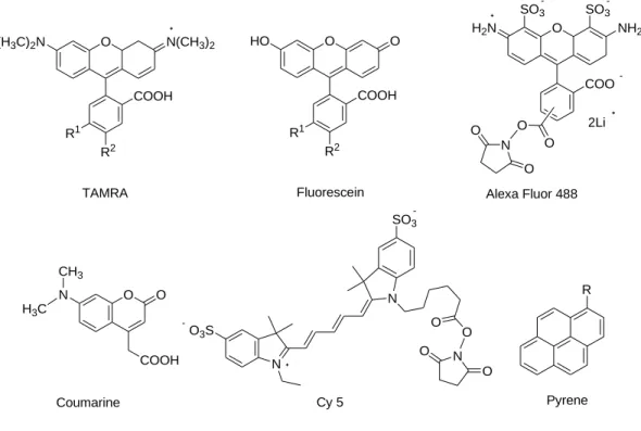
Fluorogenic nucleic acid hybridization probes are newly developed probe that generate a characteristics fluorescence signal upon hybridization with complementary target nucleic acid sequence [36]. This is a highly useful and selective probes for analysis of many bioanalytes [37]. With inclusion of potent and relatively low cost fluorescent labelling agent (Figure 1.7), it has become a very attractive research field because it has numerous applications in detection and visualization of target oligonucleotides. It is also utilized to detect non-nucleic acid analytes like protein, metal ion and other small molecules. Several fluorescent hybridization probes have been used in the detection of nucleic acid. A brief
11
discussion on different types of fluorescent hybridization probes are discussed in the following which are cited from SAE Marras - Fluorescent Energy Transfer Nucleic Acid Probes, 2006 [30].
HO O
COOH R1
R2 O N(CH3)2 O
(H3C)2N
COOH R1
R2
N CH3 H3C
O O
COOH
TAMRA Fluorescein
Coumarine
O3S
N
N SO3
O O
N O O
Cy 5
COO O
SO3 SO3
H2N
O N
O O
O
Alexa Fluor 488
Pyrene NH2
2Li
R
Figure 1.7: Frequently used fluorescent labelling agent for designing fluorogenic probes
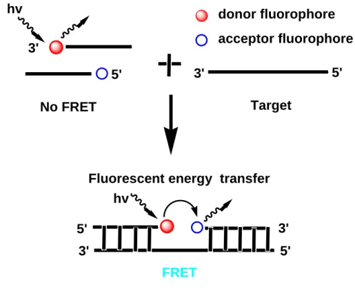
1.6.1 Adjacent probes or LightCyclerTM hybridization probes
Adjacent probe is a type of fluorescent hybridization probes that utilize two single stranded probes. One probe is labelled with a donor fluorophore molecule at one end of nucleic acid and the other is labeled with acceptor molecule at the other end. Usually the donor molecule is attached to the 3ˊ-end, whereas, the acceptor molecule is attached to the 5ˊ-end of the respective single stranded probe. There is no energy transfer between them while they stayed free condition in solution without target. However, these probes bind to the neighboring
12
sites on a target nucleic acid when they come in contact with their targets [38]. In the presence of target nucleic acid, the hybridization of the probe with target DNA is measured either by decreasing the donor fluorescence signal or by increasing the acceptor fluorescence signal through the fluorescence energy transfer from the donor to the acceptor. A schematic diagram of this kind of probes is shown in Figure 1.8.
Figure 1.8: Schematic overview of energy transfer and fluorescence signal generation in adjacent probes.
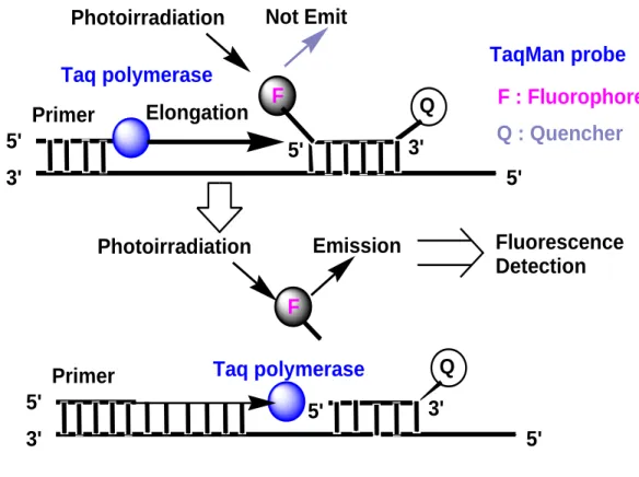
1.6.2 TaqMan® probe or 5ˊ-Nuclease probes
5ˊ-Nuclease probe (Figure 1.9) is a single stranded hybridization probes simultaneously labelled with a fluorophore acts as the donor and a quencher acts as the acceptor molecules in one strand that utilizes the inherent endonucleolytic activity of Taq DNA polymerase to
hv
No FRET
FRET
Target
Fluorescent energy transfer
donor fluorophore acceptor fluorophore
hv
3' 3'
3'
5' 5'
5' 3' 5'
13
generate a fluorescence signal [19]. The Taqman probes and the primers, designed to hybridize within the target DNA sequences, are introduced into a PCR assay. The fluorescence signal of the fluorophore is quenched by the quencher molecule via FRET.
Annealing of the probes start synthesis of the complementary strands from the primer, the Taq DNA polymerase extends the primer and reached to the probe binding downstream of the primer. Then the enzyme degrades the probe and releases the fluorophore from it which results the separation of the fluorophore from the quencher molecules. Thus an increased fluorescent signal is resulted. After completion of each cycle, a new cycle of hybridization takes place. As a result, more fluorophores are separated from the probes (quencher), resulting in increased fluorescence signal which indicates the accumulation of target DNA molecules. A schematic overview of fluorescent signal generation is shown below.
Figure 1.9: Schematic overview of energy transfer and fluorescence signal generation in 5ˊ-Nuclease probes.
F : Fluorophore Q : Quencher 5'
3'
Primer Elongation
5'
5' 3'
F Q
Taq polymerase
Photoirradiation Not Emit
5' 3'
Primer
5'
5' 3'
Taq polymerase Q Photoirradiation Emission
F
Fluorescence Detection TaqMan probe
14
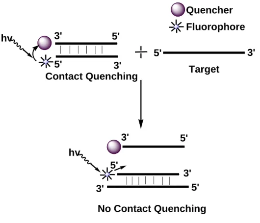
1.6.3 Strand-displacement probes or Yin-Yang probe
This probe is designed by utilizing two complementary nucleic acid probes. One of the probe is designed by labeling with fluorophore. On the other hand, the other probe is labeled with a non-fluorescent quencher molecule [39]. Fluorophore and quencher molecules come in close proximity while the probes hybridize each other and as a result it gives a very low fluorescence emission i.e. contact quenching property. By the addition of the probes into the solution of target, one of the probe forms a more stable probe-target hybrid than the other, thus two probes are being separates from each other. Due to this displacement, the fluorophore is no longer in the close contact of the quencher and an increased fluorescence signal is generated. The schematic diagram of the mechanism of this kind of probe is shown in Figure 1.10.
15
Contact Quenching
No Contact Quenching Target Quencher hv
hv
Fluorophore
5'
3'
3' 3'
5'
5' 5'
5' 5'
3'
3'
3'
Figure 1.10: Schematic overview of energy transfer and fluorescence signal generation in Strand-displacement probes.
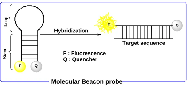
1.6.4 Molecular beacon probes
Since the first report in 1996 [29], molecular beacon probes have attracted great interests as FRET based DNA and RNA probes. Molecular beacon is a single stranded hybridization probe that forms stem-loop structure (Figure 1.11). The loop portion is complementary to the target oligonucleotide and, therefore, acts as the binding portion to the target. The loop portion is inserted between two stem sequences which are complementary to each other and, therefore, ultimately forms the double-stranded stem portion of the probe. At the same time, the probe is connected to both a fluorophore and a quencher molecule. While it stays alone (means absence of the target), it forms a stem-loop (hairpin) type of structure. In this
16
structure, the fluorophore and quencher molecules come in close proximity and thus the probe gives a low fluorescence emission due to the FRET occurs between the fluorophore and the quencher. Upon addition of target oligonucleotide, the loop-portion binds to the target to form a stable probe-target hybrid with the dissociation of the stem-portion. (Figure 1.11). As a result, the fluorophore is placed in a distant position from the quencher causing the recovery of fluorescence signal.
Figure 1.11: Schematic overview of energy transfer and fluorescence signal generation in molecular beacon probes.
F
Q
Q
Target sequence F : Fluorescence
Q : Quencher Hybridization
F
Molecular Beacon probe
LoopStem
17
1.7 Fundamentals of Molecular Beacon and its Spectroscopic Principles
1.7.1 Loop sequence
The loop sequence of molecular beacon is generally consisting of 15 to 30 nucleotide units and should not form any kind of secondary structure by themselves [29, 40-41]. Their sequences are usually complementary to a part of target oligonucleotides. This part of molecular beacon probe is also termed as sequence recognition part. Upon mixing of target and the molecular beacon probe, the loop portion binds with the target molecule in a sequence specific manner. To ensure the binding of molecular beacon probe with target oligonucleotide, the thermal stability (melting temperature) of the duplex formed by the loop portion and the target should be higher than that of stem portion. It means the stability of probe-target duplex should be sufficiently stronger than the intermediate duplex formation by molecular beacon alone. In general, the stability can be increased by increasing the length of the sequence recognition part (loop-portion).
1.7.2 Stem sequence
The stem is typically consisting of 5 to 8 base pairs which dissociates during the hybridization of molecular beacon probe with a target oligonucleotide. The thermal stability of stem depends on their length as well as its GC content. In order to ensure the stem-loop type of structure of the MB probe in the free form, the melting temperature of the stem should be 7-10 °C higher than the detection temperature [42]. The thermal stability of the complex consisting of target and molecular beacon probe with 15-25 base pairs is thought to be sufficiently higher than that of the stem [29, 40-41]. When the stem length is short, the
18
probe would have a faster hybridization kinetics to the target compared with the probe having longer length stem. However, the signal to background ratio would decreases because of undesired dissociation of the stem-portion even under the absence of the target.
On the other hand, if the stem length is too long (too stable), then the probe hardly binds to the target and the signal to background ratio would decrease too. In addition to these, the neighboring nucleotide of the fluorophore in the probe would exhibit quenching properties of the molecular beacon probe. Drake et al. recently reported that the neighboring nucleotide showed a variable degree of quenching in order of G>A>C>T [43]. Thus, one should be careful to decide about the length as well as the sequence and the neighboring nucleotide to the fluorophore in the design of stem-potion in molecular beacon type probes.
1.7.3 Fluorophore
Fluorophores, a fluorescent materials, have been used extensively in many days in material sciences as well as in biomedical sciences. Moreover, it is still focused by many researchers to find out novel fluorescent small molecules as well as making alternation in their physiochemical properties, such as absorption, excitation and emission wavelength, quantum yield and so forth. The majority of commercially available fluorescent materials are usually made from the small synthetic fluorophore. A few of genetically encoded proteins come from natural animal origin are considered as naturally occurring fluorophores.
Different types of fluorophores have been tested for their feasibility to make fluorescent oligonucleotide probes. Their emission spectra vary from near UV to near IR wavelength.
A few of the commonly used fluorophores and their analogs are discussed in the following.
19 Pyrene derivaties
Pyrene, a poly aromatic hydrocarbon of four fused benzene ring, has attracted much attention due to their unique properties. This is a chromophore of choice in fundamental and applied photochemical research. Laurent was first isolated pyrene from the residue of the destructive distillation of coal tar in the year of 1837 [44]. Initially, the synthetic dye industries used pyrene for preparing several derivatives like pyranthrone [45]. In the year of 1954, Forster and Kasper was first found the intermolecular emission (excimer emission) property of pyrene [46]. This excimer showed high quantum yield [47]. Due to these attractive properties, pyrene has become a subject of extensive research for its photophysical properties.
For the last 50 years, the fluorescence properties of pyrene have been mostly used in investigation of water soluble polymer and making dye for fluorogenic probes [48-49]. It is widely used as a probe to investigate the surfactant micelles properties, aggregates of surfactant/polymers and phospholipid vesicles. As a labelling agent, pyrene has been utilized to investigate protein and peptide structural features [50-53], lipid membrane [54- 61] and also oligonucleotide recognition [62-66]. In addition, many researchers observed the red-shift of absorption and emission wavelength and increased quantum yield by incorporation of certain functional groups [67-69]. Figure 1.12 shows the possible position of incorporation of such functional groups in the structure of pyrene for improvement of quantum yield.
20
Figure 1.12: Representative structure of 1-substituted pyrene and 1, 3, 6, 8-tetrasubstituted pyrene derivatives.
Anthracene derivatives
Anthracenes have been utilized in many practical applications as a core material for preparing potential therapeutics [70-73], polymeric material [74-75] and optical devices [76-77]. Though anthracene derivatives are well known for many days [78-81], a few studies have been done to characterize their emission wavelengths of its derivatives.
Figure 1.13: General structure of 2, 6-disubstituted anthracene and 9, 10-disubstituted anthracene derivatives.
9 1
2
3 5 4
6 7
8
10
2,6-Substitution 9,10-Substitution
1 2
3
4 5
6 7 8 9 10
1-Substitution 1,3,6,8-Substitution
21
The general structure and substitution position in the anthracene are shown in Figure 1.13.
The substitutions in 2, 6 position of the anthracene by trifluoromethylphenyl and thiophene have significant influence on their emission wavelength [82]. It is also possible to alter the emission wavelengths of anthracene 2, 6-disulfonic acid by varying organic salts in the solid state [83-84]. Kyushin and co-workers reported recently the synthesis of 9,10- disilylanthracene and they observed an intense quantum yield of this anthracene derivative (f > 0.9 ) [85].
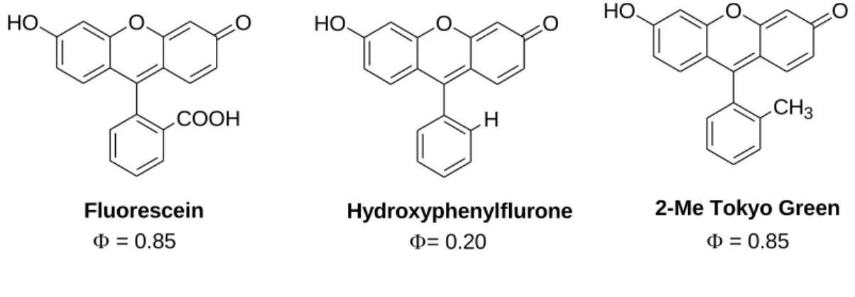
Fluorescein derivatives
Fluorescein is a main dye of choice in medicine and science for more than 100 years due to its high quantum yield, ease of handling and also for non-toxicity. It was first developed by A. Beyer in 1871 [86]. It gives an emission with reasonably long wavelength (500 to 600 nm) with relatively high quantum yield (f=0.85-0.97) upon excitation in aqueous media [87]. Fluorescein is used as fluorescent material for labeling and sensing many biomolecules [88-90]. A huge number of novel fluorescein based fluorescent materials have been synthesized by many researchers [91-95]. For example, the photophysical properties of the fluorescein improved by making modification at the aryl moiety attached to the xathene core framework [96-97]. Some structures and substituted derivatives of fluorescein with their quantum yields are presented in Figure 1.14.
22
Figure 1.14: Structure of Fluorescein and its substituted derivatives with quantum yield [cited from ref. 91]
BODIPY derivatives
BODIPY, 4,4-Difluoro-4-bora-3a,4a-diaza-s-indacene, is a molecule which was first discovered in 1968 by Treibs and Kreuzer [98]. Upon excitation, it emits sharp fluorescent peak with high quantum yield due to their tendency to strongly absorb of UV radiation. As a continuation, efficient fluorescent materials were synthesized by making a simple modification in its structure. For example, Wael et al. prepared an alkylated BODIPYs by symmetrical di-substitution with methyl function. The derivative showed a quantum yield 0.81 in ethanol. At the same time, they also prepared unsymmetrical di-substituted alkylated BODIPYs which showed a quantum yield 0.70 [99]. However, the symmetrical tetra, hexa and hepta-alkylated BODIPYs showed quantum yield 0.80, 0.56, and 0.70 respectively [99- 100]. Kim et al. also recently developed some new analogs of BODIPY which have high quantum yield (f=0.56-0.98) (Figure 1.15) [101].
O
HO O
COOH
O
HO O
H
O
HO O
CH3
Fluorescein
= 0.85
Hydroxyphenylflurone
= 0.20
2-Me Tokyo Green
= 0.85
23
Figure 1.15: Basic Structure of BODIPY and some of their analogs having high quantum yield [cited from ref. 101]
1.7.4 Quencher
Quencher is a molecule which decreases the emission intensity of a fluorescence substances.
The process is usually taking place when emission spectra of fluorescent donors have sufficiently overlap with absorption spectra of the quencher molecule (Förster Mechanism, FRET). This is only happened when both molecules come in sufficiently closed position.
The quencher molecule is widely used to design FRET based fluorogenic probes for protease activity detection [102-114], nucleic acid hybridization [29, 115], and real-time PCR [116-118]. The natural nucleobase is also used to get the quenching of fluorescence of the fluorophore. For example, the guanosine residue can effectively reduce the fluorescent intensity of the fluorophore depending on the type of fluorophore [119-120]. However, this feature of guanosine has been utilized by others to design quencher free molecular beacon probes [121].
B N N
F F
1 2
3 4
5 6 8 7
BODIPY CORE
N B N F F
O O
R1
R2 R2
R1=H, R2=Me, =0.96 R1=CF3, R2=Me, =0.98 R1=H, R2=4-methoxyphenyl, =0.86 R1=CF3, R2=4-methoxyphenyl, =0.56 R1=H, R2=Phenyl, =0.90 R1=H, R2=2-methoxyphenyl, =0.91 R1=H, R2=4-isopropylphenyl, =0.97
24
1.8 Applications of Molecular Beacon Probes
The development of MB based assay for gene identification has emerged an areas of research that were not possible previously. With time, it has become a powerful tool for many applications in human health aspects. These have included biosensor and biochip development, genetic analysis, real-time monitoring of DNA/RNA amplification during PCR, in vivo imaging of cancer cell as well as in vivo RNA detection.
1.8.1 Biosensor and biochip development
For the last couple of years, MB probes are popularly used to design highly sensitive and selective biosensors or biochips. Surface-immobilized MB sensors have been reported for various applications including ultra-small optical fiber probes using avidin–biotin binding [122], fiber optic microsphere-based biosensor [123] and microarrays [124-125]. The MB immobilized magnetic nanoparticle or genomagnetic nanocapturers (GMNC) have also designed for detecting the DNA/RNA molecules [126]. This kind of GMNC was made through avidin-biotin interaction by conjugating the probe onto the surface of magnetic nanoparticle. Martinez et al. reported a surface-immobilized MB biosensor having 2ˊ,4ˊ- locked nucleic acid (LNA) bases [127]. Due to the high binding affinity and stability of LNA, their biosensor are very stable and robust, and selective and sensitive.
The surface-immobilized MBs have a general configuration where it includes the immobilization of the hairpin onto a surface. The MBs possess a fluorophore and quencher moiety. However, the ‘on’ versus ‘off’ ratios generated by its fluorophore and quencher moieties are important factor in the immobilization of this kind of MB probes. In most cases,
25
immobilized molecular beacons showed low signal-to-background ratios because of the additional strain of the solid surface to its confirmation which ultimately makes an unstable hairpin structure. For this reason there is an increased number of ‘on’ molecules than in a homogenous assay. To overcome this problem, attempts have been taken recently by using longer stem which further force the MBs to be isolated from the solid surface [128].
1.8.2 Genetic analysis
The analysis of genetic discrepancy is an automated procedures like sequencing which requires a high throughput methods. However, the use of MBs is a simple and capable to discriminate single-base mismatches. The probes are also very selective considering the thermal stability. The perfectly matched probe-target hybrids are more stable that usually force the stem portion to open. It was also reported that the detection of mismatched using MB probes have shown their potentiality due to the formation of less stable mismatched MB probes duplexes than mismatched linear probe duplexes [115]. The stability of MB probes is also influenced by many factors like the number of complementary bases participate in the hybridization, GC content, position of the mismatches in the sequence as well as hybridization temperature. The high selectivity of MB probes to discriminate single base alternation makes them a promising probes for analyzing genetic variation [129]. Piatek et al. reported a MB which has the ability to analyze 81bp region of the rpoB gene for Mycobacterium tuberculosis mutations associated with resistance of isoniazid and rifampin [130]. In another report, Kostrikis et al. have designed a molecular beacons to distinguish allele of the -chemokine receptor 5 gene that determines susceptibility to HIV-I [129].
26
1.8.3 Real-time monitoring of DNA/RNA amplification during PCR
MBs are also used for real-time monitoring of nucleic acid amplification during PCR [28, 115]. This kind of analysis should be done at lower temperature because the MBs become dissociated at high temperature. When the annealing of primers ensured at a low temperature, the non-hybridized MB will not emit fluorescence signal because of forming stem loop type of structure. However, it generate a fluorescence signal by binding with the target amplicons.
The increase fluorescence signal indicates the degree of amplification at each annealing cycle. Recent reports indicate that real-time PCR monitoring with MBs is better than other conventional methods. For example, MBs have showed better genotyping results than TaqMan probes [131]. Moreover, it can process many samples simultaneously and consuming relatively short period of time. Considering the risk of contamination, the confirmation of amplified sample in the same tube is also an additional advantage of this method compare with time-consuming gel electrophoresis and southern blotting methods.
This method has been utilized by many researchers in many purposes like development of surface immobilized PNA-DNA hybrid probes for detecting PCR amplicons [132], quantification of viruses [133-134] as well as study of replication of hepatitis virus [135].
1.8.4 In-Vivo imaging of cancer cells
Specific and sensitive molecular beacon probe development is one of the central challenges in cancer cell imaging. In in-vivo imaging of cancer cell, the probes should have faster tissue uptake, shorter residence in the site of action and higher ratio of target accumulation. Shi et al. reported one of such novel MB probes (Figure 1.16) [136]. They named it as activated aptamer probe (AAP) which has a fluorophore and quencher moiety attached at its terminus.
In the absence of cancer cell, it remained as a hairpin structure where the fluorophore and quencher stayed in a close position resulted in quenching of the fluorescence. However, the
27
probe gave fluorescence signal upon binding with a specific protein tyrosine kinase (PTK7;
CCK4) on the membrane of the target cancer cell due to the conformational change of the probe that displaces the fluorophore from the quencher (Figure 1.16). They observed the fast restoration of fluorescence of AAP by the presence of target cancer cell in tumor site compared with the other areas.
Figure 1.16: Schematic presentation of the novel strategy for in vivo cancer imaging using AAP. CCRF-CEM tumor-bearing nude mice were intravenously injected with (a) the AAP, (b) control probe, and (c) the ‘‘always-on’’ aptamer probe, respectively. The pink circle in every image locates the tumor site [cited from ref. 136]
28 1.8.5 In vivo RNA detection
The success of in vivo RNA detection using MB probes depends on the combination of rational sequence design, ability of probe to be inserted in the cell as well as the choice of target mRNA sequence. The molecular beacon has already showed potentiality to decode the message of coded mRNA. A novel way of detecting cancer at very early stage by detecting the mRNA transcripts that comes from the gene in the living cells. Fang et al.
recently reported a molecular beacons which injected into the living cells for the detection of mRNA using an ICCD based fluorescence imaging process [137]. The cells location determination and background measurements were carried out by taking optical and fluorescence images. After injection, CCD images of the cells were collected. The fluorescence signal of the injected cells increases with time (Figure 1.17). Whereas, no fluorescence observed in the control. It indicates that MBs can be used for the real time detection of mRNA. In 1998, Matsu reported a 15-nucleotide long antisense sequence for human basic fibroblast growth factor for the detection of mRNA [138]. The injection of MB probes into K562 human leukemia cells disclosed the real-time visualization and interaction of mRNA by fluorescence confocal microscopy [138-139].
Figure 1.17: Hybridization of the MB to mRNA in living cells. (a) Optical image of a kangaroo rat kidney cell and the fluorescence images of the cell (b) 3 min (c) 12 min, and (d) 15 min after injecting MB. [cited from ref. 137]
29
1.9 Problems of Molecular Beacon Probes and Recent Solutions
Molecular beacon probe has shown its potentiality in the modern era of genomics and proteomics involving the study of cancer, genetic analysis, DNA-protein interaction, designing biosensors, in vivo RNA detection and molecular beacon aptamers etc. as described earlier. However, more improvements can also be achieved in the area like increment of signal-to-noise ratio, selection of loop and stem sequences and also overcoming the complexity of living environment. Some of the problems and their recent solutions are discussed below.
1.9.1 Signal-to-noise (S/N) ratio:
The fundamental phenomenon of molecular beacon probes is the signal increase upon hybridization to the target (increment in S/N ratio). The lower limit of the detection can only be achieved by the huge increase in fluorescence upon probe-target hybridization. On the other hand, the S/N ratio can be increased either by improving the fluorescent intensity of the fluorophore under the presence of the target or by reducing the background under the absence of the target. Many attempts have been taken until now to increase the fluorescent intensity of fluorophore. Yang et al. recently reported a water soluble poly(phenylene ethylene) (PPE) polymeric fluorophore. They conjugated PPE with an oligonucleotide [140].
They reported their synthetic method is efficient, simple, and readily controllable of water soluble PPE with an oligonucleotide. Increasing the fluorescent intensity of the fluorophore was also reported by the silylation of fluorescent aromatic compound like pyrene and anthracene [67, 85]. Recently our lab has also designed many silylated pyrene derivatives possessing dimethylsilyl function with modifiable terminal group for this purposes [69, 141-
30
143]. The fluorescent quantum yield of such pyrene derivatives showed more than double compare to that of pyerene moiety. Some of these derivatives with their quantum yield are shown in Figure 1.18. Krasnoperov et al. also developed a very impressive lanthanide based luminescent complex [144]. They designed a cross linkable lanthanide chelate probes. They showed the detection limit of their probe is 1000 times lower than that of traditional MB probe and 30 times lower than the other lanthanide based probes.
Si Me Me
NH2
Si Me Me
OH
Si Me Me
NH2 Si Me
Me
OH
CN
=0.60)
=0.67) =0.66)
=0.79) Si Me Me
Cl
Si Me Me
OH
=0.67) =0.64)
Figure 1.18: Structure of pyrene derivatives with their quantum yields
On the other hand, reducing of background fluorescence largely depends on the design of the molecular beacon probes. The design should enable the fluorophore and quencher in a very close proximity which will ultimately ensures the efficient quenching. Yang et al.
reported an approach of using multiple quencher dyes to design a molecular beacon probes
31
(Figure 1.17a) and observed an S/N ratio of upto 320 [145]. Kashida and collegues have reported another approach of improving the S/N ratio by placing the fluorophore/quencher pairs in the stem region (Figure 1.17b) and reported an S/N ratio of upto 58 [146]. Dubertret et al. used gold nanoparticles for fluorescence quenching of many fluorophore to rise the S/N ratio of the MB probe (Figure 1.17c) [147]. Using this kind of metal, they able to rise S/N ratio upto 100. Though many attempts have already taken to solve the matter of S/N ratio of the molecular beacon probes, it is still a big concern to design a molecular beacon probe with appropriate S/N ratio to ensure the lower limit of detection.
a b c
Figure 1.17: Strategies to reduce background fluorescence of MB probes (a) Superquenched MB probe contains more than one quencher (b) In-stem probe takes advantages of stacking interaction of the base pairs with fluorophore and a quencher dye to achieve efficient quenching (c) Gold nanoparticles conjugated MB probe takes advantages of super quenching abilities of gold nanoparticles. [cited from ref. 148]
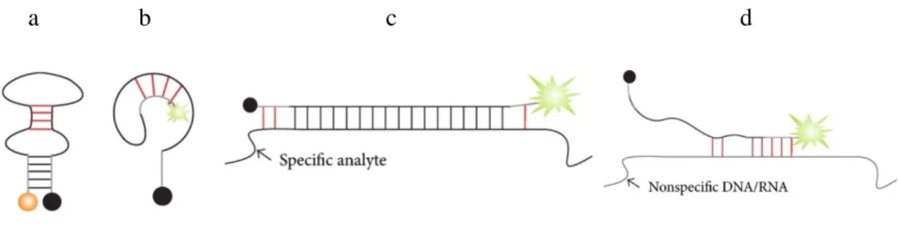
32 1.9.2 Loop and stem nucleotides:
The loop and stem nucleotides sequences influence the folding and hybridization of MB probes. Undesired structures of MB probes would be emerged by the interference of the loop as well as stem sequences. For example, the MB probes containing partially self- complementary loop sequence could hybridize each other making a secondary structure (figure 1.18a) which would be more stable and less accessible by the target DNA. Thus, it may reduce the hybridization efficiency of the MB probes with its target DNA. If the stem nucleotides have partial complementary sequence to the loop nucleotides, an alternative structure (Figure 1.18b) of MB probes may be formed. The alternate structure formation thus formed would separate the fluorophore of the MB probes from the quencher resulting in reducing the quenching efficiency of the probes. These are why one should carefully
a b c d
Figure 1.18: Examples of loop and stem interferences in MB probe. (a) Loop interference.
Complementary loop nucleotides hybridize to each other, thus making folded conformation more stable and less accessible by the target DNA. (b) Stem hybridizes to loop thus separating the fuorophore from the quencher resulting in elevated background signal. (c) Stem nucleotides can partially hybridize to the nucleotides of the targeted DNA. (d) Stem and loop nucleotides bind nonspecific nucleic acids present in a sample. Red lines indicate undesired or undersigned base pairs. [cited from ref. 148]
33
select the stem nucleotides to avoid any kind of complementarity with the loop. Stem invasion is kind of interaction of stem nucleotides with the analyte nucleotide which flank the target site. This may also happen by wrong choice of stem sequences (Figure 1.18 c,d).
In these cases, stem nucleotides can partially bind to the complementary target DNA that does not allow the target to bind perfectly with the probes resulting in the flanking of target site. At the same time, both stem and loop nucleotides also bind in a nonspecific site of DNA or RNA which may present in the sample solution.
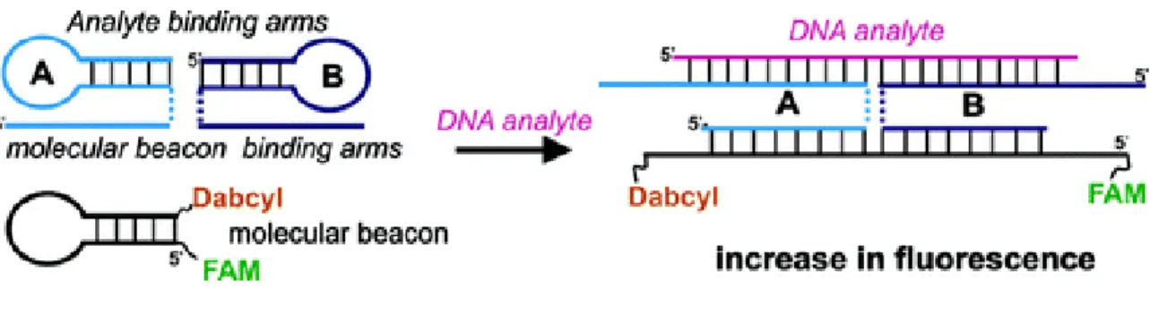
Many strategies have been adapted by researchers to overcome the aforementioned problems. Kim et al. developed a modified MB probes by using unnatural enantiomeric L- DNA in the stem [149]. They showed the incorporation of L-DNA in the stem reduces the intra- and intermolecular stem invasions. An alternate strategy was also developed by Kolpashchikovwhere he used adaptor strands (A and B strands in Figure 1.19) [150]. Each strand has a complementary part to the MB probe and another complementary part to the analyte nucleic acid. Both strands interact with the target. This complex interaction ensures the open conformation of the MB probes, thus resulting a high fluorescence.
Figure 1.19: Adaptor 𝐴 and 𝐵 strands hybridize to both analyzed sequence and MB probe.
Triethylene glycol linkers (TEG) that connect the MB-binding arms with the analyte- binding arms are shown as dashed lines. [cited from ref. 150]
34 1.9.3Complexity of living environments:
The utilization of molecular beacon probe for analyzing gene expression can frequently limited by the degradation of the backbone of the oligonucleotides of molecular beacon probes by nuclease [151-153]. In living cell, it was also previously reported that the half- life of unmodified phosphodiester oligonucleotides is very short which may vary from 15 to 20 min [154-156]. That’s why, it is necessary to design a nuclease resistant backbone in the molecular beacon probe for example using phosphorothioate backbone in place of phosphodiester backbone [157] for use in living environments. Replacing natural nucleotides of the probes by 2ˊ-OMe-modified RNA, peptide nucleic acid and locked nucleic acid have been employed to overcome these kind of problems [158, 159-160].
References
1. McCarty, M. The Transforming Principle; W. W. Norton, New York, 1985.
2. Avery, O. T.; MacLeod, C. M.; McCarty, M. J. Exp. Med., 1944, 79, 137.
3. Watson, J. D.; Crick, F. H. C. Nature, 1953, 171, 737.
4. Soyfer, V. N.; Potaman, V. N. Triple-Helical Nucleic Acids; Springer: New York, 1996.
5. Gellert, G.; Lipsett, M. N.; Davies, D. R. Proc. Natl. Acad. Sci., U.S.A. 1962, 48, 2013.
6. Zimmerman, S. B.; Cohen, G. H.; Davies, D. R. J. Mol. Biol., 1975, 92, 181.
7. Tougard, P.; Chantot, J. F.; Guschlbauer, W. Biochim. Biophys. Acta., 1973, 308, 9.
8. Wang, I.; Patel, D. J. Biochemistry, 1992, 31, 8112.
35
9. Laughlan, G.; Murchie, A. I. H.; Norman, D. G.; Moore, M. H.; Moody, P. C. E.;
Lilley, D. M. J.; Luisi, B. Science, 1994, 265, 520.
10. Phillips, K.; Murchie, A. I. H.; Lilley, D. M. J.; Luisi, B. J. Mol. Biol., 1997, 273, 171.
11. Yakovchuk, P.; Protozanova, E.; Frank-Kamenetskii, M. D. Nucleic Acids Res., 2006, 34, 564.
12. Keller, G. H.; Manak, M. M. DNA Probes, Stockton Press, New York, 1989, 1.
13. Tabares, E. Arch Virol., 1987, 92, 233.
14. Zeph, L. R.; Lin, X.; Stotzky, G. Current Microbiology, 1991, 22, 79.
15. Carey, M. F.; Peterson, C. L.; Smale, S. T. Cold Spring Harb Protoc, Cold Spring Harbor Laboratory Press, New York, 2013.
16. Wu, W.; Gong, P.; Li, J.; Yang, J.; Zhang, G.; Li, H.; Yang, Z.; Zhang, X.
RNA, 2014, 20, 580.
17. Pardue, M. L. Cold Spring Harb Protoc, Cold Spring Harbor Laboratory Press, New York, 2011.
18. Guo, J.; Ju, J.; Turro, N. J. Anal. Bioanal. Chem., 2012, 402, 3115.
19. Livak, K. J.; Flood, S. J. A.; Mamoro, J.; Giusti, W.; Deetz, K. PCR Methods Appl., 1995, 4, 357.
20. Livak, K. J.; Flood, S. J. A.; Mamoro, J.; Giusti, W.; Mullah, K. B. Hybridization Assay Using Self-quenching Fluorescence Probe, US Patent, US 6030787 A, 2004.
21. Kuhn, H.; Demidov, V. V.; Coull, J. M.; Fiandaca, M. J.; Gildea, B. D.; Frank- Kamenetskii, M. D. J. Am. Chem. Soc., 2002, 124, 1097.
22. Marks, A. H.; Bhadra, P. K.; McDowell, D. G.; French, D. J.; Douglas, K. T.;
Bichenkova, E. V.; Bryce, R. A. J. Biomol. Struct. Dyn., 2005, 23, 49.
23. Knemeyer, J. P.; Marme, N.; Sauer, M. Anal. Chem., 2000, 72, 3717.
36
24. Crockett, A. O.; Wittwer, C. T. Anal. Biochem., 2001, 290, 89.
25. Kurata, S.; Kanagawa, T.; Yamada, K.; Torimura, M.; Yokomaku, T.; Kamagata, Y.; Kurane, R. Nucleic Acids Res., 2001, 29, E34.
26. Heinlein, T.; Knemeyer, J. P.; Piestert, O.; Markus Sauer, M. J. Phys. Chem. B, 2003, 107, 7957.
27. http://chemweb.unl.edu/lai/research-area-one-biosensor-design/
28. http://www.abbottmolecular.com/products/infectious-diseases/realtime- pcr/hepatitis-hcv-assay.html
29. Tyagi, S; Kramer, F. R. Nat. Biotechnol., 1996, 14, 303.
30. Marras, S. A. E. Fluorescent Energy Transfer Nucleic Acid Probes, Humana Press, New Jersey, 2006
31. Lakowicz, R. Principles of Fluorescence Spectroscopy, 3rd Edition, Springer, New York, 2006.
32. Sauer, M.; Drexhage, K. H.; Liberwirth, U.; Muller, R.; Nord, S.; Zander, C.
Chem. Phys. Lett., 1998, 284, 153.
33. Clegg, R. M. In X. F. Wang and B. Herman (eds), Fluorescence Imaging
Spectroscopy and Microscopy, Wiley-Interscience Publication, New York, 1996.
34. Herman, B. In D. L. Taylor and Y. L. Wang (eds), Fluorescence Microscopy of Living Cells in Culture, Academic Press, San Diego, 1989.
35. Forster, T. Intermolecular Energy Migration and Fluorescence. Translated by R.
S. Knox. Ann. Phys. (Leipzig), 1948, 2, 55.
36. Lukhtanov, E. A.; Lokhov, S. G.; Gorn, V. V.; Podyminogin, M. A.; Mahoney, W.
Nucleic Acids Res., 2007, 35, e30.
37. Thompson, M. B. (ed) Fluorescence Sensors and Biosensors, Taylor and Francis, Boca Raton, 2006.
37
38. Witter, C. T.; Herrman, M. G.; Moss, A. A.; Rasmussen, R. P. Biotechniques, 1969, 22, 130.
39. Li, Q.; Luan, G.; Guo, Q.; Liang, J. Nucleic Acids Res., 2002, 30, e5
40. Kostrikis, L. G.; Tyagi, S.; Mhlanga, M. M.; Ho, D. D.; Kramer, F. R. Science, 1998, 279, 1228.
41. Ortiz, E.; Estrada, G.; Lizardi, P. M. Mol. Cell Probes, 1998, 12, 219.
42. http://www.molecular-beacons.org/MB_SC_design.html.
43. Drake, T. J.; Tan, W. Appl. Spectro., 2004, 58, 269A.
44. Laurent, A. Ann. Chim. Phys., 1837, 66, 136.
45. Welham, R. D. J. Soc. Dyers Color, 1963, 79, 98.
46. Forster, T.; Kasper, K. Z. Elektrochem., 1955, 59, 976.
47. Birks, J. B. Photophysics of Aromatic Molecules; Wiley-Interscience, London, 1970.
48. Winnik, M. A.; Winnik, F. M. Adv. Chem. Ser., 1993, 485.
49. Winnik, F. M. Chem. Rev., 1993, 93, 587.
50. Goedeweeck, R.; Vanderauweraer, M.; Deschryver, F. C. J. Am.Chem. Soc., 1985, 107, 2334.
51. Hammarstrom, P.; Kalman, B.; Jonsson, B. H.; Carlsson, U. FEBS Lett., 1997, 420, 63.
52. Sahoo, D.; Weers, P. M. M.; Ryan, R. O.; Narayanaswami, V. J. Mol. Biol., 2002, 321, 201.
53. Sahoo, D.; Narayanaswami, V.; Kay, C. M.; Ryan, R. O. Biochemistry, 2000, 39, 6594.
54. Ollmann, M.; Schwarzmann, G.; Sandhoff, K.; Galla, H. J. Biochemistry, 1987, 26, 5943.
38
55. Pap, E. H. W.; Hanicak, A.; Vanhoek, A.; Wirtz, K. W. A.; Visser, A. J. W. G.
Biochemistry, 1995, 34, 9118.
56. Kurzchalia, T. V.; Parton, R. G. Curr. Opin. Cell Biol., 1999, 11, 424.
57. Song, X. D.; Swanson, B. I. Langmuir, 1999, 15, 4710.
58. Smit, J. M.; Bittman, R.; Wilschut, J. J. Virol., 1999, 73, 8476.
59. Irurzun, A.; Nieva, J. L.; Carrasco, L. Virology, 1997, 227, 488.
60. Pillot, T.; Goethals, M.; Vanloo, B.; Talussot, C.; Brasseur, R.; Vandekerckhove, J.; Rosseneu, M.; Lins, L. J. Biol. Chem., 1996, 271, 28757.
61. Somerharju, P. Chem. Phys. Lipids, 2002, 116, 57.
62. Paris, P. L.; Langenhan, J. M.; Kool, E. T. Nucleic Acids Res., 1998, 26, 3789.
63. Lewis, F. D.; Zhang, Y. F.; Letsinger, R. L. J. Am. Chem. Soc., 1997, 119, 5451.
64. Yamana, K.; Iwai, T.; Ohtani, Y.; Sato, S.; Nakamura, M.; Nakano, H. Bioconjug.
Chem., 2002, 13, 1266.
65. Yamana, K.; Takei, M.; Nakano, H. Tetrahedron Lett., 1997, 38, 6051.
66. Tong, G.; Lawlor, J. M.; Tregear, G. W.; Haralambidis, J. J. Am. Chem. Soc., 1995, 117, 12151.
67. Maeda, H; Inoue, Y.; Ishida, H.; Mizuno, K. Chem. Lett., 2001, 30, 1224.
68. Shinozuka, K.; Moriguchi, T.; Pyrene: Chemical Properties, Biochemistry
Applications and Toxic Effect, ed. By P. Ruzicka, T. Kral, Nova Science Publishers Inc., New York, 2013.
69. Sekiguchi, T.; Ebara, Y.; Moriguchi, T.; Shinozuka, K. Bioorg. Med. Chem. Lett., 2007, 17, 6883.
70. Alfred, L. J.; Dipaolo, J. A. Cancer Res., 1968, 28, 60.
71. Srinivasan, R.; Tan, L. P.; Wu, H.; Yao, S. Q. Org. Lett., 2008, 10, 2295.
39
72. Tan, W. B.; Bhambhani, A.; Duff, M. R.; Rodger, A.; Kumar, C. V. Photochem.
Photobiol., 2006, 82, 20.
73. Piao, W. H.; Wong, R.; Bai, X. F.; Huang, J.; Campagnolo, D. I.; Dorr, R. T.;
Vollmer, T. L.; Shi, F. D. J. Immunol., 2007, 179, 7415.
74. Hargreaves, J. S.; Webber, S. E. Macromolecules, 1984, 17, 235.
75. Rameshbabu, K.; Kim, Y.; Kwon, T.; Yoo, J.; Kim, E. Tetrahedron Lett., 2007, 48, 4755.
76. Hirose, K.; Shiba, Y.; Ishibashi, K.; Doi, Y.; Tobe, Y. Chem. Eur. J., 2008, 14, 981.
77. Gimenez, R.; Pinol, M.; Serrano, J. L. Chem. Mater., 2004, 16, 1377.
78. Lee, S. K.; Yang, W. J.; Choi, J. J.; Kim, C. H.; Jeon, S. J.; Cho, B. R. Org. Lett., 2005, 7, 323.
79. James, T. D.; Sandanayake, K. R. A. S.; Iguchi, R. ; Shinkai, S. J. Am. Chem.
Soc., 1995, 117, 8982.
80. Gunnlaugsson, T.; Davis, A. P.; Glynn, M. Chem. Commun., 2001, 2556.
81. Bouas-Laurent, H.; Castellan, A.; Desvergne, J. P.; Lapouyade, R. Chem. Soc.
Rev., 2000, 29, 43.
82. Ando, S.; Nishida, J. I.; Fujiwara, E.; Tada, H.; Inoue, Y.; Tokito, S.; Yamashita, Y. Chem. Mater., 2005, 17, 1261.
83. Mizobe, Y.; Tohnai, N.; Miyata, M.; Hasegawa, Y. Chem. Commun., 2005, 1839.
84. Mizobe, Y.; Miyata, M.; Hisaki, I.; Hasegawa, Y.; Tohnai, N. Org. Lett., 2006, 8, 4295.
85. Kyushin, S.; Ikarugi, M.; Goto, M.; Hiratsuka, H.; Matsumoto, H.
Organometallics, 1996, 15, 1067.
86. Baeyer, A. Ber. Dtsch. Chem. Ges., 1871, 4, 555.
40
87. Sun, W.; Gee, K. R.; Klaubert, D. H.; Haugland, R. P. J. Org. Chem., 1997, 62, 6469.
88. Kojima, H.; Nakatsubo, N.; Kikuchi, K.; Kawahara, S.; Kirino, Y.; Nagoshi, H.;
Hirata, Y.; Nagano, T. Anal. Chem., 1998, 70, 2446.
89. Kojima, H.; Urano, Y.; Kikuchi, K.; Higuchi, T.; Nagano, T. Angew. Chem., Int.
Ed., 1999, 38, 2899.
90. Mizukami, S.; Kikuchi, K.; Higuchi, T.; Urano, Y.; Mashima, T.; Tsuruo, T.;
Nagano, T. FEBS Lett., 1999, 453, 356.
91. Urano, Y.; Kamiya, M.; Kanda, K.; Ueno, T.; Hirose, K.; Nagano, T., J. Am.
Chem. Soc., 2005, 127, 4888.
92. Tanaka, K.; Miura, T.; Umezawa, N.; Urano, Y.; Kikuchi, K.; Higuchi, T.;
Nagano, T. J. Am. Chem. Soc., 2001, 123, 2530.
93. Umezawa, N.; Tanaka, K.; Urano, Y.; Kikuchi, K.; Higuchi, T.; Nagano, T.
Angew. Chem. Int. Ed., 1999, 38, 2899.
94. Setsukinai, K.; Urano, Y.; Kakinuma, K.; Majima, H. J.; Nagano, T. J. Biol.
Chem., 2003, 278, 3170.
95. Hirano, T.; Kikuchi, K.; Urano, Y.; Higuchi, T.; Nagano, T. J. Am. Chem. Soc., 2000, 122, 12399.
96. Ueno, T.; Urano, Y. ; Setsukinai, K. I.; Takakusa, H.; Kojima, H.; Kikuchi, K.;
Ohkubo, K.; Fukuzumi, S.; Nagano, T. J. Am. Chem. Soc., 2004, 126, 14079.
97. Miura, T.; Urano, Y.; Tanaka, K.; Nagano, T.; Ohkubo, K.; Fukuzumi, S. J. Am.
Chem. Soc., 2003, 125, 8666.
98. Treibs, A.; Kreuzer, F. H. Justus Liebigs Ann. Chem., 1968, 718, 208.
99. Wael, E. V. D.; Pardoen, J. A.; Koeveringe, J. A. V.; Lugtenburg, J. Recl. Trav.
Chim. Pays-Bas, 1977, 96, 306
41
100. Bandichhor, R.; Thivierge, C.; Bhuvanesh, N. S. P.; Burgess, K. Acta. Crystallogr.
Sect. E: Struct. Rep., 2006, E62, o4310.
101. Kim, E.; Park, S. B. Chem. Asian J., 2009, 4, 1646.
102. Matayoshi, E. D.; Wang, G. T.; Krafft, G. A.; Erickson, J. Science, 1990, 247, 954.
103. Bullok, K. E.; Maxwell, D.; Kesarwala, A. H.; Gammon, S.; Prior, J. L. ; Snow, M. Stanley, S.; Piwnica-Worms, D. Biochemistry, 2007, 46, 4055.
104. Zheng, G.; Chen, J.; Stefflova, K.; Jarvi, M.; Li, H.; Wilson, B. C. Proc. Natl.
Acad. Sci., USA 2007, 104, 8989.
105. Blum, G.; Mullins, S. R.; Keren, K.; Fonovic, M.; Jedeszko, C.; Rice, M. J.;
Sloane, B. F.; Bogyo, M. Nat. Chem. Biol., 2005, 1, 203.
106. Blum, G.; Degenfeld, G. V.; Merchant, M. J.; Blau, H. M.; Bogyo, M. Nat. Chem.
Biol., 2007, 3, 668.
107. George, J.; Teear, M. L.; Norey, C. G.; Burns, D. D. J. Biomol. Screen, 2003, 8, 72.
108. Kuo, C. J.; Chi, Y. H.; Hsu, J. T.A.; Liang, P. H. Biochem. Biophysic. Research Commun., 2004, 318, 862.
109. Grahn, S.; Ullmann, D.; Jakubke, H. Anal. Biochem., 1998, 265, 225.
110. Taliani, M.; Bianchi, E.; Narjes, F.; Fossatelli, M.; Urbani, A.; Steinkuhler, C.; De Francesco, R.; Pessi, A. Anal. Biochem., 1996, 240, 60.
111. Wang, G. T.; Chung, C. C.; Holzman, T. F.; Krafft, G. A. Anal. Biochem., 1993, 210, 351.
112. Cummings, R. T.; Salowe, S. P.; Cunningham, B. R.; Wiltsie, J.; Park, Y. W.;
Sonatore, L. M.; Wisniewski, D.; Douglas, C. M.; Hermes, J. D.; Scolnick, E. M.
Proc. Natl. Acad. Sci., USA 2002, 99, 6603.