胸膜生検組織培養で診断が確定した Mycobacterium
intracellulare
による胸膜炎の 1 例
新里 彰 原永 修作 宮城 一也 健山 正男
藤田 次郎
は じ め に 結核性胸膜炎の頻度に比較して非結核性抗酸菌性胸膜 炎の頻度は少ない1) ∼ 4)。これまでの検討では結核性胸膜 炎と同様に非結核性抗酸菌性胸膜炎の胸水中 adenosine deaminase(ADA)も比較的高いと報告されており4) 5),胸 水検査だけで両者の鑑別は困難である。結核性胸膜炎で は胸膜生検が診断に寄与するが,非結核性抗酸菌性胸膜 炎で胸膜生検まで施行した症例報告は少ない6) 7)。今回 われわれは,胸水中の ADA 上昇より結核性胸膜炎が疑 われたが,局所麻酔下胸腔鏡を用いて胸膜病変を観察 し,胸膜生検にて診断した非結核性抗酸菌による胸膜炎 の 1 例を経験したので,文献的考察を加え報告する。 症 例 患者は 84 歳,女性。主訴は発熱,左胸痛。既往歴は なく,結核の家族歴もない。喫煙歴や飲酒歴はない。 現病歴:20XX 年 11 月に急性 B 型肝炎を発症し,治療 としてステロイドパルスを施行され,後療法のステロイ ドも漸減・中止されていた。同年 11 月 26 日に左気胸の 合併もあったが自然治癒したため退院となっていた。同 年 12 月 12 日,前日より続く発熱と,左胸痛が出現し当 院救急室を受診した。胸部 X 線写真で左胸水を認め,胸 膜炎が疑われ精査加療目的に入院した。 入院時現症:身長 148.5 cm,体重 44.9 kg,体温 37.8℃, 血圧 117/66 mmHg,心拍数 102 ⁄分・整,呼吸数 22 ⁄分,舌 白苔を認める。呼吸音は左下肺で呼吸音が減弱し coarse crackles を聴取するが胸膜摩擦音はなし。 入院時検査所見(Table):白血球上昇はなかったが CRP は 4.01 mg/dL と軽度上昇を認めた。肝・腎機能に異 常を認めなかった。 画像所見:胸部 X 線写真(Fig. 1-a)では大量の左胸水 が認められた。胸部 computed tomography(CT)(Fig. 1-b, c )では左胸水を認めた。舌区に気管支拡張を伴う浸潤琉球大学大学院医学研究科感染症・呼吸器・消化器内科学 連絡先 : 新里 彰,琉球大学大学院医学研究科感染症・呼吸器・ 消化器内科学,〒 903 _ 0215 沖縄県中頭郡西原町上原 207 (E-mail : [email protected])
(Received 21 Jul. 2016 / Accepted 9 Feb. 2017)
要旨:症例は 84 歳女性。急性 B 型肝炎に対するステロイドパルス治療を含む治療後の経過中に左胸痛 と,発熱が出現し救急受診した。胸部 X 線写真にて左胸水を認め肺炎および随伴性胸水を疑い入院と なった。胸水中の adenosine deaminase(ADA)が高く当初は結核性胸膜炎が強く疑われた。しかし患 側の舌区に肺非結核性抗酸菌(non-tuberculous mycobacteria ; NTM)症を示唆する気管支拡張を伴う 浸潤影および多発小粒状陰影があり,NTM による胸膜炎の可能性を考え 17 日目に局所麻酔下胸腔鏡 を施行した。肉眼的所見として胸膜は一部癒着があり,多数の結節を認めた。結節を生検したところ, 病理所見で肉芽組織しか認めなかったが,培養で Mycobacterium intracellulare が陽性となり,非結核性 抗酸菌性胸膜炎と診断した。clarithromycin(CAM),rifampicin(RFP),ethambutol(EB),streptomycin (SM)を開始後に解熱し胸痛が消失し胸部 X 線画像上も胸水の減少を認めた。SM は副作用により後 日中止した。非結核性抗酸菌性胸膜炎の報告例はあるが,胸腔鏡の所見について検討している症例は ないため報告する。 キーワーズ:非結核性抗酸菌症,胸膜炎,胸腔鏡,Adenosine deaminase(ADA)
Table Laboratory findings Hematology WBC Neu Eo Ly Mo RBC Hb PLT ESR Infectious markers CRP PCT HTLV-1 HBs-Ag HBV-DNA ββD glucan 6300 /μμL 59.7 % 0.3 % 27.8 % 11.7 % 343×104 /μμL 11.4 g/dL 16.0×10⁴ /μμL 25 mm 4.01 mg/dL 0.12 ng/mL (+) 0.13 IU/mL 2.53 log copy/mL 51.8 pg/mL Chemistry AST ALT LDH ALP T-bil BUN Cre Alb Na K Cl NTproBNP 30 IU/L 14 IU/L 311 IU/L 330 U/L 1.1 mg/dL 11 mg/dL 0.68 mg/dL 2.2 g/dL 137 mEq/L 4.3 mEq/L 101 mEq/L 288.4 pg/mL Pleural effusion Protein LDH Cell count Neut Lymph ADA PCR Culture Sputum Other bacteria Acid-fast bacilli Smear PCR Culture 3.0 g/dL 311 IU/L 4570 /μμL 4.0 % 96.0 % 56.3 IU/L (−) M.intracellulare (−) (+) M.intracellulare M.intracellulare
Fig. 1 The chest radiograph (1-a) and CT (1-b, c) on admission showed infiltration with bronchiectasis and small nodular shadows of the left lung lingular segment and left pleural effusion.
1-a 1-b 1-c 影および小粒状陰影があり,non-tuberculous mycobacteria (NTM)の感染が示唆された。2 週間前の CT では左気胸 が確認されていたが,気胸の原因になるような気腫性病 変も認めなかった。 臨床経過:当初肝臓専門医が入院を担当し肺炎および 肺炎随伴性胸水の診断で抗菌薬〔cefotaxime(CTX), tazo-bactam/piperacillin(TAZ/PIPC)〕による治療を行ったが 反応せず,急激に左胸水が増加した。そのため12 月17日 に呼吸器内科へと転科した。胸腔穿刺では黄色の滲出性 胸水を認め細胞数が 4,570/μμL と増加しており,細胞分画 ではリンパ球が 96% と増加し ADA は 56.3 IU/L と高値を 示した。これらの結果から臨床診断として結核性胸膜炎 が疑われた。しかし舌区に肺非結核性抗酸菌症を示唆す る病変があり,入院 2 週間前に左気胸を発症しているこ とを考えると,非結核性抗酸菌性胸膜炎も十分ありうる と考えた。喀痰抗酸菌検査を施行したところ,喀痰 PCR
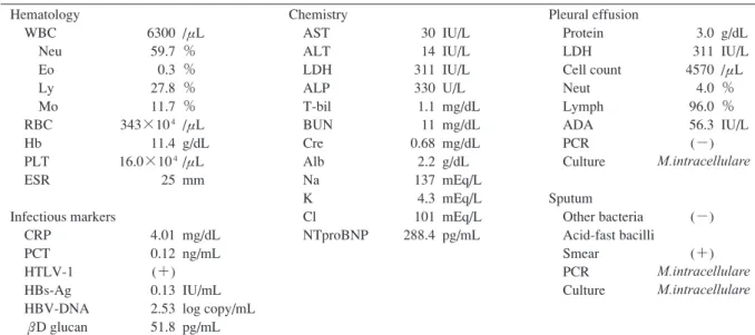
Fig. 2 Thoracoscopy revealed white fibrinous membranes (2-a) and multiple nodules (2-b) on the surface of the visceral pleura.
Fig. 3 Histopathology of pleura.
The pleural biopsy specimen showed only granulation tissue, without granuloma and Langhans giant cell. Ziehl-Neelsen stain showed no acid-fast bacilli (deta not shown).
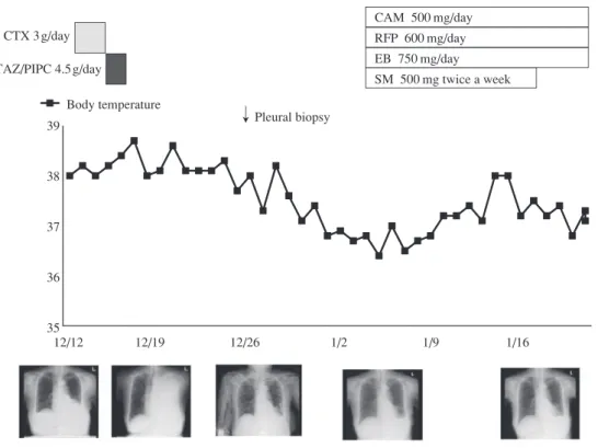
2-a 2-b の培養と PCR で M. intracellulare が陽性で結核菌は陰性 であるため,M. intracellulare による胸膜炎と診断した。 胸腔鏡前に急激に増加した左胸水は胸腔鏡施行時に排液 したが再貯留は認めなかった。確定診断後 1 月 5 日より clarithromycin(CAM)500 mg ⁄日,rifampicin(RFP)600 mg ⁄日,ethambutol(EB)750 mg ⁄日,streptomycin(SM) 500 mg ⁄週・ 2 回による治療を開始した(Fig. 4)。その後 胸痛・咳嗽も消失し胸部 X 線写真上も左胸水の改善を認 めた。胸膜生検後に一度解熱を認めていたが治療開始後 に発熱と末梢神経障害が出現した。SM による副作用と 考え SM のみ中止したところ再度解熱し末梢神経障害も 改善したため退院となった。以後外来で治療継続してい るが胸水の増加や副作用なく経過良好である。 なお,本症例において入院時のββD グルカンが高値で あった。身体所見で舌白苔を認めていたこと,アスペル ギルス抗原は陰性で CT 画像上もアスペルギルス感染を 疑わせる陰影やニューモシスチス肺炎を疑わせるすりガ ラス陰影は認めなかったことから,ββD グルカン高値は 口腔カンジダ由来と考えられた。フルコナゾール 100 mg ⁄日の 1 週間投与で舌白苔の消失を認め ββD グルカン は正常範囲内に戻っていた。 考 察 本邦における肺 NTM 症での胸膜炎の頻度は 3 ∼ 6 % と報告されており3) 5),比較的まれな疾患である。これ までの報告は胸水の培養で診断されており,胸膜炎症例 での肉眼的所見や病理像に関する検討は検索したかぎり では認めなかった。そこで,同じ抗酸菌症である結核性 胸膜炎13症例について検討した杉山ら8)の論文によると, 病初期から経過が進むにつれて,広範な発赤・腫脹が出 で 2 回以上 Mycobacterium intracellulare 陽性であり,舌 区の病変は肺 M. intracellulare 症と診断した。胸水の PCR では結核も Mycobacterium avium complex(MAC)も陰性 であり,確定診断目的に局所麻酔下胸腔鏡を施行した。舌 区近傍(Fig. 2-a)では臓側胸膜・壁側胸膜の間に白色 の線維性の癒着を認め白色の肥厚性変化を伴い,横隔膜 周囲では全体的に発赤腫脹しており,臓側胸膜・壁側胸 膜に大小不同の小結節が散在していた(Fig. 2-b)。横隔 膜内側では小結節が一部融合しており白色の肥厚性変化 を伴っていた。小結節は容易に剝離可能であり病理検査 および培養検査を施行した。病理組織(Fig. 3)で肉芽腫 の形成や Langhans 巨細胞は認めず,肉芽組織のみであっ た。抗酸菌染色では菌体は確認できなかった。生検組織
Fig. 4 Clinical course of infection
cefotaxime: CTX, tazobactam/piperacillin: TAZ/PIPC, clarithromycin: CAM, rifampicin: RFP, ethambutol: EB, streptomycin: SM
SM 500 mg twice a week EB 750 mg/day RFP 600 mg/day CAM 500 mg/day 12/12 12/19 12/26 1/2 1/9 1/16 35 36 37 38 39 CTX 3g/day TAZ/PIPC 4.5g/day Body temperature
↓
Pleural biopsy 現する時期(Ⅰ期:発赤腫脹期),壁側胸膜に粟粒大の 白色小結節が散布する時期(Ⅱ期:結節播種期),白色 のフィブリン沈着が索状または膜状に広がる時期(Ⅲ 期:線維素析出期),白色性肥厚性変化が進行し胸腔内 に癒着が出現する時期(Ⅳ期:胸膜肥厚期)の順で胸腔 鏡所見が変化すると報告している。また,病理診断率は 病初期の発赤腫脹部,白色小結節では 88.8%,90% と高 いが白色肥厚性変化部では 44.4% と低かったと報告して いる。本症例では肺 NTM 病変を認める舌区の近傍では 杉山らの結核性胸膜炎における線維素析出期に相当する 白色の索状の構造物や胸膜肥厚期に相当する白色性肥厚 性変化を認め,病変から遠い横隔膜周囲では比較的病初 期(発赤腫脹期,結節播種期)と思われる発赤腫脹,小 結節が認められた。部位によって時相の違う胸腔鏡所見 を認めており,気胸に際して舌区から直接M. intracellulare が播種し,その後胸腔内に拡がった可能性が示唆された。 肺 MAC 症に合併する胸膜炎の病態は Kotani ら9)の報告 では肺病変の直接波及または気胸による胸腔穿破である と推定されている。また水谷ら10)は肺 MAC 症の病巣が 胸膜直下にある場合,病初期であっても胸腔穿破により 気胸を発症することがあると報告している。本症例では 病巣が舌区の胸膜直下に達していたことに加え,急性 B 型肝炎に対して投与したステロイドが気胸および胸膜炎 の発症に影響した可能性が考えられる。すなわち,ステ ロイドによる免疫抑制で舌区末梢の病変が悪化したこと で炎症が胸膜へ進展し気胸を発症し,その際に M. intra-cellulareが胸腔内へ播種し胸膜炎が発症したと推測され る。また,本例では短期間にステロイドが漸減,中止と なったことで胸腔内に播種した菌体に免疫が過剰に反応 し多量の胸水が出現した可能性も考慮される。本症例で は胸腔鏡の施行に際し胸水を一度排液したのみで解熱を 得た。これは排液により胸腔の M. intracellulare の抗原量 が減少したことによる可能性が示唆される。 近年,本邦においても肺 NTM 症例の罹患率が急激に 増加していることが報告されている11)。肺 NTM 症にお ける気胸の合併率は 2.2∼2.3% と報告されており12) 13), それに伴う胸膜炎の発症も今後増加する可能性があるこ とを念頭に入れるべきである。 著者の COI(conflicts of interest)開示:本論文発表内 容に関して特になし。 文 献 1 ) 久世文幸:非定形抗酸菌症. 呼吸. 1985 ; 4 : 1298 1317. 2 ) Christensen EE, Dietz GW, Ahn CH, et al.: Pulmonarymanifestations of Mycobacterium intracellularis. Am J Roentogenol. 1979 ; 133 : 59 66.
3 ) 佐渡紀克, 中村保清, 北 英夫:肺非結核性抗酸菌症 に合併した胸膜炎の臨床的検討. 結核. 2014 ; 89 : 821 824.
Abstract A case of pleurisy from Mycobacterium
intra-cellulare is rare, and there have been no reports on the details of thoracoscopic findings. Here, we report a case of pleurisy caused by M.intracellulare in an 84-year-old woman, with a history of acute hepatitis B virus infection. Following her treatment of acute hepatitis B with steroid pulse and maintenance therapy, she visited the emergency department complaining of high fever and chest pain on her left side. She was diagnosed with pneumonia and parapneumonic effusion due to a chest X-ray showing left-sided pleural effusion, and admitted to our department for treatment. The analysis of pleural effusion revealed an increasing level of adenosine deaminase, therefore, tuberculous pleurisy was the tentative diagnosis. However, chest computed tomography findings on the lingular segment of the left lung suggested a nontuber-culous mycobacteria pulmonary infection. Thoracoscopy was performed under local anesthesia. The findings of thoracoscopy showed multiple nodules and small clusters of fibrinous mem-brane adhesion in both the visceral and parietal pleurae. After pleural biopsy was performed, M.intracellulare was identified
using polymerase chain reaction and bacterial culture. There-after, this case was diagnosed as M.intracellulare pleuritis. After combination therapy with clarithromycin, rifampicin, ethambutol and streptomycin for M.intracellulare pulmonary disease, the patient improved and clinical symptoms subsided.
Key words: Nontuberculous mycobactria, Pleurisy,
Thora-coscope, Adenosine deaminase
Department of Infectious, Respiratory, and Digestive Medi-cine, Control and Prevention of Infectious Diseases, Faculty of Medicine, University of the Ryukyus
Correspondence to: Shusaku Haranaga, Department of Infec-tious, Respiratory, and Digestive Medicine, Control and Prevention of Infectious Diseases, Faculty of Medicine, University of the Ryukyus, 207 Uehara, Nishihara-cho, Okinawa 903_0215 Japan.
(E-mail: [email protected]) −−−−−−−−Case Report−−−−−−−−
A CASE OF MYCOBACTERIUM INTRACELLULARE PLEURISY
DIAGNOSED BY PLEURAL BIOPSY
Akira SHINZATO, Shusaku HARANAGA, Kazuya MIYAGI, Masao TATEYAMA, and Jiro FUJITA
complex による胸膜炎の 2 例. 日呼吸会誌. 2010 ; 48 : 151 156. 5 ) 市木 拓, 植田聖也, 渡邊 彰, 他:胸膜炎を合併し た肺非結核性抗酸菌症の検討. 日呼吸会誌. 2011 ; 49 : 885 889. 6 ) 萩原恵里, 関根朗雅, 佐藤友英, 他:気胸を合併した 肺Mycobacterium fortuitum感染症の1例. 感染症学会誌. 2008 ; 82 : 73 76.
7 ) Gribetz AR, Damsker B, Marchevsky A, et al.: Nontuber-culous mycobacteria in pleural fluid. Chest. 1985 ; 87 : 495 498.
8 ) 杉山昌裕, 立川壮一, 堀口高彦, 他:結核性胸膜炎に対 する局所麻酔下胸腔鏡検査の有用性. 気管支学. 2001 ; 23 : 336 340.
9 ) Kotani K, Hirose Y, Endo S, et al.: Surgical treatment
of atypical Mycobacterium intracellulare infection with chronic empyema : a case report. J Thoracic Cardiovasc Surg. 2005 ; 130 : 907 908.
10) 水谷尚雄, 萱野公一:肺末梢孤立性の Mycobacterium avium intracellulare complex感染症による続発性自然気 胸の1例. 日呼外会誌. 2008 ; 22 : 943 947.
11) Namkoong H, Kurashima A, Morimoto K, et al.: Epi-demiology of pulmonary nontuberculous mycobacterial disease, Japan. Emerg Infect Dis. 2016 ; 22 : 1116 1117. 12) 荻原恵理, 椎原 淳, 榎本崇宏, 他:気胸を合併した非
結核性抗酸菌症16例の臨床的検討. 日呼吸会誌. 2010 ; 48 : 104 107
13) 高橋伸政, 星 永進, 鍵山奈保, 他:気胸を合併した抗 酸菌症症例の検討. 結核. 2012 ; 87 : 649 653.