Studies on Intermolecular Interactions,
Crystallization Behavior and Highly
Ordered/Intermediate Structures of
Poly(3-hydroxybutyrate) in the Blends and
Ultrathin Films by Infrared Spectroscopy and
Grazing Incidence X-ray Diffraction
学位名
博士(理学)
学位授与機関
関西学院大学
学位授与番号
34504甲第644号
Studies on Intermolecular Interactions, Crystallization
Behavior and Highly Ordered/Intermediate Structures
of Poly(3-hydroxybutyrate) in the Blends and Ultrathin
Films by Infrared Spectroscopy and Grazing Incidence
X-ray Diffraction
March 2017
A Dissertation
by
Khasanah
Department of Chemistry
Graduate School of Science and Technology
Kwansei Gakuin University
i
Contents
List of Symbols and Abbreviations I
General Introduction 1
Chapter 1: Intermolecular Hydrogen Bondings in the Poly(3-hydroxybutyrate) and Chitin Blends: Their effects on the Crystallization behavior and Crystal Structure of Poly(3-hydroxybutyrate)
Abstract 39
Introduction 40
Experimental Section 44
Results and Discussion 46
Conclusions 55
References 59
Appendix 65
Chapter 2: Evolution of Intermediate and Highly Ordered Crystalline States under Spatial Confinement in Poly(3-hydroxybutyrate) Ultrathin Films
Abstract 75
Introduction 76
Experimental Section 80
ii
Conclusions 93
References 94
Appendix 100
Chapter 3: Crystallization Behavior of Ultrathin Poly(3-hydroxybutyrate) Films in Blends with a Small Amount of Poly(L-lactic Acid): Correlation between Molecular Weight of Poly(L-lactic Acid) and Film Thickness
Abstract 104
Introduction 111
Experimental Section 114
Results and Discussion 116
Conclusions 128
References 129
Appendix 140
Acknowledgements 146
I
List of Symbols and Abbreviations
(020)H (020)L 2D-GIXD CAB DSC FWHM GIXD HBs HFIP IPHB IR IRRAS Ii Mw PFA PHB PLLA weight fraction
the (020) reflection at lower 2θ
the (020) reflection at lower 2θ
two-dimensional grazing incidence X-ray Diffraction
cellulose acetate butyrate
differential scanning calorimeter
full-width at half maximum
grazing incidence X-ray Diffraction
hydrogen bonds
1,1,1,3,3,3-hexafluoro-2-propanol
integrated intensity of (020) peak of neat PHB
infrared spectroscopy
infrared-reflection absorption spectroscopy
integrated intensity of (020) peak in the blends
molecular weight
perfluoroalkoxy
poly(3-hydroxybutyrate)
II PVPh Tc Tg Tm WAXD Xc free C=O inter C=O inter C=O chitin intra C=O intra C=O chitin
wt % ΔHm ΔH°PHB θc poly(4-vinyl phenol) crystallization temperature
glass transition temperature
melting temperature
wide-angle X-ray diffraction
degree of crystallinity
free C=O group of PHB (without HBs)
intermolecularly HB(s) C=O group of PHB with chitin
intermolecularly C=O∙∙∙HN HBs within chitin
intramolecularly HB(s) C=O group within PHB
intramolecularly C=O∙∙∙HO HBs within chitin
weight percentage
measured enthalphy
enthalphy of the neat 100% PHB
1
GENERAL INTRODUCTION
1. Scope of This Thesis
This thesis is mainly concerned with the study of intermolecular hydrogen bonding
interactions, crystallization behavior and crystal structures of poly(3-hydroxybutyrate)
[PHB] and blends. These three aspects are closely interrelated in determining the final
physical and mechanical properties of a polymer. Thus, they have been gaining much
attention for a long time as very important research themes in polymer science. Moreover,
controlling the crystallinity of such semicrystalline PHB is also crucial in order to fit the
best condition for practical applications. One of the most simple and economic approaches
to modify the properties of a polymer is blending. In this thesis, PHB was blended with
two biodegradable polymers, chitin and poly(L-lactic acid) PLLA, to control the
crystallinity of PHB. Several measurements were used in this thesis are differential
scanning calorimetry (DSC), infrared spectroscopy (IR), and wide-angle X-ray diffraction
(WAXD). Two surface sensitive techniques were specially employed to investigate the
crystallization and crystal structure of PHB ultrathin films, i.e. infrared-reflection
absorption spectroscopy (IRRAS) and grazing incidence X-ray diffraction (GIXD).
The novelty and originality of this thesis can be described as follows:
2
blends was revealed through the intensive analysis of various ratios of blends with the
temperature dependence of IR spectroscopy combined with the results obtained from
DSC and WAXD measurements. We systematically analyzed the IR spectra of PHB
including the intensity change, full width at half maximum (FWHM), and wavenumber
shift of C=O bands with composition and temperature dependences. Similarly, the
change of amide I and II bands of chitin is also discussed as well. The effect of
intermolecular hydrogen bonding formation on crystallization and crystal structure of
PHB also carefully discussed by monitoring the change of DSC and WAXD profiles of
PHB.
2. Through the measurement and analysis of temperature-dependent IRRAS and GIXD,
we proposed two different ordered of crystalline structures in PHB ultrathin films: less
ordered and highly ordered structure. The existence of less ordered structure was
obviously recognized in the intermediate state which generally difficult to find in bulk
PHB. The transformation from intermediate state to highly ordered state was
meticulously examined from the integrated intensity change of their corresponding IR
bands as a function of temperature. Moreover, the nucleation site, growth and preferred
orientation of crystallites PHB were elucidated from temperature-dependent of
2D-GIXD profiles.
3
interpreted through investigating of various molecular weight of PLLA and two
different film thicknesses using surface sensitive IRRAS and GIXD measurements. The
results exposed that the inhibition of crystallization of PHB by PLLA strongly depends
on the molecular weight of PLLA and thickness confinement. The crystallization
behavior of PHB in the PHB/PLLA ultrathin films behaves relatively inverse from the
PHB/PLLA bulk. The present of a very confined environment by reducing the film
thickness seems to enhance the miscibility of PHB and PLLA in the blends. Apart from
the molecular weight of PLLA and thickness confinement dependences, phase
separation due to the presence of free surface effect, entanglement of PHB and
aggregation of small molecules of PLLA are also found to be important factors that
influence the ability of a small amount of PLLAs in inhibiting the crystallization of
PHB.
2. Introduction of PHB, Chitin and PLLA 2.1 PHB
In recent years, biodegradable polymers have been gaining considerable attention
along with increasing global concern over the harmful effects of plastic derived from
petroleum in the environment. Biodegradable polymers can degraded naturally in the
environment into water and carbon dioxide, thus, they are ideal alternative for replacing
4
poly(3-hydroxybutyrate) [PHB] that belong to polyhydroxyalkanoates (PHAs) class
polyester. PHAs is produced from various bacteria in the storage granules as carbon and
energy.1-5 On the other hand, PHAs is very potential to use in the wide-range applications
because of their advantageous characteristics, such as biodegradable, biocompatible,
insoluble in water and impermeable to oxygen, nontoxic, piezoelectric, thermoplastic
and/or elastomeric.6-8
PHB was firstly isolated and characterized from Bacillus megaterium bacteria by
Maurice Lemoigne in 19259, however, its commercial production scale had wait until the
early of 1960s.1 The chemical structure of PHB is shown in Figure 1a. PHB is a
semicrystalline polymer where the crystalline molecules are arranged in an orthorhombic
structure. Yokouchi et al.10 and Marchessault et al.11 reported that the orthorhombic has
two-left handed helices along the antiparallel orientation in accordance with the
P212121-D42 space group. The crystal lattice parameters are determined with a = 5.76 Å, b
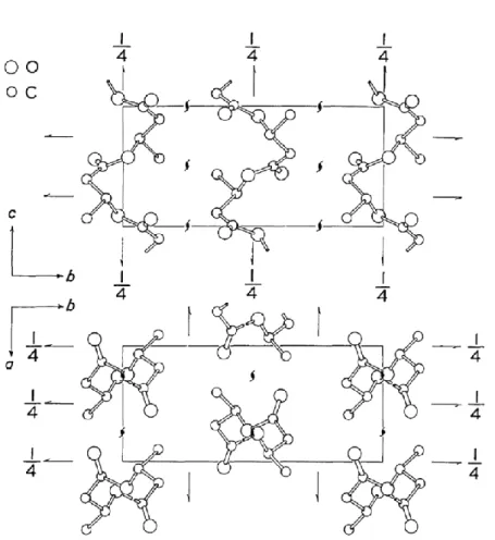
= 13.20 Å and c = 5.96 Å (fiber axis).10-12 Figure 2 is depicted the crystal structure of PHB
reproduced from Ref. 10.
In the PHB crystalline, there was found weak hydrogen bonds between methyl and
carbonyl groups (CH3···O=C).The formation of CH3···O=C hydrogen bond was firstly
proposed by Sato et al.13 on the basis of IR spectra study of an antisymmetric C−H
5
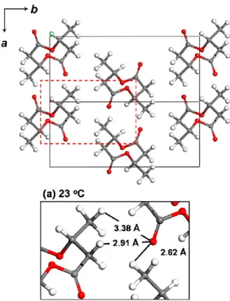
reinforced by WAXD study and chemical quantum analysis.14-16 Recently, Tashiro et al.17
reinvestigated the existence of this hydrogen bonding in the PHB α-form crystal through
the advance X-ray approach. It was reported that the abnormally short distance of methyl
group to the oxygen atom of C=O group lead the formation of CH3···O=C hydrogen bond.
The shortest H∙∙∙O distance exhibits in the C−H···O=C hydrogen bonds is found to be
2.62 Ǻ which shorter than the expected value of normal van der Waals distance (see Figure
3). Moreover, the direction of these C–H···O=C hydrogen bonds is proposed to be almost
parallel to the direction of the chain folding along the a-axis. Therefore, the presence of
these hydrogen bonds may responsible for stabilizing the chain folding in the lamellae
structure of PHB. Figure 4 displays the model of lamella structure with intermolecular
hydrogen bond interactions of PHB crystal proposed by Sato et al.
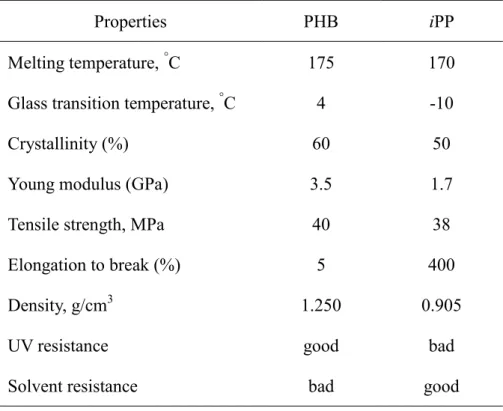
PHB has physical and mechanical properties similar to those of commercial plastics of
isotactic polypropylene (iPP),18,19 as tabulated in Table 1. However, PHB is rigid, brittle
and thermally unstable due to highly crystallinity and narrow processability temperature
that caused difficulty in the conventional processing.20-22 In order to modify those
unfavorable properties and improve the physical and mechanical properties,
copolymerization and blending approaches often used to obtain the desired properties of
PHB. The unit and composition of comonomer greatly affect the physical and
6
been developed are poly(hydroxybutyrate-co-hydroxyvalerate) [P(HB-co-HV)],26-31
poly(hydroxybutyrate-co-hydroxyhexanoate) [P(HB-co-HHx)]32-36 and
poly(hydroxybutyrate-co-hydroxypropionate) [P(HB-co-HP)].37-39
On the other hand, blending technique is more convenient and low cost for creating
new materials by combining two or more polymers. The important characteristic in
polymer blends is miscibility. According to thermodinamical behavior and compatibility
between two polymers, miscible blend refers to a single phase system (homogeneous
phase) which is equivalent with polymer-polymer solution (mix on a molecular level),
whereas, immiscible blend refers to separate phase (inhomogenous system) that do not mix
on a molecular level.40 PHB was reported to be miscible by blending with poly(vinyl
acetate) (PVAc),41-43 poly(vinyl alcohol) (PVA),44-47 poly(ethylene oxide) (PEO),48-51
cellulose acetate butyrate (CAB),52-55 poly(epichlorohydrin) (PECH),56-59 and
poly(ethylene glycol) (PEG).60 PHB formed immiscible systems by blending with
polylactic acid (PLA),61-65 poly(methylene oxide) (PMO),66 poly(butylene succinate)
(PBS)67 and polycaprolactone (PCL).68
2.2 Chitin
Chitin is the second most abundant polysaccharide in the nature after cellulose that
7
insects and the internal shells of cephalopods. It is also a biodegradable and biocompatible
polymer with excellent absorbability and non-toxicity. The chemical structure of chitin is
similar with cellulose, except a NHCOCH3 group replaces the hydroxyl group, as displays
in Figure 5a. Moreover, the infrared spectra of chitin and cellulose is also similar since
their chains conformation are same.69,70
Chitin naturally exhibits three crystalline allomorphs as -, - and -chitin. The most
abundant and stable one is -chitin which packed in the orthorhombic space group P212121
with a = 0.474, b = 1.032 and c = 1.886 nm.71 The -chitin has excessive intramolecular
and intermolecular hydrogen bonds that causes difficulty to dissolve in many solvents, see
figure 5b.72 The -chitin crystal consists of two antiparallel molecules per unit cell where
its chains arranged in sheets tightly held by a number of intra-sheet hydrogen bonds along
the a parameter of unit cell dominated by C=O···NH hydrogen bond. The inter-sheet
hydrogen bonds exist along the b parameter of the unit cell.73 Chitin has amide and
hydroxyl groups which can be expected to form intermolecular hydrogen bonding with
carbonyl or methyl groups of PHB. Therefore, chitin is beneficial to improve the properties
of PHB. Chitin and its derivatives has been reported in a wide variety of biomedical
applications, such as tissue engineering,74 suture,75 wound dressing,76 drug delivery,77
wastewater treatment,78,79 cosmetics and pharmaceutical fields,80 agriculture and food
8
2.3 PLA
Poly(lactic acid) [PLA] is a biodegradable aliphatic polyester that can be produce from
renewable resources, such as starch (from corn and potatoes) and sugar (from sugar cane
and beets).82-84 It can be readily broken down through a simple hydrolysis reaction into
water and carbon dioxide. PLA has excellent mechanical properties which is comparable or
even superior than those of petroleum-based polymers, especially the biocompatibility,
high strength, high elasticity modulus, thermoplastic, molding capability, and
printability.84-88 However, PLA is rigid, brittle, and thermally unstable because it deforms
at temperatures in excess of its glass transition temperature.88 Therefore, several
modification methods are often applied to improve those drawbacks aim to suit the
application purpose, including blending,89-91 copolymerization92,93 and using
plasticizers.94-96 In addition, the change of molecular weight, crystallinity, chain orientation,
and stereochemistry of PLA will also greatly affects the physical and mechanical properties
of PLA.88,97,98
Depending on the stereochemistry and thermal history, PLA can be either
semicrystalline or amorphous. PLA has two optically active seteroisomers, L (+)-LA and
D(−)-LA (see Figure 6), but it also can exist as a racemic mixture, DL. The arrangement of
the stereochemical L- and D-lactic acid structures can control almost all the properties of
9
[PLLA] and poly(D-lactic acid) [PDLA] are semicrystalline, whereas the racemic
poly(DL-lactic acid) [PDLLA] is amorphous.99,100 The highly crystalline PLA can be
obtained by the addition of low D or L content (< 2 %), while the presence of relatively
high D or L content (> 20%) yields the amorphous one.101,102
The semicrystalline PLLA has three polymorphism forms, α, β, and γ depending on the
method of preparation. The α-form is the most common type which can be prepared by
melt or cold crystallization. Its crystals are packed into an orthorhombic P212121 space
group with a = 10.66 Ǻ, b = 6.16 Ǻ and c = 28.88 Ǻ containing two antiparallel chains in
103 helix conformation.103 The β-form can be obtained at a high draw ratio and high
drawing temperature. The β-form crystal is considered to have a frustrated structure
packing of three 31 helices in a trigonal unit cell with a = b = 10.52 Ǻ and c = 8.8 Ǻ, space
group P32.104 The third type, γ-form was produced by epitaxial crystallization on
hexamethylbenzene substrate, containing two antiparallel helices which packed in an
orthorhombic unit cell of parameters a = 9.95 Ǻ, b = 6.25 Ǻ= and c = 8.8 Ǻ.105
In the practical application, PLA has been widely used for a long time in biomedical
field due to its excellent bioresorbability and biocompatibility in the human body, such as
suture and orthopaedic fixation.106-111 Since the PLA production cost can be reduced and
the technique to improve the properties of PLA is also developed tremendously, PLA has
10
Currently, PLA has been commercially used in many applications, as packaging
material,112,113 fiber/textile,114,115 coating,116 drug delivery,117 and foamed article.118,119
3. Polymer Thin and Ultrathin Films
Recently, polymer thin and ultrathin films have attracted increasing interest in both
research and application points of view. The confinement effect from the surface and
interface in polymer thin and ultrathin films greatly affected their physical properties,
which is considerably differ compare to their bulk form. Therefore, investigation of
polymer films under spatial confined environment will certainly provide a new insight in
the field of polymer science. On the other hand, the preferred characteristic of current
devices that is lighter, smaller, and thinner, also contributed to the increase of polymer thin
and ultrathin films technology.
The term of thin films is commonly used to refer to the films having a thickness of up
to 1000 nm, however, sometimes it also used to address the ultrathin films. In order to
distinguish the use of these terms, Ma et al.120 roughly classified the films thickness into
three categories: The first category includes the films with thicker than several hundred
nanometers (usually labeled as thin films). The second category includes the films with the
thickness close to the polymer coil size but less than 100 nm (termed as ultrathin films).
11
approaching a quasi-two-dimensional state (the so-called monolayer).121 Those confined
films with restricted geometries can be considered to be the quasi two-dimensional (2D)
system with one-dimensional (1D) confinement normal to the substrate. Along 1D
confinement on the substrate, the lamellae have preferential orientation that can be either
edge-on lamellae with the chain axis parallel to the substrate or flat-on lamellae with the
chain axis normal to the substrate. Figure 7 shows the illustration of edge-on and flat-on
lamellae. The free surface is generally dominated by edge-on lamellae that form at low
temperature,122,123 whereas the flat-on lamellae predominantly form at the interface at high
temperature.124,125 In thin films, edge-on lamellae are usually observed as the free surface
effect is predominant. Further decreasing the film thickness, both edge-on and flat-on
lamellae can be found in ultrathin films. In monolayer films, a typical diffusion-limited
crystal usually grow because the interface effect plays major role to control the growth of
the crystal, so flat-on lamellae is more favorable.120-122, 126-128 Actually, many factors can
control the lamellae orientation in the thin films, however, the thickness of the films,
crystallization temperature, and surface chemistry of the substrate are the most important
factors.121
As mentioned above, except for the lamellae orientation, the confinement and
surface/interface effects can affect almost all the physical properties of polymer thin and
12
mobility,132,133 glass transition temperature (Tg),134,135 morphology and phase
behavior,136-138 etc. The crystallinity and the kinetic of crystallization of semicrystalline
polymers were found to decrease in thin and ultrathin films. It seems the main reason is
that the polymer chains hardly to fold in thermodynamically stable nucleus or the lamellae
thickness is close to the films thickness, associated with a possible slowdown of the
diffusion of polymer chains in the melt of thin films.139 For example, the crystallinity of
poly(di-n-hexylsilane) is decreased when the film thickness is less than 50 nm, in fact, the
crystallization is almost inhibited when the thickness below 15 nm as the critical
dimensions of nuclei is difficult to develop with decreasing the film thickness.130,131 The
mobility of polymer chains in the thin and ultrathin films may be differ at its surface and
interface. The mobility of polymer chains is usually increased near the free surface region,
especially for the lower molecular weight polymers, but no obvious change is observed for
higher molecular weight polymers.140,141 In contrast, the mobility of polymer chains at
interface become limited due to the existence of interactions between polymer and solid
substrate. The difference mobility of polymer chains at surface and interface is also closely
related with the shifting of the Tg in thin and ultrathin films.142,143
Recent measurements have been developed to study the behaviors of polymer thin and
ultrathin films. Among them, IR still to be a powerful tool to extract the information about
13
films. IR has several measurement techniques that are adjusted to the kinds of samples. For
investigation of polymer thin and ultrathin films, IRRAS is the most suitable technique to
characterize solid sample with nanometer scale. IRRAS is a surface sensitive technique
where the electric field vector of light undergoes a phase change with the magnitude of
each depends on the polarization of incident light.144 Upon the reflection on the metal
substrate, the electric vector of the light polarized parallel to the plane of incidence
(p-polarized light) gives the signals of up to 90 degrees, whereas, the electric field of the
light polarized perpendicular to the plane of incidence (s-polarized light) shift of 180
degrees which is negligible at all angle of incidence/theta (). The IRRAS reflection on metal substrate is illustrated in Figure 8. In short, this mechanism is known as the surface
selection rule of IRRAS: vibration modes having transition dipole perpendicular to the
surface substrate will appear with enhanced intensity. Therefore, IRRAS is very useful to
observe the conformation and orientation of molecules on the surface of polymer thin and
ultrathin films.
In many experiments, IR technique is often combined with XRD technique to
investigate the structural properties of materials. Similar with IRRAS, one of GIXD
techniques with surface sensitive that suitable for investigation of polymer thin and
ultrathin films is called GIXD. GIXD uses a very small angle of incidence that reflects the
14
along normal and parallel to the substrate can be obtained by measuring both in-plane and
out-of plane geometries. Moreover, the depth of X-ray penetration into the film can be
control by varying the angle of incidence around the critical angle for total reflection
(c).145,146 Therefore, the specific crystalline information along out-of plane and in-plane
directions within different depth can be observed.
The study of PHB thin and ultrathin films has been conducted using the combination
of IRRAS and GIXD techniques. It has been found that the (020) reflection along out-of
plane direction is strongly observed using GIXD measurement. The appearance of
out-of-plane (020) reflection indicated that the edge-on lamellae with b-axis normal to the
substrate surface is the preferred lamellae orientation of PHB crystallites in thin and
ultrathin films.147,148 The formation of edge-on lamellae corresponds to the dominant of
free surface effect with lower nucleation barrier. Furthermore, increasing the annealing
temperature caused the buried interface effect increased, as a result, the lamellar
orientation changed from b-axis normal to substrate surface to the c-axis normal to the
substrate surface (flat-on lamellae).147 Furthermore, the crystallization of PHB is inhibited
when the film thickness decreased to tens of nanometer close to the polymer-substrate
interface.149 The weak intermolecular C−H···O=C hydrogen bonds still observed at 3009
cm-1 in IRRAS spectra of PHB thin films along the a-axis.150
15
films (thickness ~52 nm) using the combination of IRRAS and GIXD. The important
finding in this present study is the evident presence of intermediate state observed in the
melt crystallization process and the crystals transformation from intermediate state into
highly-ordered state with the assistance of thermal energy. Intermediate state is specially
appeared at 1731 cm-1 in IR frequency. It is usually difficult to detect in bulk PHB because
it only appears in the early stage and diminish after the crystallization is finished.151-153 In
chapter 3, the effect of a small amount of PLLA on the crystallization behavior of PHB
ultrathin film is investigated using the various molecular weights of PLLA. In this study,
we described the correlation between the film thickness and the molecular weights of
PLLA from the crystallization of PHB point of view.
4. Outline of each chapter
This thesis consists of three chapters. The outline of each chapter will be described as
follows.
Chapter 1 described the effect of intermolecular hydrogen-bonding interactions
formed between PHB and chitin in the blends on the crystallization behavior and
crystalline structure of PHB. The PHB/chitin blends were studied as a function of
composition and temperature by DSC, WAXD, and IR. We observed the significant
16
DSC curves, WAXD patterns and IR spectra. The temperature-dependent spectral
variations in the C=O stretching were further analyzed by calculating the intensity
changes, full width at half maximum (FWHM) and wavenumber shift. It is found that a
new band appeared at around 17101714 cm-1 which is known as the hydrogen bonded C
=O band in many polymer blends. Therefore, the appearance of this band clearly reveals
the formation of the intermolecular hydrogen bondings in the PHB/chitin blends. We
proposed that the intermolecular interactions formed between C=O groups of PHB and
the O−H and N−H groups of chitin (C=O∙∙∙H−O and C=O∙∙∙H−N) in the amorphous
phase. The formation of these intermolecular hydrogen bondings is crucially responsible
for decreasing the crystallinity of PHB in the blends. However, we found that the
crystalline structure of PHB is not much affected by the addition of chitin.
In Chapter 2, the crystallization behavior and crystalline structure of PHB were
investigated as ultrathin films (thickness 52 nm) using two surface sensitive techniques,
IRRAS and GIXD through heating and melt-cooling processes. Two kinds of crystalline
structures of PHB were observed at 1722 and 1731 cm-1 from the analysis of IRRAS
spectra that correspond to the C=O stretching of highly-ordered and intermediate states,
respectively. Increasing temperature caused the crystals in the intermediate state acquire
sufficient thermal energy to overcome the energy barrier, as a result, the transformation
17
hydrogen bonds of PHB still exist in such ultrathin films along a-axis. Furthermore, the
2D-GIXD results show that the intermediate state was dominant in edge-on-lamellae
configuration where the crystallographic b-axis is normal to the film surface. Meanwhile,
the highly-ordered state was predominant in flat-on lamellae configuration where the
b-axis is parallel to the film surface. Moreover, from a very shallow angle of incidence
measurement which only penetrates 8 nm deep from the surface reveals that the crystals
in the surface region strongly tended to align in an edge-on lamellae configuration.
Chapter 3 reported the effect of a small amount of PLLA on the crystallization
behavior of PHB ultrathin films studied by IRRAS and GIXD. In this study, the correlation
between molecular weight of PLLA and the film thickness was investigated using PLLA
having molecular weight ranging from 300,000710 g mol−1 and two different film
thicknesses, i.e. 30 and 13 nm. The PHB/PLLA ratio is fixed at 80/20 (w/w) for all blends.
The crystallization of PHB has shown a strong dependency on the molecular weight of
PLLA and film thickness. In the 30-nm-thick samples, the crystallization of PHB is
significantly reduced in the blends with molecular weight PLLAs ranging from Mw 23,000
to 13,100 g mol-1, however, the higher (Mw ≥ 50,000 g mol-1) and lower (Mw ≤ 6,900 g
mol-1) molecular weight PLLAs do not significantly affect the crystallization. In contrast,
in the 13-nm-thick films, the crystallization of PHB is remarkably inhibited in the blends
18
small addition of PLLA (Mw ≥ 13,100 g mol−1) altered the crystalline structure of PHB
only in the highly ordered state. However, such PLLAs greatly affect the PHB crystals in
both intermediate and highly ordered states in the films with the thickness of 13 nm. Both
GIXD and IRRAS results revealed some consistency that the lower molecular weights of
PLLA (Mw ≤ 3,600 g mol−1) only slightly affect the crystallinity and crystalline structure of
PHB. Furthermore, several factors such as the presence of free surface and interface effects,
entanglement of PLLA chains and molecular size of PLLA must seriously be taken into
account to comprehend the complex crystallization behavior of PHB in the PHB/PLLA
19
References
1. Hocking, P. J.; Marchessault, R. H. In Biopolymers from Renewable Resources;
Kaplan, D. L. Ed.; Springer: Berlin, 1998; p 220.
2. Muller, H. M.; Seebach, D. Angew. Chem. 1993, 32, 477-502.
3. Anderson, A. J.; Dawes, E. A. Microbiol. Rev.1990, 54, 450-472.
4. Van der Walle, G. A. M.; de Koning, G. J. M.; Weusthuis, R. A.; Eggink G. Adv.
Biochem. Eng. Biotechnol. 2001, 71, 263-291.
5. Byrom, D. In Plastic from Microbes: Microbial Synthesis of Polymers and Polymer
Precursors, Hanser: Munich, 1994; p 5-33.
6. Valappil, S. P.; Misra, S. K.; Boccaccini, A. R.; Roy, I. Expert Rev. Med. Devices
2006, 3, 853-868.
7. Philip, S.; Keshavarz, T.; Roy, I. J. Chem. Technol. Biotechnol. 2007, 82, 233-247.
8. Chen, G. Microbiol. Monogr. 2010, 14, 17–37.
9. Lemoigne, M. Ann. Inst. Pasteur 1925, 39, 144-173.
10. Yokouchi, M.; Chatani, Y.; Todokoro, H.; Teranishi, K.; Tani, H. Polymer 1973, 14,
267-272.
11. Cornibert, I.; Marchessault, R. H. J. Mol. Biol. 1972, 71, 735-756.
12. Okamura, K.; Marchessault, R. H. In Conformation of Biopolymers; Ramachandran,
20
13. Sato, H.; Murakami, R.; Padermshoke, A.; Hirose, F.; Senda, K.; Noda, I.; Ozaki, Y.
Macromolecules 2004, 37, 7203–7213.
14. Sato, H.; Dybal, J.; Murakami, R.; Noda. I.; Ozaki, Y. J. Mol. Struct. 2005, 744-747,
35-46.
15. Sato, H.; Mori, K.; Murakami, R.; Ando, Y.; Takahashi, I.; Zhang, J.; Terauchi, H.;
Hirose, F.; Senda, K.; Tashiro, K.; Noda, I.; Ozaki, Y. Macromolecules 2006, 39,
1525-1531.
16. Sato, H.; Sato, H.; Ando, Y.; Dybal, J.; Iwata, I.; Noda, I.; Ozaki, Y.
Macromolecules 2008, 41, 4305-4312.
17. Wang, H.; Tashiro, K. Macromolecules 2016, 49, 581-594.
18. Zhao, Q.; Cheng, G. In New Frontiers in Polymer Research, R. K. Bregg, Ed.; Nova
Science Publishers, Inc: New York, 2006; p 101.
19. Sudesh, K.; Abe, H.; Doi, Y. Prog. Polym. Sci. 2000, 25, 1503-1555.
20. de Koning, G.J.M.; Lemstra, P.J. Polymer 1993, 34, 4089-4094.
21. Barham, P.J. J. Mater. Sci. 1984, 19, 3826-3834.
22. El-hadi, A.; Schnabel, R.; Straube, E.; Mu, G.; Henning, S. Polym. Test. 2002, 21,
665-674.
23. Mitomo, H.; Morishita, N.; Doi, Y. Macromolecules 1993, 26, 5809-5811.
21
25. Inoue, Y. J. Mol. Struct. 1998, 441, 119-127.
26. Bloembergen, S.; Holden, D. A.; Bluhm, T. L.; Marchessault, R. H. Macromolecules
1986, 19, 2865-2871.
27. Bluhm, T. L.; Hamer, G. K.; Marchessault, R. H.; Fyfe, C. A.; Veregin, R. P.
Macromolecules 1986, 19, 2871-2878.
28. Inoue, Y.; Kamiya, N.; Yamamoto, Y.; Chujo, R.; Doi, Y. Macromolecules 1989, 22,
3800-3802.
29. Scandola, M.; Ceccoruli, G.; Pizzoli, M.; Gazzano, M. Macromolecules 1992, 25,
1405-1410.
30. Mansour, A. A.; Saad, G. R.; Hamed, A. H. Polymer 1999, 40, 5377-5391.
31. Yoshie, N.; Saito, M.; Inoue, Y. Polymer 2004, 45, 1903-1911.
32. Doi, Y.; Kitamura, S.; Abe, H. Macromolecules 1995, 28, 4822-4828.
33. Asrar, J.; Valentin, H. E.; Berger, P. A.; Tran, M.; Padgette, S. R.; Garbow, J. R.
Biomacromolecules 2002, 3, 1006-1012.
34. Tsuge, T.; Kikkawa, Y.; Doi, Y. Sci. Technol. Adv. Mater. 2004, 5, 449-453.
35. Alata, H.; Aoyama, T.; Inoue, Y. Macromolecules 2007, 40, 4546-4551.
36. Hu, Y.; Zhang, J.; Sato, H.; Noda, I.; Ozaki, Y. Polymer 2007, 48, 4777–4785.
22
38. Feng, L.; Watanabe, T.; He, Y.; Wang, Y.; Kichise, T.; Fukuchi, T.; Chen, G.; Doi,
Y.; Inoue, Y. Macromol. Biosci. 2003, 3, 310–319.
39. Fukui, T.; Suzuki, M.; Tsuge, T.; Nakamura, S. Biomacromolecules 2009, 10,
700-706.
40. George, S. C.; Thomas, S. In Handbook of Multiphase Polymer Systems; Boudenne,
A.; Ibos, L.; Candau, Y.; Thomas, S., Eds.; John Willey and Sons, Ltd.: United
Kingdom, 2011; p 124.
41. Greco, P.; Martuscelli, E. Polymer 1989, 30, 1475-1483.
42. An, Y.; Dong, L.; Li, L.; Mo, Z.; Feng, Z. Eur. Polym. J. 1999, 35, 365-369.
43. Madbouly, S. A.; Mansour, A. A.; Abdou, N. Y. Eur. Polym. J. 2007, 43, 3933-942.
44. Azuma, Y.; Yoshie, N.; Sakurai, M.; Inoue, Y.; Chûjô, R. Polymer 1992, 33,
4763-4767.
45. Yoshie, N.; Azuma, Y.; Sakurai, M.; Inoue, Y. J. Appl. Polym. Sci. 1995, 56, 17-22.
46. Ikejima, T.; Cao, A.; Yoshie, N.; Inoue, Y. Polym. Degrad. Stab. 1998, 62, 463-469.
47. Ikejima, T.; Yoshie, N.; Inoue, Y. Polym. Degrad. Stab. 1999, 66, 263-270.
48. Kumagai, Y.; Doi, Y. Polym. Degrad. Stab. 1992, 35, 87-93.
49. Avella, M.; Martuscelli, E. Polymer 1988, 29, 1731-1737.
50. Avella, M.; Martuscelli, E.; Raimo, M. Polymer 1993, 34, 3234-3240.
23
52. El-Shafee, E.; Saad, G. R.; Fahmy, S. M. Eur. Polym. J., 2001, 37, 2091-2104.
53. Scandola, M.; Ceccorulli, G.; Pizzoli, M. Macromolecules 1992, 25, 6441-6446.
54. Ceccoruli, G.; Pizzoli, M.; Scandola, M. Macromolecules 1993, 26, 6722-6726.
55. Pizzoli, M.; Scandola, M.; Ceccorulli, G. Macromolecules 1994, 27, 4755-4761.
56. Paglia, E. D.; Beltrame, P. L.; Canetti, M.; Seves, A.; Marcandalli, B.; Martuscelli,
E. Polymer 1993, 34, 99-1001.
57. Finelli, L.; Sarti, B.; Scandola, M. J. Macromol. Sci.-Pure Appl. Chem. 1997, A34,
13-33.
58. de Lima, J. A.; Felisberti, M. I. Eur. Polym. J. 2006, 42, 602-614.
59. Sadocco, P.; Canetti, M.; Seves, A.; Martuscelli, E. Polymer 1993, 34, 3368-3375.
60. Zhang, Q.; Zhang, Y.; Wang, F.; Liu, L. Chinese J. Mater. Sci. Technol. 1998, 14,
95-96.
61. Furukawa, T.; Sato, H.; Murakami, R.; Zhang, J.; Duan, Y. X., Noda, I.; Ochiai, S.;
Ozaki, Y. Maromolecules 2005, 38, 6445-6454.
62. Zhang, J.; Sato, H.; Furukawa, T.; Tsuji, H.; Noda, I.; Ozaki, Y. J. Phys. Chem. B
2006, 110, 24463-24471.
63. Furukawa, T.; Sato, H.; Murakami, R.; Zhang, J.; Noda, I.; Ochiai, S.; Ozaki, Y.
Polymer 2006, 47, 3132-3140.
24
65. Vogel, C.; Wessel, E.; Siesler, H. W. Biomacromolecules 2008, 9, 523-527.
66. Avella, M.; Martuscelli, E.; Orsello G., Raimo, M.; Pascucci, B. Polymer 1997, 38,
6135-6143.
67. Qiu, Z.; Ikehara, T.; Nishii, T. Polymer 2003, 44, 2503-2508.
68. Lovera, D.; Márquez, L.; Balsamo, V.; Taddei, A.; Castelli, C.; Müller, A. J.
Macromol. Chem. Phys. 2007, 208, 924-937.
69. Tsai, C. S. Biomacromolecules: Introduction to Structure, Function and Informatics.
John Willey and Sons, Inc: Hoboken, New Jersey, 2007; p 166.
70. Walton, A. G.; Blackwell, J. Biopolymers. Academic Press, Inc: New York, 1973; p
477
71. Minke, R.; Blackwell, J. J. Mol. Biol. 1978, 120, 167-181.
72. Kameda, T.; Miyazawa, M.; Ono, H.; Yoshida, M. Macromol. Biosci. 2005, 5,
103-106.
73. Rinaudo, M. Prog. Polym. Sci. 2006, 31, 603-632.
74. Jayakumar, R.; Prabaharan, M.; Nair, S. V.; Tamura, H. Biotechnology Advances
2010, 28, 142–150.
75. Nakajima, M.; Atsumi, K.; Kifune, K. In Chitin, Chitosan and Related Enzymes;
Zikakis, J.P. Ed.; Harcourt Brace Janovich: New York, 1984; p. 407.
25
Nair, S. V.; Jayakumar, R. J. Mater. Sci. Mater. Med. 2010, 21, 807–813.
77. Jayakumar, R.; Nwe, N.; Tokura, S.; Tamura, H. Int. J. Biol. Macromolec. 2007, 40,
175-181.
78. Aly, A. S.; Jeon, B. D.; Park, Y. H. J. Appl. Polym. Sci. 1997, 65, 1939–1946.
79. Crini, G. Prog. Polym. Sci. 2005, 30, 38-70.
80. Hirano, S.; Hirochi, K.; Hayashi, K.; Mikami, T.; Tachibana, H. In Cosmetic and
Pharmaceutical Applications of Polymers; Gebelein, C. G.; Cheng, T. C.; Yang, V. C.
Eds.; Springer: New York, 1991; p 95-104.
81. Kardas, I.; Struszczyk, M. H.; Kurcharska, M.; van den Broek, L. A. M.; van Dam, J.
E. G.; Ciechańska, D. In The European Polysaccharide Network of Excellence
(EPNOE): Research Initiatives and Result; Navard, P. Ed.; Springer: Verlag-Wien,
2012; p 329-364.
82. Kharas, G. B.; Sanchez-Riera, F.; Severson, D. K. In Plastics from Microbe; Mobley,
D. P., Ed.; Hanser Publishers: New York, 1994; pp 93-137.
83. Xiao, L.; Wang, B.; Yang, G.; Gauthier, M. In Biomedical Science, Engineering and
Technology; Ghista, D. N. Ed.; Intech, DOI: 10.5772/23927 , 2012; pp 247-282.
84. Garlotta, D. J. Polym. Environ. 2001, 9, 63-83.
85. Hartmann, M. H. In Biopolymers from Renewable Resources; Kaplan, D. L. Ed.;
26
86. Raquez, J. M.; Habibi, Y.; Murariu, M.; Dubois, P. Polylactide (PLA)-based
nanocomposites. Prog. Polym. Sci. 2013, 38, 1504-1542.
87. Kulkarni, R. K.; Moore, E. G.; Hegyeli, A. F.; Leonard, F. J. Biomed. Mater. Res.
1971, 5, 169-181.
88. Zhang, J-F.; Sun, X. In Biodegradable Polymers for Industrial Applications; Smith, R.
Ed.; Woodhead Publishing Limited: Cambrige England, 2005: pp 251-281.
89. Wang, J.; Zhai, W.; Zheng, W. J. Polym. Environ. 2012, 20, 528-539.
90. Todo, M.; Takayama, T. In Biomaterials-Physics and Chemistry; Pignatello, R. Ed.;
InTech 2011: pp 375-394.
91. Mohapatra, A. K.; Mohanty, S.; Nayak, S. K. Polym. Compos. 2014, 35, 283-293.
92. Plikk, P.; Målberg, S.; Albertsson, A-C. Biomacromolecules 2009, 10, 1259-1264.
93. Tyson, T.; Finne-Wistrand, A.; Albertsson, A-C. Biomacromolecules 2009, 10,
149-154.
94. Wang, R.; Wan, C.; Wang, S.; Zhang, Y. Polym. Eng. Sci. 2009, 49, 2414-2420.
95. Ljungberg, N.; Wesslén, B. Polymer 2003, 44, 7679-7688.
96. Martin, O.; Avérous, L. Polymer 2001, 42, 6209-6219.
97. Dorgan, J. R.; Braun, B.; Wegner, J. R.; Knauss, D. M. In Degradable Polymers and
Materials; Khemani, K., Scholz, C. Eds.; American Chemical Society: Washington
27
98. Sin, L. T.; Rahmat, A. R.; Rahman, W. A. W. A. Polylactic Acid: PLA Biopolymer
Technology and Applications. Elsevier: Oxford, United Kingdom, 2012; pp 109-131
and 177-215.
99. Jain, R. J. Biomaterials 2000, 21, 2475-2490.
100. Urayama, H.; Kanamori, T.; Fukushima, K.; Kimura, Y. Polymer 2003, 44,
5635-5641.
101. Lunt, J.; Shafer, A. L. J. Ind. Text. 2000, 29, 191-205.
102. Groot, W.; Krieken, J. V.; Sliekersl, O.; Vos, S. D. In Poly(lactic acid): Synthesis,
Structure, Properties, Processing, and Application; Auras, S., Lim, L-T., Selke, S. E.
M., Tsuji, H. Eds.; John Wiley & Sons, Inc: Hoboken, New Jersey, 2010; pp
141-154.
103. Sasaki, S.; Asakura, T. Macromolecules 2003, 36, 8385-8390.
104. Puiggali, J.; Ikada, Y.; Tsuji, H.; Cartier, L.; Okihara, T.; Lotz, B. Polymer 2000, 41,
8921-8930.
105. Cartier, L.; Okihara, T.; Ikada, Y.; Tsuji, H.; Puiggali, J.; Lotz, B. Polymer 2000, 41,
8909-8919.
106. Kulkarni, R. K.; Pani, K. C.; Neuman, C.; Leonard, F. Arch. Surg. 1966, 93, 839-843.
107. Cutright, D. E.; Hunsuck, E. E.; J. Oral Surg. 1971, 31, 134- 139.
28
109. Albertsson, A-C.; Varma, I. K. Biomacromolecules 2003, 4, 1466-1486.
110. Ikada, Y.; Tsuji, H. Macromol. Rapid Commun. 2000, 21, 117-132.
111. Suzuki, S. Ikada, Y. In Poly(lactic acid) Synthesis, Structures, Properties, Processing,
and Applications; Auras, R., Lim, L-T., Selke, S. E. M., Tsuji, H. Eds.; John Wiley &
Sons, Inc.: Hoboken, New Jersey, 2010; pp 445-456.
112. Tawakkal, I. S. M. A.; Cran, M. J.; Miltz, J.; Bigger, S. W. J. Food Sci. 2014, 79,
R1477-R1490.
113. Obuchi, S.; Ogawa, S. In Poly(lactic acid) Synthesis, Structures, Properties,
Processing, and Applications; Auras, R., Lim, L-T., Selke, S. E. M., Tsuji, H. Eds.;
John Wiley & Sons, Inc.: Hoboken, New Jersey, 2010; pp 457-467.
114. Henton, D. E.; gruber, P.; Lunt, J.; Randall, J. In Natural Fibers, Biopolymers, and
Biocomposite; Mohanty, A. K., Misra, M., Drzal, L. T. Eds.; CRC Press: Boca raton,
Florida, 2005; pp 527-578.
115. Mochizuki, M. In Poly(lactic acid) Synthesis, Structures, Properties, Processing, and
Applications; Auras, R., Lim, L-T., Selke, S. E. M., Tsuji, H. Eds.; John Wiley &
Sons, Inc.: Hoboken, New Jersey, 2010; pp 141-154.
116. Whiteman, N. “2000 Polymers, Laminations and Coatings Conference”, Chicago, IL
2000, pp 631-635.
29
118. Mikos, A. G.; Thorsen, A. J.; Czerwonka, L. A.; Bao, Y. Langer, R. Polymer 1994, 35,
1068-1077.
119. Fang, Q.; Hanna, M. A. Creal Chem. 2000, 77, 779-783.
120. Ma, Y.; Hu, W.; Reiter, G. Macromolecules 2006, 39, 5159-5164.
121. Liu, Y-X.; Chen, E-Q. Coord. Chem. Rev. 2010, 254, 1011-1037.
122. Wang, Y.; Ge, S.; Rafailovich, M.; Sokolov, J.; Zou, Y. Ade, H. Lüning, J.; Lustiger,
A.; Maron, G. Macromolecules 2004, 37, 3319-3327.
123. Muratoğlu, O. K.; Argon, A. S.; Cohen, R. E. Polymer 1995, 36, 2413-2152.
124. Lovinger, A. J.; Keith, H. D. Macromolecules 1979, 12, 919-924.
125. Kovacs, A. J.; Starupe, C. Faraday Discuss 1979, 68, 225.
126. Schönherr, H.; Frank, C. W.; Macromolecules 2003, 36, 1188-1198.
127. Wang, Y.; Chan, C-M.; Ng, K-M.; Li, L. Macromolecules 2008, 41, 2548-2553.
128. Kikkawa, Y.; Abe, H.; Iwata, T.; Inoue, Y.; Doi, Y. Biomacromolecules 2001, 2,
940-945.
129. Frank, C. W.; Rao, V.; Despotopoulou, M. M.; Pease, R. F. W.; Hinsberg, W. D.;
Miller, R. D.; Rabolt, J. F. Science 1996, 273, 912-915.
130. Despotopoulou, M. M.; Miller, R. D.; Rabolt, J. F. Frank, C. W. J. Polym. Sci. Part
30
131. Despotopoulou, M. M.; Frank, C. W.; Miller, R. D.; Rabolt, J. F. Macromolecules
1996, 29, 5797-5804.
132. Frank, B.; Gast, A. P.; Russell, T. P.; Brown, H. R.; Hawker, C. Macromolecules
1996, 29, 6531-6534.
133. Lin, E. K.; Kolb, R.; Satija, S. K.; Wu, W. Macromolecules 1999, 32, 3753-3757.
134. Tsui, O. K. C.; Russell., T. P.; Hawker, C. J. Macromolecules 2001, 34, 5535-5539.
135. Yang, C.; Onitsuka, R.; Takahashi, I. Eur. Phys. J. E. 2013, 36, 66.
136. Abe, H.; Kikkawa, Y.; Iwata, T.; Aoki, H.; Akehata, T.; Doi, Y. Polymer 2000, 41,
867-874.
137. Taguchi, K.; Miyaji, H.; Izumi, K.; Hoshino, A.; Miyamoto, Y.; Kokawa, R. Polymer
2001, 42, 7443-7447.
138. Ludwigs, S.; Krausch, G.; Magerle, R.; Zvelindovsky, A. V.; Sevink, G. J. A.
Macromolecules 2005, 38, 1859-1867.
139. Mareau, V. H.; Prud’homme, E. E. In Soft Materials Structure and Dynamics;
Dutcher, J. R., Marangoni, A. G. Eds.; Marcel Dekker: New York, 2005; pp 39-48.
140. Reiter, G. Europhys. Lett. 1993, 23, 579-584.
141. Tanaka, K.; Taura, A.; Ge, S-R.; Takahara, A.; Kajiyama, T. Macromolecules 1996,
29, 3040-3042.
31
143. Inoue, R.; Kanaya, T.; Nishida, K.; Tsukushi, I.; Telling, M. T. F.; Gabrys, B. J.;
Tyagi, M.; Soles, C.; Wu, W-I. Phys. Rev. E 2009, 80, 031802.
144. Finke, S. J. Infrared Reflection-Absorption Spectroscopy of Thin Film Structures.
Iowa State University, 1988, a Ph.D dissertation.
145. Factor, B. J.; Russell, T. P.; Toney, M. F. Macromolecules 1993, 43, 3441-3446.
146. Saraf, R. F.; Dimitrakopoulus, C.; Toney, M. F.; Kowalczyk, S. P. Langmuir 1996,
12, 2802-2806.
147. Sun, X.; Guo, L.; Sato, H.; Ozaki, Y.; Yan, S.; Takahashi, I. Polymer 2011, 52,
3865-3870.
148. Mori, K.; Mukoyama, S.; Zhang, Y.; Sato, H.; Ozaki, Y.; Terauchi, H.; Noda, I.;
Takahashi, I. Macromolecules 2008, 41, 1713-1719.
149. Capitán, M. J.; Rueda, D. R.; Ezquerra, T. A. Macromolecules 2004, 37, 5653-5659.
150. Sato, H.; Murakami, R.; Mori, K.; Ando, Y.; Takahashi, I.; Noda, I.; Ozaki, Y. Vib.
Spectrosc. 2009, 51, 132-135.
151. Zhang, J.; Sato, H.; Noda, I.; Ozaki, Y.; Macromolecules 2005, 38, 4274-4281.
152. Padermshoke, A.; Katsumoto, Y.; Sato, H.; Ekgasit, S.; Noda, I.; Ozaki, Y.
Spectrochim. Acta, Part A 2005, 61, 541-550.
153. Padermshoke, A.; Katsumoto, Y.; Sato, H.; Ekgasit, S.; Noda, I.; Ozaki, Y. Polymer
32
Table 1. Comparison of physical properties of PHB and iPP
Properties PHB iPP
Melting temperature, °C 175 170
Glass transition temperature, °C 4 -10 Crystallinity (%) 60 50 Young modulus (GPa) 3.5 1.7 Tensile strength, MPa 40 38 Elongation to break (%) 5 400 Density, g/cm3 1.250 0.905 UV resistance good bad Solvent resistance bad good
33
(a) (b)
Figure 1. (a) Chemical structures of PHB and (b) accumulated PHAs in bacteria.
Figure 2. Crystal structure of PHB with a = 5.76 Å, b = 13.20 Å and c = 5.96 Å
34
Figure 3. Intermolecular distance of PHB between O atom of C=O and H atom of
CH3 groups calculated at room temperature, reproduced from Ref. 17.
Figure 4. Intermolecular distance of PHB between O atom of C=O and H atom of CH3
groups calculated at room temperature, reproduced from Ref. 15.
35
Figure 5. (a) Chemical structure of chitin, (b) diagram of the hydrogen bonding
structure in the ac projection for -chitin (reproduces from reference 72).
(a)
36
CH
3HOOC
OH
H
HO
COOH
CH
3H
Figure 6. Two seteroisomers of lactic acid.
L (+)-LA D (−)-LA
x
Figure 7. Illustration of (a) edge-on lamella and (b) flat-on lamellae. x and y
indicated the two dimensional directions, while l indicated the lamellar thickness. l fold surface y fold surface l x y
(a)
(b)
37
X
s-polarization
surface electric field
X
p-polarizatio
surface electric field
Figure 8. General schemes of IRRAS reflection on metal substrate
Figure 9. General schemes of (a) out-of plane and (b) in-plane GIXD measurements (a)
38
Chapter 1
Intermolecular Hydrogen Bondings in the Poly(3-hydroxybutyrate) and Chitin Blends: Their Effects on the Crystallization Behavior and Crystal Structure of
39
ABSTRACT
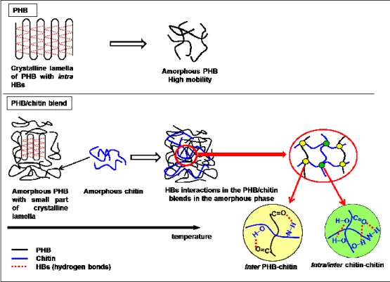
Crystallization behavior and intermolecular hydrogen-bonding interactions of
poly(3-hydroxybutyrate) (PHB)/chitin blends on as-solution cast films were studied as a
function of composition and temperature by differential scanning calorimetry (DSC),
wide-angle X-ray diffraction (WAXD) and infrared (IR) spectroscopy. The significant
changes were observed in the DSC curves, WAXD patterns and IR spectra of the blends
with PHB ≤ 50 wt %. We found that the crystallinity of PHB decreases in the blends,
however, its crystal structure is not much affected by blending with chitin. The appearance
of a new band at around 1710 1714 cm-1 clearly reveals the formation of intermolecular
hydrogen bondings between the C=O groups of PHB and the OH and NH groups of
chitin (C=O···H−O and C=O···H−N). It is very likely that these intermolecular
C=O···H−N and C=O···H−O hydrogen bondings occur in the amorphous phase because of
the reduction in the chain mobility in the blends with increasing chitin content, even above
the melting temperature of PHB. The C=O···H−N and C=O···H−O hydrogen bondings are
formed upon the cleavage of weak C=O···H3C hydrogen bondings of PHB. Thus, the
formation of the C=O···H−N and C=O···H−O hydrogen bondings is accompanied by the
40
1. INTRODUCTION
Polyhydroxyalkanoates (PHAs) are bacterially synthesized polyesters that have
attained great interest as promising biodegradable and biocompatible polymers for
wide-range applications, such as biomedical, agricultural, packaging, pharmaceutical and
paint industries.1-3 Poly(3-hydroxybutyrate) or PHB (Figure 1a) is one of the most studied
PHAs because its physical and mechanical properties are similar to those of commercial
plastic derived from petrochemical, such as isotactic poly(propylene).4-6 However, as a
bacterially synthesized product, PHB has high-ordered stereoregularity that makes it highly
crystalline and yields a narrow temperature window for processability. In addition, the
secondary crystallization on the storage at ambient temperature7 and the pre-existing crack
within the spherulites result in the brittleness of PHB.8,9 Therefore, these problems have
decreased the potential applications of PHB.
Blending technique is one of the most convenient and more economical methods for
making new materials based on the combination of two or more polymers to achieve the
desired properties. Hence, PHB has been reported to be blended with various polymers,
such as poly(vinyl acetate),10,11 poly(l-lactic acid),12,13 poly(ethylene oxide),14 cellulose
esters15 and poly(vinyl alcohol).16-18
Our group has reported a series of studies on PHB, including its copolymers and
41
combination of various experimental techniques, such as infrared, near infrared, Raman
and terahertz spectroscopy, X-ray diffraction (Wide-angle X-ray diffraction; WAXD and
Small-angle scattering; SAXS), two-dimensional correlation spectroscopy and quantum
chemical calculations.12,13,16-32 One of the most important features in our findings is the
existence of weak intramolecular interactions CH···OC hydrogen bonding between the
CH3 group of one helical structure and the CO group of the other helical structure of
PHB.19-23 Furthermore, we reported the formation of intermolecular hydrogen-bonding
interactions in the PHB blends. In the PHB/poly(4-vinylphenol) (PVPh) blends,17 the
intermolecular hydrogen bonds are formed between C=O groups of PHB and OH groups of
PVPh. The exchange between intermolecular and intramolecular hydrogen bonds are found
in those blends with PVPh content higher than the critical composition of 50 wt %. In the
PHB/cellulose acetate butyrate (CAB) blends,27 the weak intermolecular hydrogen bonds
between the OH groups in CAB and the C=O groups in the amorphous part of PHB (O–
H···OC) are formed in the blends with the high CAB content. These intermolecular
interactions in the PHB/CAB blends highly depend on temperature and affect the
crystallization kinetic of PHB in the blends.28 Accordingly, the presence of these hydrogen
bondings in the general polymer blends plays significant effects on the crystallinity,
thermal properties, solubility and miscibility of the polymer blends.33,34
42
systems of PHB and chitin (Figure 1b). Despite the studies on PHB/chitin blends are rare,
chitin was chosen as a blend partner because it is the second most abundant polysaccharide
in the nature after cellulose, which also has biodegradable and biocompatible
properties.35,36 Therefore, it is a highly potential blending source for large scale application
in the future. On the other hand, chitin has hydroxyl and amide functional groups that may
promote the formation of intermolecular hydrogen bondings with carbonyl groups of PHB.
The intermolecular interaction, such as C=O···H−O and C=O···H−N hydrogen bonds, is
an essential factor to reduce the crystallinity of PHB which further will improve the
physical properties of PHB.16,17,27,28 Therefore, the blending of PHB with chitin is expected
to fabricate a good biodegradable and biocompatible polymer with more wide-range
applications.
Previously, Lee et al.37 reported that chitin can improve the mechanical properties of
PVA with specific molecular interactions between C=O and OH. In related on another
study on PHB blending systems, Ikejima et al.38 studied thermal properties and
crystallization behavior of PHB in the blends with chitin and chitosan. They reported that
the crystallization of PHB was suppressed by blending with chitin and chitosan and
suggested the formation of hydrogen bonds between carbonyl groups of PHB and amide
NH groups of chitin from the results of 13C NMR spectra. However, their suggestion was
43
results. So far, the investigation of intermolecular hydrogen bondings in the PHB/chitin
blends has not been fully explored yet.
The present study has aimed to investigate the intermolecular interactions and
crystallization behavior of the PHB/chitin blends with the blend ratio of 100/0, 90/10,
80/20, 70/30, 60/40, 50/50, 40/60, 30/70, 20/80, 10/90 and 0/100 by combination of
various techniques: differential scanning calorimetry (DSC), wide-angle X-ray diffraction
(WAXD) and infrared (IR) spectroscopy. In this paper, our discussion focuses mainly on
the following points: (1) the corroboration of the intermolecular hydrogen-bonding
interactions in the PHB/chitin blends, particularly studies from IR spectra of the blends
through the assignments in the regions of amide I, amide II and C=O stretching; (2) the
evidences for the existence of these intermolecular hydrogen bondings in the amorphous
phase; (3) the effects of intermolecular hydrogen bondings on the crystal structure and
crystallization behavior of PHB in the PHB/chitin blends. The present study shows that the
crystallinity of PHB decreases in the blends with chitin, particularly in the blends with
PHB 50 wt % along with the formation of intermolecular C=O···H−N and C=O···H−O
hydrogen bonds between PHB and chitin. Furthermore, these intermolecular hydrogen
bonds were found to occur in the amorphous phase. The intermolecular interaction in the
44
2. EXPERIMENTAL SECTION 2-1. Materials and Sample Preparation
The bacterially synthesized PHB and chitin were purchased from Aldrich Japan Co.
and Tokyo Chemical Industry Co., respectively, and were used without further purification.
Samples of PHB/chitin blends were prepared by dissolving PHB and chitin in
1,1,1,3,3,3-hexafluoro-2-propanol (HFIP) with prescribed weight percentage. The films
were prepared by casting the solution of blend samples on a perfluoroalkoxy (PFA) petri
dish followed by evaporation at room temperature and continued by drying in a vacuum
oven at 60°C for 12h.
2-2. Differential Scanning Calorimetry (DSC)
DSC measurements were performed with a Perkin-Elmer Pyris6 under nitrogen purge
and a pure indium was used for calibration of the calorimeter. The DSC thermograms of
PHB/chitin blends were measured over a temperature range of -40 to 200°C at heating and
cooling rate of 10°C/min. The melting temperature and the heat of fusion of PHB were
obtained from the first heating process. The samples were firstly melted at 190°C and
maintained for one minute, then followed by cooling to -40°C.
The degree of crystallinity (Xc) of each blend was calculated from the enthalpy
normalized to the actual weight fraction according to:
𝑋𝑐 =
∆𝐻𝑚 ∆𝐻°
45
in which ΔHm is the measured enthalpy in each blend, ΔH°PHB is the enthalpy of the neat
100% PHB crystals (146 J g-1) [39,40] and is the weight fraction (see also Table 1).
2-3. Wide-angle X-ray Diffraction (WAXD)
WAXD profiles of the blend films were measured at room temperature by using
ULTIMA IV (Rigaku Co., Akishima, Japan) X-ray diffractometer equipped with a
scintillation detector in the scattering range of 2θ = 10° – 30°. The X-ray beam of Cu Kα
(wavelength 1.5406 Å) was employed at generator power of 40kV and 40 mA.
2-4. IR Spectroscopy
IR spectra of the blend films were measured by a Thermo Nicolet 6700 equipped with
a liquid nitrogen cooled system and a mercury cadmium telluride (MCT) detector. The
spectra were measured with 256 scans at a 2 cm-1 resolution in the region of 4000 to 650
cm-1. The film samples were sandwiched by two KBr substrates, which were connected to
a thermocouple to measure the precise temperature of film samples. The temperature was
controlled by a temperature controller unit (CHINO, Model SU). The films were step
wisely heated and cooled at a rate of 2°/min and maintained for three minutes before the
46
3. RESULTS AND DISCUSSION
3-1. Differential Scanning Calorimetry (DSC)
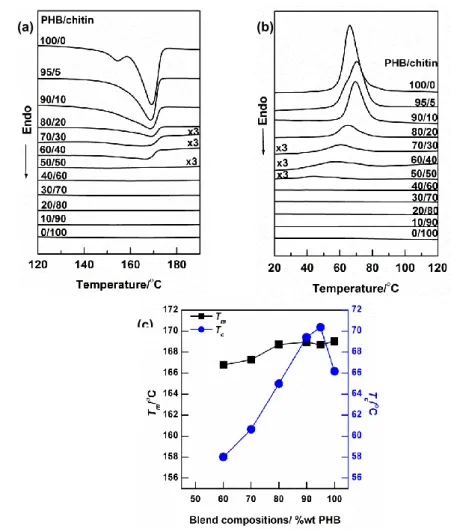
Figure 2a shows DSC thermogram of PHB/chitin blends with various compositions in
the first heating process. PHB shows double endotherm peaks, i.e. a small peak appears
because of the partial melting of imperfect crystals while a larger peak is caused by the
melting of more perfect crystals and the recrystallized crystals during the heating process.27
In contrast, chitin does not show any endotherm peak during the heating process as in the
cases of previous studies of chitin blends.37,38 Chitin most likely exists as the amorphous
phase, and therefore, chitin does not show its thermal activity in DSC. The intensity of
melting peaks of PHB decreases with increasing chitin contents in the blends, however the
melting temperature (Tm) changes a little. A clear endotherm peak cannot be observed for
the blends with PHB 50 wt % and eventually disappears when the PHB content becomes
less than 40 wt %, signifying that the crystallinity of PHB substantially decreases by
blending with chitin. However, it is noted that chitin does not much affect the Tm of the
PHB crystals in the blend samples.
Figure 2b shows DSC thermograms obtained during the cooling process to investigate
the effect of chitin matrix on the crystallization of PHB. It can be clearly seen that the
intensity of the crystallization peak of PHB in the blends decreases with increasing chitin
47
important point in Figure 2b is that the depression in the crystallization temperature (Tc) is
higher than that of Tm. The increment of Tc in the blends with the chitin up to 10 wt % is
caused by the nucleation effect of chitin. It clearly indicates that in the small loadings
chitin act as a nucleating agent that promotes the rapid growth of the PHB crystals.8,9,41 As
a result, the temperature when PHB begins to crystallize is earlier in those blends.
However, in the blends with higher chitin contents, the certain chitin chains interfere the
crystallizability of PHB by forming intermolecular interactions and hinder the growth of
the PHB crystal. Therefore, plot of Tc in Figure 2c is gradually decreased. The thermal
characteristics of blends are summarized in Table 1.
The most important factor in the reduction of crystallinity is due to the formation of
intermolecular interactions between PHB and chitin during the crystallization process,
which would be caused by reduced mobility of PHB molecules peculiar in PHB/chitin
blends. The intermolecular interactions which play a crucial role for reducing the
crystallinity of PHB has also been observed in other blends, such as PHB/CAB blend27 and
PHB/chitosan blend.38,42
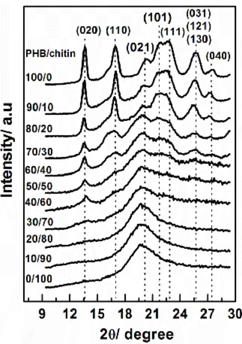
3-2. Wide-Angle X-ray Diffraction (WAXD)
Figure 3 shows X-ray diffraction patterns of PHB/chitin blends with various
48
the WAXD patterns, while chitin presents a simple broad diffraction peak (110) located
around 2 = 19.6°. It is important to highlight that chitin as a cast film from HFIP solution has crystalline volume fraction about 10%.43 It gives us another evidence that chitin cast
film reasonably exists in the amorphous phase.
The intensity of PHB diffraction peaks decreases gradually with increasing chitin
content in the blends and eventually the peaks disappear for the blends with PHB ≤ 30
wt %. The diffraction peak position of PHB in the blends is almost the same as that of PHB,
indicating that chitin little affects the crystalline structure of PHB. The WAXD results in
Figure 3 indicate a similar trend as the DSC results that the significant changes are
observed in the blends with PHB ≤ 50 wt %. Even though chitin suppresses the
crystallinity of PHB, the WAXD results suggest that the crystalline structure of PHB does
not change significantly by blending with chitin. It is noted that although DSC could not
observe the melting peak for the blend with PHB 40 wt %, the crystalline diffraction due to
(020) planes still appears in its WAXD pattern. This occurrence may be ascribed to the
different sensitivity of the DSC and WAXD measurement techniques.
Figure 3 also suggests that the formation of intermolecular interactions between PHB
and chitin occur in the amorphous phase. If the intermolecular interactions do not occur,
the diffraction of crystalline peaks of PHB should be observed together with the diffraction