Accepted : October 26, 2017 Published online : December 31, 2017
Glycative Stress Research 2017; 4 (4): 267-274 Review article
Ippei Kanazawa, Toshitsugu Sugimoto
Internal Medicine 1, Shimane University Faculty of Medicine, Izumo, Shimane, Japan
Glycative Stress Research 2017; 4 (4): 267-274 (c) Society for Glycative Stress Research
The mechanism of bone fragility in diabetes mellitus
(総説論文
-日本語翻訳版)
糖尿病における骨脆弱化機序
要旨
金沢一平、杉本利嗣 島根大学医学部内科学講座内科学第一 糖尿病患者では骨折リスクが高いことが明らかとなった。脆弱性骨折は患者の日常生活動作(activity ofdaily living: ADL)や生活の質(quality of life: QOL)の低下に影響するのみならず、生命予後も不良にす
る重大なイベントである。糖尿病では骨密度とは独立した骨折リスク上昇があることから、糖尿病関連骨粗 鬆症では骨質劣化が重要な病態と考えられている。これまでに骨質劣化の病態には骨組織への終末糖化産物
(advanced glycation end products: AGEs)蓄積、骨形成低下を伴う骨リモデリング異常、骨微細構造の異常
が関連していることが報告されている。AGEsはコラーゲン線維間に架橋(cross-links)を形成することにより
骨強度を低下させる。さらに、骨芽細胞、骨細胞にはAGEsに対する受容体Receptor for AGEs(RAGE)が発
現しており、生理活性物質として骨芽細胞、骨細胞の機能を障害する。糖尿病では酸化ストレス誘導因子であ
るhomocysteineの血中濃度が上昇しており、homocysteineが骨芽細胞や骨細胞の酸化 ストレス増強を介して
骨質劣化に影響する可能性がある。さらに骨にアナボリックに作用するインスリンやインスリン様成長因子- I
(insulin-like growth factor-I: IGF-I)作用低下も重要な要素となる。今後は病態を踏まえた糖尿病関連骨粗鬆症
の治療ストラテジーの構築が重要な課題である。
連絡先: 金沢一平
島根大学医学部内科学講座内科学第一 〒 693-8501 島根県出雲市塩冶町 89 -1
KEY WORDS:
糖尿病、骨粗鬆症、骨質、終末糖化産物(advanced glycation end products: AGEs)、 酸化 ストレスはじめに
人口の高齢化が進む中、高齢者の日常生活動作(activities
of daily living: ADL)や生活の質(quality of life: QOL) を保ち自立性を維持することは、医学的な観点のみでなく、 社会的にも重要な喫緊の課題である。骨粗鬆症は骨強度が 低下することにより軽微な外力でも容易に骨折をきたし、 その後のADLとQOLを著しく低下させる。さらには、骨 粗鬆症に伴う骨折受傷後では生命予後が不良であることも 報告されている1, 2)。 糖尿病治療の目標は、健康な人と変わらないQOLの維 持と寿命の確保である。これまで蓄積されたエビデンスに より、糖尿病患者では非糖尿病患者に比較して有意に骨折 リスクが高いことが明らかとなった3-5)。したがって糖尿 病に合併する骨粗鬆症の病態を把握し、骨折リスクに対す るアプローチを行うことは重要である。 本稿では糖尿病における骨折リスクと骨脆弱性のメカニ ズムについて概説する。
糖尿病における骨折リスク上昇
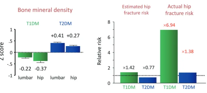
骨強度は骨量と骨質の総和と定義される6)。古くは骨粗 鬆症は骨密度が低下する疾患と考えられていたが、骨密度 の低下がなくても脆弱性骨折をきたす患者が存在すること から、骨質劣化も重要な骨粗鬆症の要因であることが認識 されるようになった。これまでにいくつかのメタ解析によ り、糖尿病患者では1型、2型ともに骨折リスクが上昇し ていることが明らかとなっている。また、いずれの病型に おいても骨密度で想定されるよりも高い骨折リスクが存在 することが示されている。Vestergaardが報告したメタ解 析では3)、1型糖尿病では同性同年齢の健常者と比較した Zスコアが腰椎で− 0.22、大腿骨で− 0.37と低下してお り、骨密度の低下から予測される大腿骨近位部骨折リスク は1.42倍であるのに対して、実際の骨折リスクは6.94倍 と予想以上に高値であった。さらに2型糖尿病のZスコア は腰椎で+0.41、大腿骨で+0.27と上昇しており、骨密度か ら予測される大腿骨近位部骨折リスクは0.77倍に低下す ることとなるが、実際には1.38倍と上昇していた(Fig. 1)。 我々の日本人を対象に骨密度と椎体骨折との関連性をみた 検討でも同様に、2型糖尿病では健常者に比較して骨密度 は高値であるにも関わらず、椎体骨折リスクは上昇してい た4)。Schwartzらの3つの前向きコホート研究を統合解 析した報告では、同等の大腿骨近位部骨折リスクに対する 大腿骨頚部骨密度は、糖尿病女性では非糖尿病に比較して + 0.59 SD、男性では+ 0.38 SD高いと報告されている5)。 したがって糖尿病患者では骨密度のみでは実際の骨折リス クを過小評価してしまう危険性があり、骨密度に依存しな い骨折リスクの上昇、すなわち骨質劣化が重要な病態と考 えられる。 糖尿病患者においても脆弱性骨折がADL、QOL、生命 予後に影響しているかの報告はなかった。我々は2型糖尿 病患者を対象にBarthel index、SF-36によるアンケート 調査を行い、椎体骨折とADL、QOLの関係について横断 検討を行った。Genant分類7)でGrade 2と3の椎体骨折 を有する患者では、年齢、性別、HbA1c、腎機能、他の 糖尿病合併症などで補正しても有意にADL、QOL(特にbodily pain、general health、vitality、social functioning、
role emotional)の低下に影響することを報告した8)。さら に、2型糖尿病患者を対象とした観察研究により、多発椎 体骨折、Grade 3椎体骨折があると、様々な交絡因子で補 正しても総死亡リスクが有意に上昇することを明らかにし た(Fig. 2)9)。したがってこれらの結果は糖尿病患者にお いても脆弱性骨折を予防することは重要な課題であること を示している。
糖尿病における骨質劣化の病態
骨質劣化の機序として、コラーゲン線維間における終 末糖化産物(advanced glycation end products: AGEs)架橋(AGE cross-links)の蓄積や骨微細構造異常が重要と考え られている。骨基質には1型コラーゲンが豊富に存在し、 コラーゲン線維間に生理的架橋を形成することにより骨の しなやかさと強度が保たれる。AGEsは蛋白質が非酵素的 に糖化反応を受けて形成されるものの総称であり、糖尿病 の状態では非生理的にAGEs架橋が形成されることにより コラーゲンのしなやかさが損なわれ、骨強度が低下する (Fig. 3)。Saitoらによる自然糖尿病発症ラットの検討では、 骨組織内AGEs架橋の蓄積により骨強度低下が惹起される ことが報告されており10)、1型糖尿病患者の骨生検を行っ た臨床研究においもAGEsのひとつであるペントシジンが 骨基質で増加していることが明らかになっている11)。した がって、糖尿病による骨質劣化の病態に、骨内の非生理的 AGEs架橋の蓄積が関与していると考えられる。このこと は骨密度が低下しなくても骨脆弱性が惹起される糖尿病関 連骨粗鬆症の病態をよく反映する仮説である。 糖尿病患者における骨構造異常として皮質骨多孔化や 海綿骨微細構造異常が関連している可能性が報告されて いる。High-resolution peripheral quantitative computed
tomography(HR-pQCT)を用いた臨床検討により、皮質 骨多孔性が骨密度とは独立して骨強度に影響する重要な要 素であることが報告され12)、注目を集めている。これまでに 2型糖尿病における検討で、脆弱性骨折を伴う患者では皮 質骨の多孔性が増加していることが報告されており13, 14)、 皮質骨多孔化が糖尿病関連骨粗鬆症の病態に関連している 可能性が示唆されている。一方、皮質骨多孔化はすべての 部位で起こっているわけではなく、部位によって皮質骨多 孔化は変化ない14)、あるいは2型糖尿病患者の方が少ない という報告もあり15)、皮質骨多孔化が糖尿病による骨脆弱 性に関与しているか否かは今後のさらなる検討が必要と考 えられる。
Fig. 2. Correlation between vertebral fractures (VF) and the overall mortality rate in patients with T2DM.
When patients have more than two VF, their cumulative survival rate was sure to lower significantly (A). This correlation was significant also after adjusting for age, gender, duration of T2DM, HbA1c, BMI, serum Cr, sBP, LDL-C, and treatment of osteoporosis (HR 2.93, 95%CI 1.42-6.02, p = 0.004). When VF became grade 3 (G3), the cumulative survival rate lowered significantly (B). This correlation was significant also, after adjusting for the confounding factors (HR 7.64, 95%CI 2.13-27.42, p = 0.002). VF, vertebral fractures; T2DM, type 2 diabetes mellitus; BMI, body mass index; Cr, creatinine; sBP, systolic blood pressure; LDL-C, low-density lipoprotein-cholesterol; HR, hazard ratio; CI, confidence interval.
Fig. 1. Bone density and fracture risks regarding diabetic patients.
In the case of type 1 diabetes (T1DM), Z scores, compared to those of healthy people of the same age and the same sex, are lower by 0.22 regarding lumbars, and 0.37 regarding femurs, while in the case of type 2 diabetes (T2DM), Z scores are on the rise by 0.41 regarding lumbars, and by 0.27 regarding femurs. While it is expected that the risks of proximal femoral fractures to be 1.42 times higher in the case of T1DM, and 0.77 times higher in the case of T2DM, the actual fracture risks were each 6.94 times and 1.38 times the risk for T1DM and T2DM which were higher than expected. Author’s drawing based on Reference 3.
Logrank p < 0.001 month month Logrank p = 0.002 VF 0 VF 1 VF 2£ 0 .2 .4 .6 .8 1.0 0 20 40 60 80 G0 G1G2 G3 0 0.2 0.4 0.6 0.8 1.0 0 20 40 60 80
Number of VF
Grade of VF
Cum
ul
at
iv
e
sur
vi
va
l
Cum
ul
at
iv
e
sur
vi
va
l
VF 2< vs none
HR 2.93(1.42-6.02), p = 0.004 Adjusted for age, gender, T2DM duration, HbA1c, BMI, Cr, sBP, LDL-C, osteoporosis treatmentsGrade 3 vs none
HR 7.64(2.13-27.42), p = 0.002 Adjusted for age, gender, T2DM duration, HbA1c, BMI, Cr, sBP, LDL-C, osteoporosis treatments Figure 2 A BTrabecular Bone Score(TBS)は海綿骨微細構造の緻 密度を評価する指標であり、骨密度とは独立して骨折リス
ク評価に有用である。TBS測定は腰椎骨密度を測定する
際に用いられるX線骨密度測定装置(dual energy X-ray
absorptiometry: DXA)の画像を再解析して行うため新た に侵襲を加える必要がなく、今後の臨床応用が期待される ツールである。これまでに2型糖尿病患者では非糖尿病に 比較してTBSが低下しており、さらに糖尿病患者におい てもTBSが低いことが骨折リスクに関連することが報告 されている16)。Ikiらは日本人男性1,683人を対象とした 検討により、糖尿病患者では高血糖、インスリン抵抗性が TBSの低下と有意に関連することを報告している17)。した がって糖尿病関連骨粗鬆症に海綿骨微細構造の異常も関与 している可能性がある。 糖尿病における骨微細構造異常のメカニズムは明らかと なっていないが、我々は後述する骨芽細胞機能異常、骨リ モデリング障害や骨細胞アポトーシスが関連している可能 性があると考えている。
糖尿病における骨芽細胞分化障害の
メカニズム
骨は骨芽細胞による骨形成と破骨細胞による骨吸収のバ ランスにより恒常性が保たれている。骨芽細胞は骨型アル カリホスファターゼ(BAP)と1型コラーゲンを発現する とともにカルシウムやリンを石灰沈着させる(石灰化)こ とにより骨を形成する。また、コラーゲンの生理的架橋の形成には骨芽細胞が産生するlysyl oxidase(LOX)が必須
であることから、骨強度・骨質を維持するために骨芽細胞 は重要な役割を担っている。 糖尿病では骨芽細胞の初期から後期への分化・成熟が障 害されると考えられる。糖尿病では非糖尿病に比較して分 化初期のマーカーであるBAPに有意な差はないが、後期 のマーカーであるオステオカルシンが有意に低下してい る18)。糖尿病における骨芽細胞分化障害の存在は、オステ オカルシン/BAP比が低下するほど骨折リスクが増大する ことや19, 20)、短期的糖尿病治療介入によりBAPは低下し、 オステオカルシンは上昇することからも示唆される21)。糖 尿病における骨芽細胞分化障害のメカニズムとして、高血 糖やAGEs、homocysteineなどの重要性が報告されている。
1)AGEs
AGEsはコラーゲン架橋を形成する以外にも受容体であるreceptor for AGEs(RAGE)を介して作用する生理活
性を有する。骨芽細胞にもRAGEは発現しており、AGEs は骨芽細胞に直接作用してアポトーシス促進や分化、石灰 化障害を引き起こす。我々は高血糖状態がRAGE発現を 増強することによりAGEs作用を増強し22)、AGEsが骨 芽細胞分化に重要であるRunx2とosterixの発現低下を 介して石灰化を著明に抑制することを明らかにした23, 24)。 さらにその機序に小胞体ストレスやtransforming growth factor-β (TGF-β)発現上昇が関連していることを報告し ている(Fig. 4)24, 25)。
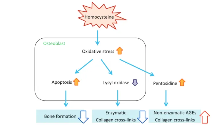
2)Homocysteine
Homocysteineは必須アミノ酸であるメチオニンやシス テインの生成に重要なアミノ酸であるが、homocysteine の過剰により酸化ストレスが誘導されることはよく知ら れている。糖尿病ではインスリン作用不足に伴い肝臓で Fig. 3. Deterioration of collagen cross-links in diabetes mellitus.Crosslinkings physiologically formed in collagen arrays enhance bone strength. On the other hand, non-physiologically formed AGE crosslinkings, which damage suppleness of bones, play a part in lowering bone strength. AGE, advanced glycation end product.
の糖新生が亢進することによりビタミンB群の不足が生 じ、血中homocysteine濃度が上昇するといわれている。 Liらは糖尿病124人、非糖尿病115人を対象に、血中ホ モシステインと骨折リスクとの関連性を検討した横断研 究を報告している26)。糖尿病群では非糖尿病群に比較し て血中homocysteine濃度が上昇しており、さらに骨折 の既往のある糖尿病群ではない群に比較して有意に血中 homocysteineが高値であった。さらに年齢や性別、ビタ ミンB12、葉酸、腎機能などで補正したロジスティック回 帰分析において、血中homocysteine高値が独立して骨折 リスクに関連することを明らかにしている。 我 々 は 骨 芽 細 胞 様 細 胞 MC3T3-E1 を 用 い て、 homocysteineが骨芽細胞の酸化ストレスを亢進してアポ トーシスを誘導すること、さらにアポトーシスを誘導しな い低濃度においてもLOX発現を抑制し、細胞外AGEs蓄積 を増強することを報告した27)。したがってhomocysteine は骨芽細胞に直接影響して酸化ストレス増強を介して骨芽 細胞機能の低下とコラーゲン架橋異常(生理的架橋低下、 AGEs架橋増加)を引き起こすことにより骨質劣化を惹起 すると考えられる(Fig. 5)。また、homocysteineは骨芽細 胞への影響だけでなく、コラーゲンへの直接作用により架 橋形成を抑制することも報告されている28)。
3)
インスリン、insulin-like growth factor-I
(IGF-I
)インスリン作用は骨芽細胞の分化やコラーゲン産生に 重要な役割を担っていると考えられている。骨芽細胞特異 的インスリン受容体ノックアウトマウスを用いた検討で は、骨芽細胞数の低下による骨形成減弱により著明な骨量 低下が惹起されることが報告されている29, 30)。骨芽細胞 におけるインスリンシグナルの阻害は骨芽細胞増殖の抑制 やアポトーシス誘導を引き起こし、さらにRunx2阻害因 子であるTwist2の発現を上昇することにより骨芽細胞分 化を抑制することが明らかになっている30)。また、インス
リンシグナルの下流分子であるinsulin receptor substrate-1
(IRS-1)やIRS-2欠損マウスでも骨形成低下を伴う骨量 減少が報告されている31)。1型糖尿病患者では骨形成低 下、骨密度低下が認められるという臨床像からもインスリ ンが骨にアナボリックに作用することは支持される。ま た、IGF-Iも古くから骨にアナボリックに作用するホルモ ンとして知られている。IGF-Iは骨芽細胞でも産生される ため局所因子として重要である。一方、血中IGF-Iは成長 ホルモンによる刺激により主に肝臓で合成、分泌され、内 分泌ホルモンとして骨に作用する。骨芽細胞特異的IGF-I 受容体欠損マウスにおいて、有意な石灰化減弱、骨量の低 下が認められており32)、肝臓特異的IGF-Iノックアウトマ ウスの解析においても有意な骨量減少が認められることか ら33)、IGF-Iの内分泌ホルモンとしての役割も重要である ことが認識されている。我々は2型糖尿病閉経後女性にお いて、血中IGF-I値は骨形成マーカーと有意な正の相関を 認め、さらに血中IGF-I低値が椎体骨折、多発椎体骨折 のリスク上昇に関与することを報告している34, 35)。また、
AGEsは骨芽細胞におけるIGF-IやIGF-I受容体の発現を
抑制することによりIGF-Iシグナルを低下することが報告 されており36-38)、内分泌因子としてのIGF-I作用の低下と 骨局所におけるIGF-Iシグナルの抑制が糖尿病における骨 脆弱性の病態に重要である可能性が考えられる。
糖尿病による骨細胞機能異常のメカニズム
骨細胞は骨を形成する細胞の90%以上を占め、骨内に 最も多く存在する細胞である。骨芽細胞は自らが分泌し た骨基質に埋め込まれていく過程で、最終分化形態であ る骨細胞となる。骨細胞の多くは皮質骨に埋没しており、 骨細管と呼ばれるトンネル様の構造に樹状突起を伸ばし て隣接する骨細胞や骨表面の骨芽細胞や破骨細胞との情 報交換を行っている。 また、骨細胞は液性因子を分泌し、骨芽細胞分化抑制因 子であるsclerostinやDickkopf-1、破骨細胞分化制御因子であるreceptor activator of NF-кB ligand (RANKL)
やosteoprotegerinを発現することにより、骨芽細胞と破 骨細胞のカップリングを制御している。したがって、骨 細胞機能は骨代謝制御、骨リモデリングに重要と考えら れている。骨はリモデリングを繰り返すことにより3~4 カ月単位で常に新しい骨へと生まれ変わっているが、糖 尿病では骨リモデリングが低下しているためAGEs架橋 や微小骨折(micro crackle)が蓄積されやすい状態にあ ると考えられる。 RAGEは骨細胞にも発現していることからAGEsが骨 細胞に直接影響する可能性がある。我々は骨細胞様細胞
MLO-Y4-A2細胞を用いた検討において、AGEsはRAGE
を介して骨細胞のアポトーシスを誘導し、sclerostin発現 を上昇し、RANKL発現を抑制することを初めて報告し た39)。さらに、AGEsのアポトーシス誘導とsclerostin発 現増強はTGF-βシグナルを介していることを明らかにし た40)。また、我々はhomocysteineも骨細胞へ直接作用し、 酸化ストレス亢進を介してアポトーシスを誘導することも 報告している41)。Vijayanらはhomocysteineを30日間マ ウスに投与した検討で、骨細胞のアポトーシスが増強され 皮質骨空隙数が増加、さらにsclerostin陽性骨細胞数も増 加し、生体力学的特性(ヤング率)は低下することを報告 している42)。したがってhomocysteineの骨細胞へ影響が 高homocysteine血症による骨折リスク上昇に関連してい る可能性が考えられる。 糖尿病における骨細胞機能異常がどの程度骨脆弱性に 寄与しているかは不明であるが、皮質骨における骨細胞 アポトーシスは皮質骨多孔化に影響する可能性が考えら れる。また、sclerostinやRANKL発現を介した骨リモ
デリング抑制により、AGEs架橋やmicro crackleが蓄積 した古い骨を代謝することができないため、骨質の劣化
Fig. 4. Impacts of AGEs on osteoblasts.
While AGEs accelerate apoptosis via RAGE that exists in osteoblasts, they suppress the proliferation of cells. Additionally, AGEs suppress differentiation and mineralization of osteoblasts in their mechanism. During the process, the abnormality of endoplasmic reticulum stress proteins and increase of TGFβ expression are seen. Although AGEs suppress Runx2 expression in the initial stage of differentiation, they suppress differentiation of osteoblasts by strengthening them in the later stage of differentiation. AGEs, advanced glycation end products; RAGE, Receptor for AGEs; ER, endoplasmic reticulum; IRE1α, inositol-requiring enzyme 1α; ATF6, activating transcription factor 6; OASIS, old astrocyte specifically induced substance; TGFβ, transforming growth factor-β.
Fig. 5. Impacts of homocysteine on osteoblasts.
Homocysteine induces osteoblasts apoptosis, resulting in inhibition of function of osteoblasts. Furthermore, it prevents physiological collagen from forming cross-links outside cells and increases extracellular AGE cross-cross-links. AGE, advanced glycation end product.
AGEs
RAGE
ER stress
IRE1a
ATF6
OASIS
TGFb
Runx2
Osterix
Differentiation
Mineralization
Apoptosis
Cell growth
Osteoblast
Oxidative stress
Apoptosis
Lysyl oxidase
Pentosidine
Osteoblast
Homocysteine
Bone formation
Enzymatic
Collagen cross-links
Non-enzymatic AGEs
Collagen cross-links
Figure 5
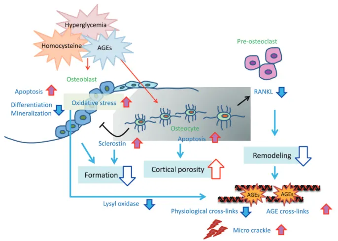
Fig. 6. Mechanism of bone fragility caused by diabetes mellitus.
In the case of diabetic patients, blood concentrations of AGEs and homocysteine increase. AGEs and homocysteine cause apoptosis or suppression of differentiation regarding osteoblasts, resulting in reducing bone formation rate. Furthermore, they suppress osteoblast differentiation by enhancing sclerostin expression in osteocytes. On the other hand, it causes RANKL expression to lower from osteocytes, which suppresses osteoclasts differentiation, resulting in damaging of remodeling. Owing to this, old bones which should be metabolized will not be resorbed, resulting in accumulation of AGE cross-links, and micro crackles. It can be thought that an increase of apoptosis in osteocytes induces porosity in cortical bones. AGEs, advanced glycation end products; RANKL, receptor activator of nuclear factor kappa-B ligand.
糖尿病における破骨細胞への影響
前述のように、糖尿病における骨形成低下、骨リモデリ ング低下の機序は徐々に明らかになってきているが、糖尿 病における破骨細胞活性や骨吸収機能については未だ一定 の見解がない。これまでの報告では、糖尿病患者では骨吸 収マーカーは上昇しているという報告もあるが、メタ解析 ではむしろ非糖尿病患者に比較して低下していると報告さ れている43)。AGEsやhomocysteineの破骨細胞への影響 についても分化誘導を抑制するという報告44)や活性を増 強するという報告もあり45, 46)、一定の見解はない。糖尿 病患者では骨密度があまり低下しない3)ということを考慮 すると、骨吸収が著明に亢進しているということは考えに くく、おそらく骨形成低下に比較して相対的亢進の程度と 推測される。おわりに
糖尿病では骨質劣化に伴う骨脆弱性が重要な病態であ ることが明らかとなっている。AGEsによるコラーゲン架 橋の異常、骨芽細胞と骨細胞の機能障害は重要と考えら れることから、骨脆弱性を予防、改善するためにはAGEs 産生を抑制するための長期的な血糖管理や酸化ストレス の低減は極めて重要と考えられる。また、糖尿病に伴う homocysteine上昇や酸化ストレス亢進はAGEs形成に寄 与するのみならず骨形成低下、骨リモデリング異常にも 影響する。また、糖尿病によるインスリンやIGF-Iの作 用低下による内分泌環境の異常も重要と考えられる。 糖尿病による骨脆弱性の機序は徐々に明らかになりつ つある。しかしながら日常診療においてどのような血糖 管理手法が骨脆弱性を改善しうるか、あるいはどの骨粗 鬆症治療薬が有効であるかについての検討は少なく、エ ビデンスが欠如しているのが現状である。糖尿病に合併 する骨粗鬆症はADL、QOLの低下に加えて生命予後 にも関わる重要な疾患であることを踏まえると、今後の さらなる検討を行い糖尿病関連骨粗鬆症の治療ストラテ ジーを構築することは急務と考えられる。利益相反申告
本論文に関して利益相反に該当する事項はない。1) Haentjens P, Magaziner J, Colon-Emeric CS, et al. Meta-analysis: excess mortality after hip fracture among older women and men. Ann Intern Med. 2010; 152: 380-390. 2) Ensrund KE, Thompson DE, Cauley JA, et al. Prevalent
vertebral deformities predict mortality and hospitalization in older women with low bone mass. J Am Geriatr Soc. 2009; 48: 241-249.
3) Vestergaard P. Discrepancies in bone mineral density and fracture risk in patients with type 1 and type 2 diabetes: A meta-analysis. Osteoporos Int. 2007; 18: 427-444. 4) Yamamoto M, Yamaguchi T, Yamauchi M, et al. Diabetic
patients have an increased risk of vertebral fractures independent of BMD or diabetic complications. J Bone Miner Res. 2009; 24: 702-709.
5) Schwartz AV, Vittinghoff E, Bauer DC, et al. Association of BMD and FRAX score with risk of fracture in older adults with type 2 diabetes. JAMA. 2011; 305: 2184-2192.
6) Kanis JA. Assessment of fracture risk and its application to screening for postmenopausal osteoporosis: Synopsis of a WHO report. WHO Study Group. Osteoporos Int. 1994; 4: 368-381.
7) Genant HK, Wu CY, van Kuijk C, et al. Vertebral fracture assessment using a semiquantitative technique. J Bone Miner Res. 1993; 8: 1137-1148.
8) Kanazawa I, Takeno A, Tanaka KI, et al. Osteoporosis and vertebral fracture are associated with deterioration of ADL and QOL in patients with type 2 diabetes independently of other diabetic complications. 53rd EASD Annual Meeting, Lisbon, Portugal, 2017. Diabetologia. 2017; 60(S1): 1210. (abstract)
9) Miyake H, Kanazawa I, Sugimoto T. Association of bone mineral density, bone turnover markers, and vertebral fractures with all-cause mortality in type 2 diabetes mellitus. Calcif Tissue Int. 2018; 102: 1-13.
10) Saito M, Fujii K, Mori Y, et al. Role of collagen enzymatic and glycation induced cross-links as a determinant of bone quality in spontaneously diabetic WBN/Kob rats. Osteoporos Int. 2006; 17: 1514-1523.
11) Farlay D, Armas LA, Gineyts E, et al. Nonenzymatic glycation and degree of mineralization are higher in bone from fractured patients with type 1 diabetes mellitus. J Bone Miner Res. 2016; 31: 190-195.
12) Bjornerem A. The clinical contribution of cortical porosity to fragility fractures. Bonekey Rep. 2016; 5: 846.
13) Burghardt AJ, Issever AS, Schwart AV, et al. High-resolution peripheral quantitative computed tomographic imaging of cortical and trabecular bone microarchitecture in patients with type 2 diabetes mellitus. J Clin Endocrinol Metab. 2010; 95: 5045-5055.
14) Samelson EJ, Demissie S, Cupples LA, et al. Diabetes and deficits in cortical bone density, microarchitecture, and bone size: Framingham HR-pQCT study. J Bone Miner Res. 2017 Sep 20.
15) Nilsson AG, Sundh D, Johansson L, et al. Type 2 diabetes mellitus is associated with better bone microarchitecture but lower bone material strength and poorer physical function in elderly women: A population-based study. J Bone Miner Res. 2017; 32: 1062-1071.
16) Dhaliwal R, Cibula D, Ghosh C, et al. Bone quality assessment in type 2 diabetes mellitus. Osteoporos Int. 2014; 25: 1969-1973.
17) Iki M, Fujita Y, Kouda K, et al. Hyperglycemia is associated with increased bone mineral density and decreased trabecular bone score in elderly Japanese men: The Fujiwara-kyo osteoporosis risk in men (FORMEN) study. Bone. 2017; 105: 18-25.
18) Starup-Linde J, Eriksen SA, Lykkeboe S, et al. Biochemical markers of bone turnover in diabetes patients: A meta-analysis, and a methodological study on the effects of glucose on bone markers. Osteoporos Int. 2014; 25: 1697-1708.
19) Okazaki R, Totsuka Y, Hamano K, et al. Metabolic improvement of poorly controlled noninsulin-dependent diabetes mellitus decreases bone turnover. J Clin Endocrinol Metab. 1997; 82: 2915-2920.
20) Kanazawa I, Yamaguchi T, Yamauchi M, et al. Adiponectin is associated with changes in bone markers during glycemic
control in type 2 diabetes mellitus. J Clin Endocrinol Metab. 2009; 94: 3031-3037.
21) Kanazawa I, Yamaguchi T, Yamamoto M, et al. Serum osteocalcin/bone-specific alkaline phosphatase ratio is a predictor for the presence of vertebral fractures in men with type 2 diabetes. Calcif Tissue Int. 2009; 85: 228-234. 22) Ogawa N, Yamaguchi T, Yano S, et al. The combination
of high glucose and advanced glycation end-products (AGEs) inhibits the mineralization of osteoblastic MC3T3-E1 cells through glucose-induced increase in the receptor for AGEs. Horm Metab Res. 2007; 39: 871-875. 23) Okazaki K, Yamaguchi T, Tanaka K, et al. Advanced
glycation end products (AGEs), but not high glucose, inhibit the osteoblastic differentiation of mouse stromal ST2 cells through the suppression of osterix expression, and inhibit cell growth and increasing cell apoptosis. Calcif Tissue Int. 2012; 91: 286-296.
24) Notsu M, Yamaguchi T, Okazaki K, et al. Advanced glycation end product 3 (AGE3) suppresses the mineralization of mouse stromal ST2 cells and human mesenchymal stem cells by increasing TGF-β expression and secretion. Endocrinology. 2014; 155: 2402-2410. 25) Tanaka K, Yamaguchi T, Kaji H, et al. Advanced glycation
end products suppress osteoblastic differentiation of stromal cells by activating endoplasmic reticulum stress. Biochem Biophys Res Commun. 2013; 438: 463-467. 26) Li J, Zhang H, Yan L, et al. Fracture is additionally
attributed to hyperhomocysteinemia in men and premenopausal women with type 2 diabetes. J Diabetes Investing. 2014; 5: 236-241.
27) Kanazawa I, Tomita T, Miyazaki S, et al. Bazedoxifene ameliorates homocysteine-induced apoptosis and accumulation of advanced glycation end products by reducing oxidative stress in MC3T3-E1 cells. Calcif Tissue Int. 2017; 100: 286-297.
28) Kang AH, Trelstad RL. A collagen defect in homocystinuria. J Clin Invest. 1973; 52: 2571-2578. 29) Ferron M, Wei J, Yoshizawa T, et al. Insulin signaling
in osteoblasts integrates bone remodeling and energy metabolism. Cell. 2010; 142: 296-308.
30) Fulzele K, Riddle RC, DiGirolamo DJ, et al. Insulin receptor signaling in osteoblasts regulates postnatal bone acquisition and body composition. Cell. 2010; 142: 309-319.
31) Ogata N, Chikazu D, Kubota N, et al. Insulin receptor substrate-1 in osteoblast is indispensable for maintaining bone turnover. J Clin Invest. 2000; 105; 935-943.
32) Zhang M, Xuan S, Bouxsein ML, et al. Osteoblast-specific knockout of the insulin-like growth factor (IGF) receptor gene reveals an essential role of IGF signaling in bone matrix mineralization. J Biol Chem. 2002; 277: 44005-44012.
33) Yakar S, Rosen CJ, Beamer WG, et al. Circulating levels of IGF-I directly regulate bone growth and density. J Clin Invest. 2002; 110: 771-781.
34) Kanazawa I, Yamaguchi T, Yamamoto M, et al. Serum insulin-like growth factor-I level is associated with the presence of vertebral fractures in postmenopausal women with type 2 diabetes mellitus. Osteoporos Int. 2007; 18: 1675-1681.
35) Kanazawa I, Yamaguchi T, Sugimoto T. Serum insulin-like growth factor-I is a marker for assessing the severity of vertebral fractures in postmenopausal women with type 2 diabetes mellitus. Osteoporos Int. 2011; 22: 1191-1198. 36) McCarthy AD, Etcheverry SB, Cortizo AM. Effect of
advanced glycation endproducts on the secretion of insulin-like growth factor-I and its binding proteins: Role in osteoblast development. Acta Diabetol. 2001; 38: 113-122.
37) Napoli N, Chandran M, Pierroz DD, et al. Mechanisms o f diabetes mellitus-induced bone fragility. Nat Rev Endocrinol.
2017; 13: 208-219.
38) Qin Y, Ye J, Wang P, et al. miR-223 contributes to the AGE-promoted apoptosis via down-regulating insulin-like growth factor 1 receptor in osteoblasts. Biosci Rep. 2016; 36: e00314.
39) Tanaka K, Yamaguchi T, Kanazawa I, et al. Effects of high glucose and advanced glycation end products on the expressions of sclerostin and RANKL as well as apoptosis in osteocyte-like MLO-Y4-A2 cells. Biochem Biopjys Res Commun. 2015; 461: 193-199.
40) Notsu M, Kanazawa I, Takeno A, et al. Advanced glycation end product 3 (AGE3) increases apoptosis and the expression of sclerostin by stimulating TGF-β expression and secretion in osteocyte-like MLO-Y4-A2 cells. Calcif Tissue Int. 2017; 100: 402-411.
41) Takeno A, Kanazawa I, Tanaka K, et al. Activation of AMP-activated protein kinase protects against homocysteine-induced apoptosis of osteocytic MLO-Y4 cells by regulating the expressions of NADPH oxidase 1 (Nox1) and Nox2. Bone. 2015; 77: 135-141.
42) Vijayan V, Gupta S. Role of osteocytes in mediating bone mineralization during hyperhomocysteinemia. J Endocrinol. 2017; 233: 243-255.
43) Hygum K, Starup-Linde J, Harslof T, et al. Mechanisms in endocrinology: Diabetes mellitus, a state of low bone turnover: A systematic review and meta-analysis. Eur J Endocrinol. 2017; 176: R137-157.
44) Valcourt U, Merle B, Gineyts E, et al. Non-enzymatic glycation of bone collagen modifies osteoclastic activity and differentiation. J Biol Chem. 2007; 282: 5691-5703. 45) Dong XN, Qin A, Xu J, et al. In situ accumulation of
advanced glycation endproducts (AGEs) in bone matrix and its correlation with osteoclastic bone resorption. Bone. 2011; 49: 174-183.
46) Herrmann M, Umanskaya N, Wildemann B, et al. Stimulation of osteoblast activity by homocysteine. J Cell Mol Med. 2008; 12: 1205-1210.