ミオドコーピナ亜目に属する貝虫類2種の幼生期の形態について
24
0
0
全文
(2) Journal of Hokkaido University of Education (Section II B) Vol. 30, No. 2 March, 1980. -fbySiatint^ffiS (^2$|SB) ^30^ ^2-f BSW 55 ^ 3 ^. Morphology of the Larval Stages of Vargula kilgendorfii (G. W. Muller) and Euphilomedes nipponica Hiruta from Japan (Ostracoda : Myodocopina). Shinlchi HIRUTA Biological Laboratory, Kushiro College, Hokkaido University of Education, Kushiro 085. ^ —: $ t K^-b°-^-^g^^t^MAZ. 2m^±m^m^^^r. WWK^MW^W^ Abstract The morphology of all the larval stages of two myodocopid ostracods : Vargula hilgendorfii (G. W. Miiller) and Euphilomedes nipponica Hiruta was studied. Both species have a total of five different larval stages, and the morphological differentiation of the first antenna, sixth limb, and seventh limb almost accords with that of other myodocopid ostracods so far known.. Introduction Up to the present, the post-embryonic development of several myodocopid ostracods had. been investigated in detail by some workers (Skogsberg, 1920 ; Poulsen, 1962 ; Kornicker, 1969 ; Hiruta, 1977, 1978, 1979 a, b). The present paper deals with the morphology of all the larval stages of Vargula hilgendorfii (G. W. Miiller, 1890) and Euphilomedes nipponica Hiruta, 1976, as a fifth report on the ontogeny of myodocopid ostracods. The former species belongs to the family Cypridinidae and the latter belongs to the family Philomedidae. Accordingly, the ontogeny of six species of five genera of four different families from Japan was clarified.. Descriptions Vargula hilgendorfii (G. W. Muller, 1890) (Figs. 1-11) Cypridina hilgendorfti G. W. Muller 1890, p. 228-230, pl. 25, fig. 9, pl. 26, fig. 1-3, pl. 27,. (145).
(3) Sh. HIRUTA. Fig. 1 Vargula hilgendorfii. Male. 1. first antenna ; 2. ditto, distal part. Female. 3. distal part of first antenna ; 4. upper lip ; 5. rod-shaped organ and median eye.. fig. 23, 30 ; Kajiyama, 1912, p. 610, 611, pl. 9, fig. 1-8. Cypridina (Vargula) hilgendorfii : Skogsberg, 1920, p. 247. Vargula hilgendorfii : Poulsen, 1962, p. 178-181, fig. 90. With regard to the complete bibliography for this species, see Hanai et al., 1977 . Supplementary description of adults. Female. Campace about 3.43 mm in length, about 2.33 mm in height. First antenna (Fig. 1—3). Sensory bristle of fifth segment with nine long proximal and two short distal filaments and two terminal filaments. Second antenna (Fig. 2—1—3). Protopodite with medial bristle.. (146).
(4) Larvae of Vargula and Euphilomedes. Kg. 2 Vargula httgendorfii. Female. 1. second antenna ; 2. ditto.. endopodite ;3. exopodite of second antenna ; 47mandTble75.' part of maxillar endopodite ; 6. main tooth of-fifth'iimb.'. SES^r=^^^ s s and t^o p.. ^p^:;':u::':u:,eXZ^ ^slc:;:ting of antenor unpa-red part ."feJF18;2:^ .Exopodite as Iong asdorsal m^'°°"'"t endopodite segment: th,rd. endopod,te s^ent ..th three-claws and'fou; b"s;:'°Z^^^'t^^^^ (147).
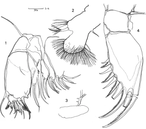
(5) Sh. HIRUTA. Fig. 3 Vargula hilgendorfii. Female. 1. sixth limb ; 2. seventh limb ; 3. furca ; 4. genitalia. Male. 5. copulatory appendage.. segment with two large teeth on cutting edge, one pulmose a—bristle, and two /?—bristles, of. which one is short, slender ; second segment with four a—bristles and eight pectinate bristles along distal margin. Fifth limb (Fig. 2—6). Main tooth with six constituent teeth and proximal peg. Sixth limb (Fig. 3—1). Epipodial appendage with five short bristles. Seventh limb (Fig. 3—2) with a total of 23 bristles. Furca (Fig. 3—3). Each lamella with about 13 claws ; claw 2 continuous with lamella, others separated from lamella by suture. Genitalia (Fig. 3—4) as shown in figure. Rod-shaped organ and median eye (Fig. 1—5). Rod-shaped organ short, pear-shaped. Male. Carapace about 2.78 mm in length, about 1.90 mm in height. First antenna (Fig. 1—. 1,2) : b— bristle with stout proximal filament furnished with a swollen basis and a large sucker, and with two following filaments which are longer and slenderer, and provided with six and five small suckers respectively ; c—bristle with sucking organs as in b—bristle. Second antenna to. seventh limb similar to female. Copulatory limb (Fig. 3—5) as shown in figure. Specimens examined. A number of specimens were collected by Dr. I. Okubo from. Mukaijima Islet, Hiroshima Pref. (11—VIII—76). Three females and two males were selected and examined.. First larval stage (A—5 instar ; sex undetermined). Carapace (Fig. 4—1) about 0.88 mm in length, about 0.59 mm in height, similar to adult in shape. First antenna (Fig. 5—1) similar to adult in total appearance ; surface of second segment. with short hairs and a row of hairs along distal margin ; fourth segment without bristles ; sensory. (148).
(6) Larvae of Vargula and Euphilomedes. Fig. 4 Vargula hilgendorfii. 1-5. first to fifth larval stages. 1-4. sex undetermined ; 5. male.. bristle of fifth segment without filaments. Second antenna (Fig. 5—2,3) similar to adult ; first endopodite segment without bristles ; bristle of second exopodite segment with a row of several teeth along ventral margin ; ninth segment with basal spine and two bristles, of which one is short.. Mandible (Fig. 5—4). Coxale endite semitriangular, with a number of spinules ; dorsal margin of basale with one midbristle and two subterminal bristles ; ventral margin of basale with one proximal and two distal bristles. Exopodite as long as dorsal margin of first endopodite segment, with a hairy process and two bristles. Ventral margin of first endopodite segment with two bristles ; second segment long, with one proximal stiff hair, two short bristles and three long bristles on proximal half of dorsal margin, and with one short subterminal bristle on ventral margin ; end segment small, with four bristles of an equal length. Maxilla (Fig. 6—1,2). First endite with four bristles ; second one with four bristles ; third one with six bristles. Basale with one bristle on ventral margin. Exopodite with one proximal and two distal bristles, of which one is pulmose. Endopodite : first segment with one a—bristle, one /?—bristle, and two processes near base of /?—bristle ; end segment with two a—bristles and. three pectinate terminal bristles. Fifth limb (Fig. 6—3). First endite with one bristle and one process terminating in a sharp point ; second one with three bristles and one process ; third one with four bristles. Exopodite : first segment with one stout bristle and main tooth which is composed of one tooth ; second one with two stout bristles ; third one with two bristles on outer lobe and one bristle on inner part ; fourth one with two distal bristles and a small hairy process. Sixth limb (Fig. 6—4) small, leaf-like, with long marginal hairs. Seventh limb not observed. Furca (Fig. 6—5). Each. lamella with four or five claws. Rod-shaped organ (Fig. 5—5) similar, to adult. Upper Up (Fig. 6—6) similar to adult except for small number of glandular openings.. (149).
(7) Sb. WRUTA. ... 47man^ble'-0-. (150).
(8) Larvae of Vargula and Euphilomedes. Fig. 6 Vargula hilgendorfii. First larval stage (A-5 instar). 1. endopodite and exopodite of maxilla ; 2. endite of maxilla ; 3. distal part of fifth limb ; 4. sixth limb ; 5. furca ; 6. upper lip.. Second larval stage (A—4 instar ; sex undetermined).. Carapace (Fig. 4—2) about 1.12 mm in length, about 0.73 mm in height. First antenna (Fig. 7— 1). One dorsal bristle added to fourth segment; sensory bristle of fifth segment with two long proximal and two distal filaments, terminating in bifurcate tip ; c—, f—, and g—bristle with filaments. Second antenna (Fig. 7—2—4). First endopodite segment with one bristle. Ninth segment of exopodite with three bristles ; fourth to ninth segments with basal spines. Mandible (Fig. 7—5). Ventral margin of basale with two proximal bristles and three distal bristles, of which one is very short ; dorsal margin of second endopodite segment with four bristles and two hairy short bristles ; end segment with two claws and three bristles. Maxilla (Fig. 8—1). Second endopodite segment with three a—bristles and six pectinate bristles along distal margin. Fifth limb. Main tooth with two constituent teeth. Sixth limb (Fig. 8—2) with two lobes ; small. (151).
(9) Sh. HIRUTA. Fig. 7 Vargula hilgendorfii. Second larval stage (A-4 instar). 1. first antenna ; 2. endopodite of second antenna ; 3. distal part of exopodite of second antenna ; 4. bristle of second exopodite segment of second antenna ; 5. mandible.. antenor one with one bristle and some marginal long hairs ; large posterior one covered with long hairs on surface ; one proximal bristle present on posterior margin near base of large lobe". Seventh Umb (Fig. 8-3) forming a small thumb-like process. Furca (Fig. 8-4). EachTamella. with five claws.. Third larval stage (A ~ 3 instar ; sex undetermined). Carapace (Fig. 4-3) about 1.46 mm in length, about 0.96 mm in height. First antenna (Fig. 9-1). One ventrodistal bristle added to fourth segment ; sensory bristle of fifth segment with four long proximal and two short distal filaments. Second antenna (Fig. 9-2). First"endopodite. segment with one proximal and one distal bristles. Mandible (Fig. 9-3). Ventral margin of basale with two proximal bristles and five bristles, of which two are very short and distalmost one. (152).
(10) Larvae of Vargula and Euphilomedes. Fig. 8 Vargula hilgendorfii. Second larval stage (A—4 instar). 1. maxilla ; 2. sixth limb ; 3. seventh limb ; 4. furca.. is long with stiff hairs in the midst. Maxilla (Fig. 9—4). First endopodite segment with two ,3—bristles ; end segment with three a—bristles and total seven pectinate bristles along distal margin. Fifth limb. Main tooth with three constituent teeth. Sixth limb (Fig. 9—5) similar to adult; epipodial appendage with two bristles ; first endite with one bristle ; second one with three bristles ; third one with three bristles ; fourth one with one bristle ; end segment with five bristles and long hairs along ventral margin. Seventh limb (Fig. 9—6) elongate, bare. Furca. Each lamella with six or seven claws.. Fourth larval stage (A—2 instar ; sex undetermined).. Carapace (Fig. 4—4) about 1.90 mm in length, about 1.23 mm in height. First antenna (Fig. 10—1). Sensory bristle of fifth segment with six proximal and two short distal filaments. First endopodite segment of second antenna (Fig. 10—2) with two proximal and one distal bristles. Mandible similar to adult. Maxilla (Fig. 10—3). Second endopodite segment with four a— bristles, seven pectinate bristles and one short slender bristle. Fifth limb. Main tooth with four constituent teeth. Sixth limb (Fig. 10—4). Epipodial appendage with three bristles ; first endite with one bristle ; second one with four bristles ; third one with three bristles ; fourth one with two bristles ; end segment with six pulmose bristles. Seventh limb (Fig. 10—5) with two cleaning. (153).
(11) Sh.HWUTA. ^3 instar). ^ L. stage ^^ :"3 central. "•' len£po^;5-!. (154).
(12) Larvae of y,. ar"'aandEWo,. 'edes. Fi8-10 Varg^^. (155).
(13) Sh. HIRUTA. Fig. 11 Vargula hilgendorfii. Fifth larval stage (A-l instar). Mzde. 1. sixth limb ; 2. seventh limb ; 3. copulatory appendage.. bristles ; terminal comb with three teeth ; one peg present opposing comb. Furca. Each lamella with nine claws.. Fifth larval stage (A—l instar). Male. Carapace (Fig. 4—5) about 2.43 mm in length, about 1.65 mm in height. Sensory. bristle of first antenna with seven or eight long proximal filaments. First endopodite segment of second antenna with three proximal and one distal bristles. Mandible and maxilla similar to adult. Fifth limb. IVtain tooth with five constituent teeth. Sixth limb (Fig. 11—1) similar to adult ; epipodial appendage with four bristles. Seventh limb (Fig. 11—2) similar to adult ; each cleaning bristle tapering distally, with two to four bells ; terminal comb with six teeth ; one peg present opposing comb. Furca. Each lamella with ten claws. Copulatory limb (Fig. 11—3) composed of three lobes, which have one or two short bristles. Female. Structures of carapace and appendages except for genital organ were almost same as in male of A—1 instar.. Remarks. Okada and Kato (1946) have already reported that the present species certainly has five different larval stages, in the study on the life history of this species. Specimens examined. A—5—A—1 instars (11—VIII—'76) IVEukaijima, I. Okubo leg.. Euphilomedes nipponica Hiruta, 1976 (Figs. 12-23) Euphilomedes nipponica Hiruta, 1976, p. 580—589, fig. 1—6. First larval stage (A—5 instar ; sex undetermined). Carapace (Fig. 12—1) similar to adult female in shape, about 0.63 mm in length, about 0.46. (156).
(14) Larvae of Vargula and Euphilomedes. Fig. 12 Euphilomedes nipponica. 1—5. first to fifth larval stage. 1,2. sex undetermined ; 3—5. female.. mm in height. First antenna (Fig. 13—1) similar to about female in general appearance ; second and fourth segments without bristles ; a row of hairs present along distal margin of second segment ; bristles of fifth to end segments without filaments. Second antenna (Fig. 13—2) similar to adult female. Endopodite two-segmented ; first segment without bristles. Mandible (Fig. 13—3) similar to adult femade ; coxale endite well-developed ; ventral margin of basale with some groups of hairs, and without bristles ; exopodite well-developed ; ventral margin of first endopodite segment with two bristles ; dorsal margin of second segment with four bristles ; ventral margin of second segment with one subterminal bristle ; end segment with two claws and two bristles. Maxilla (Fig. 13—4) similar to adult female ; first endite with six bristles ; second one with five bristles ; third one with four bristles ; basale with one ventral bristle ; exopodite with three terminal bristles of different lengths ; first endopodite segment with one a—bristle and one /3— bristle ; end segment with five bristles, of which one is short, claw-like. Fifth limb (Fig. 13—5) similar to adult female except for small number bristles ; inner lobe of third exopodite segment with one short bristle ; outer lobe of third segment with two bristles ; fourth and fifth segments with two stout terminal bristles. Sixth limb (Fig. 13—6) small, leaf-like, with six protuberances which are provided with long hairs. Seventh limb not observed. Furca (Fig. 13—7). Each lamella with two claws and one proximal claw-like process, which is hairy and continuous with lamella ; posterior margin of lamella with hairs. Rod-shaped organ (Fig. 13—8) elongate, as shown in figure. Second larval stage (A—4 instar ; sex undetermined).. Carapace (Fig. 12—2) about 0.75 mm in length, about 0.55 mm in height. First antenna (Fig.. (157).
(15) Sh. HIRUTA. - sSSff^S5^ median eye' . »^p..t •, one dorsodistal br. ^^SSis^iS .- °"ir:.^:;^s=S^:S. ^^S^i^S^ S^!sS^===':?. Veriral mar? "i basall7^7n oTsecond ^^^^e br>stle and three d. T^ one is short.^ MWU^I!°^ t,m6 (Fig. 14-6)as^ee'pr°ot"berances «h:. ;ovidedw^au3',la^-.- ^.
(16) Lar. WO^.W,^EupM. omedes. KE-U^^^J '"' '^/^ 3u ~" ° /•' /• J\VM^. " §?ssS£?»"^s.'.^ Kmb ;9:fa^aa& •• 6. fif"'^Tna7;',tTC?nd ^ennT,^. a°d some „„„... .. ""'urca' ^"""o''7'suy"'mb~^e'^. ':=!=9ss^^^te. ni^^^ ^^._ ~ ~ """" ws- u~l) s"""ar"to":duTe"a with f've ^. pemaie. ^rZuge (A^3 ins^).. sy^'sssSss ^"^. 2TT& 12:^bout 0.93 _ ,,. rl& 16~1)' and^^"'OT,STent ?itA two. (159) "" ^" ^~~2^ similar to.
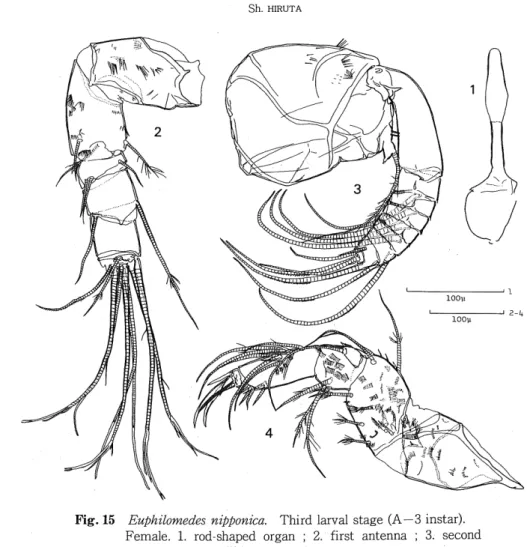
(17) Sh. HIRUTA. Fig. 15 Euphilomedes nipponica. Third larval stage (A—3 instar). Female. 1. rod-shaped organ ; 2. first antenna ; 3. second. antenna ; 4. mandible.. adult female except for somewhat smaller number of bristles. Sixth limb (Fig. 16—3) welldeveloped, similar to adult female. Seventh limb (Fig. 16-4) elongate, bare. Furca (Fig. 16-5) similar to adult female ; third claw short, as long as fifth to seventh ones. Rod-shaped organ (Fig. 15—1) similar to adult female. Male. Second antenna. Endopodite (Fig. 17—1) weakly three-segmented ; first segment. with three short bristles ; end segment with one short distal bristle. Other structures almost same as in female of A—3 instar.. Fourth larval stage (A—2 instar). Female. Carapace (Fig. 12-4) about 1.21 mm in length, about 0.86 mm in height First antenna (Fig. 18—2) almost Same as in adult. Second antenna (Fig. 18—3). First endopodite segment with three proximal and one distal bristles ; ninth segment of exopodite with five bristles of different lengths. Median eye and rod-shaped organ (Fig. 18—1), mandible (Fig. 19—1), maxilla (Fig. 19-2), fifth limb (Fig. 19-3), sixth limb (Fig. 19-4), and furca (Fig. 19-6) similar to adult female. Seventh limb (Fig. 19—5) furnished with total eight cleaning bristles ; each. (160).
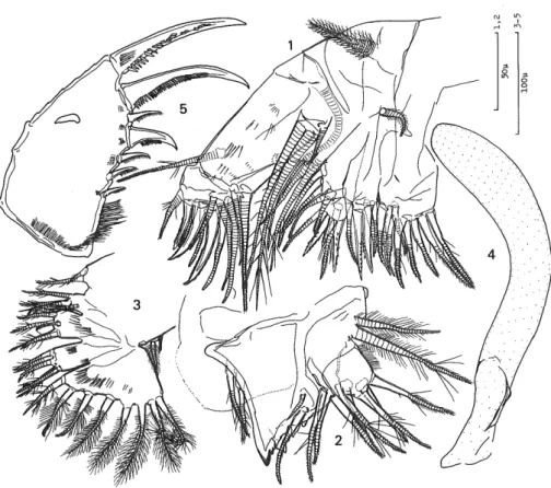
(18) Larvae of Vargula and Euphilomedes. Fig. 16 Euphilomedes nipponica. Third larval stage (A—3 instar). Female. 1. maxilla ; 2. fifth limb ; 3. sixth limb ; 4. seventh limb ; 5. furca.. bristle tapering distally, with marginal spines and one to three bells. Male. Endopodite of second antenna (Fig. 17-2) three-segmented ; first segment with three proximal and one distal bristles ; second one longer than third, with a distal short bristle and proximal long one ; third one with minute bristle on middle of posterior margin and short distal bristle. Other structures almost same as in female of A—2 instar.. Fifth larval stage (A—l instar). Female. Carapace (Fig. 1.5) about 1.50 mm in length, about 1.11 mm in height. First antenna (Fig. 20-2), mandible (Fig. 20-5), maxilla, fifth Limb (Fig. 20-6), sixth limb (Fig. 207), furca, and rod-shaped organ similar to adult female. Second antenna (Fig. 20—3,4). First. endopodite segment with four proximal bristles and one distal bristle ; ninth segment of exopodite with six bristles. Seventh limb (Fig. 20—8) with ten bristles, six distally, four proximally. Male. Second antenna (Fig. 21—3,4). Endopodite three-segmented ; first segment with. four proximal bristles and a distal one ; second segment elongate, somewhat longer than third, with long proximal bristle and two short bristles on middle of anterior surface ; third one with one short bristle near middle of posterior surface and two short juxtaposed terminal bristles. Lateral. (161).
(19) Sh. HIRUTA. Fig. 17 Euphilomedes nipponica. Male. 1. endopodite of second antenna of third larval stage (A—3 instar) ; 2. endopodite of second antenna of fourth larval stage (A—2 instar).. Fig. 18 Euphilomedes nipponica. Fourth larval stage (A—2 instar). Female. 1. rod-shaped organ and median eye ;2. first antenna 3. second antenna.. (162).
(20) Larvae of Vargula and Euphilomedes. Fig. 19 Euphilomedes nipponica. Fourth larval stage (A—2 instar). Female. 1. mandible ; 2. maxilla ; 3. fifth limb ; 4. sixth limb 5. seventh limb ; 6. furca.. eye (Fig. 21-5) developed, with ommatidia. Seventh limb (Fig. 23—3) with a total of eight cleaning bristles, four distally, four proximally. Other structures were almost same as in female. of A-l instar : first antenna (Fig. 21-2), mandible (Fig. 22-1). maxilla (Fig. 22-2-5), fifth limb (Fig. 23-1), sixth limb (Fig. 23-2), and furca (Fig. 23-4). Remarks. Since five different larval stages were detected by the investigation of the shell size and appendage morphology and, further, specimens which had just hatched out were also examined, the present species certainly passes through five moults before reaching maturity.. Sex is easily determinable by the structure of the endopodite of the second antenna in the last three larval stages.. Specimens examined. The specimens were collected from the bottom sediment of muddy sand (0-4 m depths) sampled at Oshoro, on the Japan Sea coast of Hokkaido by means of the decanting and sieving method. A-5 instars hatched from eggs of a female (11-VI-75) ; A-. (163).
(21) Sh. HIRUTA. 20 SUMmede\nippomca- Flfth larval stage (A-l instar). e.^,rcdjhapedorgan and medlan eye =2. first"antenna. ienjlopoditeofsecon^ '• ^.'nmtoseg^nFof^^nT. i^Tna; 5-mandlble; 6. fifth limb ; 7. sixth'UmbTrseven^th. (164).
(22) Larvae of Vargula and Euphilomedes. Fig. 21 Euphilomedes nipponica. Fifth larval stage (A—l instar). Male. 1. rod-shaped organ ; 2. first antenna ; 3. exopodite of second antenna ; 4. protopodite and endopodite of second antenna ; 5. lateral eye.. 4 instars (11-VI-75) ; A-3 instars (10-IX-75) ; A-2 instars (10-IX-75) ; A-l instars (27-V-75) Sh. Hirutaleg.. Discussion. It is ascertained that the present two species, as described above, pass through five moults (A—5 to adult stage) before reaching maturity. In the family Cypridinidae, to which Vargula hilgendorfii belongs, some members possessing five or six different larval stages are hitherto known (Doloria pectinata, Macrocypridina castanea, and Gigantocypris- species), and in the family Philomedidae, which contains Euphilomedes nipponica, it is known that Philomedes globosus has five different larval stages (Kornicker, 1969). The morphological differentiation of the sixth and seventh limbs in the present two species almost accords with that designated in the key to early myodocopid instars (Kornicker, op. cit). However, it is noticeable that in Vargula hilgendorfii, the sixth limb of the second larval stage (A—4 instar) is provided with two bristles, of which one arises from the anterior lobe and the other from the posterior margin of the limb, while in Kornicker's key, the "6th limb with one. (165).
(23) Sh. HIRUTA. Fig. 22 Euphilomedes nipponica. Fifth larval stage (A—l instar). Male. 1. mandible ; 2. endopodite of maxilla ; 3. first endite of maxilla ; 4. second endite of maxilla ; 5. third endite of maxilla.. bristle" characterizes the second larval stage. The occurrence of bristles on the fourth segment. ourth of the first antenna in these species entirely accords with that of other early myodocopid instars so far known. Namely, in the first larval stage, the fourth segment is without any bristles.. In the second stage, the segment is provided with one dorsodistal bristle, and then in the next stage, one bristle is added to the ventrodistal edge. Therefore, this characteristic of the first antenna seems to be also useful for the identification of early myodocopid instars.. Acknowledgements I would like to express my sincere gratitude to Professor Mayumi Yamada of Hokkaido University for his advice and guidance. Cordial thanks are also due to Dr. I. Okubo of Biological Laboratory, Okayama Shujitsu Junior College, who kindly sent me many specimens of Vargula hilgendorfii from Mukaijima Islet, Hiroshima Pref.. (166).
(24) Larvae of Vargula and Euphilomedes. Fig. 23 Euphilomedes nipponica. Fifth larval stage (A—instar). Male. 1. fifth limb ; 2. sixth limb ; 3. seventh limb ; 4. furca.. References Hanai, T., Ikeya, N., Ishizaki, K., Sekiguchi, Y., and Yajima, M. 1977. Checklist of Ostracoda from Japan and its adjacent seas. Bull., Univ. Mus. Univ. Tokyo 12, 120 pp.. Hiruta, Sh. 1976. Euphilomedes nipponica n. sp. from Hokkaido, with a redescription of E. sordida (G. W. Muller) (Ostracoda : Myodocopina). J. Fac. Sci. Hokkaido Univ. Ser. 6, Zool. 20 : 579-599. Hiruta, Sh. 1977. A new species of the genus Sarsiella Norman from Hokkaido, with reference to the larvel stages (Ostracoda : Myodocopina). Ibid. 21 : 44—60, pl. 4.. Hiruta, Sh. 1978. Redescription of Sarsiella misakiensis Kajiyama from Hokkaido, with reference to the larval stages (Ostracoda : Myodocopina). Ibid. 21 : 262-278. Hiruta, Sh. 1979 (a). A new species of the genus Bathyleberis Kornicker from Hokkaido, with reference to the larval stages (Ostracoda : Myodocopina). Ibid. 22: 99—121. Hiruta, Sh. 1979 (b). Redescription of Asteropteron fuscum (G. W. Muller) from Amakusa, Kyushu, with reference to the larval stages (Ostracoda : Myodocopina). Proc. Jap. Soc. Syst. Zoology. 17 : 15—30. Kajiyama, E. 1912. The Ostracoda from Misaki ; Part 2, Myodocopa. Zool. Mag., Tokyo (Dobutsugaku-zasshi) 24 : 609-619, pl. 9, (in Japanese). Kornicker, L. S. 1969. Morphology, ontogeny, and intraspecific variation of Spinacopia, a new genus of myodocopid ostracod (Sarsiellidae). Smiths. Contr. Zool. 8 : 1—42, 4 pis. Muller, G. W. 1890. Neue Cypridiniden. Zool. Jahrb. System. 5 : 211-252, pl. 25-27.. Okada, Y. and Kato, K. 1946. Studies on luminous animals in Japan, III. Preliminary report on the life history of Cypridina hilgendorfii. Kagaku 16 : 64—66, (in Japanese). Poulsen, E. M. 1962. Ostracoda-Myodocopa ; Part 1. Cypridiniformes-Cypridinidae. Dana Reports 57 : 1—414.. Skogsberg, T. 1920. Studies on marine Ostraeods, 1. Cypridinids, Halocyprids, and Polycopids. Zool. Bidrag fran Uppsala, suppl. 1 : 1—784.. (167).
(25)
図


+7


関連したドキュメント
Lomadze, On the number of representations of numbers by positive quadratic forms with six variables.. (Russian)
We then introduce the notion of compression of a graph Γ which plays an important role in the study of partially commutative groups and prove that the lattices of closed sets for
We describe a filtration of Pic( L I ) in the last section as well as the proofs of some facts. We also discuss there the small objects in some local stable homotopy categories...
As fun- damental groups of closed surfaces of genus greater than 1 are locally quasicon- vex, negatively curved and LERF, the following statement is a special case of Theorem
Then the strongly mixed variational-hemivariational inequality SMVHVI is strongly (resp., weakly) well posed in the generalized sense if and only if the corresponding inclusion
Lemma 1.11 Let G be a finitely generated group with finitely generated sub- groups H and K , a non-trivial H –almost invariant subset X and a non-trivial K –almost invariant subset
Finally, we use results from the well-developed theory of permutation groups and modular permutation representations to give a description of the primitive permuta- tion groups
[r]
