老化による脂質吸収能変化の解析とその遅延方法の
探索に関する研究
著者
山本 和史
学位授与機関
Tohoku University
学位授与番号
11301甲第18744号
URL
http://hdl.handle.net/10097/00125730
1
博士論文
老化による脂質吸収能変化の解析と
その遅延方法の探索に関する研究
Resarch on analysis of lipid absorption
ability change with aging and search for
method to delay this problem
東北大学大学院農学研究科
生物産業創成科学専攻
山本 和史
指導教官 都築 毅 准教授
平成 30 年度
2
Contents
Introduction・・・・・・・・・・・・・・・・・・・・・・・・・・・・・4
References・・・・・・・・・・・・・・・・・・・・・・・・・・・・・・8
Chapter 1: Analysis of the mechanism of lipid absorption ability change with aging 1.1. Abstract・・・・・・・・・・・・・・・・・・・・・・・・・・9
1.2. Introduction・・・・・・・・・・・・・・・・・・・・・・・・10
1.3. Materials and methods・・・・・・・・・・・・・・・・・・・・12
1.4. Results・・・・・・・・・・・・・・・・・・・・・・・・・・17
1.5. Discussion・・・・・・・・・・・・・・・・・・・・・・・・・26
1.6. References・・・・・・・・・・・・・・・・・・・・・・・・・30
Chapter 2: Effect of high fat diet intake on lipid absorption ability with aging 2.1. Abstract・・・・・・・・・・・・・・・・・・・・・・・・・・36
2.2. Introduction・・・・・・・・・・・・・・・・・・・・・・・・37
2.3. Materials and methods・・・・・・・・・・・・・・・・・・・・39
2.4. Results・・・・・・・・・・・・・・・・・・・・・・・・・・43
2.5. Discussion・・・・・・・・・・・・・・・・・・・・・・・・・54
2.6. References・・・・・・・・・・・・・・・・・・・・・・・・・60
Chapter 3: Effects of a moderate-fat diet enriched with fish oil on intestinal lipid absorption and various biochemical parameters with aging
3.1. Abstract・・・・・・・・・・・・・・・・・・・・・・・・・・67
3.2. Introduction・・・・・・・・・・・・・・・・・・・・・・・・68
3.3. Materials and methods・・・・・・・・・・・・・・・・・・・・70
3.4. Results・・・・・・・・・・・・・・・・・・・・・・・・・・76
3
3.6. References・・・・・・・・・・・・・・・・・・・・・・・・・93
Chapter 4: The effect of moderate-fat diet enriched with fish oil on gut microbiota change due to aging
4.1. Abstract・・・・・・・・・・・・・・・・・・・・・・・・・・98
4.2. Introduction・・・・・・・・・・・・・・・・・・・・・・・・99
4.3. Materials and methods・・・・・・・・・・・・・・・・・・・・102
4.4. Results・・・・・・・・・・・・・・・・・・・・・・・・・・107
4.5. Discussion・・・・・・・・・・・・・・・・・・・・・・・・・122
4.6. References・・・・・・・・・・・・・・・・・・・・・・・・・129
Chapter 5: The effect of moderate-fat diet enriched with fish oil and vitamin E on lipid absorption ability and other biochemical parameters due to aging
5.1. Abstract・・・・・・・・・・・・・・・・・・・・・・・・・・138
5.2. Introduction・・・・・・・・・・・・・・・・・・・・・・・・139
5.3. Materials and methods・・・・・・・・・・・・・・・・・・・・141
5.4. Results・・・・・・・・・・・・・・・・・・・・・・・・・・147
5.5. Discussion・・・・・・・・・・・・・・・・・・・・・・・・・162
5.6. References・・・・・・・・・・・・・・・・・・・・・・・・・169
Conclusion・・・・・・・・・・・・・・・・・・・・・・・・・・・・・・175
4
Introduction
Aging is a decrease in function dependent on age, and a decrease in function has been
observed in various organs due to aging [1-3]. It is important for the elderly to retard
aging and to delay the decline in tissue function in order to realize productive life and
finishing their long life.
To delay aging, our group focused on the diet (Japanese diet) that Japanese people
usually eat, and studied this. We used mice. As a result, it was revealed that Japanese
diet in 1975 is effective as a Japanese diet that can delay aging and delay the
deterioration of organs [4] (Figure 1). However, though the onset of lifestyle diseases
such as obesity was seen early in mice which ingested Japanese diet in 1990 which has
higher lipid content than that of Japanese food in 1975, the time reaching the end of
their lives was long, so the Japanese diet in 1990 seemed to delay the aging. Therefore,
it seemed that the decrease in energy intake due to aging was supplemented by ingesting
a diet containg high lipid. In addition, we also observed an interesting phenomenon that
in mice, when aging progressed and mice eventually died, weight was significantly
reduced and visceral fat disappeared. From the above, it is considered that aging may
cause a change in the gastrointestinal absorption ability of lipids, which may have an
adverse effect on the elderly. Moreover, elderly people with a BMI of 20 or less are
malnutritional trends, and malnutrition of elderly people has become a serious problem
in recent years. Furthermore, the number of elderly people judged to be malnutritional
trends continues to increase, and it is expected that they will increase further because of
progression of aging society [5] (Figure 2). Based on the above, it was thought that by
investigating the change in lipid absorption ability due to aging, we can be cause a stir
5
the elderly. However, little research has been done on the change in gastrointestinal
absorption ablity of lipids due to aging.
Therefore, in this study, we focused on changes in gastrointestinal absorption ability
of lipids due to aging, especially; we focused on clarifying its mechanism and searching
6
Figure 1. Effect of Japanese foods on survival curves and mean life span in senescence-accelerated prone mouse-8 mice. Values are means ± SEM n = 20.
7
Figure 2. Trends and forecasts of the number of elderly people with malnutrition trends in Japan.
2005
2011
2017
3000
6000
9000
Thousand people
2023
Year
8
Referernces
[1] Honma T, Yanaka M, Tsuduki T, Ikeda I., 2011. Increased lipid accumulation in
liver and white adipose tissue in aging in the SAMP10 mouse. J Nutr Sci Vitaminol
(Tokyo). 57:123-129.
[2] Cuesta S, Kireev R, García C, Rancan L, Vara E, Tresguerres JA., 2013. Melatonin
can improve insulin resistance and aging-induced pancreas alterations in
senescence-accelerated prone male mice (SAMP8). Age (Dordr). 35:659-671. doi:
10.1007/s11357-012-9397-7
[3] Drozdowski L, Woudstra T, Wild G, Clandinin MT, Thomson AB., 2005. Dietary
lipids modify the age-associated changes in intestinal uptake of fructose in rats. Am
J Physiol Gastrointest Liver Physiol. 288:G125-134.
[4] Yamamoto K, E S, Hatakeyama Y, Sakamoto Y, Honma T, Jibu Y, Kawakami Y,
Tsuduki T (2016) The Japanese diet from 1975 delays senescence and prolongs life
span in SAMP8 mice. Nutrition. Jan;32(1):122-128. doi: 10.1016/j.nut.2015.07.002
[5] Ministry of Health, Labour and Welfare., 2017. National Health and Nutrition
Examination Survey. URL:
9
Chapter 1: Analysis of the mechanism of lipid absorption ability change with aging
1.1. Abstract
Malnutrition due to aging is partly caused by decreased absorption of nutrients by the
gastrointestinal tract. However, the underlying mechanism for age-associated
malnutrition is unclear and changes in lipid absorption with aging are poorly understood.
In this study, changes in lipid absorption with aging were examined in mice aged 3 and
25 months. After overnight fasting, blood samples were collected from snipped tails and
then soybean oil was administered orally. Three hours later, mice were sacrificed by
decapitation and the liver, pancreas, small intestine and blood were collected. The
increase in serum triacylglycerol after soybean oil administration was significantly
lower in the older mice, indicating a decrease in lipid absorption with aging.
Measurement of mRNA levels for triacylglycerol absorption-related molecules showed
that mRNA for pancreatic lipase tended to decrease in 25-month-old mice. There was no
significant difference in the protein level of pancreatic lipase, but the enzyme activity
showed a significant decrease in the older mice. To examine this mechanism, expression
levels of mRNA for protein turnover-related molecules in the pancreas were measured.
The level of a proteasomal mRNA showed a significant decrease in 25-month-old mice.
This suggests that the ability to degrade unfolded protein decreases in the aging
pancreas, and that this leads to reduction of pancreatic lipase activity and a decrease in
lipid absorption.
10
1.2. Introduction
The population of elderly persons has increased worldwide and this has emphasized
the importance of studying mechanisms of aging. Senescence refers to age-related
changes in physiological function and aging depresses tissue functions such as lipid
metabolism in the liver and insulin secretion in the pancreas [1-4]. Delay of senescence
and maintenance of quality of life in elderly persons requires careful attention to diet
and nutrition.
Malnutrition is a concern in elderly persons because it induces loss of motor function,
delayed recovery from illness, and increased rates of complications and mortality [5, 6].
Malnutrition may arise because of a decrease in the ability of the gastrointestinal tract to
facilitate absorption of nutrients [7]. The functions of tissues related to gastrointestinal
tract absorption, such as those in the pancreas and small intestine, decrease with aging.
Exocrine secretion of digestive enzymes decreases with reduction of acinar cells in the
pancreas. This induces a decrease in the secretion volumes of amylase and lipase, which
process carbohydrates and lipids, respectively [8]. In the small intestine, the surface area
decreases due to degeneration of villi and the number of nerve cells related to digestive
absorption also decreases [9-11]. Carbohydrate absorption decreases with aging,
including reduced uptake of glucose [9, 10, 12]. Absorption of essential minerals such
as calcium and magnesium also decreases with aging [13]. However, few studies have
examined the absorption properties of the gastrointestinal tract with aging because there
are several inconsistent comment reports for absorption and metabolism in the small
intestine. For example, previous studies were shown that glucose absorption in small
intestine increased or decreased with aging [9, 14, 15]. To understand aging and small
11
In this study, we examined lipid absorption in 3 and 25-month-old mice. After
overnight fasting, blood samples were collected from snipped tails and soybean oil was
then administered orally. Three hours later, mice were sacrificed by decapitation and
tissues and blood were collected. Senescence indicators were examined in tissues
associated with triacylglycerol (TG) absorption. Serum TG levels were measured to
examine changes in lipid absorption with aging. Mechanisms underlying changes in
lipid absorption with aging were examined based on measurement of expression levels
of mRNA for TG absorption-related genes. The results showed that decreased
12
1.3. Materials and methods Animals
All procedures were performed in accordance with the Animal Experiment
Guidelines of Tohoku University. The animal protocol was approved by the Animal Use
Committee at Tohoku University [16]. Male ICR mice (1-month-old) were obtained
from Japan SLC (Hamamatsu, Japan). Thirteen mice [3-month-old mice (n = 7) and
25-month-old mice (n = 4, two mice died on the way)] were used. The mice were
sacrificed for analysis at age 3 and 25 months. The mice were housed in individual
cages with access to a control diet (CE-2) and distilled water ad libitum in a
temperature- and humidity-controlled room with light cycles of 12 h on and 12 h off
until age 3 or 25 months [17]. The mice were weighed and then sacrificed for analysis at
3 and 25-month-old by decapitation. The liver, pancreas, small intestine and serum were
collected and stored at -80°C until performance of assays.
Triacylglycerol assays in serum and small intestine
After overnight fasting of mice aged 3 and 25 months, 5 g of soybean oil per kg body
weight (about 200 µL) was administered orally. Serum TG levels were measured at 0
and 3 h with an enzyme kit (Wako Pure Chemical, Osaka, Japan). Serum samples were
collected in 1.5 ml tubes for the assay. The small intestine of mice sacrificed at 3 hours
after soybean oil administration was homogenized with four volumes of ice-cold saline.
Total lipids from the small intestine homogenate were extracted using the Bligh-Dyer
method [18]. TG in total lipids of the small intestine was measured using an enzyme kit
13
Thiobarbituric acid active substance assay
To examine oxidative stress caused by aging, the levels of thiobarbituric acid active
substances (TBARS) in serum, liver, pancreas and small intestine were measured as
described previously [19, 20].
mRNA expression analysis
For real-time quantitative reverse transcriptase PCR (qRT-PCR), total RNA was
isolated from liver, pancreas and small intestine using an RNeasy Mini Kit (Qiagen,
Valencia, CA) [2, 21], eluted with 30 µL RNase-free water, and stored at -80°C until
use. To quantify the expression levels of genes, mRNA levels for beta-actin (Actb),
activating transcription factor 6 (Atf6), colipase (Clps), cholesterol 7α-hydroxylase
(Cyp7a1), fatty acid-binding protein 2 (Fabp2), glucose-related protein 78 (Grp78), heat
shock protein 40 (Hsp40), heat shock protein 70 (Hsp70), inositol-requiring 1 (Ire1),
microsomal triglyceride transfer protein (Mttp), p21, pancreatic lipase-related protein 2
(Plrp2), proteasome (prosome) subunit, beta type 5 (Psmb5) and pancreatic lipase (Ptl)
in liver, pancreas and small intestine were determined with a Thermal Cycler Dice Real
Time System® (Takara Bio, Otsu, Japan). This system allows real-time quantitative
detection of PCR products by measuring the increase in fluorescence caused by binding
of SYBR green to double-stranded DNA [2, 22]. In brief, cDNA was made using Prime
Script® RT Master Mix (Perfect Real Time) (Takara Bio, Otsu, Japan) from total RNA
in liver, pancreas and small intestine. The cDNA was subjected to PCR amplification
using SYBR® Premix Ex TaqTM (Perfect Real Time) (Takara Bio, Otsu, Japan) and
gene-specific primers for Actb, Atf6, Clps, Cyp7a1, Fabp2, Grp78, Hsp40, Hsp70, Ire1,
14
an activation step at 95oC for 10 s, followed by 40 cycles at 95oC for 5 s (denaturation)
and 60oC for 31 s (extension), and a dissociation stage at 95oC for 15 s, 60oC for 30 s
and 95oC for 15 s for each gene. Melting curve analysis was performed following each
reaction to confirm the presence of only a single reaction product. The threshold cycle
(CT) represents the PCR cycle at which an increase in reporter fluorescence above a
baseline signal can first be detected. The ratio between the Actb content in standard
samples and test samples was defined as the normalization factor [23].
Western blot analysis
Western blot analysis was performed using antibodies that specifically recognize
proteins including pancreatic lipase (PTL), proteasome (prosome) subunit, beta type 5
(PSMB5), and actin beta (β-actin) [23]. The pancreas was homogenized, proteins were
extracted, and 10 μg of extracted protein was loaded for sodium dodecyl
sulfate-polyacrylamide gel electrophoresis (SDS-PAGE) immunoblot analysis. Protein
bands were then transferred to polyvinylidene fluoride membranes (Bio-Rad
Laboratories, Hercules, CA). After blocking of nonspecific sites with skim milk, the
membrane was probed with primary antibodies, followed by a horseradish
peroxidase-conjugated secondary antibody (Abcam, Cambridge, MA). Detection of
antibody reactions was performed with ECL Western blotting Detection Reagents (GE
Healthcare, Little Chalfont, UK). Each band was normalized using the corresponding
value of β-actin as an internal control. The antibodies used were β-actin [primary
antibody (Abcam, mouse monoclonal to beta Actin) dilution is 1:10000, secondary
antibody (Abcam, rabbit polyclonal) dilution is 1:4000], PTL [primary antibody (Santa
15
dilution is 1:4000] and PSMB5 [primary antibody (Abcam, rabbit polyclonal) dilution is
1:1000, secondary antibody (Abcam, goat polyclonal) dilution is 1:3000] [23-25].
Reaction times were 4oC over night in all antibodies.
Pancreatic lipase activity assay
The activity of pancreatic lipase was measured as described previously [26, 27]. The
frozen pancreas was thawed and homogenized in 4 vol of ice-cold 154 mmol/L NaCl.
The homogenate was centrifuged (13,800 × g for 20 min at 4°C), and aliquots of the
supernatant were stored at −80°C (enzyme liquid). Enzymatic activity was measured by determining the free fatty acid (FFA) level. In this procedure, 80 mg of triolein, 10 mg
of lecithin and 9 ml of bile acid were mixed in 0.1 mol/L Tris-HCl buffer (pH 7.0), and
then sonicated for 10 min (substrate liquid). Subsequently, 0.1 ml of substrate liquid and
0.05 ml enzyme liquid were mixed and incubated for 30 min at 37°C. FFA was
measured using an enzyme kit (Wako Pure Chemical, Osaka, Japan).
Statistical analysis
All statistical analyses were performed using Ekuseru-Toukei 2012 (SSRI, Tokyo,
Japan). Data are reported as mean ± SE. Statistical analysis was performed by one-way
16
Table 1.1. Primer pairs used for the quantitative RT-PCR analysis.
Genbank ID Target gene Primer Primer sequence (5’–3')
NM_007393 Actb F GAAATCGTGCGTGACATCAAAG R TGTAGTTTCATGGATGCCACAG NM_001081304 Atf6 F CCCAAGCTCTCCGCATAGTC R TAAAATGCCCCATAACTGACCAA NM_025469 Clps F GCTCTTGCCTTCTGCTGTCTGA R ATGGCGCCGATGATGCTCCTGT NM_007824 Cyp7a1 F GGGATTGCTGTGGTAGTGAGC R GGTATGGAATCAACCCGTTGTC NM_007980 Fabp2 F AGAGGAAGCTTGGAGCTCATGACA R TCGCTTGGCCTCAACTCCTTCATA NM_001163434 Grp78 F TCATCGGACGCACTTGGAA R AACCACCTTGAATGGCAAGAA NM_018808 Hsp40 F AGGCTCTCTGTGGTTGCACT R CTCAGGTGTTTTGGGGAGAG NM_010478 Hsp70 F AGAAGGTGCTGGACAAGTGC R CTGGTACAGCCCACTGATGA NM_023913 Ire1 F CATGAGGAACAAGAAGCACCACTA R TCGCTGTGTGAAGTACTGAATGAA NM_008642 Mttp F AGTGCAGTTCTCACAGTACCCGTT R AGCATATCGTTCTGGTGGAAGGGA NM_001111099 p21 F CGAGAACGGTGGAACTTTGAC R CAGGGCTCAGGTAGACCTTG NM_011128 Plrp2 F ATGCCTATGGATGTCCGTGGA R TGCCCAGGGCTTGTCATTG NM_011186 Psmb5 F CGCGAATCGAAATGCTTCA R TGTGGCCCGGGAATCC NM_026925 Ptl F CTGGGAGCAGTAGCTGGAAG R AGCGGGTGTTGATCTGTGC
Actb, actin beta; Atf6, activating transcription factor 6; Clps, colipase; Cyp7a1, cholesterol
7α-hydroxylase; Fabp2, fatty acid-binding protein 2; Grp78, glucose-related protein 78; Hsp40, heat shock protein 40; Hsp70, heat shock protein 70; Ire1, inositol-requiring 1; Mttp, microsomal triglyceride transfer protein; Plrp2, pancreatic lipase-rerated protein; Psmb5, proteasome (prosome) subunit, beta type 5; Ptl, pancreatic lipase.
17
1.4. Results
Growth parameters
To examine the difference in body weight, food intake and tissue weight between
young mice and old mice, growth parameters were measured. There were no significant
differences in body weight (35.4 ± 0.35 vs. 40.7 ± 3.89 g), food intake (5.19 ± 0.05 vs.
5.19 ± 0.03 g/day), and weights of liver (4.41 ± 0.08 vs. 4.37 ± 0.33 g/100 g body
weight) and pancreas (0.63 ± 0.08 vs. 0.81 ± 0.04 g/100 g body weight) between 3 and
25-month-old mice.
Senescence indicators in serum, liver, pancreas and small intestine
To examine progression of senescence in 25-month-old mice, lipid peroxidation
(TBARS) and p21 mRNA levels were examined (Table 1.2). The TBARS level in the
pancreas of 25-month-old mice was 141% of that in 3-month-old mice, showing a
significant increase with aging. In contrast, there were no significant difference in
TBARS levels in serum, liver and small intestine between 3 and 25-month-old mice.
p21 mRNA levels in the liver and pancreas of 25-month-old mice were 8.10 times and
1.85 times of those in 3-month-old mice, respectively, showing a significant increase
with aging. There was no significant difference in the p21 mRNA level in the small
intestine between 3 and 25-month-old mice.
Triacylglycerol levels in serum and small intestine
To examine lipid absorption in mice with aging, soybean oil (5 g/kg body weight)
was administered orally and the serum TG level was measured at 0 and 3 h (Table 1.3).
18
showing a tendency to decrease with age (P = 0.07). The serum TG level at 3 h in
25-month-old mice was 12% of that in 3-month-old mice, showing a significant
decreasewith aging. The difference in serum TG at 0 h and 3 h after administration of
soybean oil (ΔTG) in 25-month-old mice was 5% of that in 3-month-old mice, also showing a significant decreasewith aging. To examine the location of unabsorbed lipid
in senescence, the small intestine TG level was measured (Table 1.3). However, there
was no significant difference in small intestine TG levels between 3 and 25-month-old
mice.
mRNA levels for TG absorption-related genes in liver, pancreas and small intestine
To examine the mechanism underlying decreased lipid absorption with aging in mice,
mRNA levels for TG absorption-related genes in liver, pancreas and small intestine
were measured (Table 1.4). There was no significant difference in the mRNA level for
Cyp7a1, which is crucial for bile acid synthesis in the liver, between 3 and
25-month-old mice. The mRNA level for Ptl, which is required for TG hydrolysis in the
pancreas, in 25-month-old mice was 45% of that in 3-month-old mice, showing a
tendency for a decrease with age (P = 0.12). There were no significant differences in the
mRNA levels for Clps and Plrp2, which are required for TG hydrolysis in the pancreas,
between 3 and 25-month-old mice. There were also no significant differences in the
mRNA levels for Fabp2, which transports free fatty acids, and Mttp, which brings TG
into chylomicrons in the small intestine, between 3 and 25-month-old mice.
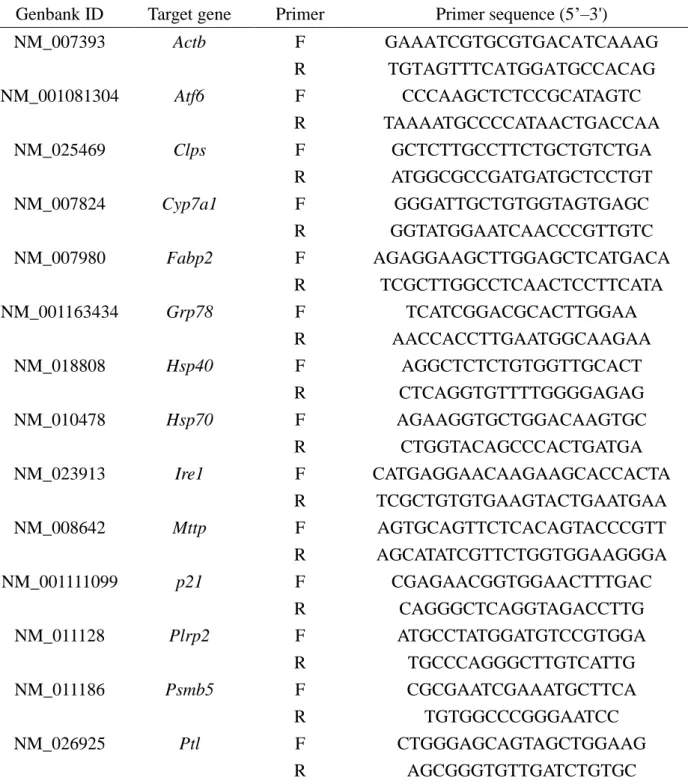
Protein level and enzymatic activity of pancreatic lipase
19
evaluate the pancreatic lipase function, the protein level and enzymatic activity were
measured (Figure 1.1). There was no significant difference in the protein level of
pancreatic lipase between 3 and 25-month-old mice (Figure 1.1A), but the pancreatic
lipase activity in 25-month-old mice was 62% of that in 3-month-old mice, showing a
significant decreasewith aging (Figure 1.1B).
mRNA levels for protein turnover-related genes in the pancreas
Expression levels of mRNA for protein turnover-related genes in the pancreas were
measured to examine why the protein level of pancreatic lipase was unchanged, but the
enzyme activity decreased, with aging (Table 1.5). In 3 and 25-month-old mice, there
were no significant differences in mRNA levels for Grp78, Hsp40 and Hsp70, which are
molecular chaperones that assist with protein folding, and for Atf6 and Ire1, which
maintain cell homeostasis by responding to endoplasmic reticulum (ER) stress caused
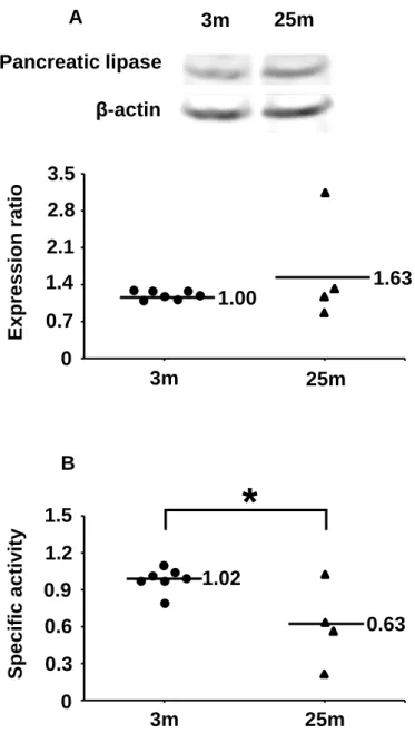
by accumulation of unfolded protein. In contrast, mRNA for Psmb5, a proteasomal
protein that facilitates elimination of unfolded protein in the pancreas, in 25-month-old
mice was 55% of the level in 3-month-old mice, showing a significant decreasewith
aging. The protein level of PSMB5 (Figure 1.2) in the pancreas of 25-month-old mice
20
Table 1.2. Senescence indicator in mice.
3m 25m p
TBARS
Serum (µmol/mmol lipid) 1.73 ± 0.52 0.91 ± 0.27 0.283 Liver (nmol/g tissue) 79.7 ± 8.17 92.1 ± 8.07 0.346 Pancreas (nmol/g tissue) 43.1 ± 3.73 60.8 ± 7.42* 0.040 Small intestine (nmol/g tissue) 37.5 ± 10.9 33.8 ± 9.95 0.827
p21 mRNA expression
Liver (Ratio) 1.00 ± 0.15 7.97 ± 2.12* 0.002 Pancreas (Ratio) 1.00 ± 0.13 1.85 ± 0.45* 0.045 Small intestine (Ratio) 1.00 ± 0.32 2.06 ± 0.73 0.153 Values are mean ± SE. *p <0.05 (vs 3-month-old mice). 3m, 3-month-old mice (n =7); 25m, 25-month-old mice (n = 4).
21
Table 1.3. Serum TG and small intestine TG levels in mice.
3m 25m p
Serum TG levels (mmol/L)
Before administering soy bean oil 1.38 ± 0.21 0.71 ± 0.23 0.069 After administering soy bean oil 8.33 ± 1.06 1.04 ± 0.47* <0.001
ΔTG 6.94 ± 1.00 0.34 ± 0.35* <0.001
Small intestine TG (µmol/g tissue) 21.0 ± 8.68 30.5 ± 22.6 0.651 Values are mean ± SE. *p <0.05 (vs 3-month-old mice). 3m, 3-month-old mice (n =7); 25m, 25-month-old mice (n = 4).
ΔTG, the differences in the serum TG level at 0h and after 3h in administration of soy bean oil. TG, triacylglycerol.
22
Table 1.4. mRNA expression levels for triacylglycerol absorption-related genes in liver, pancreas and small intestine of mice.
3m 25m p Liver (Ratio) Cyp7a1 1.00 ± 0.26 0.49 ± 0.18 0.202 Pancreas Clps 1.00 ± 0.18 1.17 ± 0.21 0.570 Plrp2 1.00 ± 0.21 0.74 ± 0.22 0.437 Ptl 1.00 ± 0.23 0.45 ± 0.11 0.123 Small intestine Fabp2 1.00 ± 0.11 0.88 ± 0.22 0.582 Mttp 1.00 ± 0.27 0.55 ± 0.22 0.290 Values are mean ± SE. 3m, 3-month-old mice (n =7); 25m, 25-month-old mice (n = 4).
Cyp7a1, cholesterol 7α-hydroxylase; Clps, colipase; Plrp2, pancreatic
lipase-related protein; Ptl, pancreatic lipase; Fabp2, fatty acid-binding protein 2; Mttp, microsomal triglyceride transfer protein.
23
Figure 1.1. Effects of aging on expression of pancreatic lipase in mice (A). Expression levels were measured by western blotting. The β-actin level in each sample was used to normalize the result. Effect of aging on pancreatic lipase activity in mice (B). Activities were normalized to protein levels of pancreatic lipase. 3m, 3-month-old mice (n = 7); 25m, 25-month-old mice (n = 4). *p <0.05 vs. 3-month-old mice.
3m 25m 0 0.3 0.6 0.9 1.2 1.5 S pe c ific a c tiv ity 3m 25m 0 0.7 2.8 3.5 1.4 2.1 E x pre s s ion ra tio β-actin Pancreatic lipase 3m 25m A B 1.00 1.63 1.02 0.63
*
24
Table 1.5. mRNA expression levels for protein turnover-related genes in pancreas of mice. 3m 25m p Function (Ratio) Grp78 1.00 ± 0.16 0.81 ± 0.20 0.476 Chaperone Hsp40 1.00 ± 0.12 0.79 ± 0.11 0.293 Hsp70 1.00 ± 0.23 0.73 ± 0.22 0.466 Atf6 1.00 ± 0.15 0.63 ± 0.13 0.136
Unfolded protein response
Ire1 1.00 ± 0.28 0.77 ± 0.29 0.609
Psmb5 1.00 ± 0.09 0.55 ± 0.01* 0.006 Protein degradation
Values are mean ± SE. *p <0.05 (vs 3-month-old mice). 3m, 3-month-old mice (n =7); 25m, 25-month-old mice (n = 4).
Grp78, glucose-related protein 78; Hsp40, heat shock protein 40; Hsp70, heat
shock protein 70; Atf6, activating transcription factor 6; Ire1, inositol-requiring 1; Psmb5, proteasome (prosome) subunit, beta type 5.
25
Figure 1.2. Effects of aging on expression of PSMB5 in the pancreas of mice. Expression levels were measured by western blotting. The actin content in each sample was used to normalize the results. 3m, 3-month-old mice (n = 3); 25m, 25-month-old mice (n = 4). *p <0.05 vs. 3-month-old mice.
0 0.3 0.6 0.9 1.2 1.5 3m 25m E x pre s s ion ra tio PSMB5 β-actin 3m 25m 1.00 0.60
*
26
1.5. Discussion
The results of this study suggest that lower lipid absorption with aging may occur due to decreased pancreatic lipase activity associated with reduced degradation of unfolded protein in the pancreas.
The degree of senescence in TG absorption-related tissues (liver, pancreas and small intestine) in 25-month-old mice was examined by measuring lipid peroxide (TBARS) and p21 mRNA levels. TBARS is a marker of oxidative stress that increases in aging tissues [28-30]. p21 is a cyclin dependent kinase inhibitor that inhibits cell growth and also increases with aging [31, 32]. In 25-month-old mice, TBARS significantly increased in the pancreas and p21 mRNA significantly increased in the liver and pancreas (Table 1.2). These results confirm progression of senescence in the pancreas of 25-month-old mice. However, in this study, there was no significant difference in the TBARS level in the small intestine. As this reason, it was thought that TBARS is the marker of low sensitivity and the accumulation of lipid peroxide is inhibited because small intestine has the high metabolic capacity [33].
Lipid absorption with aging was examined by measurement of serum TG levels after administration of soybean oil. TG is absorbed in the small intestine and discharged into the blood via the lymph [34]. In mammals, serum TG is gradually increased by fat intake and peaks at 3-4 hours, after which the serum TG level gradually decreases [35, 36]. In this study, the increase in serum TG at 3 h was suppressed in 25-month-old mice (Table 1.3), showing a decrease in lipid absorption with aging. There was no significant difference in the TG level in the small intestine between 3 and 25-month-old mice (Table 1.3), which shows that the lipid did not stagnate in small-intestinal epithelial cells. In this study, we could not show the data of feces because we didn’t collect feces, but in mice and rats with suppressed lipid absorption, the TG level in feces is increased [26, 37]. Therefore, unabsorbed lipid is likely to be excreted in the feces.
To examine the mechanism underlying the decrease in lipid absorption with aging, the levels of mRNA for TG absorption-related genes in the liver, pancreas, and small intestine were measured. The mRNA level for Ptl (pancreatic lipase), which is required for TG hydrolysis [38], showed a tendency to decrease in the pancreas of 25-month-old mice (Table 1.4). In contrast, there were no significant differences between 3 and 25-month-old mice in the mRNA levels for Cyp7a1, which is important for bile acid
27
synthesis in the liver [39]; Clps and Plrp2, which are required for TG hydrolysis in the pancreas [38]; Fabp2, a transporter of free fatty acids in the small intestine [40]; and
Mttp, which brings TG into chylomicrons in the small intestine [41].
In lipid absorption, TG is first hydrolyzed by pancreatic lipase. The lipid is then absorbed in the small intestine and resynthesized into TG. TG is then brought into chylomicrons and discharged into the blood via the lymph [42]. Therefore, our results suggest that the function of the pancreas decreased with aging due to decreased expression of pancreatic lipase and a consequent reduction in TG hydrolysis. To evaluate this mechanism further, the protein level and enzymatic activity of pancreatic lipase were measured. There was no significant difference in the protein level of pancreatic lipase between 3 and 25-month-old mice, but the enzymatic activity showed a significant decrease in 25-month-old mice (Figure 1.1). This is of importance because inhibition of the enzymatic activity of pancreatic lipase has previously been shown to influence lipid absorption [26, 37, 43]. These results suggest that the decrease in TG absorption ability with aging is due to the decrease in pancreatic lipase activity. In common with all enzymes, pancreatic lipase must be properly folded to exert its activity and physiological effects [44]. Thus, the activity of pancreatic lipase may decrease because of accumulation of unfolded proteins with aging in the pancreas.
To examine the mechanism of pancreatic lipase folding, mRNA levels for protein turnover-related genes in the pancreas were measured. The mRNA level of Psmb5, a proteasomal protein that promotes elimination of unfolded proteins [45, 46], showed a significant decrease in 25-month-old mice (Table 1.5). In contrast, there were no significant differences in mRNA levels for Grp78, Hsp40 and Hsp70, which are molecular chaperones assisting protein folding [45, 47, 48]; and Atf6 and Ire1, which maintain cell homeostasis by responding to ER stress caused by accumulation of unfolded proteins [49], between 3 and 25-month-old mice. The protein level of PSMB5 also showed a significant decrease in 25-month-old mice (Figure 1.2). Unfolded protein accumulates in various tissues such as liver and brain due to decreased proteasome levels with aging and this can induce neurodegenerative disease and cause aging-associated disease such as Alzheimer and Parkinson disease [47, 48, 50]. Proteasome expression is also related to life-span. For example, the naked mole rat has higher proteasome levels than other rodents and has a greater ability to degrade
28
unfolded proteins. The naked mole rat can avoid cellular damage and lives 8 times longer than other rodents because it inhibits accumulation of unfolded protein [51]. In this study, there was no significant difference in the protein level of pancreatic lipase. However, the pancreatic lipase activity that normalized to protein levels of pancreatic lipase significantly decreased. Therefore, in aged mice, it was thought that inactive pancreatic lipase of unfolding state was increased and decreased superficial pancreatic lipase activity because the proteasome (Psmb5) decreased with aging. There is no evidence that shows relationship of the decrease of pancreatic lipase activity and the decrease of Psmb5 yet. However, there is the report that unfolded protein increases and enzyme activity decreases by reduction of the proteasome expression [50]. In this study, it is thought that the relationship of the decrease of pancreatic lipase activity and the decrease of Psmb5 is strong. It was suggested that unfolded proteins in 3-month-old mice was lower than that in 25-month-old mice because unfolded proteins ware dissolved immediately by proteasome in 3-month-old mice. 20S proteasome, 19S conjugate and 11S conjugate dissolve unfolded proteins. 20S proteasome has the most important activity in the proteasome system [52]. So, we paid attention to Psmb5 in 20S proteasome in this study. The current study shows that the ability to degrade unfolded protein in the pancreas decreases with aging. This reduces lipid absorption because unfolded pancreatic lipase has reduced enzyme activity. On the other hand, the endoplasmic reticulum (ER) stress occurs by accumulation of unfolded protein in ER and Atf1 and Ire1 are upregulated. However, in the study, there were no significant differences in the protein level of pancreatic lipase and the mRNA levels of Atf1 and
Ire1 between 3 and 25-month-old mice. So, it was thought that ER stress response gene
didn’t change because unfolded protein was secreted in the extracellular and was not accumulative in ER. Therefore, it was suggested that unfolded protein that has not was degraded by Psmb5 was secreted in the extracellular and the function of pancreatic lipase was inhibited in the small intestine because many unfolded proteins existed in 25-month-old mice.
Other factor of the decrease in lipid absorption is the mutant of pancreatic lipase gene. Misfolding proteins relate to mutant genes and mutant genes accumulate with aging [53]. In this study, it was also thought that pancreatic lipase activity decreased with the increase in misfolding proteins by the increase in pancreatic lipase mutant gene.
29
Although we examined only male mice in the study, sex difference is an important point. The ability of lipid absorption is decreased in both males and females with aging [54]. So, it is suggested that female mice also decrease the ability of lipid absorption with aging. However, to demonstrate this, we need to examine the lipid absorption of female mice.
As the mechanism of elderly malnutrition, our results were suggested that the ability of lipid absorption decreased by the reduction of pancreatic lipase activity with the increase in unfolded proteins with aging. It was thought possible that humans also cause elderly malnutrition by the mechanism of this study because humans and mice have many common systems [55, 56]. The reduction of lipid absorption ability induces the reduction of body weight. It is reported that elderly persons may have high fat diet to prevent this and this may induce long life [57]. Indeed, it is known that elderly persons who are the long life consume a lot of fats [58]. In addition, it is also known that females are a long life by taking high fat diet [57]. To clarify this, it will be necessary to verify this in the human in the future.
The decrease in lipid absorption with aging may also be due to decreases in the digestive tract pancreatic lipase level and in the surface area of the small intestine. These changes are caused by decreased exocrine function of the pancreas and loss of villi in the small intestine [8-10], respectively. The results of the current study indicate a new mechanism underlying the decrease in lipid absorption with aging due to a decrease in pancreatic lipase activity caused by decreased degradation of unfolded proteins in the pancreas. Resolution of undernutrition in elderly persons by delaying the decrease in lipid absorption function with aging will require further study of the mechanisms underlying lipid absorption [59].
30
1.6. References
[1] Honma T, Yanaka M, Tsuduki T, Ikeda I., 2011. Increased lipid accumulation in liver and white adipose tissue in aging in the SAMP10 mouse. J Nutr Sci Vitaminol
(Tokyo). 57:123-129.
[2] Honma T, Shinohara N, Ito J, Kijima R, Sugawara S, Arai T, Tsuduki T, Ikeda I., 2012. High-fat diet intake accelerates aging, increases expression of Hsd11b1, and promotes lipid accumulation in liver of SAMP10 mouse. Biogerontology. 13:93-103. doi: 10.1007/s10522-011-9363-2
[3] Cuesta S, Kireev R, García C, Rancan L, Vara E, Tresguerres JA., 2013. Melatonin can improve insulin resistance and aging-induced pancreas alterations in senescence-accelerated prone male mice (SAMP8). Age (Dordr). 35:659-671. doi: 10.1007/s11357-012-9397-7
[4] Zhou Y, Dong Y, Xu QG, Zhu SY, Tian SL, Huo JJ, Hao TT, Zhu BW., 2014. Mussel oligopeptides protect human fibroblasts from hydrogen peroxide (H2O2)-induced premature senescence. Arch Gerontol Geriatr. 58:293-299. doi: 10.1016/j.archger.2013.10.005
[5] Wijnhoven HA, Schilp J, van Bokhorst-de van der Schueren MA, de Vet HC, Kruizenga HM, Deeg DJ, Ferrucci L, Visser M., 2012. Development and validation of criteria for determining undernutrition in community-dwelling older men and women: The Short Nutritional Assessment Questionnaire 65+. Clin Nutr. 31:351-358. doi: 10.1016/j.clnu.2011.10.013
[6] Benković V, Kolčić I, Ivičević Uhernik A, Vranešić Bender D, Oreb I, Stevanović R, Krznarić Z., 2013. The economic burden of disease-related undernutrition in selected chronic diseases. Clin Nutr. pii: S0261-5614(13)00244-6. doi: 10.1016/j.clnu.2013.09.006
[7] Winter TA, O'Keefe SJ, Callanan M, Marks T., 2007. Effect of severe undernutrition and subsequent refeeding on gut mucosal protein fractional synthesis in human subjects. Nutrition. 23:29-35.
[8] Ishibashi T., 1999. Aging and exocrine pancreatic function evaluated by endoscopic retrograde aspiration of pure pancreatic juice. Okayama Igakkai Zasshi. 111:3-8. [9] Drozdowski L, Woudstra T, Wild G, Clandinin MT, Thomson AB., 2005. Dietary
31
J Physiol Gastrointest Liver Physiol. 288:G125-134.
[10] Drozdowski L, Thomson AB., 2006. Aging and the intestine. World J
Gastroenterol. 12:7578-7584.
[11] Cirilo CP, Schoffen JP, de Santi-Rampazzo AP, Zapater MC, Vicentini FA, Comar JF, Natali MR., 2013. Dietary restriction interferes with oxidative status and intrinsic intestinal innervation in aging rats. Nutrition. 29:673-680. doi: 10.1016/j.nut.2012.09.004
[12] Feibusch JM, Holt PR., 1982. Impaired absorptive capacity for carbohydrate in the aging human. Dig Dis Sci. 27:1095-1100.
[13] Coudray C, Feillet-Coudray C, Rambeau M, Tressol JC, Gueux E, Mazur A, Rayssiguier Y., 2006. The effect of aging on intestinal absorption and status of calcium, magnesium, zinc, and copper in rats: a stable isotope study. J Trace Elem
Med Biol. 20:73-81.
[14] Thompson JS, Crouse DA, Mann SL, Saxena SK, Sharp JG., 1988. Intestinal glucose uptake is increased in aged mice. Mech Ageing Dev. 46:135-143.
[15] Ferraris RP, Hsiao J, Hernandez R, Hirayama B., 1993. Site density of mouse intestinal glucose transporters declines with age. Am J Physiol. 264:G285-293. [16] Tsuduki T, Tokuyama Y, Igarashi M, Miyazawa T., 2004. Tumor growth
suppression by alpha-eleostearic acid, a linolenic acid isomer with a conjugated triene system, via lipid peroxidation. Carcinogenesis. 25:1417-1425.
[17] Tsuduki T, Kuriyama K, Nakagawa K, Miyazawa T., 2013. Tocotrienol (unsaturated vitamin E) suppresses degranulation of mast cells and reduces allergic dermatitis in mice. J Oleo Sci. 62:825-834.
[18] Bligh EG, Dyer WJ., 1959. A rapid method of total lipid extraction and purification.
Can J Biochem Physiol. 37:911-917. doi: 10.1139/o59-099
[19] Ohkawa H, Ohishi N, Yagi K., 1979. Assay for lipid peroxides in animal tissues by thiobarbituric acid reaction. Anal Biochem. 95:351-358.
[20] Shinohara N, Tsuduki T, Ito J, Honma T, Kijima R, Sugawara S, Arai T, Yamasaki M, Ikezaki A, Yokoyama M, Nishiyama K, Nakagawa K, Miyazawa T, Ikeda I., 2012. Jacaric acid, a linolenic acid isomer with a conjugated triene system, has a strong antitumor effect in vitro and in vivo. Biochim Biophys Acta. 1821:980-988. doi: 10.1016/j.bbalip.2012.04.001
32
[21] Tsuduki T, Kambe T, Shibata A, Kawakami Y, Nakagawa K, Miyazawa T., 2007. Conjugated EPA activates mutant p53 via lipid peroxidation and induces p53-dependent apoptosis in DLD-1 colorectal adenocarcinoma human cells.
Biochim Biophys Acta. 1771:20-30.
[22] Tsuduki T, Kawakami Y., 2008. Tumor angiogenesis suppression by alpha-eleostearic acid, a linolenic acid isomer with a conjugated triene system, via peroxisome proliferator-activated receptor gamma. Carcinogenesis. 29:797-806. doi: 10.1093/carcin/bgm298
[23] Honma T, Tsuduki T, Sugawara S, Kitano Y, Ito J, Kijima R, Tsubata M, Nakagawa K, Miyazawa T., 2013. Aging decreases antioxidant effects and increases lipid peroxidation in the Apolipoprotein E deficient mouse. J Clin
Biochem Nutr. 52:234-240. doi: 10.3164/jcbn.12-85
[24] Jia J, Yan M, Lu Z, Sun M, He J, Xia C., 2010. Regulated expression of pancreatic triglyceride lipase after rat traumatic brain injury. Mol Cell Biochem. 335:127-136. doi: 10.1007/s11010-009-0249-4
[25] D'Souza AJ, Desai SD, Rudner XL, Kelly MN, Ruan S, Shellito JE., 2013. Suppression of the macrophage proteasome by ethanol impairs MHC class I antigen processing and presentation. PLoS One. 8:e56890. doi: 10.1371/journal.pone.0056890
[26] Han LK, Li DX, Xiang L, Gong XJ, Kondo Y, Suzuki I, Okuda H., 2006. Isolation of pancreatic lipase activity-inhibitory component of spirulina platensis and it reduce postprandial triacylglycerolemia. Yakugaku Zasshi. 126:43-49.
[27] Hedemann MS, Jensen BB, Poulsen HD., 2006. Influence of dietary zinc and copper on digestive enzyme activity and intestinal morphology in weaned pigs. J
Anim Sci. 84:3310-3320.
[28] Manikonda PK, Jagota A., 2012. Melatonin administration differentially affects age-induced alterations in daily rhythms of lipid peroxidation and antioxidant enzymes in male rat liver. Biogerontology. 13:511-24. doi: 10.1007/s10522-012-9396-1
[29] Sugawara S, Honma T, Ito J, Kijima R, Tsuduki T., 2013. Fish oil changes the lifespan of Caenorhabditis elegans via lipid peroxidation. J Clin Biochem Nutr. 52:139-145. doi: 10.3164/jcbn.12-88
33
[30] Schoffen JP, Santi Rampazzo AP, Cirilo CP, Zapater MC, Vicentini FA, Comar JF, Bracht A, Natali MR., 2014. Food restriction enhances oxidative status in aging rats with neuroprotective effects on myenteric neuron populations in the proximal colon.
Exp Gerontol. 51C:54-64. doi: 10.1016/j.exger.2014.01.001
[31] Villeneuve NF, Sun Z, Chen W, Zhang DD., 2009. Nrf2 and p21 regulate the fine balance between life and death by controlling ROS levels. Cell Cycle. 8:3255-3256. [32] Choudhery MS, Khan M, Mahmood R, Mehmood A, Khan SN, Riazuddin S., 2012.
Bone marrow derived mesenchymal stem cells from aged mice have reduced wound healing, angiogenesis, proliferation and anti-apoptosis capabilities. Cell Biol
Int. 36:747-753. doi: 10.1042/CBI20110183
[33] Aw TY., 1997. Luminal peroxides in intestinal thiol disulfide balance and cell turnover. Comp Biochem Physiol B Biochem Mol Biol. 118:479-485.
[34] Uchida A, Lee HJ, Cheng JX, Buhman KK., 2013. Imaging cytoplasmic lipid droplets in enterocytes and assessing dietary fat absorption. Methods Cell Biol. 116:151-166. doi: 10.1016/B978-0-12-408051-5.00014-0
[35] Watson TD, Mackenzie JA, Stewart JP, Barrie J., 1995. Use of oral and intravenous fat tolerance tests to assess plasma chylomicron clearance in dogs. Res Vet Sci. 58:256-262.
[36] Wu D, Liu Q, Wei S, Zhang YA, Yue F., 2014. A preliminary report on oral fat tolerance test in rhesus monkeys. Lipids Health Dis. 13:11. doi: 10.1186/1476-511X-13-11
[37] Sheng L, Qian Z, Zheng S, Xi L., 2006. Mechanism of hypolipidemic effect of crocin in rats: crocin inhibits pancreatic lipase. Eur J Pharmacol. 543:116-122. [38] Ren J, Chen Z, Zhang W, Li L, Sun R, Deng C, Fei Z, Sheng Z, Wang L, Sun X,
Wang Z, Fei J., 2011. Increased fat mass and insulin resistance in mice lacking pancreatic lipase-related protein 1. J Nutr Biochem. 22:691-698. doi: 10.1016/j.jnutbio.2010.06.002
[39] Murashita K, Yoshiura Y, Chisada S, Furuita H, Sugita T, Matsunari H, Yamamoto T., 2013. Postprandial response and tissue distribution of the bile acid synthesis-related genes, cyp7a1, cyp8b1 and shp, in rainbow trout Oncorhynchus mykiss. Comp Biochem Physiol A Mol Integr Physiol. 166:361-369. doi: 10.1016/j.cbpa.2013.07.015
34
[40] Venold FF, Penn MH, Thorsen J, Gu J, Kortner TM, Krogdahl A, Bakke AM., 2013. Intestinal fatty acid binding protein (fabp2) in Atlantic salmon (Salmo salar): Localization and alteration of expression during development of diet induced enteritis. Comp Biochem Physiol A Mol Integr Physiol. 164:229-240. doi: 10.1016/j.cbpa.2012.09.009
[41] Wetterau JR, Lin MC, Jamil H., 1997. Microsomal triglyceride transfer protein.
Biochim Biophys Acta. 1345:136-150.
[42] Clement G., 1964. The digestion and absorption of fats. J Physiol (Paris). 56:111-192.
[43] Vincent M, Philippe E, Everard A, Kassis N, Rouch C, Denom J, Takeda Y, Uchiyama S, Delzenne NM, Cani PD, Migrenne S, Magnan C., 2013. Dietary supplementation with Agaricus blazei murill extract prevents diet-induced obesity and insulin resistance in rats. Obesity (Silver Spring). 21:553-561. doi: 10.1002/oby.20276
[44] Kovacs D, Szabo B, Pancsa R, Tompa P., 2013. Intrinsically disordered proteins undergo and assist folding transitions in the proteome. Arch Biochem Biophys. 531:80-89. doi: 10.1016/j.abb.2012.09.010
[45] Muchowski PJ, Wacker JL., 2005. Modulation of neurodegeneration by molecular chaperones. Nat Rev Neurosci. 6:11-22.
[46] Kwak MK, Huang B, Chang H, Kim JA, Kensler TW., 2007. Tissue specific increase of the catalytic subunits of the 26S proteasome by indirect antioxidant dithiolethione in mice: enhanced activity for degradation of abnormal protein. Life
Sci. 80:2411-2420.
[47] Naidoo N, Ferber M, Master M, Zhu Y, Pack AI., 2008. Aging impairs the unfolded protein response to sleep deprivation and leads to proapoptotic signaling.
J Neurosci. 28:6539-6548. doi: 10.1523/JNEUROSCI.5685-07.2008
[48] Naidoo N., 2009. ER and aging-Protein folding and the ER stress response. Ageing
Res Rev. 8:150-159. doi: 10.1016/j.arr.2009.03.001
[49] Zhu Y, Zhu Y, Yin H, Zhou H, Wan X, Zhu J, Zhang T., 2012. All-trans-retinoic acid induces short forelimb malformation during mouse embryo development by inhibiting chondrocyte maturation rather than by evoking excess cell death. Toxicol
35
[50] Löw P., 2011. The role of ubiquitin-proteasome system in ageing. Gen Comp
Endocrinol. 172:39-43. doi: 10.1016/j.ygcen.2011.02.005
[51] Pérez VI, Buffenstein R, Masamsetti V, Leonard S, Salmon AB, Mele J, Andziak B, Yang T, Edrey Y, Friguet B, Ward W, Richardson A, Chaudhuri A., 2009. Protein stability and resistance to oxidative stress are determinants of longevity in the longest-living rodent, the naked mole-rat. Proc Natl Acad Sci U S A. 106:3059-3064. doi: 10.1073/pnas.0809620106
[52] Chondrogianni N, Sakellari M, Lefaki M, Papaevgeniou N, Gonos ES., 2014. Proteasome activation delays aging in vitro and in vivo. Free Radic Biol Med. 71C:303-320. doi: 10.1016/j.freeradbiomed.2014.03.031
[53] Khaidakov M, Heflich RH, Manjanatha MG, Myers MB, Aidoo A., 2003. Accumulation of point mutations in mitochondrial DNA of aging mice. Mutat Res. 526: 1-7.
[54] Woudstra T, Thomson AB., 2002. Nutrient absorption and intestinal adaptation with ageing. Best Pract Res Clin Gastroenterol. 16:1-15.
[55] Oh S, Tseng GC, Sibille E. Reciprocal phylogenetic conservation of molecular aging in mouse and human brain., 2011. Neurobiol Aging. 32: 331-5. doi: 10.1016/j.neurobiolaging.2009.08.004
[56] Hasty P., 2005. The impact of DNA damage, genetic mutation and cellular responses on cancer prevention, longevity and aging: observations in humans and mice. Mech Ageing Dev. 126:71-77
[57] Shibata H. The Relationship of Fat Intakes to Health in the Elderly., 2013.
Oleoscience. 13:17-23
[58] Shibata H, Nagai H, Haga H, Yasumura S, Suzuki T, Suyama Y., 1992. Nutrition for the Japanese elderly. Nutr Health. 8:165-175.
[59] Yamamoto K, Kitano Y, Shuang E, Hatakeyama Y, Sakamoto Y, Honma T, Tsuduki T., 2014. Decreased lipid absorption due to reduced pancreatic lipase activity in aging male mice. Biogerontology. 15:463-473. doi: 10.1007/s10522-014-9512-5
36
Chapter 2: Effect of high fat diet intake on lipid absorption ability with aging
2.1. Abstract
We examined the effect of a high-fat diet from senescence as a means of preventing malnutrition among the elderly. The senescence-accelerated mouse P8 was used and divided into three groups. The 6C group was given a normal diet until 6 months old. The 12N group was given a normal diet until 12 months old. The 12F group was given a normal diet until 6 months old and then a high-fat diet until 12 months old. In the oral fat tolerance test, there was a decrease in area under the curve for serum triacylglycerol level in the 12N group and a significant increase in the 12F group, suggesting that the attenuation of lipid absorption ability with aging was delayed by a high-fat diet from senescence. To examine this mechanism, histological analysis in the small intestine was performed. As a result, the degeneration of villi with aging was inhibited by the high-fat diet. There was also a significant decrease in length of villus in the small intestine in the 12N group and a significant increase in the 12F group. The high-fat diet from senescence inhibited the degeneration of villi with aging in the small intestine, and inhibited the attenuation of lipid absorption ability.
37
2.2. Introduction
The population of elderly persons is increasing worldwide, making it important to study mechanisms of aging. Senescence is defined as age-related changes in physiological function and aging depresses tissue functions such as decreasing lipid metabolism ability in the liver and insulin secretion ability in the pancreas [1-4]. To delay senescence, maintain quality of life for the elderly and die a natural death, it is important to consider diet and nutrition [5].
Malnutrition is one of the nutrition problems of the elderly. Malnutrition induces loss of motor function, delays recovery from disease and increases the rate of complications and death [6, 7]. One cause of malnutrition is the decreased ability of the gastrointestinal tract to absorb nutrients [8]. However, this mechanism is not well understood. In our previous study, we examined lipids, which are an important nutrient, and showed the progression of senescence of the pancreas, which is related to lipid absorption ability, and found that lipid absorption ability was reduced by decreased expression of pancreatic lipase in aged mice [5]. In addition, we necropsied deceased mice and observed the interesting phenomenon that body weight decreased dramatically and visceral fat disappeared in mice that were approaching death. This suggests that delaying the phenomenon by slowing the attenuation of lipid absorption ability with aging may prolong life.
A high-fat diet is a diet which includes many lipids. Mice fed a high-fat diet from a young age are susceptible to fatty liver and diabetes, and a high-fat diet is considered poor for health [3, 9]. However, it was shown that elderly people who consume much fat lived longer, prompting a second look at high-fat diets [10]. Although a high-fat diet may have a beneficial effect of increasing life expectancy, few studies have examined this mechanism. It was reported that lipid absorption ability in the small intestine increases in mice fed a high-fat diet from 6 weeks to 9 weeks of age [11]. Therefore, a high-fat diet may delay the attenuation of lipid absorption ability with aging and reduce malnutrition in the elderly. However, a high-fat diet from a young age induces aging-related diseases such as fatty liver and diabetes.
In this study, we examined the effect of a high-fat diet from senescence as a means of delaying the attenuation of lipid absorption ability with aging. We used the senescence-accelerated mouse (SAM) P8. SAM was developed in 1981 at Kyoto
38
University and there are various senescence-prone inbred strains (SAMP1, P2, P3, P6, P7, P8, P9, P10) [12, 13]. The SAMP8 mouse shows normal growth and then senescence progresses from 6 months of age. In addition, this mouse exhibits aging amyloidosis, the attenuation of immune function and learning and memory disorder [14, 15]. The SAMP8 mouse has a lifespan of about a year and it is widely used for analysis of mechanisms of aging and dietary components [16, 17]. In this study, we examined the effect of lipid absorption ability in the SAMP8 mouse fed a high-fat diet from senescence (6 months of age).
39
2.3. Materials and methods Animals and diets
All procedures were performed in accordance with the Animal Experiment Guidelines of Tohoku University. The animal protocol was approved by the Animal Use Committee at Tohoku University [18]. Male SAMP8 mice (11 weeks of age) were obtained from Japan SLC (Hamamatsu, Japan). After acclimatization to a commercial diet (CE-2; CLEA Japan, Tokyo, Japan) for 1 week, the mice received a control diet (CE-2) until age 6 months, then they were randomly divided into three groups that were sacrificed or received different commercial diets: a group given control diet (CE-2) and a group given high-fat diet (Quick Fat; CLEA Japan, Tokyo, Japan). The control diet or high-fat diet composition (g/100g diet) was nitrogen-free extract, 51.0 or 46.7; crude protein, 24.9 or 24.8; crude fat, 4.6 or 14.4; crude ash, 6.6 or 5.0; crude fiber, 4.1 or 2.5; moisture, 8.9 or 6.8. The control diet or high-fat diet calorie content (kcal/100 g diet) was 345 or 415. The energy of the high-fat diet was about 20% higher than that of the control diet. Twenty mice which received the control diet were sacrificed for analysis at age 6 months (6C; n = 9, one mouse died on the way) and 12 months (12N; n = 8, two mice died on the way). Ten mice which received the high-fat diet from age 6 months were sacrificed for analysis at age 12 months (12F; n = 6, four mice died on the way). The mice were housed in individual cages with free access to commercial diets and distilled water in a temperature- and humidity-controlled room with light cycles of 12 h on and 12 h off [19]. At the appropriate time point, the mice were weighed and then sacrificed by decapitation, and brain, heart, kidney, lung, liver, pancreas, spleen, thymus, mesenteric adipose tissue, perirenal adipose tissue, epididymal adipose tissue, small intestine and serum were collected and stored at −80°C until the assays were performed.
Oral fat tolerance tests
After overnight fasting of mice aged 6 and 12 months, 5 g of soybean oil per kg body weight was administered orally. Blood samples were collected by cutting tissue from the tail tip and then massaging the tail. Serum triacylglycerol (TG) levels were measured at 0, 1, 2, 3, 4, 5 and 6 hours with an enzyme kit (Wako Pure Chemical, Osaka, Japan). Serum TG levels were determined using the TG kit (Wako Pure Chemical, Osaka, Japan), and the area under the curve for blood TG (AUC) was
40
calculated.
Thiobarbituric acid active substance assay
To examine oxidative stress caused by aging, the levels of thiobarbituric acid active substances (TBARS) in serum, liver, pancreas and small intestine were measured as described previously [20, 21].
mRNA expression analysis
For real-time quantitative reverse transcriptase PCR (qRT-PCR), total RNA was isolated from liver, pancreas and small intestine using an RNeasy Mini Kit (Qiagen, Valencia, CA) [3, 22], eluted with 90–200 µL RNase-free water, and stored at −80°C until use. To quantify the expression levels of genes, mRNA levels for beta-actin (Actb), colipase (Clps), cholesterol 7α-hydroxylase (Cyp7a1), fatty acid-binding protein 2 (Fabp2), microsomal triglyceride transfer protein (Mttp), p21, pancreatic lipase-related protein 2 (Plrp2) and pancreatic lipase (Ptl) in liver, pancreas and small intestine were determined with a Thermal Cycler Dice Real Time System® (Takara Bio, Otsu, Japan). This system allows real-time quantitative detection of PCR products by measuring the increase in fluorescence caused by binding of SYBR green to double-stranded DNA [3, 23]. In brief, cDNA was made using Prime Script® RT Master Mix (Perfect Real Time) (Takara Bio, Otsu, Japan) from total RNA in liver, pancreas and small intestine. The cDNA was subjected to PCR amplification using SYBR® Premix Ex TaqTM (Perfect Real Time) (Takara Bio, Otsu, Japan) and gene-specific primers for Actb, Clps, Cyp7a1,
Fabp2, Mttp, p21, Plrp2 or Ptl (Table 2.1). The PCR amplification was performed with
an activation step at 95°C for 10 s, followed by 40 cycles at 95°C for 5 s (denaturation) and 60°C for 31 s (extension), and a dissociation stage at 95°C for 15 s, 60°C for 30 s and 95°C for 15 s for each gene. Melting curve analysis was performed following each reaction to confirm the presence of only a single reaction product. The threshold cycle (CT) represents the PCR cycle at which an increase in reporter fluorescence above a baseline signal can first be detected. The ratio between the Actb content in standard samples and test samples was defined as the normalization factor [24].
41
For histological analysis of the small intestine, the boundary between duodenum and jejunum of each mouse were fixed in 10% formalin and embedded in paraffin [25]. Vertical sections were cut, mounted on a glass slide, stained with hematoxylin and eosin, and observed using a microscope (BZ-9000; Keyence, Osaka, Japan). The length of villus in the small intestine was measured by using image analysis software (BZ-9000; Keyence, Osaka, Japan). We calculated means by measuring 40 sites at random for each sample.
DNA microarray analysis
Total RNA was isolated from the small intestine using an RNeasy Mini Kit (Qiagen, Valencia, CA) [3, 22], eluted with 90–200 µL RNase-free water, and stored at −80°C until use. DNA microarray analysis (Super Print G3 Mouse GE 8x60K Microarray (Agilent)) using total RNA was performed by Takara Bio (Otsu, Japan). Total RNA was pooled for each group and subjected to DNA microarray analysis [2]. Gene expression ratio is shown as “Log2 Ratio”.
Biochemical analyses in plasma and liver
The lipid compositions in the liver and serum were measured as described previously [26, 27]. TG and total cholesterol (TC) levels in serum and liver, and phospholipid (PL) and glucose levels in serum were measured using commercial enzyme kits (Wako Pure Chemical, Osaka, Japan) according to the manufacturer’s protocol. Insulin was determined using ELISA kits (Shibayagi, Shibukawa, Japan) [27]. PL levels in liver were determined using the method described by Rouser [28].
Statistical analysis
All statistical analyses were performed using Ekuseru-Toukei 2012 (SSRI, Tokyo, Japan). Results were expressed as means ± SE. Data were analyzed by a one-way ANOVA with a Tukey post hoc test. Food intake and caloric intake were analyzed by Student’s t test. We estimated survival curves for each diet group using the Kaplan-Meier method and tested for differences in survival among the groups with a log rank tests. A difference was considered to be significant at P < 0.05.
42
Table 2.1. Primer pairs used for the quantitative RT-PCR analysis.
Genbank ID Target gene Primer Primer sequence (5’–3')
NM_007393 Actb F GAAATCGTGCGTGACATCAAAG R TGTAGTTTCATGGATGCCACAG NM_025469 Clps F GCTCTTGCCTTCTGCTGTCTGA R ATGGCGCCGATGATGCTCCTGT NM_007824 Cyp7a1 F GGGATTGCTGTGGTAGTGAGC R GGTATGGAATCAACCCGTTGTC NM_007980 Fabp2 F AGAGGAAGCTTGGAGCTCATGACA R TCGCTTGGCCTCAACTCCTTCATA NM_008642 Mttp F AGTGCAGTTCTCACAGTACCCGTT R AGCATATCGTTCTGGTGGAAGGGA NM_001111099 p21 F CCTGGTGATGTCCGACCTG R CCATGAGCGCATCGCAATC NM_011128 Plrp2 F ATGCCTATGGATGTCCGTGGA R TGCCCAGGGCTTGTCATTG NM_026925 Ptl F CTGGGAGCAGTAGCTGGAAG R AGCGGGTGTTGATCTGTGC
Actb, actin beta; Clps, colipase; Cyp7a1, cholesterol 7α-hydroxylase; Fabp2, fatty acid-binding
protein 2; Mttp, microsomal triglyceride transfer protein; Plrp2, pancreatic lipase-rerated protein;
43
2.4. Results
Oral fat tolerance tests
To examine the alteration of lipid absorption ability by a high-fat diet from senescence, we conducted oral fat tolerance tests. First, 5 g of soybean oil per kilogram body weight was administered orally and serum TG levels were measured at 0, 1, 2, 3, 4, 5 and 6 hours (Figure 2.1A). As a result, serum TG levels gradually increased by consumption of soybean oil and peaked at 3–4 hours in all groups. At all times, serum TG levels showed the highest values in the 12F group and the lowest values in the 12N group. There was a significant increase in AUC for serum TG levels in the 12F group compared to the 12N group (Figure 2.1B). This suggested that the attenuation of lipid absorption ability with aging can be delayed by a high-fat diet from senescence.
Senescence indicator in serum, liver, pancreas and small intestine
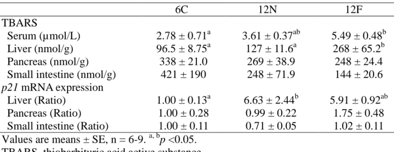
To examine the degree of aging in SAMP8 mice, the lipid peroxide (TBARS) and
p21 mRNA levels were examined in serum and TG absorption-related tissues (liver,
pancreas and small intestine) (Table 2.2). There was a significant increase in TBARS for serum in the 12F group compared to the 6C group. There was a significant increase in TBARS for liver in the 12F group compared to the 6C and 12N groups. There was no significant difference in TBARS for pancreas and small intestine. There was a significant increase in p21 mRNA level for liver in the 12N group compared to the 6C group. There was no significant difference in p21 mRNA level for pancreas and small intestine. These results showed clear progression of senescence in the liver of 12-month-old SAMP8 mice.
mRNA levels for TG absorption-related genes in liver, pancreas and small intestine To examine the effect of a high-fat diet on the attenuation of lipid absorption ability with aging, the expression of mRNA for TG absorption-related genes in liver, pancreas and small intestine was measured (Table 2.3). There was no significant difference in the mRNA level of Cyp7a1 which is crucial for bile acid synthesis in the liver. There was no significant difference in the mRNA levels of Clps, Plrp2 and Ptl which are required for efficient TG hydrolysis in the pancreas. There was no significant difference in the mRNA levels of Fabp2 which transports free fatty acid and Mttp which brings TG into