九州大学学術情報リポジトリ
Kyushu University Institutional Repository
PRIP (phospholipase C-related but catalytically inactive protein)による開口分泌の制御に関する研 究
張, 釗
九州大学大学院歯学府
https://doi.org/10.15017/26322
出版情報:Kyushu University, 2012, 博士(歯学), 課程博士 バージョン:
権利関係:
Studies on the regulatory role of PRIP
(phospholipase C-related but catalytically inactive protein) in dense-core vesicle exocytosis
– Inhibition by direct interactions with syntaxin 1 and SNAP-25 through its C2 domain –
2012 Zhao Zhang
Laboratory of Molecular and Cellular Biochemistry Graduate School of Dental Science
Kyushu University
Supervisors Dr. Hiroshi Takeuchi
Laboratory of Molecular and Cellular Biochemistry, Faculty of Dental Science Kyushu University
(Current post: Professor of Kyushu Dental College)
Professor Masato Hirata
Laboratory of Molecular and Cellular Biochemistry, Faculty of Dental Science Kyushu University
Part of this thesis has been submitted and in a revision:
PRIP (phospholipase C-related but catalytically inactive protein) inhibits exocytosis by direct interactions with syntaxin 1 and SNAP-25 through its C2 domain
Zhao Zhang, Hiroshi Takeuchi, Jing Gao, DaGuang Wang, Declan J. James, Thomas F. J.
Martin and Masato Hirata
- 3 -
Abbreviations used:
CBB: Coomassie Brilliant Blue GFP: green fluorescence protein GST: glutathione S-transferase
Ins(1,4,5)P3: D-myo-inositol 1,4,5-trisphosphate KO: knock out
NA: noradrenalin PLC: phospholipase C
PRIP: phospholipase C-related but catalytically inactive protein PtdIns(4,5)P2: phosphatidylinositol 4,5-bisphosphate
SDS-PAGE: sodium dodecyl sulfate-polyacrylamide gel electrophoresis SNAP-25: synaptosome-associated protein of 25 kDa
SNARE: soluble NSF-attachment protein receptors VAMP: vesicle-associated membrane protein WT: wild type
Contents
ABSTRACT・・・・・・・・・・・・・・・・・・・・・・・・・・・・・・・5
INTRODUCTION・・・・・・・・・・・・・・・・・・・・・・・・・・・・・・6
EXPERIMENTAL PROCEDURES・・・・・・・・・・・・・・・・・・・・・・・8
RESULTS・・・・・・・・・・・・・・・・・・・・・・・・・・・・・・・・15
DISCUSSION・・・・・・・・・・・・・・・・・・・・・・・・・・・・・・・30
ACKNOWLEDGEMENTS・・・・・・・・・・・・・・・・・・・・・・・・・33
REFERENCES・・・・・・・・・・・・・・・・・・・・・・・・・・・・・・・34
- 5 - ABSTRACT
Membrane fusion for exocytosis is mediated by SNAREs (soluble N-ethylmaleimide-sensitive factor attachment protein receptors), forming trans-ternary complexes to bridge vesicle and target membranes. There is an array of accessory proteins that directly interact with and regulate SNARE proteins. PRIP (phospholipase C-related but catalytically inactive protein) is likely one of these proteins; PRIP consisting of multiple functional modules including pleckstrin homology and C2 domains, inhibited exocytosis, probably via the binding to membrane phosphoinositides through the pleckstrin homology domain. However, the roles of the C2 domain have not yet been investigated. In this study, we found that the C2 domain of PRIP directly interacts with syntaxin 1 and SNAP-25 but not with VAMP2. The C2 domain promoted PRIP to co-localize with syntaxin 1 and SNAP-25 in PC12 cells. The binding profiles of the C2 domain to SNAP-25 were comparable with that of synaptotagmin I, and PRIP inhibited synaptotagmin I in the binding with SNAP-25 in mouse brain. It was also shown that the C2 domain was also required for PRIP to suppress SDS-resistant ternary SNARE complex formation, and inhibit high-K+-induced noradrenaline release from PC12 cells. These results suggest that PRIP resides at proper sites where SNARE executes membrane fusion and inhibits regulated exocytosis through the interaction of the C2 domain with syntaxin 1 and SNAP-25, by competing with other SNARE-binding accessory proteins bearing C2 domain that promote exocytosis, and also, by directly inhibiting strong trans-SNARE complex formation.
INTRODUCTION
Exocytosis is one of fundamental cellular events by which cells secrete neurotransmitters, neuropeptides, and peptide hormones, and also distribute membrane proteins such as receptors, channels, and transporters to the cell surface. The final step of exocytosis, membrane fusion, is mediated by hetero-trimeric complexes of SNARE [soluble NSF (N-ethylmaleimide-sensitive factor)-attachment protein receptors] proteins (1-3) consisting of members of the VAMP (vesicle-associated membrane protein, also called synaptobrevin) family on the vesicular membrane (v-SNARE), and syntaxin and synaptosome-associated protein of 25 kDa (SNAP-25) families on the target plasma membrane (t-SNARE). α-helical SNARE motifs from VAMP and syntaxin and two from SNAP-25 forming parallel coiled-coil bundles are believed to promote fusion of vesicular and target membranes (4,5). A number of accessory proteins regulating SNARE-mediated membrane fusion have been shown to interact directly with individual SNARE proteins and/or with assembled SNARE protein complexes (6).
Phospholipase C-related but catalytically inactive protein (PRIP), consisting of type 1 and type 2, was originally isolated as a novel D-myo-inositol 1, 4, 5-trisphosphate [Ins(1,4,5)P3] binding protein in our laboratory. It was named for its lack of catalytic activity in spite of structural similarity to phospholipase C (PLC)-δ1 (7,8), comprising a pleckstrin homology (PH) domain, EF-hand motifs, X and Y motifs, and a C2 domain. In addition to Ins(1,4,5)P3 and phosphatidylinositol 4,5-bisphosphate [PtdIns(4,5)P2] binding to the PH domain (9,10), a number of interacting partners have been identified for PRIP including GABARAP [γ-aminobutyric acid type A (GABAA) receptor-associated protein] (11,12), β subunit of GABAA receptor (13,14), the catalytic subunit of protein phosphatase 1α (PP1α) and PP2A (13,15), and phosphorylated (active) form of Akt (16). To explore the biological functions of PRIP in relation to these interacting proteins, we generated PRIP-1 and/or PRIP-2 knock-out (KO) mice, and found that the mice exhibited increased exocytosis of various peptide hormones such as gonadotropins and insulin (17,18) suggesting that PRIP exerted inhibitory effects on
- 7 -
exocytosis. We subsequently investigated the molecular mechanism by which PRIP inhibited dense-core vesicle exocytosis. PtdIns(4,5)P2 is required for vesicle exocytosis (19-21), and we found that PtdIns(4,5)P2 binding to its PH domain was required for PRIP to suppress exocytosis (Takeuchi, H., Gao, J., Zhang, Z., James, D., Marin, T. and Hirata, M., manuscript in preparation). By binding to PtdIns(4,5)P2, PRIP localizes to sites of exocytosis and competes with other molecules such as CAPS (Ca2+-activated protein for secretion) for PtdIns(4,5)P2
binding required for exocytosis. In the course of these experiments, however, we noticed that other mechanisms besides PtdIns(4,5)P2 binding of the PH domain are also needed for PRIP to exert full inhibition.
In this study, we investigate the role of the PRIP-C2 domain in the inhibition of exocytosis in the light of many reports that a variety of proteins with C2 domain participate in exocytosis (22,23). We found that the PRIP-C2 domain showed little interaction with phospholipids but interacted with t-SNARE proteins in a Ca2+-dependent manner. The C2 domain was required for the co-localization of PRIP with t-SNAREs in cells. Moreover, the C2 domain also had direct inhibitory effects on ternary SNARE complex formation. Thus, we propose that PRIP is a new member of C2 domain-containing proteins that regulate membrane traffic by its negative regulation of exocytosis through a combination of PtdIns(4,5)P2 binding by the PH domain and t-SNARE binding by the C2 domain.
EXPERIMENTAL PROCEDURES
Reagents and Antibodies – [3H]noradrenalin (NA) was obtained from GE Healthcare (Montreal, Canada). Duolink in situ kit for proximity ligation assay (PLA) was from Olink Bioscience (Uppsala, Sweden). Antibodies used were as follows: SNAP-25 (Sigma-Aldrich, St. Louis, MO), VAMP2 (Synaptic systems, Göttingen, Germany), Munc18 (BD Transduction Laboratories, Franklin Lakes, NJ), and syntaxin 1, synaptotagmin I, GFP (green fluorescent protein) and GST (glutathione S-transferase) (Santa Cruz Biotechnology, Santa Cruz, CA). Alexa Fluor 488-anti-rabbit antibody was from Invitrogen (Carlsbad, CA). Cy3-anti-mouse antibody, normal rabbit and mouse globulins were purchased from Jackson ImmunoResearch Laboratories (West Grove, PA). Anti-PRIP-1 mouse monoclonal antibody (2F9) and antigen-purified rabbit polyclonal antibody were prepared in this laboratory as described previously (11,24).
DNA Constructs – The plasmid to express EGFP-PRIP-1 in mammalian cells and the full-length (PRIP-WT) and deletion mutant (PHXY, amino acid residues; 82-704) of His-tagged PRIP-1 for baculovirus expression system were prepared as described previously (10). EGFP-PRIP-1 lacking the C2 domain (PRIP∆C2) was prepared as follows; Both 5’- and 3’-end region corresponding to outside the C2 domain of PRIP-1 was amplified by PCR and HindIII/SalI fragment of 5’-end region was first subcloned into HindIII/SalI digested vector, pEGFP-C3 (Clontech, Palo Alto, CA), followed by subcloning the XhoI/SalI fragment of 3’-end region into SalI site of the plasmid prepared as above. The resulting construct was used to express PRIP-1 lacking the residues 714-850 (PRIP∆C2). PRIP, whose Arg at 134 from the N-terminus was replaced with Gln (R134Q mutant and R134Q/∆C2 mutant) to diminish PtdIns(4,5)P2 binding, were prepared from the templates, EGFP-PRIP-WT and EGFP-PRIP∆C2, respectively, using Quick Change site-directed Mutagenesis kit (Stratagene, La Jolla, CA) as described previously (25). Domain organization of PRIP-1 and the related protein used in this study is depicted in Fig.
1.
- 9 -
FIGURE 1. Schematical representation of PRIP-1 used in this study. Domain organization of PRIP-1 and the related proteins used in this study is depicted. EGFP-tagged PRIP-WT, PRIP∆C2, PRIP R134Q, PRIP∆C2/R134Q were expressed in PC12 cells, while His-tagged PRIP-WT, PHXY, PRIP-C2 and GST-tagged PRIP-C2 were expressed in baculovirus or bacterial expression systems and purified for in vitro assays.
Plasmids to express recombinant C2 domain proteins in the bacterial expression system were prepared by subcloning cDNA amplified by PCR from reverse transcripts of rat brain total RNAs into BamHI/SalI site of pGEX (GE Healthcare) or pET-His30 (11) vectors.
The primers to amplify each cDNA were as follows; the C2 domains of PRIP-1 (amino acid
residue: 709-849); 5’-TAGGATCCATGGCAAACACAAAGG-3’ and 5’-CCCAGCTGCCAATAACGATAC-3’, PLC-δ1 (residue: 630-755);
5’-GTGGATCCAGGCTCCGTGTCC-3’ and 5’-GCCAGCTGCAGGACCTACCTCTAG-3’, rabphilin-3A (residue: 529-685); 5’-TAGAATTCCATGGAGCAGGTGGAGCGGATC-3’ and 5’-CGCAGCTGGATCAGCGAGCTGTGG-3’, synaptotagmin I (C2A; residue: 142-263);
5’-GCGGATCCCTGGGAAAGCTCCAATATTC-3’ and 5’-CGCAGCTGGACCTCTAGTGC GGTG-3’, (C2B; residue: 272-408); 5’-GCGGATCCCTGGGTGACATCTGCTTCTC-3’ and
PRIP-WT PH X Y C2 D
1 111 222 399 702 723 849 1096
PH X Y
PHXY
704 82
PRIP-C2 C2
709 849
PH X Y
PRIP∆C2
713 1
D 851 1096
PRIP
R134Q PH X Y C2 D
1 R134Q 1096
*
PH X Y
PRIP∆C2 /R134Q
713 1
D 851 1096 R134Q
*
PRIP-WT PH X Y C2 D
1 111 222 399 702 723 849 1096
PRIP-WT PH X Y C2 D
1 111 222 399 702 723 849 1096
PH X Y
PHXY
704 82
PH X Y
PHXY
704 82
PRIP-C2 C2
709 849
PRIP-C2 C2
709 849
PH X Y
PRIP∆C2
713 1
D 851 1096
PH X Y
PRIP∆C2
713 1
D 851 1096
PRIP
R134Q PH X Y C2 D
1 R134Q 1096
*
PRIP
R134Q PH X Y C2 D
1 R134Q 1096
*
PH X Y
PRIP∆C2 /R134Q
713 1
D 851 1096 R134Q
*
PH X YPRIP∆C2 /R134Q
713 1
D 851 1096
PH X Y
PRIP∆C2 /R134Q
713 1
D 851 1096 R134Q
*
5’-CGCAGCTGGACGTCTCACACGGTGAC-3’. The GenBank accession numbers to prepare C2 domain constructs are available in Fig. 2.
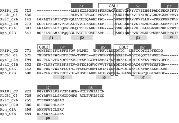
FIGURE 2. Sequence alignment of C2 domains. Amino acid sequences of C2 domains were aligned using ClustalW2 (http://www.ebi.ac.uk/Tools/msa/clustalw2/) and minor correction was applied manually. The positions of conserved aspartate residues for coordinating Ca2+ are boxed. The secondary structures of type I and type II topologies are schematically shown above and below the sequence, respectively. The three loops involved in Ca2+ binding are shown above as CBL1, 2 and 3. GenBank accession numbers of parental proteins are rat PRIP-1; NP445908, rat PLC-δ1; NP058731, rat synaptotagmin I;
NP001028852, rat rabphilin 3A; NP598202.
The plasmid to express SNARE proteins including the syntaxin 1A lacking the C-terminal transmembrane region has been described elsewhere (26). All constructs were fully sequenced to confirm their integrity at the Research Support Center, Graduate School of Medical Sciences, Kyushu University.
- 11 -
Expression and Purification of Recombinant Proteins – Recombinant full-length SNARE proteins with transmembrane region were prepared by bacterial expression system as described previously in the presence of β-octylglucoside (27). Other recombinant proteins were prepared as described elsewhere by bacterial (28,29) or baculovirus expression system (10).
Lipid-protein overlay assay – One nmol of 1,2-dipalmitoyl-sn-glycero-3-phosphocholine (PC), 1,2-dipalmitoyl-sn-glycero-3-phosphoethanolamine (PE), 1,2-dipalmitoyl-sn-glycero-3- phosphoserine (PS) and phosphatidylinositol 4,5-bisphosphate (PIP2) were blotted on nitrocellulose membranes. The membranes were air-dried overnight at 4ºC, then were immersed in blocking buffer (50 mM Tris-HCl, pH 8.0, 150 mM NaCl, 0.02% tween 20 and 3% bovine serum albumin) to reduce background, followed by an incubation with purified GST or GST-fused PRIP-C2, PLCδ-PH or Syt (synaptotagmin)-C2A at 10 µg/ml in blocking buffer containing free Ca2+ of 10 µM. After extensive washing with the buffer (50 mM Tris-HCl, pH 8.0, 150 mM NaCl and 0.02% tween 20, 10 µM CaCl2), the membranes were immunoblotted for bound GST-C2 proteins using anti-GST antibody.
Immunoprecipitation and Western Blotting – The cerebrum of wild-type or PRIP-KO mouse was homogenized in the lysis buffer (20 mM Tris-HCl, pH 7.5, 150 mM NaCl, 1 mM EDTA, 1% Triton X-100, and 1 mM dithiothreitol) containing protease inhibitors (5 µg/ml pepstatin A, 10 µM leupeptin, 1.7 µg/ml aprotinin, and 50 µM 4-amidinophenylmethanesulfonyl fluoride hydrochloride). The lysates were cleared by centrifugation and incubated with antibody of interest or control immunoglobulin at 4ºC overnight, followed by incubation with protein G beads at 4ºC for 1 h. Then the beads were washed with the lysis buffer four times, and the proteins bound to the beads were separated by SDS-polyacrylamide gel electrophoresis (PAGE), followed by transfer to polyvinylidene fluoride membranes (Merck-Millipore, Billerica, MA).
After blocking, the membrane was blotted with the appropriate antibody, and horseradish peroxidase-conjugated anti-rabbit or anti-mouse IgG (GE Healthcare), and detected for
chemiluminescent signals using an LAS-3000 mini gel documentation system (Fujifilm, Tokyo, Japan). Digital images were analyzed with Image Gauge software (Fujifilm) or NIH Image J software (http://rsb.info.nih.gov/ij/) to measure the density of each band. The handling of mice and all procedures were approved by the Animal Care Committee of Kyushu University, which follows the guidelines of the Japanese Council on Animal Care.
t-SNARE liposome binding assay – Preparation of t-SNARE-incorporated liposomes and the floatation assays were performed as described previously (27), except that GST alone or GST-fused C2 domain was used instead of CAPS. All phospholipids used in this study were purchased from Avanti Polar Lipids (Alabaster, AL).
Cell culture, Transfection and Stable cell lines expressing PRIP – Rat pheochromocytoma (PC12) cells were maintained and used to establish stable cell lines expressing wild-type or several mutants of PRIP as described previously (29). Briefly, PC12 cells were cultured in Dulbecco’s modified Eagle’s medium (DMEM) supplemented with 10% horse serum and 5%
fetal bovine serum. The generation of PC12 clonal cells stably expressing GFP-PRIP-1 was performed as follows: PC12 cells were first transfected with phGFP105, PRIP-1 (wild type)/phGFP105, PRIP-1 (∆C2)/phGFP105, PRIP-1 (R134Q)/phGFP105 or PRIP-1 (∆C2/R134Q)/phGFP105 with Lipofectamine2000 (Invitrogen) according to the manufacturer’s instructions. Transfected cells were maintained in growth medium containing 400 µg/ml G418 (Invitrogen) for about a month. The growing colonies were visually selected for GFP fluorescence and picked up.
Immunofluorescence and Proximity Ligation Assay (PLA) – The cells were plated onto poly-D-lysine-coated glass coverslips at a density of 4 x 10⁴ cells/well in a 12-well plate and subjected to immunofluorescent observation as described elsewhere (30). In some experiments, the cells were transiently transfected with plasmid DNA using Lipofectamine2000 (Invitrogen,
- 13 -
Carlsbad, CA) and OPTI-MEM (Invitrogen) according to the manufacturer's protocol and 24 h after transfection, the cells were plated onto glass coverslips as described above.
In situ PLA was performed using ‘Duolink in situ kit’ following to the manufacture’s protocol, but the cells were prepared, permeabilized and blocked in the same manner as for immunofluorescent studies with the combination of primary antibodies produced in mouse and rabbit. The confocal images were obtained using LSM510 META (Carl Zeiss), and analyzed using Duolink Image Tool (Olink Bioscience) to obtain objective quantification of PLA signals.
GST Pull-down Assay – PC12 cell lysate was prepared by homogenizing in the lysis buffer (50 mM Hepes-KOH, pH 7.3, 150 mM NaCl, 1 mM MgCl2, 1 mM dithiothreitol, and 1% Triton X-100) containing protease inhibitors. Cell lysate or recombinant SNARE protein, whose GST-tag had been removed by thrombin digestion, was mixed with GST alone or GST-fused C2 domain at an equal molar ratio in the binding buffer (10 mM Hepes-KOH, pH 7.3, 100 mM KCl, 3.5 mM MgCl2, 1 mM EDTA, 0.1% nonidet P-40) and incubated at 4ºC for 2 h. Then glutathione Sepharose 4B (GE Healthcare) equilibrated with appropriate buffer was added to the mixture, and incubated at 4ºC for another 1 h. At the end of the incubation, the beads were washed with the same buffer four times and boiled in a sample buffer for 5 min, then subjected to SDS-PAGE followed by Western blotting. Similar experiments in a reverse mode using GST-fused SNARE protein and His-tagged PRIP protein were performed in the same condition as described above. The internal standard sample with known protein concentration was included in each blot and used to calculate the amount of protein of interest by fitting to the standard curve. To examine the effect of free Ca2+ concentration on the binding of the C2 domains to syntaxin 1 or SNAP-25, the pull-down assay was performed in the same binding buffer as above for that in the presence of calculated amount of Ca2+ to give the free Ca2+
concentration of 10 µM or the buffer containing 1 mM EGTA.
In vitro SDS-resistant SNARE complex Formation – The mixture of 10 pmol each of the purified GST-SNAP25, Stx∆C-His and His-VAMP2∆C were incubated overnight in the binding buffer (10 mM Hepes-KOH, pH 7.3, 100 mM KCl, 3.5 mM MgCl2, 1mM EDTA and 0.1% nonidet P-40). At the end of incubation, the mixture was either boiled or incubated in 37ºC for 5 min in a SDS sample buffer and immediately subjected to SDS-PAGE followed by immunoblotting with antibodies indicated in the figures. Additional proteins were included in the mixture of SNAREs during the incubation period. To test the effect of free Ca2+ on the SNARE complex formation, the mixture was incubated in the binding buffer containing calculated amounts of CaCl2 to give a free concentration of 10 µM with 1 mM EGTA (in place of EDTA).
Measurement of [3H]noradrenaline (NA) Release – [3H]NA secretion from the stable PC12 cell lines were measured as previously described (29). PC12 cells were seeded in a 12-well plate coated with 10 µg/ml poly-L-lysine (3 x 10⁵ cells). Twenty-four h later, cells were labeled with 0.5 µCi of [3H]NA (GE Healthcare) per well in 1 ml of complete culture medium containing serum in the presence of 0.5 mM ascorbic acid for 12–16 h. The medium was replaced with fresh complete medium, and the cells were further incubated for 1–2 h to remove unincorporated [3H]NA. For the intact cell assay, cells were washed with physiological salt saline (PSS) containing 145.4 mM NaCl, 5.6 mM KCl, 2.2 mM CaCl2, 0.5 mM MgCl2, 5.6 mM
D-glucose, and 15 mM Hepes, pH 7.4, and the secretion of [3H]NA was triggered with high K+/PSS (95 mM NaCl, 56 mM KCl). At the end of the secretion assay, the medium was transferred into a scintillation vial, and cells were collected to assess the remaining radioactivity in 0.3 ml of 0.5% Triton X-100, enabling calculation of the ratio of secretion relative to the total radioactivity. The radioactivity of [3H]NA remaining in cells and secreted into the medium was measured by a liquid scintillation counter.
Statistical Analysis – All statistical analyses were performed by Students’ t-test, with a two- tailed value of P< 0.05 considered significant using GraphPad Prism (GraphPad Software).
- 15 - RESULTS
Interaction of PRIP with t-SNARE proteins through the C2 domain – We first examined phospholipid binding of the PRIP-C2 domain in a lipid-protein overlay assay (30). The isolated C2 domain did not show more binding than GST alone to phospholipids tested in the presence of Ca2+ (Fig. 3). By contrast, strong lipid binding as positive controls was observed as follows:
PtdIns(4,5)P2 binding by the PH domain of PLC-δ1, and phosphatidylserine binding by the C2A domain of synaptotagmin I, which was abolished in the absence of Ca2+ (Fig. 3).
FIGURE 3. Phospholipid binding of PRIP-C2. Lipid-overlay assay was performed using nitrocellulose membrane on which 1 nmol of PC (phosphatidylcholine), PE (phosphatidylethanolamine), PS (phosphatidylserine) and PIP2 were blotted. Each membrane was probed with GST-fused protein indicated on the left followed by immunodetection with anti-GST antibody. GST-fused PRIP-C2, PLCδ-PH or Syt (synaptotagmin)-C2A were used.
Ca2+ (-) indicates the incubation with 1 mM EGTA alone.
As there are several C2 domain-containing proteins that promote SNARE-mediated membrane fusion by direct SNARE protein interactions (31-35), we examined whether PRIP binds to SNARE proteins via its C2 domain. Anti-PRIP-1 antibody precipitated PRIP-1 along with syntaxin 1 and SNAP-25 from brain lysate of wild-type but not from that of PRIP-KO mouse (Fig. 4A), indicating that PRIP-1 interacts with syntaxin 1 and SNAP-25 either in a
GST
GST-PRIP-C2 GST-PLCδ-PH
PC
GST-Syt-C2A
PE PS PIP2
Ca2+
+ + +
- +
GST
GST-PRIP-C2 GST-PLCδ-PH
PC
GST-Syt-C2A
PE PS PIP2
Ca2+
+ + +
-
+
direct or indirect manner. This result was further confirmed and the region responsible for the binding was identified using the recombinant PRIP-C2 domain protein. The lysate from PC12 cells was applied to GST alone and GST-fused C2 domains of PRIP-1 (PRIP-C2) or GST-C2B of rabphilin 3A (Rph-C2B) as a positive control (known to bind to SNAP-25, Ref. 33) immobilized on glutathione beads, followed by immunoblotting with the indicated antibodies (Fig. 4B). The result clearly showed that both syntaxin 1 and SNAP-25 bound to PRIP-C2, but VAMP2 and Munc-18 did not, while only SNAP-25 bound to Rph-C2B as previously reported (33,34). Thus, the C2 domain of PRIP interacts with two t-SNARE proteins, i.e., syntaxin 1 and SNAP-25, but not with the v-SNARE protein, VAMP2. To clarify whether the interactions of PRIP-C2 with syntaxin 1 and SNAP-25 are direct or indirect, since the experiments shown in Fig. 4A and B were done using crude samples, i.e., the lysates from brain or PC12 cells, we next performed GST pull-down assays using recombinant purified protein samples. GST alone or GST-C2 domain proteins immobilized on glutathione-beads were incubated with soluble syntaxin 1 lacking the C-terminal transmembrane region (Stx∆C) or SNAP-25. Both syntaxin 1 and SNAP-25 bound to PRIP-C2 (Fig. 4C). The binding was comparable to that of the C2B domain of synaptotagmin I (Syt-C2B) as a positive control. The Rph-C2B interacted with SNAP-25, but not with syntaxin 1, agreeing with previous reports (33,34) and Fig. 4B. The C2 domain of PLC-δ1 (PLCδ-C2), albeit with a high homology to PRIP, did not bind to syntaxin 1 or SNAP-25 (Fig. 4C). The GST pull-down assay in a reverse mode was also performed.
His-tagged forms of full-length PRIP-1 (PRIP-WT), the PRIP-C2 or PHXY (see Fig. 1) lacking the C2 domain and the N-terminal extension were assayed using immobilized GST alone, GST-SNAP-25 or GST-Stx∆C. PRIP-WT and PRIP-C2, but not PHXY, showed binding to both SNAP-25 and syntaxin 1 (Fig. 4D) confirming that PRIP directly interacts with t-SNARE proteins through the C2 domain.
- 17 -
FIGURE 4. Interaction of PRIP-C2 with SNARE proteins. A, PRIP-1 was immunoprecipitated using mouse anti-PRIP-1 monoclonal antibody or normal IgG (Cont. IgG) from brain lysates of PRIP-WT and PRIP-KO mice. The lysates and immunoprecipitates were subjected to Western blotting by the antibodies against the proteins indicated on the left. Two or 20 % of the total amount of brain lysates or immunoprecipitates, respectively, was applied to SDS-PAGE. Typical blot was shown, and two other blots yielded similar results. Syntaxin 1 was seen in double bands; the lower band would be a degraded product. B, GST or GST-fused C2 domains immobilized on glutathione beads were mixed with PC12 cell lysate, followed by extensive washing and the bound proteins were analyzed by Western blotting using the antibodies indicated in the figure. The bottom panel indicates the blot of the beads, probed with anti-GST antibody. GST-C2 proteins appear to be the top band in each lane based on the expected molecular size as indicated by arrowhead with some degraded proteins below the band. C, The mixture of 100 pmol each of GST or GST-fused C2 domains and SNAP-25 or syntaxin 1∆C were incubated at 4ºC for 2 h and then applied to glutathione beads. After another hour incubation, the beads were washed extensively followed by Western blotting. SNAP-25 and syntaxin 1∆C bound to the beads were probed with anti-SNAP-25 and anti-syntaxin 1 antibodies, respectively. The bottom panel indicates the blot of the beads mixed with SNAP-25, which was probed with anti-GST antibody. GST-C2 protein appears as the top band in each lane with some degraded proteins below the band. The identical result was obtained for the amount of immobilized GST-proteins from the beads mixed with syntaxin 1∆C. A band in the lane of PLCδ-C2 detected by anti-SNAP-25 antibody was also detected in the control beads at a similar density, indicating that non-specific background interaction. D, GST-pull down assay was performed in the reverse direction shown in C. GST, GST-SNAP-25 or GST-syntaxin 1∆C was mixed with purified PRIP-WT, PRIP-C2 or PHXY (PRIP-1 lacking the C2 domain; see materials and methods), then bound proteins on the beads were analyzed as same as in “B” using anti-PRIP-1 polyclonal antibody which recognizes all PRIP-1 constructs.
25 kD
20 kD
GST-PRIP-C2 syntaxin 1
GST-Rph-C2B
GST SNAP-25
VAMP2 Munc18
25 kD
75 kD 50 kD 37
25
Input GST Rph-C2B PRIP-C2
25 kD
20 kD
GST-PRIP-C2 syntaxin 1
GST-Rph-C2B
GST SNAP-25
VAMP2 Munc18
25 kD
75 kD 50 kD 37
25
Input GST Rph-C2B PRIP-C2
A B
C D
Input Beads
PRIP- C2PRIP -WT
Input Beads
GST SNAP-25 Stx∆C
20
75 100 kD
Input Beads
PHXY
100
20
75
GST GST-Stx∆C
50 37 25 GST-SNAP-25
75 Input
Beads
PRIP- C2PRIP -WT
Input Beads
GST SNAP-25 Stx∆C
20
75 100 kD
Input Beads
PHXY
100
20
75 Input
Beads
PRIP- C2PRIP -WT
Input Beads
GST SNAP-25 Stx∆C
20
75 100 kD
Input Beads
PHXY
100
20
75
GST GST-Stx∆C
50 37 25 GST-SNAP-25
75
GST GST-Stx∆C
50 37 25 GST-SNAP-25
75 Input
Beads
SNAP-25Stx∆C Input Beads
GST GST-C2
GST PRIP-C2 PLCδ-C2 Rph-C2B Syt-C2B
50 kD 25 kD 25 kD 25 kD 25 kD
37 25 Input
Beads
SNAP-25Stx∆C Input Beads
GST GST-C2
GST PRIP-C2 PLCδ-C2 Rph-C2B Syt-C2B
50 kD 25 kD 25 kD 25 kD 25 kD
37 25 WTWTKO WTWTKO
IP
Lysate
PRIP syntaxin 1
SNAP25 25 kD
25 kD 100 kD Cont.
IgG anti- PRIP-1
WTWTKO WTWTKO IP
Lysate
PRIP syntaxin 1
SNAP25 25 kD
25 kD 100 kD Cont.
IgG anti- PRIP-1
As the binding of syntaxin 1 and SNAP-25 to PRIP-C2 shown in Fig. 4 was assayed by pull-down method using truncated soluble SNARE proteins, we further confirmed the interaction using full-length membrane-integrated SNARE proteins which might exhibit properties different from truncated soluble SNARE proteins (36,37). Proteoliposomes incorporating full-length syntaxin 1 and SNAP-25 were prepared and incubated with PRIP-WT, PRIP-C2 or GST alone, followed by a gradient centrifugation to detect protein samples in the liposome fraction. Both PRIP-WT and PRIP-C2, but not GST alone, were detected in the floating liposome fraction in the case of proteoliposomes, but not in the protein-free (Pf) liposome, indicating that PRIP and PRIP-C2 proteins bound to membrane-associated t-SNARE proteins (Fig. 5).
FIGURE 5. Interaction of PRIP with t-SNAREs incorporated in liposomes. One µmol of GST, GST-PRIP-C2 (PRIP-C2), or His-PRIP-WT (PRIP-WT) was incubated with protein-free (Pf) or t-SNARE containing phosphatidylcholine/phosphatidylserine liposomes and bound PRIP (fraction 1 and 2) was separated from free PRIP (fractions 6-8) by gradient centrifugation. Fractions were analyzed by immunoblotting for PRIP-1. Representative result of three experiments is shown.
PRIP-C2
syntaxin 1/SNAP-25 liposome
1 2 3 4 5 6 7 8
GST
input
Pf liposome
PRIP-WT SNAP-25 syntaxin 1
PRIP-WT Syntaxin 1
syntaxin 1
syntaxin 1 Fraction No.
Gradient
PRIP-C2GSTPRIP-WT
25
25 37 25 25 3725 50 kD
150
25 37
150 kD
25 37
PRIP-WT
- 19 -
The C2 domains binding at increasing amounts with t-SNARE proteins were examined by a GST pull-down assay (Fig. 6). The Kd value of 1.1 µM for PRIP-C2 binding to syntaxin 1 was comparable to that of Syt-C2B (2.4 µM), while Rph-C2B showed no binding to syntaxin 1 (Fig. 6A). On the other hand, the binding of PRIP-C2 to SNAP-25 (Kd = 1.9 µM) was lower than that of Rph-C2B (Kd = 0.4 µM), although it was still comparable to the affinity of Syt-C2B to SNAP-25 (Kd = 2.1 µM) (Fig. 6B). Mol ratio for the binding was smaller compared to the positive control; To SNAP-25, the Bmax were 0.18 and 0.18 mol for PRIP-C2 and Syt-C2B, respectively, while 0.35 mol for Rph-C2B as a positive control. To syntaxin 1, the Bmax was 0.17 mol for PRIP-C2, while 0.55 mol for Syt-C2B as a positive control. These results, however, indicate that the binding of PRIP-C2 to both syntaxin 1 and SNAP-25 are permissive in the regulation of SNARE-mediated membrane fusion (26,33).
FIGURE 6. Comparison of binding profile of C2 domains to t-SNARE proteins. Purified C2 domains were incubated with GST-syntaxin 1∆C (A) or GST-SNAP-25 (B) immobilized on glutathione beads at the indicated concentrations and the C2 domains bound to the beads were determined by Western blotting. The blots shown are typical of three independent experiments giving similar results. The proteins bound to the beads were quantified as described in “Experimental Procedures” and expressed as mol ratio of the C2 domain of PRIP-1 (PRIP-C2; closed circle), synaptotagmin I (Syt-C2B; closed square) or rabphilin 3A (Rph-C2B; closed triangle) to syntaxin 1∆C (A) or SNAP-25 (B) bound on the beads. The Kd and Bmax values described in “Results” were obtained by non-linear regression curve fits to the data (means ± SD, n=3).
mol C2 bound /mol Stx∆C
C2 (µM)
A
GST-Stx∆C PRIP-C2
0.005µM 0.015 0.05 0.15 0.45 1.35 4.05 12.15
Syt-C2B Rph-C2B
mol C2 bound /mol Stx∆C
C2 (µM) mol C2 bound /mol Stx∆C
C2 (µM)
A
GST-Stx∆C PRIP-C2
0.005µM 0.015 0.05 0.15 0.45 1.35 4.05 12.15
Syt-C2B Rph-C2B GST-Stx∆C PRIP-C2
0.005µM 0.015 0.05 0.15 0.45 1.35 4.05 12.15
Syt-C2B Rph-C2B
mol C2 bound /mol SNAP-25
C2 (µM)
B
GST-SNAP-25 PRIP-C2 Syt-C2B Rph-C2B
0.005µM 0.015 0.05 0.15 0.45 1.35 4.05 12.15
mol C2 bound /mol SNAP-25
C2 (µM) mol C2 bound /mol SNAP-25
C2 (µM)
B
GST-SNAP-25 PRIP-C2 Syt-C2B Rph-C2B
0.005µM 0.015 0.05 0.15 0.45 1.35 4.05 12.15
GST-SNAP-25 PRIP-C2 Syt-C2B Rph-C2B GST-SNAP-25 PRIP-C2 Syt-C2B Rph-C2B
0.005µM 0.015 0.05 0.15 0.45 1.35 4.05 12.15
0.005µM 0.015 0.05 0.15 0.45 1.35 4.05 12.15
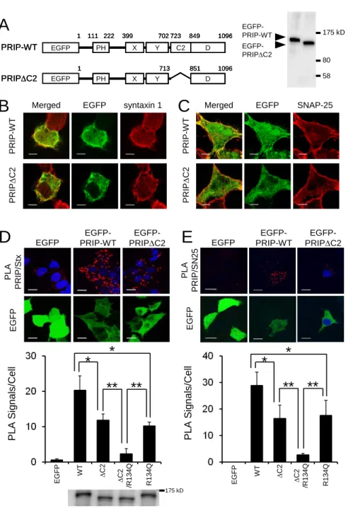
Co-localization of PRIP with t-SNARE proteins in cells – We next examined the subcellular localization of PRIP with SNAP-25 and syntaxin 1 in rat pheochromocytoma PC12 cells where we observed PRIP-mediated down-regulation of NA release (29). As PC12 do not express detectable level of endogenous PRIP, we established stable PC12 cell lines expressing wild-type or the mutant version of PRIP-1 fused to EGFP (Fig. 7A). The cells were processed for immunofluorescence of EGFP-PRIP-1 and endogenous SNAP-25 or syntaxin 1 with respective antibodies. PRIP-WT appeared abundant because of exogenous expression, but localized to both cytosol and plasma membrane, while both syntaxin 1 (Fig. 7B) and SNAP-25 (Fig. 7C) were mainly present at the plasma membrane. Both syntaxin 1 and SNAP-25 showed co-localization with PRIP-1 at the plasma membrane as observed in the merged images (Fig. 7B and C). By contrast, PRIP-1 lacking its C2 domain (PRIP∆C2) showed less localization at the plasma membrane with diffuse distribution in the cytosol resulting in less co-localization with either SNAP-25 or syntaxin 1 (Fig. 7B and C, lower panels). These results indicated that PRIP-C2 partly contributes to the co-localization with t-SNAREs in the cells.
We further confirmed the importance of PRIP-C2 in co-localization with t-SNARE proteins by using in situ proximity ligation assay (PLA) technology, which visualizes protein-protein interactions quantitatively as fluorescent spots by rolling-circle amplification reactions dependent on the close proximity (<40 nm) of the target proteins (38). PC12 cells were probed with a combination of mouse antibody against SNAP-25 or syntaxin 1 and rabbit antibody against PRIP-1 as primary antibodies, followed by further probing with PLA probes for mouse and rabbit primary antibodies with ligation and amplification reactions. As shown in Fig. 7D, red fluorescent spots indicating co-localization of PRIP and syntaxin 1 were observed only in the PC12 cells expressing EGFP-PRIP-WT and, to a smaller extent, expressing PRIP∆C2, with none in the cells expressing EGFP alone (upper panels). Similar results were obtained for SNAP-25 proximity with PRIP or PRIP∆C2 (Fig. 7E). Consistent with the results in Fig. 7B and C, the number of signal per cell were significantly decreased by the lack of the C2 domain of PRIP, suggesting that the C2 domain partly contributes to the co-localization of PRIP with SNAP-25 and syntaxin 1 (Fig. 7D and E, bar graphs).
- 21 -
FIGURE 7. Co-localization of PRIP with t-SNARE proteins in PC12 cells. A, Proper expression with the expected molecular weight of each construct in PC12 cells was confirmed by Western blotting with anti-GFP antibody. B and C, PC12 cells expressing EGFP-PRIP-WT or EGFP-PRIP∆C2 were cultured on coverslips and visualized in green for EGFP and red for intrinsic syntaxin 1 (B) or SNAP-25 (C) by indirect immunofluorescence using a combination of rabbit antibody against GFP and Alexa488-conjugated rabbit IgG, and mouse antibodies against SNAP-25 or syntaxin 1 and Cy3-conjugated mouse IgG, respectively. The yellowish staining in the merged picture indicates co-localization of EGFP-PRIP-WT or EGFP-PRIP∆C2 with the SNARE proteins. Scale bars, 5 µm. D and E, PC12 cells expressing EGFP-fused PRIP-WT or the mutant constructs indicated in the graphs were cultured on coverslips and subjected to PLA assay using the combination of the antibodies against PRIP and syntaxin 1 (Stx) or SNAP-25 (SN25) and visualized in red for PLA signals, green for EGFP signals, and blue for counter stained nucleus. Scale bars, 10 µm. Typical images of the cells expressing EGFP, EGFP-PRIP-WT and EGFP-PRIP∆C2 are presented in top panels. In each image, the number of PLA signals per cell was counted using Duolink Image Tool with manual corrections and presented as bar graphs including the data from the cells expressing additional mutants (see "Results"). More than 30 cells for each experiment were counted and the data are the means ± SD of three experiments. Significance by Student’s t-tests is represented by * or ** for p<0.05 or p<0.01, respectively. The cell lysates used in the experiment were subjected to Western blotting and the image detected with anti-GFP antibody is shown in the bottom to show the comparable expression levels of the constructs in the cells.
A
B C
Merged EGFP SNAP-25PRIP-WTPRIP∆C2
PRIP-WTPRIP∆C2
syntaxin 1 EGFP
Merged
PRIP-WT PH X Y C2 D
1 111 222 399 702 723 849 1096
PH X Y
PRIP∆C2
713 1
D 851 1096
PRIP-WT PH X Y C2 D
1 111 222 399 702 723 849 1096
PRIP-WT PH X Y C2 D
1 111 222 399 702 723 849 1096
PH X Y
PRIP∆C2
713 1
D 851 1096
PH X Y
PRIP∆C2
713 1
D 851 1096 EGFP
EGFP
80 175 kD
58 EGFP-
PRIP∆C2 EGFP- PRIP-WT
EGFP- PRIP-WT EGFP
PLA PRIP/StxEGFP
EGFP- PRIP∆C2
D E
EGFP PRIP-WTEGFP-PLA PRIP/SN25EGFP
EGFP- PRIP∆C2
PLA Signals/Cell EGFP WT ∆C2 ∆C2 /R134Q R134Q
**
*
**
*
PLA Signals/Cell EGFP WT ∆C2 ∆C2 /R134Q R134Q
**
*
**
*
175 kD
We found that PtdIns(4,5)P2 binding to the PH domain is required for PRIP to inhibit exocytosis (Takeuchi, H., Gao, J., Zhang, Z., James, D., Martin, T. and Hirata, M., manuscript in preparation). Therefore, further experiments using PRIP with the mutation (R134Q) in the PH domain, which lacks binding to PtdIns(4,5)P2, were performed. Double mutation of ∆C2 and R134Q showed almost complete loss of the co-localization with t-SNARE proteins in PC12 cells (Fig. 7D and E; graphs).
Increased binding of synaptotagmin I to SNAP-25 in the absence of PRIP – Binding parameters
of the C2 domains of PRIP to t-SNARE was comparable to those of synaptotagmin I, suggesting the possibility that PRIP competes with synaptotagmin for binding to t-SNARE proteins. To test this possibility, brain lysates prepared from WT and PRIP-KO mice were immunoprecipitated by anti-SNAP-25 antibody, followed by immunoblotting with antibodies of interest. From the band density in the precipitates compared to that in the lysates, we estimated that about 60 % of SNAP-25 present in the lysates was immunoprecipitated by anti-SNAP-25 antibody, and the value was almost the same both in WT and PRIP-KO mice. The amount of synaptotagmin I (SytI) precipitated along with SNAP-25 was apparently increased in PRIP-KO mouse by two-fold (WT; 4.8 ± 0.8 %, KO; 10.2 ± 1.9 %), while the amount of syntaxin 1 (WT; 24.1 ± 2.5 %, KO; 24.6 ± 1.8 %) was not affected by the absence of PRIP (Fig. 8). A similar effect of the absence of PRIP in increasing Munc18 in SNAP-25 immunoprecipitates was observed. It should also be noted that PRIP (5.1 ± 1.3 %) was found in the immunocomplex among syntaxin 1, synaptotagmin I and SNAP-25 in WT mice. The results suggest that PRIP partially inhibits the participation of synaptotagmin I in t-SNARE complex formation with syntaxin 1 and SNAP-25, leading to the inhibition of regulated exocytosis.
- 23 -
FIGURE 8. Effect of PRIP on the binding of synaptotagmin I with SNAP-25. Immunoprecipitates of brain lysates prepared from WT and PRIP-KO mice by anti-SNAP-25 antibody were immunoblotted by antibodies indicated. Two or 20 % of the brain lysates or immunoprecipitates, respectively, was applied to SDS-PAGE. The blots shown are typical of three independent experiments and the other experiments gave similar results. Arrowheads in the top panel indicate the bands of synaptotagmin I (SytI), while the bands with 50 kDa are the heavy chain of the IgG used for immunoprecipitation. Graph was shown as follows; the amount of each protein in the immunocomplex by anti-SNAP-25 antibody was measured from the band intensities and expressed as percent of total amount in the lysate. The data are the means ± SD of three experiments. Significance by Student’s t-tests is represented by ** for p<0.01.
Effect of PRIP on ternary SNARE complex formation – The formation of SDS-resistant heterotrimeric SNARE complexes consisting of VAMP2, SNAP-25 and syntaxin 1 was assayed because there is growing evidence to indicate that the amount of the SNARE complex correlates well with the extent of exocytosis (6,39-41). To examine the effect of PRIP on SNARE complex formation, we used recombinant SNAP-25, soluble syntaxin 1∆C, and soluble VAMP2∆C
WT WT KO WT WT KO
IP Lysate Cont.SNAP-25
IP ( % of t ot al l y s at e)
SNAP-25
SytI syntaxin 1
**
IP ( % of t ot al l y s at e)
SNAP-25
SytI syntaxin 1
IP ( % of t ot al l y s at e)
SNAP-25
SytI syntaxin 1
**
25 kD
syntaxin 1 SNAP-25 PRIP-1
30 kD 100 kD
SytI
50 kD
75 kD
Munc18
25 kD
syntaxin 1 SNAP-25 PRIP-1
30 kD 100 kD
SytI
50 kD
75 kD
Munc18 Munc18
prepared from bacterial expression system. An equimolar mixture of these three SNARE proteins were incubated and treated with SDS-sample buffer with or without boiling, followed by SDS-PAGE analysis and Western blotting. Two major high molecular weight bands (~110 and 220 kDa) were detected with antibodies against GST-SNAP-25, syntaxin 1, and VAMP2, which disappeared by boiling (Fig. 9A). The band densities of individual SNARE proteins were more intense following boiling, indicating that high molecular weight bands prior to boiling represented SNARE complexes as previously reported (4,39). We then examined the effect of PRIP and the related proteins on the complex formation. PRIP-WT inhibited the SNARE complex formation in a dose-dependent manner, while PHXY showed no effect (Fig. 9 B and C).
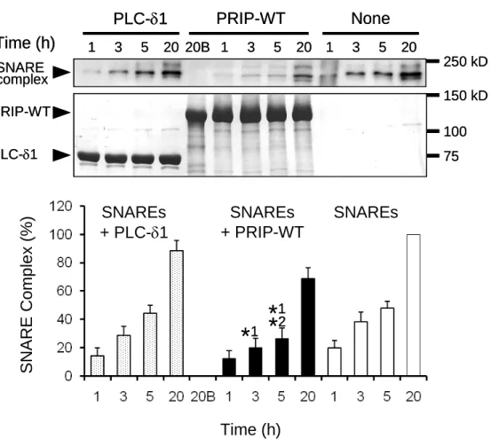
Isolated PRIP-C2 at higher concentrations showed inhibitory effects, but PLC-δ1 even at high concentrations had no effect. The result indicates that PRIP inhibits the SDS-resistant SNARE complex formation in a dose-dependent manner, likely by binding to SNARE proteins through its C2 domain. Studies of the time-dependent formation of SDS-resistant SNARE complexes revealed that PRIP, but not PLC-δ1, delayed complex formation (Fig. 10).
Ca2+-dependence of the binding to t-SNAREs and the inhibitory effect on SNARE complex formation by PRIP – As the binding of the C2 domain of synaptotagmin I to t-SNARE proteins is Ca2+-dependent (31), and the aspartate residues for Ca2+ binding are relatively well conserved in PRIP-C2 (Fig. 2), we tested whether the binding of PRIP-C2 to t-SNARE proteins is Ca2+-dependent. The GST pull-down assay was performed in the presence or absence of 10 µM Ca2+. Both the binding of PRIP-C2 to syntaxin 1 and SNAP-25 exhibited stimulation by Ca2+
with overall Ca2+-dependence less than observed for the positive control, Syt-C2B (Fig. 11A).
The Ca2+-dependence of the effect of PRIP and PRIP-C2 in SDS-resistant SNARE complex formation was assayed. The inhibition of SNARE complex formation by full-length PRIP and PRIP-C2 was stronger in the presence of 10 µM Ca2+ (Fig. 11B and C). These results suggest that PRIP or PRIP-C2 might inhibit exocytosis in both a Ca2+-independent and Ca2+-dependent manner.
- 25 -
FIGURE 9. Effect of PRIP on SDS-resistant SNARE complex formation. A, Mixture of the purified recombinant proteins of His-VAMP2, syntaxin 1∆C and GST-SNAP-25 was incubated overnight at 4ºC, then subjected to SDS-PAGE with or without boiling after the addition of sample buffer, followed by Western blotting using the indicated antibodies. Black arrowheads indicate high molecular weight bands observed only in the unboiled sample, while white arrowheads indicate the bands with the expected molecular weights of monomeric proteins. B, Mixture of the three SNARE proteins were incubated for 3 h at 4ºC in the presence or absence of the indicated concentrations of purified PRIP-1 wild-type, PRIP-1 mutants or PLC-δ1, followed by Western blotting without boiling. Only the bands of 220 kDa detected with anti-VAMP2 antibody are shown in the figure, but the bands of 110 kDa behaved similarly with 220 kDa bands detected with either antibody. C, The amount of SNARE complex formed shown in “B” was expressed as relative to that in the absence of PRIP or PLC-δ1. All data are the means ± SE of four experiments. Closed circle; PRIP-WT, closed triangle; PRIP-C2, open triangle; PHXY, open diamond; PLC-δ1.
VAMP2 syntaxin 1
GST- SNAP-25
SNARE complex
50 Boiling: +
25
+
15 100 250 kD 150
75
+
A
GST- syntaxin 1 VAMP2SNAP-25
SNARE complex
50 50 Boiling: +
25 25
+
15 15 100 250 kD 150
75 100 250 kD 150
75
+
A
C
Protein Added (pmol)
SNARE Complex (%)
Protein Added (pmol)
SNARE Complex (%)
PRIP-WT
0 pmol 40 80 120
SNARE complex
PHXY SNARE complex
PLC-δ1 SNARE complex
B
PRIP-C2 SNARE complex
0 pmol 120 200 300 500
PRIP-WT
0 pmol 40 80 120
0 pmol 40 80 120
SNARE complex
PHXY SNARE complex
PLC-δ1 SNARE complex
B
PRIP-C2 SNARE complex
0 pmol 120 200 300 500
FIGURE 10. Time course of SDS-resistant SNARE complex formation and the delay by PRIP. A mixture of SNARE proteins was incubated for the indicated times at 4ºC in the presence or absence of 100 pmol of full-length PRIP-1 (PRIP-WT) or PLC-δ1, and was subjected to immunoblotting. Bands of 220 kDa detected by anti-VAMP2 antibody are shown in top panel and bottom panel shows proteins on the same membrane visualized by Coomassie staining. SNARE complex formation was calculated from the band density and expressed as in Fig. 9C. Significance by Student’s t-tests between the values at same time point between none and PRIP-WT, or between PLC-δ1 and PRIP-WT, are represented by *1, *2, respectively, for p<0.05. “20B”
in PRIP-WT indicates the lane for boiling (B) after 20 h.
1 3 5 20 20B 1 3 5 20 1 3 5 20 Time (h)
PLC-δ1 PRIP-WT None
SNARE complex PRIP-WT PLC-δ1
250 kD 150 kD 100 75 1 3 5 20 20B 1 3 5 20 1 3 5 20 Time (h)
PLC-δ1 PRIP-WT None
SNARE complex PRIP-WT PLC-δ1
250 kD 150 kD 100 75
Time (h)
SNARE Complex (%)
SNAREs + PRIP-WT SNAREs
+ PLC-δ1 SNAREs
*
1*
1*
2*
2*
1*
1- 27 -
FIGURE 11. Effect of Ca2+ on the binding of PRIP-C2 to SNARE proteins. A, GST-pull-down assays were performed in the binding buffer containing 1 mM EGTA for Ca2+ (-) or 10 µM free Ca2+ for Ca2+ (+). The blot by anti-GST antibody shows GST or GST-C2 proteins on the beads and the bands corresponding to GST or GST-C2 are indicated by arrowheads, while the antibody detected degraded products of GST-C2 proteins with lower molecular weight. B and C, Mixture of SNARE proteins were incubated in the presence of 100 pmol of PRIP-1 (B) or 300 pmol of PRIP-C2 (C) as in Fig. 9, but in the binding buffer containing 1 mM EGTA for Ca2+ (-) or 10 µM free Ca2+ for Ca2+ (+). Formation of SDS-resistant SNARE complex was expressed as in Fig. 9. All data are the means ± SE of four experiments. Significance by Student’s t-tests is represented by * or ** for p<0.05 or p<0.01, respectively.
Ca2+:
+ + + +
PRIP-WT :
Ca2+:
+ + + +
PRIP-C2 :
PRIP-WT SNARE complex
PRIP-C2 SNARE complex 250 kD
150
250 kD 20 Ca2+:
+ + + +
PRIP-WT :
Ca2+:
+ + + +
PRIP-C2 :
PRIP-WT SNARE complex
PRIP-C2 SNARE complex 250 kD
150
250 kD 20
SNARE complex (%)
** * **
SNARE complex (%)SNARE complex (%)
** * **
SNARE complex (%)
* * * **
SNARE complex (%)SNARE complex (%)
* * * **
B C
25 kD
A
Beads Input
GST-C2
GST Beads Input
Ca2+:
GST GST- PRIP-C2
GST- Syt-C2B
+ +
+
SNAP-25Stx∆C
25 kD 25 kD
50 kD 37
25 25 kD 25 kD
A
Beads Input
GST-C2
GST Beads Input
Ca2+:
GST GST- PRIP-C2
GST- Syt-C2B
+ +
+
SNAP-25Stx∆C Beads Input
GST-C2
GST Beads Input
Ca2+:
GST GST- PRIP-C2
GST- Syt-C2B
+ +
+
SNAP-25Stx∆C
25 kD 25 kD
50 kD 37
25 25 kD