結核は近年増加傾向にあり,急性呼吸促迫症候群
(ARDS)を合併した粟粒結核の報告も散見される.
粟粒結核に ARDS を合併すると予後不良であり,
また粟粒結核は ARDS の原因の 1 つである.粟粒 結核は結核菌の血行性播種により,少なくとも 2 臓 器以上に細菌学的または病理学的に活動的な散布性 病巣を形成する重篤な病態である.本症例は気道吸 引物培養と尿培養で結核菌が陽性であった.初感染 後のリンパ血行性に生じる早期蔓延型と治癒後結核 病巣の再燃により結核菌が血行に入り生じる晩期蔓 延型がある.今回われわれは,後者に ARDS が発 症し,重篤な状態に陥ったにもかかわらず救命しえ た症例を経験したので文献的考察を加えて報告す る.
症 例 患者:84 歳.男性.
主訴:全身倦怠感,高熱.
既往歴:20 歳肺結核(治療歴あるも詳細不明).
75 歳高血圧症.
家族歴:特になし.
現病歴:従来他院にて高血圧症に対する管理を受 けており,2005 年 12 月上旬より全身倦怠感を自覚 していた.12 月 20 日より 38 度台の発熱を認めた.
続いて食欲不振,咳嗽も出現したため,12 月 27 日 夜間に当科を受診した.
入院時現症:身長 165 cm,体重 47 kg,BMI 17.3
(著明な痩せ),血圧 162/80 mmHg,脈拍 84/分,体 温 38.0℃,SpO2 92%(room air),呼吸数 22 回/分,
急性呼吸促迫症候群を発症したが 救命することのできた粟粒結核の 1 例
昭和大学附属豊洲病院内科
小川 玄洋 桂 隆 志 野村 憲弘 佐 藤 雅 林 量 司 佐藤 貴俊 久保 定徳 関川 高志 松川 正明
昭和大学附属豊洲病院放射線科
長谷川 真
要約:症例は 84 歳男性.他院にて高血圧症に対する管理を受けていたが,1 か月ほど続く全 身倦怠感と食欲不振に加えて咳嗽,高熱も出現したため,当科を受診し精査加療目的で入院し た.入院時,SpO2 92%(room air)と呼吸状態悪く,非定型感染を疑う血液検査所見を認め,
胸部 X 線検査と胸部 CT 検査で,両肺野にスリガラス状陰影とびまん性粒状影を認めたこと から粟粒結核を含む肺感染症を念頭に,抗結核薬と塩酸シプロフロキサシン(CPFX)の投与 を開始した.後日,喀痰・胃液および尿より結核菌検出の報告があり,粟粒結核の診断が確定 した.入院第 3 病日には,低酸素血症が進行し,急性呼吸促迫症候群(Acute Respiratory Distress Syndrome;ARDS)を発症し,直ちに人工呼吸器を装着するとともに副腎皮質ステ ロイド薬の投与を開始した.治療は奏功し,挿管後 6 日で抜管し得た.その後の経過も順調で あった.粟粒結核の播種性血管内凝固症候群(DIC)や ARDS 発症例では死亡率 70 〜 80%と 極めて予後不良とする報告が多いが,本症例では早期治療(治療的診断)により救命すること が出来た.高齢者が原因不明の高熱を来した場合には,粟粒結核を念頭に置いた鑑別診断が必 要で時機を逸せず抗結核薬療法を行うことが肝要である.
キーワード:粟粒結核,ARDS,びまん性粒状影 症例報告
意識レベル清.皮膚乾燥あり.頸部表在リンパ節は 触知せず.心雑音は聴取せず.呼吸音に異常を認め ず.腹部は平坦,軟,圧痛を認めず.四肢浮腫な し.
入院時検査所見:核左方移動を認めるものの白血 球は正常値で,CRP の軽度上昇を認めた.軽度の 低蛋白血症,軽度の肝障害,電解質異常を認めた.
インフルエンザ抗原迅速検査 A,B 共に(−),咽 頭アデノウィルス抗原迅速検査(−),血清マイコ プラズマ IgM 抗体迅速検査(−),尿中肺炎球菌抗 原迅速検査(−),レジオネラ尿中抗原迅速検査
(−),ガフキー陰性であった(Table 1,2).
胸部 X 線検査では,左肺尖部に陳旧性肺結核を 思わせる(fibrotic scar change)硬化像を認め,両 下肺野を中心に微小粒状影を認めた.また,胸部 CT にて小葉中心性にスリガラス影とびまん性粒状 影の散在を認めた(Fig. 1).
入院後経過(Fig. 2):入院時の喀痰塗抹検査では 抗酸菌は陰性であったが(Table 2),肺結核の既往 と血液検査,画像所見より粟粒結核と非定型肺炎を 疑い,12 月 28 日からニューキノロン系抗菌薬,
isoniazid(INH) 200 mg/ 日,rifampicin(RFP)
450 mg/日を用いた治療を開始した.入院当初の SpO2は 92%(room air)で動脈血ガス分析では,
Table 1 のようにⅠ型呼吸不全を認めた.酸素投与 を開始したが,呼吸状態は徐々に悪化し,リザー バ ー 付 き マ ス ク に て 酸 素 10 L/分 を 投 与 し て も SpO2 90%を維持できず,12 月 30 日(入院第 4 病日)
の胸部 X 線検査(Fig. 3)で両側肺野に微小粒状影 と浸潤影の広がりを認めるに至り,患者本人と家族 と相談し同意を得た上で,気管内挿管を行い人工呼 吸器を装着した.一回換気量を 7 ml/kg に設定し SIMV モードで人工呼吸管理を開始した.
1994 年に発表されたアメリカ胸部疾患学会と ヨーロッパ集中治療医学会の合同カンファレンス で提唱された診断基準より PaO2/FiO2=121(≦
200 mmHg) と 低 下 を 認 め,ARDS と 診 断 し た1)
(Table 3).パルス療法を施行し,その後プレドニ ゾロンを 40 mg より点滴投与した.内服可能後は経 口投与へ変更し漸減した.治療は著効し,2006 年 1 月 5 日に抜管した.胸部 CT の再検査では,全肺野 に広がった浸潤影の減少が認められた(Fig. 4).
挿管時の吸引気道分泌物,挿管後挿入した経鼻胃 管より採取した胃液,尿道カテーテルより採取した Table 1 Laboratory findings upon admission
< Blood count > < Biochemistry > < Arterial blood gas analysis >
WBC 4370/μl Nuetro 82.3%
Lynph 10.0%
RBC 386×104/μl Hb 10.7g/dl Hct 32.1%
Plt 14.1×l04/μl
TP 6.1g/dl Alb 2.7g/dl T-bil 1.0mg/dl AST 75U/l ALT 39U/l γ-GTP 78U/l ALP 371U/l LDH 379U/l CPK 70U/l
T-chol. 135mg/dl BUN 19.1mg/dl Cr 0.9mg/dl Na 129.6mEq/l
K 3.4mEq/l
Cl 93.0mEq/l CRP 4.6mg/dl
room air
pH 7.469
PCO2 34.5(torr)
PO2 57.8(torr)
HCO3− 25.0(mmol/l)
B.E. 2.5(mmol/l)
O2 SAT 91.9(%)
(ESR 18mm/60m.)
Tuberculin skin test, neg.
Rapid influenza diagnostic test A(−), B(−) Rapid throat swab test for adenovirus antigens (−)
Rapid test to detect IgM antibodies against Mycoplasma pneumoniae in sera (−) Rapid test to detect Streptococcus pneumoniae antigens in urine (−)
Rapid test to detect urinary Legionella antigens (−)
WBCs were normal but neutrophils were elevated and lymphocytes were reduced. Erythrocyte sedimentation was 18 mm and CRP was elevated to 4.6; an inflammatory response was noted.
Hypoproteinemia and hypoalbuminemia, hyponatremia, an AST level of 75, and slightly abnormal liver function were noted.
In addition, blood gas results revealed type I respiratory failure.
尿検体から,後日ガフキー 1 号,PCR 法で結核菌群 陽性の結果が得られたことから粟粒結核に ARDS が 発症し合併したものと考え,1 月 8 日より ethambutol
hydrochloride(EB) 750 mg/day を追加し,3 剤併 用の標準治療法(B 法)による抗結核治療を施行し た.さらに,抗酸菌培養 6 週で 20 コロニーを認め,
Table 2 Mycobacterium tuberculosis smears and cultures of various specimens
[Sputum] (Dec. 27th‑29th) Smear (−), Gaffky count of 0
Culture (−)
(The specimen material cannot be gathered though the examination of sputum is enforced on the 28th and December 29.)
[Suctioned airway secretions]
(Dec. 30th)Smear (±), Gaffky count of 1
PCR (+)
Culture: 20 colonies (6W germicidal lamp)
Identification of Mycobacterium tuberculosis complex (+)/Hybridization protection assay (HPA)
[Gastric juices]
(Dec. 30th) Smear (±), Gaffky count of 1
PCR (+)
Culture: 20 colonies (6W germicidal lamp)
Identification of Mycobacterium tuberculosis complex (+)/HPA
[Blood] (Dec. 27th) Culture (−)
In terms of bacteriological testing, a sputum examination prior to intubation resulted in a Gaffky count of 0 and Mycobacterium tuberculosis was not detected, but endotracheal suctioning was done after intubation on Dec. 30th.
The collected sputum and morning gastric juice revealed a Gaffky count of 1; a 6W germicidal lamp was used in culturing and 20 colonies were subsequently noted. Tuberculosis was identified via an HPA to detect Mycobacteri- um tuberculosis complex.
Fig. 1 Chest x-ray and CT findings upon admission
Fibrotic changes (scarring) thought to be signs of old tuberculosis were noted in the apex of the left lung, and fine granular opacities were noted primarily in both lower lung fields. In addition, CT scan revealed a centrilobularly distribution of ground glass appearance, and the presence of inflammatory changes could not be ruled out.
結核菌群 HPA 法にて結核菌と同定された.(薬剤 感受性は INH,RFP,EB すべて sensitive であっ た.)
抜管後より嚥下訓練,ADL 改善目的に座位保持,
起立,歩行訓練などのリハビリテーションを理学療 法士の協力を得て施行した.抗結核薬に伴う副作用 も認めず順調に経過し,1 月 2 日以降の喀痰検査で はガフキー陰性,抗酸菌 8 週培養でも陰性化が確認 された.胸部 X 線検査,胸部 CT 検査で浸潤影は 消失し(Fig. 5),血液検査でも炎症反応は陰性化 し,ADL も改善を認めたため,外来治療可能と判 断し,3 月 19 日に退院した.
考 察
粟粒結核は結核症の約 1 〜 2%を占めると報告さ れ2‑4),粟粒結核に ARDS を合併する頻度は 7%以下 で,ARDS の原因としての粟粒結核は 2%を占める Fig. 2 Chart of the course of treatment. The anti-TB drugs INH (200 mg/day) and RFP (450 mg/day) were
started and EB (750 mg/day) was added starting Jan. 8th. Standard therapy (therapy B) with 3 agents was given. The patient developed ARDS on Dec. 30th and was intubated. The patient was placed on a ventilator in SIMV mode and underwent steroid mini-pulse therapy. The patient was subsequently giv- en a stress dose of prednisolone intravenously. Once the patient was able to take the corticosteroid orally, the dose was decreased as appropriate through oral administration. The patient responded well to these treatment regimen, and he was weaned off from the ventilator to be extubated on Jan. 5th.
Fig. 3 Chest x-ray(Dec. 30th)
Chest x-ray on Dec. 30th. The x-ray revealed worsening infiltrates throughout both lung fields.
と報告されている5).
また臨床症状では,呼吸器症状に乏しいことが多 く,主要な徴候は発熱のことが多いとされる3).本 症例でも発熱,全身倦怠感が主な症状であり,咳嗽 は認めるものの目立つものではなかった.
検査所見では白血球数は正常範囲で,CRP の上
昇を認め,リンパ球の減少と核左方移動を伴うと指 摘されている6,7).また,粟粒結核では他の結核症 に比較して栄養状態が不良であり低蛋白血症,低ア ルブミン血症を認め,電解質異常,軽度肝機能障害 などの所見がある7).
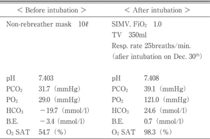
ツベルクリン反応も 65 歳以上では陰性であること Table 3 Arterial blood gas measurements (Dec. 30th)
< Before intubation > < After intubation > Non-rebreather mask 10ℓ
pH 7.403
PCO2 31.7(mmHg)
PO2 29.0(mmHg)
HCO3 −19.7(mmol/l)
B.E. −3.4 (mmol/l)
O2 SAT 54.7(%)
SIMV, FiO2 1.0 TV 350ml
Resp. rate 25breaths/min.
(afier intubation on Dec. 30th)
pH 7.408
PCO2 39.1(mmHg)
PO2 121.0(mmHg)
HCO3 24.6(mmol/l)
B.E. 0.7(mmol/l)
O2 SAT 98.3(%)
Blood gas results before and after intubation on Dec. 30th. Blood gas results revealed improvement with administration of 100% O2 following intubation, but PO2 was 121.0 (mmHg) and the P/F ratio was below 200 mmHg, so the patient was diag- nosed with ARDS.
Fig. 4 Chest CT (Jan. 6th)
Chest CT on the day following extubation. The exam revealed that the exten- sive infiltrates in both lungs had improved.
が多く 15.4%が陰性とされ8),本症例でも陰性で あった.喀痰塗抹染色の陽性率は 33 〜 46%,培養 陽性率は 62 〜 77%,といわれ9‑12),最も有効であっ たとする報告もある3).本症例のように,塗抹検査 で陰性であっても,核酸増幅同定などの高感度な抗 酸菌検出法を併用することによって陽性率の向上が 期待できる.本症例では挿管後の吸引気道分泌物の 細菌学的検査で結核菌を検出したが,気管支肺胞洗 浄による結核菌の検出も有用と考えられた.また,
尿,血液,骨髄からの結核菌の検出率は比較的高 く,特に前 2 者は検体採取が容易であり積極的に施 行すべきとされていて3),本症例でも尿 PCR 法,
尿培養にて陽性であった.
粟粒結核に ARDS を合併すると予後が極めて不 良で,死亡率 76%と報告されている13).ステロイ ドの有効性については議論のあるところで,その薬 理作用は IL-6 や TNF-α等の炎症性メディエーター 産生調節作用で,この抗炎症効果が ARDS に対し ても有効と考えられる14).発症早期のステロイド大 量療法は否定的な見解もあるが15‑17),急性期での ステロイドパルス療法にて症状の軽快を認めた報告
も多い18‑21).ARDS の発症と病態の悪化は急激で,
臨床的にはパルス療法に頼らざるを得ないことが多
いと考えられる.また,発症早期のステロイド少量 投与(メチルプレドニゾロン 1 mg/kg/ 日より漸減)
による肺損傷スコアの有意な改善傾向を認めた報告 もある22).本症例でもパルス療法後,前述の少量投 与を漸減しつつ継続した.しかし,粟粒結核の症状 出現後にその診断がなされる前に,抗結核薬の併用 が無いままステロイド単独使用された症例で病態を 悪化させた可能性を示唆する報告もある3). 本症例では当初より結核症を強く疑い,抗結核薬 による治療を開始した後,ARDS を発症したため,
ステロイド投与療法を併用した.そのため,呼吸状 態の改善を得ることができ,本質的な抗結核薬治療 を継続でき,幸い救命することができたと考える.
高齢者に体重減少などの低栄養の傾向を伴った原因 不明の発熱が持続した場合,血液検査所見で非定型 感染を疑う所見を認めた際には当初から粟粒結核も 視野に入れた検査を進め,早期に診断し,抗結核薬 による治療を開始することが肝要であると考えられ た.
文 献
1) Bernard GR, Artigas A, Bringham KL, : The American-European Consensus Conference on ARDS. Definitions, mechanisms, relevant Fig. 5 Chest x-ray and CT(Mar. 6th)
Chest x-ray and chest CT scan both revealed fibrotic changes (scarring) in the left upper lobe of the lung, but infiltrates that had spread to both lung fields disappeared, and the patient had clinically improved.
outcomes, and clinical trial coordination.
149:818‑824, 1994.
2) 永井英明,倉島篤行,赤川志のぶ,ほか:粟粒 結核症の臨床的検討.結核 73:611‑617,1998.
3) 平塚雄聡,有村保次,伊井敏彦:当院における 粟 粒 結 核 の 臨 床 的 検 討. 感 染 症 誌 78:929‑
934,2004.
4) 石井 学,石川雅健,武田宗和,ほか:診断に 苦慮した粟粒結核の 1 例.日救急医会関東誌 23:22‑23,2002.
5) Dyer RA, Chappel WA and Potgieter PD: Adult respiratoly distress syndrome associated with military tuberculosis. 13:12‑15, 1985.
6) 森谷知恵,有田浩之,吉岡宏治,ほか:急性呼 吸促迫症候群を合併した粟粒結核症の 1 例.広 島医 55:504‑509,2002.
7) 原田 進,永田忍彦,原田泰子:粟粒結核.化 療の領域 17:1428‑1434,2001.
8) 毛利昌史,四元秀毅,倉島篤行,編:結核 Up to Date:結核症+非定型抗酸菌症+肺アスペルギ ルス症.南江堂,東京,1999.
9) Maartens G, Willcox PA and Benatar SR: Mil- iary tuberculosis: rapid diagnosis, hematologic abnormalities, and outcome in 109 treated adults. 89:291‑296, 1990.
10) Munt PW : Miliary tuberculosis in the chemo- therapy era: with a clinical review in 69 Ameri- can adults. (Baltimore) 51:139‑155, 1972.
11) Sahn SA and Neff TA: Miliary tuberculosis.
56:494‑505, 1974.
12) Kim JH, Langston AA and Gallis HA : Miliary tuberculosis : epidemiology, clinical manifesta- tions, diagnosis and outcome.
12:583‑590, 1990.
13) 河端美則,和田雅子,岩井和郎,ほか:粟粒結 核症の病理―有用な臨床情報と DIC,急性呼吸
不 全 に 焦 点 を 当 て て―. 呼 吸 5:576‑583,
1986.
14) 石倉宏恭:治療の実際 SIRS-associated coagul- opathy(SAC)病態下における ALI/ARDS の 発症機序と治療戦略.臨と研 86:1202‑1208,
2009.
15) Lefering R and Neugebauer EAM: Steroid con- troversy in sepsis and septic shock : a meta- analysis. 23:1294‑1303, 1995.
16) Bernard GR, Luce JM, Sprung CL, : High- dose corticosteroids in patients with the adult respiratory distress syndrome.
317:1565‑1570, 1987.
17) Bone RC, Fisher CJ, Clemmer TP, : Early methylprednisolone treatment for septic syn- drome and the adult respiratory distress syn- drome. 92:1032‑1036, 1987.
18) 巽浩一郎,栗山喬行,高松芳郎,ほか:粟粒結 核症に続発した ARDS の 1 例.日胸疾患会誌 25:574‑577,1987.
19) 川山智隆,澤亜希子,吉住尚志,ほか:メチル プレドニゾロンパルス療法が有効であった成人 呼吸促迫症候群を呈した粟粒結核の 1 例.日胸 臨 56:58‑63,1997.
20) Murray HW, Tuazon CU, Kirmani N, : The adult respiratory distress syndrome associated with military tuberculosis. 73:37‑43, 1978.
21) Meduri GU, Headley AS, Golden E, : Effect of prolonged methylprednisolone therapy in un- resolving acute respiratory distress syndrome:
a randomized controlled trial. 280:159‑
165, 1998.
22) Meduri GU, Golden E, Freire AX, : Methyl- prednisolone infusion in early severe ARDS: re- sults of a randomized controlled trial.
131:954‑963, 2007.
A RARE CASE OF MILIARY TUBERCULOSIS DEVELOPING TO ACUTE RESPIRATORY DISTRESS SYNDROME
IN WHICH THE PATIENT S LIFE WAS SAVED
Genyo OGAWA, Takashi KATSURA, Norihiro NOMURA, Masashi SATOH, Ryouji HAYASHI, Takatoshi SATOH, Sadanori KUBO, Takashi SEKIKAWA and Masa-aki MATSUKAWA
Depertment of Internal Medicine, Showa University Toyosu Hospital
Makoto HASEGAWA
Depertment of Radiology, Showa University Toyosu Hospital
Abstract The patient was an 84-year-old male who was treated for hypertension in another hos- pital. In addition to general malaise and anorexia lasting for about a month, the patient had a high fever, which prompted the referral to our department. Chest x-ray and chest CT scan upon admission revealed ground glass appearance and diffuse granular infiltration in both lung fields. We suspected a pulmonary infection, such as miliary tuberculosis, and an anti-TB regimen and ciprofloxacin CPFX were adminis- tered. Mycobacterium tuberculosis in the sputum, gastric juices, and urine were detected several days later by bacteriological tests, confirmed the diagnosis of miliary tuberculosis. On the third day of hospi- talization, hypoxemia worsened and developed into acute respiratory distress syndrome ARDS; the pa- tient was immediately placed on a ventilator and corticosteroids were administered. The patient re- sponded well to these treatments, and was successfully extubated 6 days later. The patient had slow but steady, uneventful recovery after extubation, and he was discharged home on hospital day 83. Review of literature indicates an extremely poor prognosis of miliary tuberculosis developing to ARDS, with mortal- ity of 70‑80. We believe that this patient survived due the to early introduction of anti-TB drug therapy, even before the diagnosis was confirmed. When an elderly individual develops a high fever of unknown origin with pulmonary infiltrate, a differential diagnosis must be made with miliary tuberculosis in mind and anti-TB drug therapy should be initiated even before the diagnosis is confirmed.
Key words: miliary tuberculosis, ARDS, diffuse granular infiltration
〔受付:10 月 5 日,受理:11 月 9 日,2010〕