緒 言
Occipital sinus(OS)は,通常 venous confluence から 起始し,marginal sinus(MS)や sigmoid sinus(SS)もし くは vertebral venous plexus に流出する静脈洞である4). OS は胎児期に発達するが,出生後は消退するのが通常 であり,認めても小さな静脈洞か,その遺残である5).我々 が渉猟した限りでは,OS のみに main shunt point もし く は fistulous pouch を 認 め る 硬 膜 動 静 脈 瘻(dural arteriovenous fistula;dAVF)は報告されていない.我々 は,OS のみに shunt point を持つ dAVF を経験したの で,OS に関する解剖学的な考察および治療戦略を交え て報告する.
症例呈示
51歳女性.1年前から続く拍動性の耳鳴りが悪化し,
さらに頭痛が出現したため当科外来を受診した.以前に 明らかな外傷の既往は認めなかった.両側の耳介後方に bruit を聴取したが,明らかな神経脱落症状は認めなか った.頭部 MRA では左側により強い両側 transverse sinus(TS)の異常高信号を認め,dAVF が疑われた.
脳血管撮影では,両側の後頭動脈(occipital artery),両 側 の 中 硬 膜 動 脈(middle meningeal artery) か ら の feeder が正中部で OS に流入し,両側の TS へ流出する dAVF を認めた.cortical reflux は認めなかった(Fig.
1, 2). 右 総 頚 動 脈 か ら の3-dimensional rotation
症例報告
JNET 6:141-145, 2012Occipital sinus のみに shunt point を認めた dAVF の1例:解剖学的考察および治療戦略について
髙木俊範 酒井秀樹 加藤貴之 西村康明
Dural arteriovenous fistula of the occipital sinus: a case report
Toshinori TAKAGI Hideki SAKAI Takayuki KATO Yasuaki NISHIMURA
Department of Neurosurgery, Toyohashi Medical Center
●Abstract●
Objective: There have been no reports on the dural arteriovenous fistula (dAVF) of the occipital sinus (OS). We present a case of dAVF involving the OS.
Case presentation: A 51-year-old woman suffered from pulsating tinnitus for a year and gradually worsening tinnitus. Angiograms showed the presence of a dAVF fed by the bilateral occipital arteries and bilateral middle meningeal arteries. The fistula was located in the occipital sinus and drained into the bilateral transverse sinuses. Cortical venous reflux was not observed. We chose transvenous embolization (TVE). A microcatheter was introduced into the fistulous pouch of the OS via the left transverse sinus (TS). Six detachable coils were placed in the fistulous pouch, and an angiogram showed complete occlusion of the dAVF. After the procedure, there was no observable neurological deficit or tinnitus.
Conclusion: dAVF draining into the OS is very rare. It is important to identify the fistulous point by 3-dimensional rotational angiography (3D-RA). Hemodynamic function of the OS, in particular, should be evaluated to perform transvenous embolization.
●Key Words●
dural arteriovenous fistula, occipital sinus, transvenous embolization 独立行政法人国立病院機構豊橋医療センター 脳神経外科
<連絡先:髙木俊範 〒440-0836 愛知県豊橋市飯村町字浜道上50 E(Received October 24, 2011:Accepted August 13, 2012)-mail: ttakagi@gifu-u.ac.jp >
A B Fig. 1
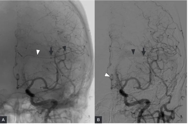
The left common carotid angiograms (A:anteroposterior view, B:lateral view) show dural arteriovenous fistula fed by the occipital artery and the middle meningeal artery. The fistula is located at the occipital sinus and drains into the bilateral transverse sinuses. No cortical venous reflux is observed.
Fig. 2
The right common carotid angiograms (A:anteroposterior view, B:lateral view) show a dural arteriovenous fistula fed by the occipital artery and the middle meningeal artery. Note these feeding arteries enter the same fistulous portion of the occipital sinus as shown in Fig. 1.
A B
Takagi T, et al
angiography(3D-RA) で は,OS に お け る fistulous pouch と両側 TS への流出経路が明瞭に描出された
(Fig. 3).Borden 分類および Cognard 分類では type 1 と 考 え ら れ た.Type 1の dAVF は 経 動 脈 的 塞 栓 術
(transarterial embolization;TAE)の適応となることが 一般的と考えられているが,本症例では,3D-RA の評 価により,TS から fistulous pouch までカテーテルが誘 導 可 能 と 推 測 し, 経 静 脈 的 塞 栓 術(transvenous embolization;TVE)を第一選択と考えた.また患者も 強度の耳鳴りによる不眠など ADL が障害されており,
治療を強く希望されたので塞栓術を行った.
1.治療手技
治療は局所麻酔下に行った.全身ヘパリン化ののち,
90 cm 長の6Fr ガイディングカテーテル(Envoy, Corids Endovascular, Johnson & Johnson, Miami, FL, USA)を 左内頚静脈に留置し,これを介して125 cm 長の4Fr カ テーテル(IPS, カテックス , 大阪)をコアキシャルに左 TS に誘導した.マイクロカテーテル(Rapid Transit, Corids Endovascular)を TS から OS へ誘導し,比較的 容易に fistulous pouch に留置できた(Fig. 4A, B).計 6本の detachable coil により fistulous pouch と OS を閉 塞した(Fig. 5A).確認造影に dAVF の完全閉塞を確 認し(Fig. 5B, C),手技を終了した.
シャントの消失とともに耳鳴は消失し,他覚的な bruit も消失した.治療後,神経脱落所見は認めず,ま た MRI diffusion weighted image では陽性所見を認めな かった.
考 察
Occipital sinus は通常,venous confluence に起始し,
MS や SS もしくは vertebral venous plexus に終わると される4).OS は胎児期に発達するが,多くの症例では 成長と共に消退することが多く,通常は認めても小さな ものであることが多い5).その詳細については MRI を 用いた検討が Kobayashi らにより報告されている3).こ の報告によれば,OS は造影 MR venography にて37.7% にその存在が認められる.OS が繋ぐ静脈の variation に つ い て は, 頭 側 で は transverse sinus(59.4%),dural vein(26.5%),confluence(11.4%)の順に多い.一方,
尾 側 で は,dural vein(53%) が 最 も 多 く, 次 い で vertebral venous plexus(44.3%),SS(11.9%),MS(11.4
%)の順であった.また,OS を認めた症例のうち4.8% に2本の OS を認め,多数の分枝を持つ variation も観察 された.また,Kiyosue らは自検例23例につき報告し ており,MS へ流出するものが最多で,続いて posterior condylar vein と MS の 両 方 に 続 く も の,posterior Fig. 3
A 3-dimensional rotation angiogram of the right common carotid artery (A:posteroanterior view, B:left oblique view) clearly demonstrates the anatomical relationship between the fistula, occipital sinus, and bilateral transverse sinuses.
A B
Fig. 4
A:A microcatheter has been inserted into the left transverse sinus through a 4 Fr catheter.
B: Note the tip of the microcatheter has reached the fistulous pouch of the occipital sinus.
(tip of 4 Fr catheter [large arrow], tip of guidewire [small arrow], first and second markers of microcatheter [white and black arrowheads])
A B
A B C
Fig. 5
The left common carotid angiograms (A: anteroposterior view, B: lateral view) and the right common carotid angiogram (C: lateral view) after the transvenous embolization reveal complete obliteration of the fistula.
Takagi T, et al
condylar vein へ続くものの3パターンであった2).なお,
この報告では,全症例に2本の OS を認めていた.この ように,OS は一般的に消退していることも少なくなく,
また頭側尾側にさまざまな連絡を持っており,個人差の 大きい静脈洞である.この部位の疾患として,他の dAVF の流出路として OS が関与している症例はある が,OS のみに shunt point を持つ dAVF は我々が渉猟 した限りでは報告されておらず,非常に稀な症例と考え られた.前述のような OS の解剖学的特徴から考えると,
OS のみに shunt point を持つ本症例のような dAVF の 場合,TVE により OS ごと shunt point を塞栓しても正 常灌流への影響は少ないと推察される.ただし,稀には OS が深部静脈系の main drainage route であることもあ り得るので注意が必要である.また OS の流入する venous confluence が plexus 状で複雑な形態をとる場合 には TS から OS へのアプローチが困難な場合もありう るため,その術前の画像評価が大切である.本症例の治 療戦略であるが,一般的には Borden 分類もしくは Congard 分類での type 1の症例では TAE が第一選択と されることが多い1).しかし,type 1の病変であっても shunt point の位置を正確に把握することができれば,
低いリスクで TVE により完治させることが可能な症例 もあると考えられる6).最近では3D-RA を用いること で静脈系の血管構築や shunt point の詳細な把握が行え るようになってきた.本症例でも OS の dAVF という 稀な症例であったが,3D-RA により venous confluence
の構造や shunt point の正確な評価ができ,少量のコイ ルにて完治することができた.dAVF では関与する静脈 系の variation のため,ときに shunt point の把握が不十 分なまま治療に臨むこともありうるが,有効な塞栓術を 行うには事前の画像診断の重要性が再確認された.
本論文に関して,開示すべき利益相反状態は存在しない.
文 献
1) Guedin P, Gaillard S, Boulin A, et al: Therapeutic management of intracranial dural arteriovenous shunts with leptomeningeal venous drainage: report of 53 consecutive patients with emphasis on transarterial embolization with acrylic glue. 112:603-610, 2010.
2) Kiyosue H, Okahara M, Sagara Y, et al: Dural arteriovenous fistula involving the posterior condylar canal.
28:1599-1601, 2007.
3) Kobayashi K, Suzuki M, Ueda F, et al: Anatomical study of the occipital sinus using contrast-enhanced magnetic resonance venography. 48:373-379, 2006. 4) Newton TH, Potts DG: Radiology of the Skull and Brain. St
Louis, Mosby, 1974, 1866-1869.
5) Okudera T, Huang YP, Ohta T, et al: Development of posterior fossa dural sinuses, emissary veins, and jugular bulb: morphological and radiologic study. 15:1871- 1883, 1994.
6) Yoshioka T, Kitagawa N, Yokoyama H, et al: Selective transvenous coil embolization of dural arteriovenous fistula.
A report of three cases. 13 Suppl 1:123- 130, 2007.
JNET 6:141-145, 2012
要 旨
【目的】我々は,occipital sinus(OS)のみに shunt point を持つ硬膜動静脈瘻(dural arteriovenous fistula:dAVF)
を経験したので,OS に関する解剖学的な考察および治療戦略を交えて報告する.【症例】51歳女性.1年前から続
く拍動性の耳鳴りがあり,その精査のために施行された頭部 MRA にて両側の transverse sinus(TS)に異常高信 号が指摘された.脳血管撮影で両側の後頭動脈(occipital artery),両側の中硬膜動脈(middle meningeal artery)
から OS のみに流入する dAVF を認めた.動静脈瘻は両側の TS へ流出しており,cortical venous reflux は認めな かった.本症例では,TS から fistulous pouch にカテーテルが誘導可能と考えられ,経静脈的塞栓術(TVE)を第 一選択と考えた.マイクロカテーテルを TS から OS の fistulous pouch まで誘導し,そこに計6本の detachable coil
を留置した.dAVF の完全閉塞が得られ,治療直後より耳鳴は消失し,他覚的な bruit も消失した.【結論】OS の
みに shunt point を有する dAVF は非常に稀である.shunt point の位置を正確に把握し,周囲の静脈や静脈洞との 解剖学的関係および OS の血行動態をよく理解することが本部位の dAVF の治療においては重要と考えられた.