Studies on GeneScan analysis of immunoglobulin and
T cell receptor genes in canine lymphoproliferative diseases
T
GeneScan
CONTENTS
Page GENERAL INTRODUCTION - - - -3
Chapter 1 - - - -7 Association between lymphocyte antigen receptor gene rearrangements and
histopathological evaluation in canine chronic enteropathy
Chapter 2 - - - 29 Detection of circulating tumor cells using GeneScan analysis of antigen receptor gene rearrangements in canine lymphoma
Chapter 3 - - - 47 GeneScan analysis for the differentiation of clonal origin of cells in dogs that developed two different types of lymphoid malignancies
CONCLUSION - - - -66 ACKNOWLEDGEMENTS - - - 71 REFERENCES - - - -73
Lymphoid malignancies are defined as clonal expansion of lymphoid cells originated from one transformed cell. They are one of the most common neoplastic diseases in dogs. Lymphoid neoplasms are generally diagnosed based on histopathological or cytological examination with or without immunostaining of cellular antigens (Fournel-Fleury et al., 1997; Valli et al., 2011). However, their pathological diagnosis is sometimes difficult when specimens include only small number of tumor cells or neoplastic population comprising small lymphoid cells, which is difficult to distinguish from reactive ones. Therefore, more objective diagnostic strategy has been required for the accurate diagnosis of lymphoid neoplasms.
For attaining more accurate diagnosis of lymphoid malignancies, assessment of the
rearrangements of lymphocyte antigen receptor genes, immunoglobulin (Ig) and T cell receptor (TCR) genes, has been commonly performed as a useful adjunctive method. Antigen receptor genes consist of randomly combined variable (V), diverse (D), and joining (J) gene segments in early lymphoid development. During the process of recombination, several nucleotides are randomly inserted or deleted in junctional regions between each gene segments, producing diverse length and sequence among each lymphoid cells, which is called complementary determining region 3 (CDR3) (Jung et al., 2006). In normal tissues, since various lymphocytes heterogeneously exist, polyclonal patterns of antigen receptor gene rearrangements are found. On the other hand, since lymphoid malignancies are composed of a clonally expanded cell
population, all of the neoplastic cells in each tumor have a uniform antigen receptor gene rearrangement.
Clonality of lymphocytes are commonly analyzed by amplifying CDR3 using polymerase chain reaction (PCR) with primers annealing to V and J segments, which is generally called PCR for antigen receptor gene rearrangements (PARR). PARR is followed by separation of the PCR products according to its nucleotide length using polyacryl amide gel electrophoresis (PAGE). This PAGE method has been widely used in dogs (Burnett et al., 2003; Tamura et al., 2006; Valli et al., 2006; Yagihara et al., 2007). Several studies have been conducted to improve its accuracy by designing multiple primer sets. In addition, heteroduplex analysis has been generally employed to avoid false positive results. However, it is difficult to discriminate fragments with differences less than 5 bp when using PAGE.
To avoid this pitfall, GeneScan analysis has been introduced in human medicine (van Dongen et al., 2003). GeneScan technique involves separation of fragments through capillary electrophoresis together with detection of fluorescent-labeled primers. There are several advantages in GeneScan compared with PAGE. First, higher resolution ability, which can separate fragments by only 1 nucleotide, can avoid false positive or negative results when assessing samples with high background. Second, nucleotide length of the amplified fragments can be defined based on size standards, which can be helpful to confirm identification among
multiple samples in a same individual. Third, since the GeneScan analysis employs PCR system, it requires only a small amount of cell or tissue sample. Finally, it is a rapid and high throughput method applicable for large numbers of samples. GeneScan analytical system of antigen
receptor genes was recently introduced into veterinary medicine and shown to be effective as an adjunctive diagnostic tool for detecting clonally expanded lymphoid cells in dogs (Gentilini et al., 2009; Keller and Moore, 2012). In addition, multiplex GeneScan analytic system has been recently constructed. In this newly developed system, fragments are amplified in small number of PCR conditions by using primers with multicolor fluorogenic probe, which is considered to be more suitable for clinical settings.
The present study was conducted for the purpose of application of multiplex GeneScan analysis of antigen receptor gene rearrangements to the clinical settings for the diagnosis and further understanding of canine lymphoproliferative diseases. In Chapter 1, association between clonal antigen receptor gene rearrangements and histopathological evaluation of endoscopic biopsies was investigated in order to understand the value of GeneScan analysis in diagnosing gastrointestinal (GI) lymphoma. In Chapter 2, usefulness of GeneScan analysis for detecting circulating tumor cells (CTCs) in canine lymphoma was investigated. In Chapter 3, GeneScan analysis was applied to distinguish the origin of tumor cells in dogs that developed 2 different types of lymphoid neoplasms.
Chapter 1
Association between lymphocyte antigen receptor gene
rearrangements and histopathological evaluation
ABSTRACT
Although definitive diagnosis of chronic enteropathy (CE) and gastrointestinal (GI) lymphoma requires histopathological evaluation of the GI tract, these conditions are often still difficult to differentiate from each other. Polymerase chain reaction for antigen receptor gene rearrangements (PARR) has been applied recently as an adjunctive for diagnosis of lymphoid tumors; however, its clinical value in canine CE and GI lymphoma remains unclear. The purpose of this study was to investigate the relationship between PARR and histopathological diagnosis, degree of enteritis or lymphoma, and long-term prognosis in dogs, in order to evaluate the clinical significance of PARR. Endoscopic biopsy specimens obtained from 96 dogs with chronic enteritis (mild, n = 14; moderate, n = 20; marked, n = 62) and 21 dogs with GI lymphoma were used. Clonal rearrangement in PARR was observed in 51% of the animals with chronic enteritis; interestingly, it was found in 29% of those with only mild enteritis. In dogs with marked enteritis, the rate of clonal rearrangements was higher in those with epitheliotropism of lymphocytes than in those without epitheliotropism. The sensitivity of PARR in animals with GI lymphoma was 76%. There was no significant prognostic difference between chronic enteritis with or without clonal rearrangements. In contrast, dogs histopathologically diagnosed with marked enteritis had a significantly shorter survival time than did those with mild or moderate enteritis. While the significance of PARR in the diagnosis
of GI lymphoma remains uncertain, the pathological roles of clonally expanding lymphocytes in canine CE should be investigated further.
INTRODUCTION
Chronic enteropathy (CE) is one of the common clinical diagnoses in dogs, characterized by persistent or recurrent gastrointestinal (GI) symptoms such as diarrhea, vomiting, and weight loss. The common causes of CE include food-responsive enteropathy, antibiotic-responsive enteropathy, and inflammatory bowel disease (IBD) (German et al., 2003). The diagnosis of canine CE requires various diagnostic evaluations in order to rule out other diseases causative of chronic GI symptoms, including metabolic diseases, infection, parasitism, or neoplastic diseases (Allenspach, 2013). Although the pathogenesis of canine CE is not yet fully elucidated, interaction between the intestinal microenvironment (bacterial or dietary antigens), mucosal immune system dysfunction, and genetic factors are considered to be involved in its etiology (German et al., 2003; Simpson and Jergens, 2011). Therapeutic trials of dietary modification, antibiotics, or immunomodulatory agents are needed to improve diagnosis and control of the disease (Allenspach, 2013). If animals with the condition are treated appropriately, the prognosis of canine CE is relatively favorable, as it has been reported that the 3-year survival rate was 97% in food-responsive dogs with diarrhea and 57% in steroid-treated dogs with IBD (Allenspach et al., 2007).
GI lymphoma should also be taken into consideration in dogs with chronic GI symptoms. Although GI lymphoma seems to be less common in dogs than in cats, it accounts for
approximately 7% of all canine GI neoplasms (Patnaik et al., 1977). Despite treatment with multidrug chemotherapy, the prognosis of dogs with GI lymphoma is usually poor, as median survival time was reported to be 77 days (Rassnick et al., 2009).
Because of differences in treatment strategies and outcomes, it is essential to distinguish between CE and GI lymphoma. As there is no specific laboratory test for differentiating between these 2 diseases, GI biopsies are used to make histopathological diagnosis. Flexible endoscopy is the preferred method for obtaining specimens as it is less invasive than surgery (Washabau et al., 2010). However, as endoscopically obtained tissues are often limited to mucosa, this can lead to pathologists overlooking cases of lymphoma, since this condition can involve transmural expansion of neoplastic cells. In addition, GI lymphoma is frequently accompanied by infiltration of inflammatory lymphocytes and plasma cells that can cause misdiagnosis of lymphocytic-plasmacytic enteritis (Kleinschmidt et al., 2006).
Since its advent, polymerase chain reaction (PCR) for antigen receptor gene rearrangements (PARR) has been introduced into veterinary practice as a useful adjunctive for the diagnosis of lymphoma. It has a high accuracy for the detection of clonal lymphocytes, and its sensitivity in diagnosing lymphoid malignancies was reported to be more than 90% (Burnett et al., 2003; Gentilini et al., 2009). There are several studies on the application of PARR in GI biopsy specimens. One of these reports suggested that PARR was a useful diagnostic tool for detecting
latent GI lymphoma, which cannot be histopathologically diagnosed using endoscopic biopsy specimens (Kaneko et al., 2009). In another study, the sensitivity of PARR in diagnosing GI lymphoma was reported to be 66.7%, which was lower than that for lymphoma affecting other anatomical sites; thus, it may be useful for detecting lymphoma when combined with histopathological examination (Fukushima et al., 2009). On the other hand, a more recent study indicated that clonal lymphocytic infiltration was also detected in the GI tract of dogs with IBD, and reduced diversity of lymphocytic infiltrates significantly correlated with one-year survival rate (Olivero et al., 2011).
The purpose of this study was to investigate the relationship between PARR and histopathological diagnosis, degree of enteritis or lymphoma, and long-term prognosis in dogs with persistent or recurrent GI symptoms, in order to evaluate the clinical significance of PARR.
MATERIALS AND METHODS Cases
Medical records of dogs investigated for chronic GI diseases at the Veterinary Medical Center of the University of Tokyo (from May 2011 to December 2012) and the Japan Small Animal Medical Center (from April 2012 to March 2013) were reviewed retrospectively. The
following inclusion criteria were used: (1) a history of chronic GI signs with a duration of >3 weeks and/or hypoalbuminemia (<2.7 g/dL); (2) exclusion of other causes of chronic GI signs (metabolic diseases, infection, and parasitism) or hypoalbuminemia (hepatic disease, renal disease, and blood loss) by complete blood cell count, serum biochemistry, urinalysis, fecal examination for parasites and bacteria, and abdominal ultrasonography; (3) availability of histopathological evaluation of mucosal specimens obtained via upper and/or lower GI tract endoscopic examination; and (4) availability of genomic DNA extracted from duodenal mucosa obtained via endoscopy. Food-responsive enteropathy and antibiotic-responsive enteropathy were not ruled out before endoscopic examination if worsening of clinical signs was observed in animals with suspected neoplastic disease. Cases were included regardless of treatments given. According to the inclusion criteria, the medical records of 120 dogs were reviewed. Three of the animals were histopathologically diagnosed with gastric adenocarcinoma and so were excluded from the present study. Of the remaining 117 dogs, 96 were diagnosed with chronic enteritis and 21 were diagnosed with GI lymphoma by histopathological evaluation. Cases were excluded from survival analysis if concurrent neoplastic diseases other than GI lymphoma were confirmed. The referring veterinarians or the owners were contacted to obtain follow-up data.
Endoscopy of the upper GI tract was performed in all dogs, and 6 mucosal biopsy specimens were collected from both the stomach and the duodenum. In cases undergoing lower GI tract endoscopy, 6 mucosal biopsies were also obtained from both the ileum and the colon. Tissue samples were submitted for histopathological examination according to the method described in a previous report (Washabau et al., 2010). Biopsy specimens were oriented with the submucosal side facing downwards and the villi side facing upwards, so that the entire depth of the mucosa could be evaluated. The specimens were then placed in histopathological cassettes and fixed in 10% formalin for 48 h, processed and paraffin embedded, cut into sections, and stained with hematoxylin and eosin (HE). Histopathological diagnoses made from HE-stained sections were reviewed in the archives, and cases were classified as described below. Final diagnoses were defined as the most severe histopathological classification for all of the bioptic sites in each case of chronic enteritis. GI lymphoma was diagnosed when at least one of the sites was diagnosed with lymphoma.
Chronic enteritis was diagnosed according to the histopathological standards for endoscopic biopsy samples proposed by the World Small Animal Veterinary Association (WSAVA) (Day et al., 2008). Microscopic inflammatory changes defined by morphological abnormalities and inflammatory cell infiltration were assessed for each of the anatomical regions, and each of the cases was categorized as (1) normal, (2) mild, (3) moderate, or (4) marked, based on the grade
of histopathological severity. For chronic enteritis with marked changes, infiltration of intraepithelial lymphocytes was evaluated and classified into two groups according to its severity: epitheliotropism [−] or epitheliotropism [+]. This classification was provisionally performed in order to attempt to distinguish lymphoma (small cell lymphoma, if it exists) from chronic inflammation, because the increase observed in intraepithelial lymphocytes was found to be significantly different between cases of GI lymphoma and IBD in cats (Kiupel et al., 2011).
GI lymphoma was diagnosed histopathologically based on severe infiltration of large lymphoid cells (nuclear diameter > 2 blood cell diameters) associated with mucosal injury, in line with the previous report on feline GI lymphoma of large cell type (Moore et al., 2012).
Polymerase Chain Reaction for Antigen Receptor Gene Rearrangements
Genomic DNA was extracted from endoscopically obtained duodenal tissues from each of the animals using a QIAamp DNA Blood Mini Kit (Qiagen, Valencia, CA), according the manufacturer’s instructions. Rearrangements of the IgH/TCRγ genes were assessed by amplifying complementarity determining region 3 (CDR3) to evaluate lymphocyte clonality using primers derived from V segments and J segments of each of the genes. Two control genes (IgM C region and HGF) were also amplified. All of the forward primers were labeled with
fluorescent dyes at the 5’ end. PCR reactions were carried out in a final volume of 25 µL. The PCR mixture was composed of 1 × Amplitaq Gold 360 Master Mix (Applied Biosystems, Foster City, CA), forward primer (200 nM), reverse primer (200 nM), and 100 ng of DNA template. Cycle conditions consisted of an initial denaturation and enzyme activation step at 95°C for 5 min, followed by 40 cycles of denaturation at 94°C for 15 sec, annealing at 62°C or 56°C for 30 sec and extension at 72°C for 30 sec. A final extension at 72°C for 30 min was included in 6 of the 7 reactions. Each PCR reaction was performed in duplicate. GeneScan analysis was carried out on the ABI 3130xl Genetic Analyzer (Applied Biosystems) as a multiplex run. One microliter of PCR product mixture diluted 40-fold was combined with 8.5 µL of Hi-Di formamide (Applied Biosystems) and 0.5 µL of 600 LIZ size standard (Applied Biosystems) in an optical 96-well plate. The products were denatured at 95°C for 5 min, immediately placed on ice for 15 min, and then subjected to the analysis. Resulting data were analyzed with Peak scanner software (Applied Biosystems). Distinct peaks that were at least 2-fold higher than other background peaks were judged to show the presence of clonally expanded lymphoid cells. Presence of a single or two peaks was interpreted as the clonal rearrangements.
Statistical Analysis
evaluation were analyzed using the Kaplan-Meier method and log-rank test.
A P value <0.05 was defined as statistically significant. Significance level was corrected by Bonferroni’s method for multiple comparison.
RESULTS Signalment
According to the inclusion criteria, 117 dogs were included in this study. Dogs with chronic enteritis (n = 96) consisted of 56 males (neutered, 32) and 40 females (neutered, 22). The median age at the time of diagnosis was 7.9 years (range, 1.9−14.3 years). The dog breeds were as follows: Miniature Dachshund (n = 11), Shiba (n = 9), Yorkshire Terrier (n = 9), Chihuahua (n = 6), French Bulldog (n = 6), Toy Poodle (n = 6), Maltese (n = 4), Pomeranian (n = 4), Boston Terrier (n = 3), Pug (n = 3), and others (n = 35).
Dogs with GI lymphoma (n = 21) consisted of 9 males (neutered, 4) and 12 females (neutered, 7). The median age was 10.3 years (range, 5.5−15.1 years). The dog breeds were as follows: Miniature Dachshund (n = 5), Pug (n = 4), Shiba (n = 3), and others (n = 9).
Histopathological Evaluation
mild (n = 14), moderate (n = 20), and marked (n = 62) categories based on the histopathological severity of disease (Figs. 1-1,2 and 3). In addition, cases of marked enteritis were classified as epitheliotropism [−] (n = 21) or epitheliotropism [+] (n = 41) based on the severity of intraepithelial lymphocytic infiltration. Intraepithelial lymphocytes were found in clusters or as diffuse infiltrates in epithelia (Fig. 1-3b). In GI lymphoma (n = 21), the lamina propria showed diffuse infiltration by large lymphoid cells accompanied by alterations of the mucosal architecture including marked villous blunting and epithelial injury (Fig. 1-4).
Association between PARR and Histopathology
The results of PARR analysis are shown in Table 1-1. Clonal rearrangements of antigen receptor genes were detected in 49 of 96 dogs (51%) with chronic enteritis and 16 of 21 dogs (76%) with GI lymphoma. With respect to histopathological severity, clonal rearrangements were detected in 4 of 14 dogs (29%) with mild enteritis, 8 of 20 dogs (40%) with moderate enteritis, 10 of 21 dogs (48%) with marked enteritis classified as epitheliotropism [−], and 27 of 41 dogs (66%) with marked enteritis classified as epitheliotropism [+].
Survival Analysis
significantly different from that in dogs without clonal rearrangements in chronic enteritis (P = 0.35) (Fig. 1-5).
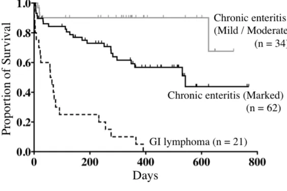
The results of survival analysis according to histopathological classification are shown in Figure 1-6. Survival time in dogs with marked enteritis was significantly shorter than that in dogs with mild or moderate enteritis (P = 0.0088). Survival time in dogs with GI lymphoma was significantly shorter than that in those with chronic enteritis, regardless of histopathological severity (P <0.0001). The median survival time was 61 days in dogs with GI lymphoma and 542 days in dogs with marked enteritis. The median survival time was not defined in all of the dogs with chronic enteritis as a whole. There was no significant difference in survival time between marked enteritis with and without epitheliotropism (P = 0.61) or clonal rearrangements (P =0.42) (data not shown).
DISCUSSION
In the current study, I assessed the association between lymphocyte antigen receptor gene rearrangements and histopathological evaluation in dogs with CE. The findings in this study revealed that even 29% of dogs with mild enteritis had clonal rearrangements. Moreover, there was no significant association between the results of PARR analysis and long-term prognosis in CE. Meanwhile, the significance of PARR in the diagnosis of canine GI lymphoma remains
questionable.
There are several possible explanations for the clonal rearrangements seen in dogs diagnosed with chronic enteritis. First, persistent stimulation by specific antigens may have caused monoclonal proliferation of lymphocytes. PARR has been reported to have high sensitivity for detection of lymphoid tumors, while studies focusing on the specificity of this method have not yet been published in veterinary medicine. In humans, monoclonality is also detected in benign skin lesions (drug eruption, insect bite reactions, and Borreliosis), some kinds of food allergy, Helicobacter pylori gastritis, various autoimmune diseases, and viral infections (Bakakos et al., 2001; Boer et al., 2008; Calvert et al., 1996; Engels et al., 2007; Hoeve et al., 2000). Furthermore, monoclonal or oligoclonal repertoires of antigen receptor genes in ehrlichiosis and leishmaniasis have also been reported in dogs (Burnett et al., 2003; Gentilini et al., 2009). Second, GI lymphoma may have been overlooked by histopathological examination, because observation was limited to the mucosal architecture since only endoscopic biopsy specimens were obtained in the present study. Full-thickness biopsy enables pathologists to evaluate transmural alterations and has diagnostic value for GI lymphoma comprising transmural lesions (Evans et al., 2006; Kleinschmidt et al., 2006). However, this explanation is less likely, because no significant difference in long-term prognosis due to the results of PARR in CE was observed.
An additional explanation is that small cell GI lymphoma could be detected by PARR. T-cell lymphoma of small cell type is the dominant GI lymphoma found in cats and is characterized by increased infiltration of mature-appearing lymphocytes into the mucosa (Moore et al., 2012). As their histological characteristics are similar to each other, small cell GI lymphoma is often difficult to differentiate from severe enteritis (Kiupel et al., 2011). In contrast, there is no extensive study clearly describing this entity in dogs thus far. Since an increase in intraepithelial lymphocytes is one of the significant histopathological findings that differentiates small cell lymphoma from enteritis in cats (Kiupel et al., 2011), I compared clonal rearrangements in cases of severe enteritis with and without epitheliotropism. The results showed that severe enteritis with epitheliotropism had a higher detection rate of PARR than cases without epitheliotropism. On the other hand, neither epitheliotropism nor PARR had a significant prognostic association in dogs with severe enteritis. The prognosis of cats with small cell GI lymphoma is favorable; median survival time has been reported to be 704 days (Kiselow et al., 2008), and median remission time was 786 days when animals were treated with prednisolone and chlorambucil (Stein et al., 2010). Considering the prognosis of cats with small cell GI lymphoma, the possibility remains that the investigation period of the present study was not long enough to distinguish between small cell GI lymphoma and chronic enteritis. Further studies are warranted in order to validate the presence and diagnostic definition of small cell GI
lymphoma in dogs.
Association between prognosis or clinical findings and histopathological severity has been reported to be equivocal so far when using endoscopic biopsy specimens (Allenspach et al., 2007; Garcia-Sancho et al., 2007; Schreiner et al., 2008), which may result in part from a lack of standardized histological scoring systems and guidelines for sample submission technique (Willard et al., 2008). In contrast to the previous studies, the present study revealed that the histopathological severity of the intestinal mucosa was significantly associated with prognosis in canine CE. In the present study, the procedure for biopsy, sample submission, and histopathological evaluation of enteritis was based on the WSAVA guidelines (Washabau et al., 2010). Considering that a significant association between histopathological severity and prognosis was observed in this study, it is reasonable to recommend following these standardized criteria for histological examination in canine CE. However, the differences between enteritis and GI lymphoma are not described in the WSAVA guidelines; thus, further investigation of the diagnostic criteria of small cell GI lymphoma in particular is needed. In the present study, the rate of PARR was 76% in GI lymphoma, which was higher than that of enteritis of any severity. However, this rate was lower than that found for lymphoma affecting other anatomical sites, similar to the results of a previous study (Fukushima et al., 2009). There are several explanations for why the sensitivity of PARR was lower in GI
lymphoma compared with lymphoma affecting other sites. First, as GI lymphoma can have a patchy distribution, differences in biopsy site may lead to inconsistencies between PARR and histopathological examination. Second, the primers used in this study did not cover all of the possible rearrangements of antigen receptor genes. Third, as the primers were for IgH and TCRγ, they could not detect non-T, non-B lymphoma. Considering the highly positive rate of PARR when all cases of enteritis were taken together, the detection of a clonal population of lymphocytes does not always confirm the diagnosis of lymphoma, nor does the absence of such a population indicate benignity.
In conclusion, clonal lymphocytic expansion was detected in about half of the animals with chronic enteritis in the present study. With regard to clinical applications, interpretation of PARR using intestinal biopsy specimens is still controversial, and further studies are needed to clarify its clinical significance. On the other hand, the present study revealed a significant association between histopathological severity and survival time in canine CE for the first time. It may be important to evaluate the histopathological severity in chronic enteritis in order for clinicians to predict a patient’s prognosis. While the significance of PARR in the diagnosis of GI lymphoma remains uncertain, the pathological roles of clonally expanding lymphocytes in canine CE should be investigated further.
Table 1-1. Detection rate of clonal rearrangements of antigen receptor genes by histopathological category.
Histopathological diagnosis (n)
Total number positive for clonal rearrangements (%)
Clonally rearranged gene(s)
TCRγ a IgH b TCRγ + IgH Chronic enteritis (96) 49 (51%) 40 3 6 Mild (14) 4 (29%) 4 0 0 Moderate (20) 8 (40%) 7 0 1 Marked (62) 37 (60%) 29 3 5 Epitheliotropism [-] (21) 10 (48%) 8 0 2 Epitheliotropism [+] (41) 27 (66%) 21 3 3 GI c lymphoma (21) 16 (76%) 13 2 1
Figs.1-1. Section of duodenum mucosa from a dog with mild chronic enteritis stained with hematoxylin and eosin. Inflammatory changes are mild and the mucosal architecture is relatively preserved (a). There is a mild increase in intraepithelial and lamina propria lymphocytes (b).
Figs. 1-2. Section of duodenum mucosa from a dog with moderate chronic enteritis stained with hematoxylin and eosin. Moderate crypt distention and lacteal dilation are observed (a). There is a mild increase in intraepithelial and lamina propria lymphocytes (b).
Figs. 1-3. Section of duodenum mucosa from a dog with marked enteritis stained with hematoxylin and eosin. Marked villous blunting and moderate lacteal dilation are observed (a). Small and mature-appearing lymphocytes heavily infiltrate into the epithelia and lamina propria. This case was classified as epitheliotropism [+] (b).
Figs. 1-4. Section of duodenum mucosa from a dog with intestinal lymphoma stained with hematoxylin and eosin. The mucosal architecture is altered by villous blunting with epithelial injury (a). Diffuse infiltration of pleomorphic large lymphoid cells with multiple nucleoli can be observed (b).
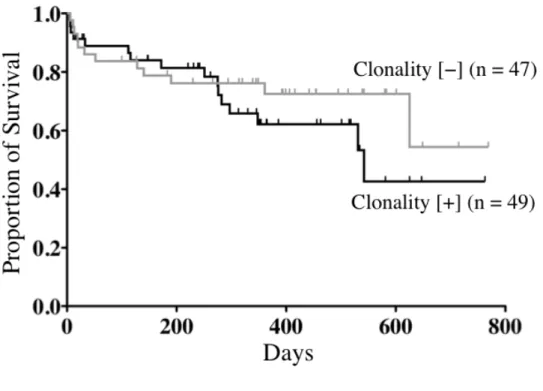
Fig. 1-5. Kaplan-Meier curves for dogs with chronic enteritis comparing animals with and without clonal rearrangements of antigen receptor genes (49 and 47 dogs, respectively). Survival time was not significantly different between dogs with and without clonal rearrangements (P = 0.35).
Fig. 1-6. Kaplan-Meier curves for dogs with mild/moderate chronic enteritis, marked enteritis, and gastrointestinal (GI) lymphoma (34, 62, and 21 dogs, respectively). Survival time in dogs with mild or moderate enteritis was significantly longer than that in animals with marked enteritis (P = 0.0088). Survival time in dogs with chronic enteritis was significantly longer than that in animals with GI lymphoma (P <0.0001).
Chapter 2
Detection of circulating tumor cells using GeneScan analysis of
antigen receptor gene rearrangements in canine lymphoma
ABSTRACT
Detection of circulating tumor cells (CTCs) is known to be useful in predicting relapse, prognosis, or evaluating treatment efficacy in lymphomas both in humans and dogs. Although several molecular techniques to detect CTCs have been proposed, each method has some limitations. The purpose of this study is to analyze the ability of GeneScan in detecting CTCs, and to investigate the prognostic significance depending on CTCs status in canine lymphoma. Samples from the primary lesions and peripheral blood mononuclear cells (PBMCs) were obtained in 32 dogs with lymphoma at the initial diagnosis. All the samples were subjected to polymerase chain reaction (PCR) for antigen receptor gene rearrangements (PARR) and analyzed by GeneScan. As a result, in 13 of the 17 dogs (76%) with high-grade multicentric lymphoma, all of the 3 dogs with low-grade multicentric lymphoma, and 4 of the 12 dogs (33%) with GI lymphoma, PARR results were positive and identical size of PCR products were detected between the primary lesions and PBMCs. Identical sequences of the PCR products were confirmed between the lesional sample and PBMCs, indicating the presence of CTCs. The GeneScan analysis employed in this study would facilitate the study on the clinical significance of CTCs in dogs with lymphoma.
INTRODUCTION
Lymphoma is one of the most common haematopoietic neoplasms in dogs with incidence of 13-24% among all canine neoplasms (Dorn et al., 1970). Multidrug chemotherapy based on CHOP (prednisolone, doxorubicin, vincristine, and cyclophosphamide) protocol has been standardized regimen in canine lymphoma, showing complete remission (CR) rates more than 80% (Burton et al., 2013; Garrett et al., 2002; Keller et al., 1993). However, most of the dogs experience relapse and die from disease progression.
For predicting outcome and monitoring of the disease, detection of circulating tumor cells (CTCs) can provide valuable information. CTCs are defined as tumor cells in the peripheral blood derived from the primary or secondary tumor lesion. CTCs are the cause of disseminated malignant cells to other distant organs and eventually become clinically evident as metastatic tumors (Joosse et al., 2014). In addition, CTCs which escape anti-cancer therapy are considered to be the source of relapse (Kern et al., 2008; Pott et al., 2006). Assessment of CTCs after chemotherapy is known to be useful for the prediction of the prognosis and the disease relapse, as well as monitoring the treatment efficacy in canine lymphoma (Sato et al., 2011a; Sato et al., 2011b; Sato et al., 2013; Yamazaki et al., 2010).
Detection of neoplastic cells by microscopic evaluation of peripheral blood smear is available in routine laboratory examination. However, it is less objective which can lead to
misjudgments when peripheral blood involves only small number of malignant cells. Therefore, several molecular techniques for detecting CTCs have been recently proposed. PCR for antigen receptor gene rearrangements (PARR) is reported to be a reliable and sensitive method to detect CTCs (Keller et al., 2004; Lana et al., 2006; Thilakaratne et al., 2010). Conventional method of PARR is generally conducted by using consensus primers followed by separation of nucleotide fragments using polyacryl amide gel electrophoresis (PAGE). However, the positive result does not always indicate the presence of CTCs especially in the cases of inflammatory diseases, since these primers were not tumor-specific.
Recently, quantitative assessment method of CTCs in canine lymphoma by real-time PCR was established (Yamazaki et al., 2008). Since antigen receptor genes were amplified by tumor-specific primers, this method is highly accurate. In addition, it is sensitive enough to detect small number of residual tumor cells in clinical remission, which is called minimal residual disease (MRD). On the other hand; however, the establishment of tailored assay for each individual is cumbersome and expensive, which is difficult to be applied in routine clinical settings.
Recently, GeneScan analysis was introduced to examine the clonal expansion of lymphoid cells in dogs with lymphoproliferative disorders (Gentilini et al., 2009; Keller and Moore, 2012). One of the advantages of GeneScan analysis over conventional PAGE method is that nucleotide
length of the amplified fragments can be defined based on size standards, which can be helpful to confirm identification among multiple samples in a same individual. Therefore, I hypothesized that even consensus primers can strongly suggest the existence of CTCs if PCR products with identical nucleotide length were detected between the primary tumors and peripheral blood by means of GeneScan analysis. In addition, most of the studies on CTCs detection in canine lymphoma have been focused on multicentric type. Therefore, in this study, GemeScan analysis was used to examine the presence of CTCs in gastrointestinal (GI) lymphoma as well as multicentric lymphoma in dogs.
The purpose of this study was to investigate the usefulness of GeneScan analysis in detecting CTCs, and prognostic significance of CTCs at the time of diagnosis in canine lymphoma.
MATERIALS AND METHODS Cases
Medical records of dogs diagnosed with lymphoma at the Medical Center of the University of Tokyo (VMC-UT) from June 2011 to November 2013 were reviewed retrospectively. The following inclusion criteria were used: (1) Clonal rearrangements of T-cell receptor γ chain gene (TCRγ) and/or immunoglobulin heavy chain gene (IgH) were detected in the primary lesions;
(2) Genomic DNA extracted from both primary lesions and corresponding peripheral blood mononuclear cells (PBMCs) obtained at the time of diagnosis were available; (3) Absence of clear evidence of concurrent inflammatory diseases including infection, parasitism, and autoimmune diseases at the time of diagnosis.
According to the inclusion criteria, 17 dogs with high-grade multicentric lymphoma, 3 dogs with low-grade multicentric lymphoma, 12 dogs with GI lymphoma, were included in this study.
All dogs with high-grade multicentric lymphoma and one dog with GI lymphoma were diagnosed by cytology of enlarged superficial lymph node and intestinal mass, respectively, based on updated Kiel classification (Fournel-Fleury et al., 1997). Remaining 11 dogs with GI lymphoma were histopathologically diagnosed based on severe infiltration of large lymphoid cells (nuclear diameter > 2 blood cell diameters) associated with intestinal mucosal injury. Three dogs with low-grade multicentric lymphoma were histopathologically classified into T-zone lymphoma based on the World Health Organization (WHO) classification of canine malignant lymphoma (Valli et al., 2011).
Samples
all dogs with high-grade multicentric lymphoma. In dogs with low-grade multicentric lymphoma, enlarged superficial lymph nodes were surgically resected for histopathological evaluation. Intestinal biopsies were obtained by flexible endoscopy in dogs with GI lymphoma except for 2 dogs. Surgical resection or fine needle aspiration were performed to obtain the samples of intestinal mass in the remaining 2 dogs with GI lymphoma.
Peripheral blood (3 mL) was collected in EDTA-treated tubes at the time of initial diagnosis and PBMCs were isolated by density gradient centrifugation with Ficoll-Paque PLUS (specific
gravity, 1.077; GE Healthcare, Buckinghamshire, UK).
DNA Extraction
Genomic DNA was extracted from both primary lesions and PBMCs using a QIAamp DNA Blood Mini Kit (Qiagen, Hilden, Germany), according to the manufacturer’s instructions, and subjected for PARR and sequencing.
PCR for Antigen Receptor Gene Rearrangements
Rearrangements of the IgH/TCRγ genes for all specimens were assessed by GeneScan analytical system to determine lymphocyte clonality as described in the Materials and Methods section in Chapter 1.
Cloning and Sequencing of Complementary Determining Region 3 (CDR3)
PCR products amplified by the method described above were cloned in the pGEM T-Easy Vector using the TA cloning system (Promega Corporation, Madison, WI) according to the manufacturer’s instructions. DNA Sequencing was performed from the plasmid preparation using a BigDye terminator v3.1 Cycle Sequencing Kit (Applied Biosystems, Foster City, CA) and Applied Biosystems 3130xl genetic analyzer (Applied Biosystems).
Statistical Analysis
Differences in survival time between dogs with and without detectable CTCs were analyzed using the Kaplan-Meier method and log-rank test. A P value <0.05 was defined as statistically significant.
RESULTS Signalment
According to the inclusion criteria, 32 dogs were included in this study. Dogs with high-grade multicentric lymphoma (n = 17) consisted of 7 males (neutered, 6) and 10 females (neutered, 2). The median age at the time of diagnosis was 9.1 years (range, 2.8−13.4 years).
The dog breeds were as follows: Pembroke Welsh Corgi (n = 4), French Bulldog (n = 3), Miniature Schnauzer (n = 3), Miniature Dachshund (n = 2), Shi Zhu (n = 2), Labrador Retriever (n =1), Rough Collie (n =1), and West Highland White Terrier (n = 1).
Dogs with low-grade multicentric lymphoma (n = 3) consisted of 1 male and 2 females (neutered, 2). The age at the time of diagnosis was ranged from 11.7 to 12.8 years. The dog breeds were as follows: Pembroke Welsh Corgi (n =1), Pomeranian (n =1), and Shi Zhu (n =1). Dogs with GI lymphoma (n =12) consisted of 5 males (neutered, 2) and 7 females (neutered, 6). The median age at the time of diagnosis was 9.7 years (range, 7.6 – 15.1 years). The dog breeds were as follows: Pug (n = 4), Miniature Dachshund (n = 2), Shiba (n = 2), Bichon Frise (n = 1), Chihuahua (n =1), Jack Russell Terrier (n = 1), and Maltese (n = 1).
GeneScan Analysis in Primary Tumors and PBMCs
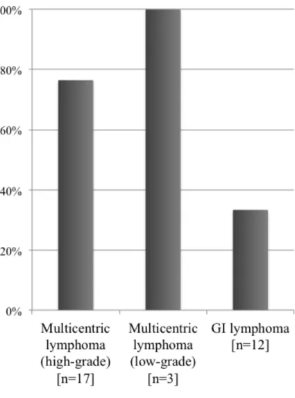
The results of GeneScan analysis are shown in Fig. 2-1. The nucleotide lengths of clonal PCR products are identical between primary lesions and corresponding PBMCs in 13 of 17 dogs (76%) with high-grade multicentric lymphoma, 3 of 3 dogs (100%) with low-grade multicentric lymphoma, and 4 of 12 dogs (33%) with GI lymphoma. Interestingly, in 5 dogs (42%) with GI lymphoma, PARR was positive; however, the nucleotide lengths of the PCR products were different between primary lesions and PBMCs.
CDR3 Sequence
To confirm the detection of CTCs, two cases (one each from dogs with high-grade multicentric lymphoma and GI lymphoma) with identical size of PCR products between primary lesions and PBMCs were selected and CDR3 sequences were analyzed (Fig. 2-2a,b). CDR3 sequences of PBMCs were identical to that of the primary lesions in 7 of 7 clones in a case with high-grade multicentric lymphoma, and 5 of 12 clones in a case with GI lymphoma. In addition, one case with GI lymphoma which had PCR products with different size between the samples was also subjected for CDR3 sequencing (Fig. 2-2c). No sequence which was identical to the duodenal samples was detected from 12 clones analyzed from PBMCs.
Survival Analysis
The results of survival analysis are shown in Fig. 2-3. There was no significant difference in survival time between dogs with and without detectable CTCs at the time of diagnosis in multicentric lymphoma (P = 0.28) (Fig. 2-3a). Median survival times were201 and 345.5 days in dogs with and without detectable CTCs at diagnosis, respectively. Survival time was significantly shorter in dogs without detectable CTCs compared with detectable CTCs in GI lymphoma (P = 0.037) (Fig. 2-3b). Median survival time was206 and 57 days in dogs with and
without detectable CTCs, respectively.
DISCUSSION
In current study, usefulness of GeneScan analysis in detecting CTCs in canine lymphoma was investigated. PCR products with identical size between lymph node and peripheral blood were detected in 80% of dogs with multicentric lymphoma and 33% of dogs with GI lymphoma which were indicative of the presence of CTCs.
Detection rate of CTCs in dogs with GI lymphoma was lower than that in dogs with multicentric lymphoma. CTCs level in peripheral blood are reported to be observed in parallel with the changes in the total lymph node volumes, indicating CTCs volume reflect tumor burden in one’s body (Yamazaki et al., 2008). Therefore, it is considered that total tumor burden was small in GI lymphoma compared with multicentric lymphoma at the time of diagnosis. Since even localized tumor cells in GI tract likely to result in readily identifiable symptoms, patients at the time of diagnosis might be relatively in early tumor stage. Another possible explanation is that tumor cells in GI lymphoma would tend to localize into epithelium rather than into circulation. Certain type of T-cells in GI tract are known to have nature of homing to epithelium (Luckschander et al., 2009). It is regulated by interaction between lymphocytes and enterocytes, and several molecules associated with epithelial homing were identified in human
medicine (Johansson-Lindbom and Agace, 2007). The expression of CD103, which is one of the homing associated molecule, was previously demonstrated in epitheliotropic T-cell lymphoma in cats (Roccabianca et al., 2006). Since the epithelial tissues were heavily injured in GI lymphoma, epitheliotriopism is not always evident in all the cases from histopathological examination. However the clonal rearrangements were detected in TCRγ in most of the dogs with GI lymphoma, tumor cells are presumed to be consist of T-cell population. Behavioral difference between tumor cells in GI lymphoma and multicentric lymphoma has to be investigated further.
Survival time was not significantly different between dogs with and without detectable CTCs at the initial diagnosis of multicentric lymphoma. It was previously reported that there is no prognostic significance in the results of conventional PARR using consensus primers or the amount of CTCs using tumor-specific primers at the initial diagnosis in canine multicentric lymphoma (Lana et al., 2006; Sato et al., 2013). The finding of the present study was consistent with the previous reports. In previous studies, assessment of CTCs is reported to be useful for prognostic marker in early phase of treatment, prediction of the disease relapse, and monitoring the treatment efficacy in canine lymphoma rather than biomarkers at the time of diagnosis (Sato et al., 2011a; Sato et al., 2011b; Sato et al., 2013).
significantly shorter in dogs without detectable CTCs compared with detectable CTCs. Since our study population was small, definitive conclusion can not made from this result. Therefore, value of GeneScan CTC monitoring have to be further investigated in larger population.
Interestingly, clonally expanded lymphocyte populations which were different from lymphoma clone were detected in high proportion of dogs with GI lymphoma. In humans, some kinds of autoimmune diseases and food allergy result in clonal expansion of lymphocytes in peripheral blood (Bakakos et al., 2001). Furthermore, ehrlichiosis can be cause of positive results in PARR of peripheral blood in dogs (Burnett et al., 2003). Although dogs with inflammatory diseases such as inflammatory bowel disease (IBD) were tried to be excluded, inflammatory changes of the intestine are often accompanied with GI lymphoma. Therefore, GI lymphoma cases concomitant with chronic enteritis were possibly included in the patient group in this study, and it may be one possible reason of this phenomenon. Some of the confusing results were demonstrated also in other study conducting PARR in canine lymphoma (Thilakaratne et al., 2010); while clonality of TCR was detected in peripheral blood, clonality of IgH was detected in lymph node at the same time. In addition, one dog did not have clonality rearrangements from lymph node, but clonal IgH rearrangement was detected from peripheral blood. Taken together, positive results do not always indicate the presence of CTCs when using consensus primers in PARR. Therefore, GeneScan analysis has an advantage over conventional
method from the aspects that it can suggest more strongly of CTCs by comparing the size of PCR products in primary lesions with peripheral blood even in use of consensus primers. In conclusion, I revealed that 80% of dogs with multicentric lymphoma and 33% of dogs with GI lymphoma were shown to have CTCs as detected by GeneScan analysis. Clinical significance of monitoring CTCs on prognosis or relapse prediction by using this novel technology has to be investigated further.
Fig. 2-1. Proportion of cases with identical length of clonal PCR products between primary lesions and peripheral blood mononuclear cells (PBMCs), indicating the presence of circulating tumor cells (CTCs) by GeneScan analysis. 13/17 (76%) dogs with high-grade multicentric lymphoma, and 3/3 (100%) dogs with low-grade lymphoma, and 4/12 (33%) dogs with GI lymphoma had detectable CTCs.
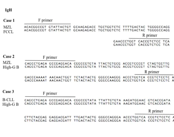
Figs. 2-2. Representative results of GeneScan analysis and comparison of sequence of the PCR products between the primary lesions and peripheral blood mononuclear cells (PBMCs). The line in the top of each alignment show the major sequence from the primary lesions, and alignment below are the sequence analyzed from PBMCs. Background of different residues are colored. Sequence identification between corresponding samples was confirmed in one each of the cases of high-grade multicentric lymphoma (a) and GI lymphoma (b), which were detected identical size of clonal products by GeneScan analysis. Meanwhile, Only different sequences were detected from PBMCs in one case of GI lymphoma, which were detected different size of clonal PCR products by GeneScan analysis (c). IgH, immunoglobulin heavy chain gene; TCRγ, T cell receptor gamma chain gene; LN, lymph node; PBMCs, peripheral blood mononuclear cells; F primer, forward primer; R primer, reverse primer.
Figs. 2-3. Kaplan-Meier curves for dogs with and without detectable CTCs in high-grade multicentric lymphoma (13 and 4 dogs, respectively) (a) and GI lymphoma (4 and 8 dogs, respectively) (b). Survival time was not significantly different between the 2 groups in high-grade multicentric lymphoma (P = 0.28). The survival time was significantly shorter in dogs without detectable CTCs compared with dogs with detectable CTCs (P = 0.037).
Chapter 3
GeneScan analysis for the differentiation of clonal origin
of cells in dogs that developed two different types
ABSTRACT
Development of secondary high-grade lymphoma from indolent lymphoid malignancies has been reported in humans, commonly referred to Richter’s syndrome. Similar conditions have been also reported in dogs. Herein, we used GeneScan analysis to differentiate the clonal origin of cells in dogs that developed 2 different types of lymphoid malignancies. One dog with splenic marginal zone lymphoma (MZL), a dog with B-cell chronic lymphocytic leukemia (B-CLL), and 2 dogs diagnosed with T-zone lymphoma (TZL) developed secondary high-grade B-cell lymphoma (diffuse large B-cell lymphoma, DLBCL). One dog with an initial diagnosis of nodal MZL developed follicular center cell lymphoma (FCCL) III more than 1 year after the initial diagnosis. GeneScan analysis revealed a single peakof the IgH gene with the same length in both the primary (MZL and B-CLL) and secondary tumors in 3 dogs, indicating that the secondary tumors evolved from the pre-existing primary indolent neoplasm clone. A single TCRγ peak was identified in the 2 TZL samples. However, subsequent DLBCL samples from the same 2 dogs revealed a single peak of the IgH gene instead of the TCRγ peak, thereby suggesting a different origin between the 2 lymphoid neoplasms developed in the same subjects. This study indicated that GeneScan analysis would be a convenient and accurate method to determine the clonal origin of lymphoid neoplasms in dogs.
INTRODUCTION
Neoplasms are formed as a result of monoclonal proliferation originated from a single transformed cell. A normal ancestral cell acquires neoplastic phenotype through tumor progression, which causes accumulation of genetic or epigenetic alteration of genes associated with cell proliferation, survival, or other malignant characteristics. This process contributed to uncontrollable cell proliferation and eventually results in formation of primary neoplasms. This process is further responsible for progressive evolution of neoplasms toward greater degrees of aggressive and invasive behavior through a multi-step process.
Such process in canine hematopoietic malignancies has not been fully demonstrated so far. However, several reports have been published on lymphoid malignancies developing into more aggressive one in dogs. One case report described a dog with chronic lymphocytic leukemia (CLL) that subsequently progressed to acute lymphoblastic leukemia (ALL) (Takahashi et al., 2007). Additionally, Comazzi et al. reported that three dogs secondarily developed high-grade lymphoma among 43 dogs that had been previously diagnosed with CLL (Comazzi et al., 2011). Such clinical course in dogs is apparently similar to that in humans commonly referred to Richter’s syndrome (RS). In human medicine, 2 to 10% of cases of CLL and small lymphocytic lymphoma (SLL) developed diffuse large-cell lymphoma (DLL) over time (Maddocks-Christianson et al., 2007; Mauro et al., 1999; Rossi et al., 2008; Tsimberidou et al.,
2006). Molecular mechanisms of RS such as the clonal relationship between the original and subsequent neoplasms has been intensively debated. Diverse studies have been conducted to characterize neoplasms for immunophenotype, immunoglobulin (Ig) gene rearrangement, Ig nucleotide sequence, or Ig isotype. It has been reported that in 67 (69%) of the 97 RS cases, two corresponding neoplasms in each individual were of the same clonal origin, and in remaining 30 (31%), second neoplasm originated from unrelated clone (Smit et al., 2006). Thus, molecular analyses of RS suggest that development of secondary malignancies does not occur in a single homogenous manner in humans.
Studies on the clonal origin of 2 distinct lymphoid malignancies developed in the same individual can provide valuable information for management and understanding pathogenesis of lymphoid neoplasms; however, detailed analysis has not been conducted in veterinary medicine so far. Thus no information is available whether such course in dogs results from transformation of single clone or secondary diseases originated from independent clone.
Herein, I report a case series of 5 dogs with lymphoid malignancies subsequently developed into secondary lymphomas. The purpose of this study was to investigate the clonal relationship between original lymphoid malignancies and the subsequent lymphoma in dogs by means of PCR for antigen receptor gene rearrangements (PARR) and sequencing of complementary determining region 3 (CDR3).
MATERIALS AND METHODS Cases
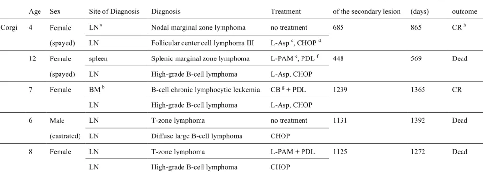
Four dogs with lymphoma (marginal zone lymphoma [MZL], n = 2; T-zone lymphoma [TZL], n = 2), and 1 with B-cell chronic lymphocytic lymphoma (B-CLL) in the initial diagnosis were included in this study. The 2 dogs with MZL consisted of 1 dog with splenic MZL and 1 dog with nodal MZL. Progression to secondary lymphoma was confirmed in all dogs by re-examination at the time of recurrence of lymphadenopathy or clinical worsening more than 1 year after the initial diagnosis. All dogs had been referred to the Veterinary Medical Center of the University of Tokyo (VMC-UT) and medical records were reviewed for collecting patient’s information. The clinical data of the animals are summarized in Table 3-1. In all animals, DNA specimens of both primary and secondary lesions were available for analyses described below.
Histopathology and Cytology
Splenic and/or lymph node tissues were surgically resected for the primary lesions in 4 cases (Case 1, 2, 4, 5) and 2 of the secondary lesions (Case 1, 4). The specimens were fixed in 10% formalin for 48 h, processed and paraffin embedded, cut into sections, and stained with
hematoxylin and eosin (HE). Immunohistochemistry was performed by streptavidin-biotin method using anti-CD3 antibody (polyclonal rabbit anti-human A0452; DAKO, Glostrup, Denmark; 1:50 dilution) for T-cell and anti-CD20 antibody (polyclonal rabbit anti-human RB-9013-P; Thermo Fisher Scientific, Waltham, MA; 1:400 dilution) for B-cell.
Bone marrow aspiration was performed in Case 3 for cytology of the primary lesions. Aspirate of enlarged lymph nodes were used to cytologically diagnose secondary lesions in 3 dogs (Cases 2, 3, and 5). Cytological diagnoses were made from Wright−Giemsa-stained smear. Histological or cytological diagnoses were reviewed from the archives. Diagnoses were made according to the World Health Organization (WHO) classification in the case with lymphoid malignancies (Valli et al., 2011). When the morphology of the cells in the FNA samples clearly indicated typical high-grade lymphoma (centroblastic type in updated Kiel classification (Fournel-Fleury et al., 1997)), resection biopsy of the lymph node was not performed.
DNA Extraction
Genomic DNA was extracted from fresh specimens except for Cases 1, 2, and 5. Since fresh specimens were not available, paraffin-embedded tissues or smear preparation were used for DNA extraction in Cases 1, 2, and 5. DNA extraction was performed using a QIAamp DNA
Blood Mini Kit (Qiagen, Hilden, Germany), according to the manufacturer’s instructions, and subjected for PARR and sequencing.
PCR for Antigen Receptor Gene Rearrangements
Rearrangements of the IgH/TCRγ genes for all specimens were assessed by GeneScan analytical system to determine lymphocyte clonality as described in the Materials and Methods section in Chapter 1.
Cloning and Sequencing of CDR3
The specimens in which clonal rearrangements were detected in the same gene between primary and secondary lesions were subjected to sequencing of the CDR3. PCR products amplified by the method described above were cloned in the pGEM T-Easy Vector using the TA cloning system (Promega Corporation, Madison, WI) according to the manufacturer’s instructions. DNA Sequencing was performed from the plasmid preparation using a BigDye terminator v3.1 Cycle Sequencing Kit (Applied Biosystems, Foster City, CA) and Applied Biosystems 3130xl genetic analyzer (Applied Biosystems). If diagnosis was made from several lesions, at least one of them was selected as sample for cloning and sequencing.
RESULTS Clinical Course
Case 1: A 4-year-old spayed female Pembroke Welsh Corgi was presented with enlargement of the right popliteal lymph node. Since regression was not observed by treatment with antibiotics, the lymph node was surgically resected for biopsy. Histopathological examination of the node indicated the characteristic appearance of MZL. There was no clinical sign or abnormality on examination, thus the dog was followed closely without treatment. On day 685, development of generalized lymphadenopathy was observed. On ultrasonography, splenomegaly and multiple hypoechoic nodules in spleen were detected. The right mandibular lymph node was surgically removed. Histopathological examination revealed follicular center cell lymphoma (FCCL) III in WHO classification. Treatment based on CHOP (prednisolone, doxorubicin, vincristine, and cyclophosphamide) protocol was given and complete remission (CR) was achieved. The dog was still alive without any clinical signs at the time the study was concluded (day 865).
Case 2: A 12-year-old spayed female Shi Zhu was presented with persistent lymphocytosis. On examination, total lymphocyte count of peripheral blood was 11,340/µL. Lymphocytes in peripheral blood were small and mature with aggregated chromatin. Enlargement of mandibular and popliteal lymph node was observed, while splenomegaly and multiple hypoechoic lesions
were detected in spleen by ultrasonography. Surgical resection of spleen and lymph nodes was performed, as well as bone marrow examination. The bone marrow was normoplastic, and the ratio of small lymphocytes in all nucleated blast cells was approximately 20%. MZL was histologically diagnosed. Although the dog was monitored without treatment, on day 58 decreased activity and progression of lymphocytosis was observed. By treatment with melpharan, the dog eventually achieved CR, and the drug was withdrawn on day 177. On day 412, lymphadenopathy relapsed, and high-grade B-cell lymphoma was diagnosed based on cytology of mandibular lymph node. Although treatment was given based on CHOP protocol, the dog died on day 569of disease progression.
Case 3: A 7-year-old female Shiba was presented with persistent lymphocytosis. On examination, total white blood cell count was 235,400/µL (small lymphocytes, 230,600/µL). Hepatomegaly and splenomegaly were observed on ultrasonography. Since rapid clinical worsening was observed, bone marrow examination could not be performed. Therefore diagnostic therapy with chlorambucil and prednisolone was conducted, presumptively diagnosing CLL. Since CR was achieved, dosage was gradually tapered and completely withdrawn on day 273. On day 1,077, since increase of lymphocyte (14,100/µL) was observed again, bone marrow examination was performed. The bone marrow was hyperplastic, and the ratio of small lymphocytes in all nucleated blast cells was approximately 30%, and CLL was
definitively diagnosed. Although treatment with chlorambucil and prednisolone was reinstituted, generalized lymphadenopathy was subsequently found on day 1,239. Total lymphocytes count in peripheral blood was 46,100/µL, and enlargement of intraabdominal lymph nodes were detected on ultrasonography. High-grade B-cell lymphoma was diagnosed based on cytology of superficial lymph nodes. Treatment based on CHOP protocol was given and CR was achieved. The dog was still alive without any clinical signs at the time the study was concluded (day 1,360).
Case 4; A 6-year-old castrated male Golden Retriever was presented with generalized lymphadenopathy. The right popliteal lymph node was surgically resected, and TZL was histologically diagnosed. There is no clinical sign or abnormality on examination, thus the dog was followed closely without treatment. On day 1,120, rapid progression of lymphadenopathy was observed. The left popliteal lymph node was surgically removed, and DLBCL was histologically diagnosed. The dog was given treatment based on CHOP protocol, and obtained partial remission (PR). Since lymphadenopathy relapsed on day 1,316, L-asparaginase and lomustine combination therapy was given as rescue regimen (Saba et al., 2007). However the dog died on day 1,381from clinical worsening.
Case 5; A 8-year-old female Shi Zhu was presented with history of decreased activity and anorexia. On examination, generalized lymphadenopathy, anemia (PCV 27%), and
lymphocytosis (26,600/µl) were observed. Lymphocytes in the peripheral blood appeared as small and mature with aggregated chromatin in the nucleus. Surgical resection of the left superficial cervical lymph node was performed, as well as bone marrow aspiration. The bone marrow was normoplastic, and the ratio of small lymphocytes in all nucleated blast cells was 11.1%. TZL was diagnosed based on histology of lymph node. Although the dog was given treatment with prednisolone and melpharan, lymphadenopathy recurred and anorexia was observed on day 1,125. High-grade B-cell lymphoma was cytologically diagnosed from LN aspirate. Although treatment based on CHOP protocol was given, disease progression was observed after remission for 91 days. There was no response to various rescue treatment (L-asparaginase, dacarbazine, cytosine arabinoside, and actinomycin D) and the dog died on day 1,272.
Assessment of Antigen Receptor Gene Rearrangements
The results of PARR analysis are shown in Table 3-2. Representative pattern of GeneScan analysis are shown in Fig. 3-1. All samples included in this study displayed monoclonal gene rearrangements in GeneScan analysis for antigen receptor gene rearrangements. Clonally rearranged genes were identical between primary and secondary lesions in 3 of the 5 dogs (Cases 1-3). Of these, all cases had identical size of PCR amplicons between two corresponding
lesions. In Cases 4 and 5, clonal rearrangements of TCRγ were detected in the primary lesion and IgH in the secondary lesion, indicating different clonal origin of the two corresponding lesions.
Sequence of CDR3
Representative results of CDR3 sequence are shown in Fig. 3-2. All dogs (Cases 1-3) which showed identical size of PCR products between the primary and the secondary lesions also had identical CDR3 sequence, indicating the common clonal origin of the 2 corresponding lesions.
DISCUSSION
In current study, I assessed the clonal relationship of the primary and the secondary lesions in 5 dogs with sequential development of morphologically distinct lymphoid malignancies. Three cases demonstrated identical CDR3 sequence between 2 corresponding lesions, indicating of the common clonal origin. Meanwhile, remaining 2 cases had clonal rearrangements in different genes between two corresponding lesions, indicating of the unrelated clonal origin. From these results, sequential development of 2 lymphoid malignancies in the same individual does not occur in homogenous pattern in dogs.
however, there are many conflicting reports regarding clonal relationship between CLL/SLL and DLL in RS. A number of studies suggest that clonal evolution is underlying mechanism of RS (Cherepakhin et al., 1993; Cofrancesco et al., 1993; Traweek et al., 1993). Other studies suggest that the two neoplasms are originated from unrelated clone (Kerim et al., 1993; Sun et al., 1990; Tohda et al., 1990). These reports suggest that the lymphoma of RS patients had diverse clonal origin. Furthermore, patients with clonally related RS had a shorter survival time than patients with clonally unrelated RS in human (Rossi et al., 2011). Although the present study included only a small number of cases, heterogenous manner of sequential development of 2 different neoplasms was observed in dogs, being similar to human RS. Accumulation of data on clonal relationship may be important to reveal clinical significance as well as pathogenesis of the disease also in veterinary medicine.
Of the 5 dogs with sequential development of 2 lymphoid malignancies, 1 dog developed B-cell high-grade lymphoma from MZL. Frantz et al. reported that gene expression profile was very similar to each other between MZL and DLBCL in dogs by genomewide microarray analysis (Frantz et al., 2013). Thus, it was suggested that MZL and DLBCL is sequential diseases, and DLBCL may represent more advanced stage of the same disease. Although we did not perform histological evaluation on the secondary lesion of Case 2, our findings may be consistent with such clinical course.
The results in this study indicate that lymphoma which is generally considered to have indolent course can transform into more aggressive type of lymphoma. While the incidence of malignant transformation in canine lymphoid malignancies has not been well understood, it has been reported that two B-cell high-grade lymphoma was developed among 17 B-CLL patients and one T-cell high-grade lymphoma among 19 T-CLL patients (Comazzi et al., 2011). Although it is generally considered that aggressive chemotherapy is not always necessary for indolent lymphoid malignancies, appropriate follow-up is essential for the cases with lymphoma even if it is recognized as indolent type.
In this study, 2 cases with TZL were found to develop secondary malignancies of different clonal origin. To our knowledge, there is only one case report describing development of two neoplasms originated from different clone in dog, which was developed high-grade B-cell lymphoma concurrent with T-CLL (Okawa et al., 2012). Occurrences of two lymphoid malignancies originated from unrelated clone have been defined also in humans by studies of immunophenotype or gene rearrangements, although its occurrence is less frequently than identical clone (Lee et al., 1995; Novogrudsky et al., 2001). Although the molecular mechanism of this phenomenon is unclear, immunosuppressive environment produced by primary neoplasms or chemotherapy are considered to be responsible for progression of secondary malignancies (Gottesman et al., 1999; Novogrudsky et al., 2001). Since alkylating agents play a