Study on Development in Medical Bone
Regeneration and Biodegradable of Mg-Based
Alloys for Biomedical Applications
骨の再生医療における Mg 基合金の開発と
生分解性に関する研究
Haijian Wang
Contents
Abstract ... i
Chapter 1 Introduction ... 1
1.1 Biomedical Materials ... 1
1.2 Biomedical metallic materials ... 3
1.2.1 Stainless steel bone reinforcement plate ... 4
1.2.2 Bone reinforcement plate of CoCr alloy ... 6
1.2.3 Ti and Ti alloy ... 8
1.3 Biodegradable magnesium alloy ... 10
1.3.1 Degradation characteristics ... 11
1.3.2 Mechanical properties ... 13
1.3.3 Biocompatibility ... 14
1.3.4 Advantages and disadvantages in biomedical applications ... 15
1.4 Modification of magnesium alloy materials ... 17
1.4.1 Surface modification ... 17
1.4.2 Alloying of bone reinforcement materials ... 19
1.4.3 Research status of magnesium alloys... 20
1.5 The purpose of this research ... 21
1.6 References ... 24
Chapter 2 The microstructure, composition design and experiment methods of Mg-based alloy ... 31
2.1 The microstructure of Mg-based alloy design ... 31
2.1.1 The microstructure of amorphous / nanocrystalline alloy ... 31
2.1.2 Rapid solidification in producing amorphous / nanocrystalline alloys ... 32
2.1.3 Rapid solidification of twin roll casting technique ... 33
2.2 The composition of Mg-based alloy design ... 35
2.2.1 The glass forming ability rules in alloys ... 35
2.2.2 Compositions of the alloys ... 36
II
2.4 Microscopic characterization ... 41
2.5 Preparation of TEM foils by focused ion beam (FIB) milling ... 41
2.6 Electrochemical characterization ... 42
2.6.1 Introduction of Electrochemical Workstation ... 42
2.6.2 Tafel linear extrapolation ... 44
2.6.3 Electrochemical impedance spectroscopy (EIS) ... 45
2.7 Animals tests ... 46
2.7.1 Implant method ... 46
2.7.2 Experimental animals ... 47
2.7.3 Surgical procedure ... 47
2.8 Methods of in vivo test analysis ... 49
2.9 Concluding remarks ... 50
2.10 References ... 51
Chapter 3 Preparation and characterization of Mg-RE by TRC for biomedical application ... 53
3.1 Introduction ... 53
3.2 Materials and methods... 55
3.3 Results and discussion ... 58
3.3.1 Microstructure characteristic ... 58 3.3.2 Electrochemical measurements ... 64 3.3.3 Immersion test ... 68 3.3.4 In vivo implantation ... 71 3.4 Concluding remarks ... 74 3.5 References ... 75
Chapter 4 In vivo degradation behaviour and bone response of cast-rolled alloy in rat femoral model ... 78
4.1 Introduction ... 78
4.2 Materials and Methods ... 80
4.2.1 Material preparation and characterization ... 80
4.2.3 Electrochemical characterization ... 82
4.2.4 Micro-CT analysis ... 82
4.2.5 In vivo corrosion assessment ... 83
4.2.6 Histological observation ... 83
4.2.7 Statistical analysis ... 83
4.3 Results and discussion ... 84
4.3.1 Microstructures analysis ... 84
4.3.2 In vitro corrosion properties ... 91
4.3.3 Animal test ... 93
4.3.4 Degradable implant cross-sectional examination ... 93
4.3.5 Bone response and histological examination ... 97
4.4 Concluding remarks ... 101
4.5 References ... 102
Chapter 5 Influence of casting speed on microstructure and biodegradation properties of Mg-RE alloy ... 108
5.1 Introduction ... 108
5.2 Experimental methods ... 110
5.2.1 Materials preparation ... 110
5.2.2 Electrochemical tests ... 111
5.2.3 In vivo bone implantation ... 111
5.2.4 Micro-CT and histological assessments ... 112
5.2.5 In vivo degradation tests ... 112
5.2.6 Statistical analysis ... 112
5.3 Experimental results ... 113
5.3.1 Investigation of microstructures ... 113
5.3.2 Electrochemical characterization ... 116
5.3.3 Micro-CT and histological characterization ... 118
5.3.4 In vivo degradation ... 120
5.4 Discussion of experimental results ... 121
IV
5.4.2 Electrochemical test analyses ... 123
5.4.3 In vivo degradation property analyses ... 125
5.5 Concluding remarks ... 127
5.6 References ... 128
Chapter 6 Conclusions ... 131
Related publications... 134
Abstract
Abstract
Nowadays, the approved and commonly metallic biomaterials include stainless steels, titanium alloys, cobalt-chromium-based alloys and noble metals. The limitations of these current metallic biomaterials are the possible release of toxic metallic ions or particles through corrosion or wear processes and the long-term biological incompatibility caused by interactions between tissues and permanent metallic implants. Magnesium (Mg) alloys are considered as the new generation of biomedical implant materials due to their good biocompatibility, biodegradability and excellent mechanical properties. This paper reports an exploratory study on the biodegradation of Mg-RE (RE - rare-earth elements) alloy sheets with fine grain around amorphous zone produced by vertical twin roll casting (TRC) and its interaction with rat femur was observed by micro-CT, which provides a novel method of crystalline/amorphous structuring alloy design and an important evidence of the potential use of Mg-RE for future surgical implant applications.
Abstract
ii
Furthermore, the Mg-RE group induced more newly formed bones, which met the necessary conditions for the prevention of pathological fractures.
Two casting speeds of 10 rpm and 30 rpm were used in vertical twin-roll casting (TRC) to obtain Mg-rare earth (Mg-RE) alloys. The results indicated that the roll-castings of TRC-30-rpm exhibited a finer grain size and higher volume fraction of non-crystallization than those in castings of TRC-10-rpm. Moreover, the results of electrochemical impedance spectroscopy (EIS) and potentiodynamic polarization indicated that the castings of 30 rpm displayed a higher corrosion resistance compared to those in the castings of TRC-10-rpm. Animal tests showed that a higher degree of newly formed bone tissues was achieved by implants of TRC-30-rpm. Additionally, in vivo tests displayed that degradation properties of the TRC-30-rpm implants were better than those of the TRC-10-rpm implants; furthermore, the degradation layer was a two-layer structure, and P and Ca were enriched in the outer degradation layer. In summary, these findings elucidated that casting speed has a substantial effect on the microstructure and degradation property of Mg-based implants, and the degradation property performs better with increased casting speed.
Chapter 1 Introduction
Chapter 1 Introduction
For a long time, the biological materials, such as artificial joints and sutures, have been implanted in body to heal damaged tissue. The artificial joints as a permanent implant require high durability, while the sutures as a biodegradable implant are broken down and absorbed by the body after tissue repair.In the past, the implants were usually durable and permanent, so they had to be surgically removed after tissue repair. However, the biodegradable materials have been developed and used as fixation materials for fractures, such as polylactic acid materials (PLA) are used for suture and as a fixed material for fractures in low-burden areas, thus reducing the burden of reoperation. In addition, the calcium cyanate ceramics can also be used as the bone - free compensation materials. However, only polymers and ceramic materials are developed, and their applications in biomedical materials are limited. Because these biodegradable absorbent materials lack the necessary mechanical strength, they cannot be overused in stressed areas. Therefore, it is desirable to develop biomaterials with better mechanical and better biodegradable properties.
In recent years, the application of magnesium (Mg) alloy in biodegradable materials have attracted the attention of researchers and medical personnel. Mg has high specific strength and is the lightest of the practical metal materials used as structural materials.In addition, as Mg is a bio-essential element, the biological safety of magnesium alloys remains high after in vivo degradation.
1.1 Biomedical Materials
Chapter 1 Introduction
2
ligate wounds and prevent arterial blood loss. With the rapid development of social progress and modern science and technology, the biomedical material industry has also entered its rapid development stage, which plays an important role in improving people's quality of life and guaranteeing human health. It has gradually become an emerging industry with extremely rapid development.
Fig. 1.1 Various biomedical materials and their functions [1].
Chapter 1 Introduction
Table 1-1 Commonly used biomaterials [2]
Materials category Advantage Shortcoming Application Metallic materials
(Titanium and its alloy; CO-Cr alloy; Stainless
steel; Au; Pt, etc)
High strength; stiffness;
flexible
Easy-corrosive high density
Joint replacement; Bone nail and bone plate
High polymer material
Good resilience; Easy preparation
Low intensity; Prone creep
Stylolite; blood vessel, ear, soft tissue Ceramics (aluminum
oxide; calcium phosphate; base apatite, etc)
Good biocompatibility; inert, high compression
strength
Too brittle, poor elasticity; Not easy processing
Surface coatings for dental, femoral head, dental and orthopedic
implants Composites
(carbon-carbon composites, carbon fiber reinforced
bone cement, etc.)
High strength, can be cut Not easy preparation Joint implants, heart valves
1.2 Biomedical metallic materials
Metal materials are applied as biological materials due to their excellent mechanical properties and good electrical conductivity and thermal conductivity. Because the metal bonds in metal materials are non-directional, they can change the position of metal atoms without destroying the crystal structure. Therefore, metal materials show the characteristics of plastic deformation.
Chapter 1 Introduction
4
implants. It can also be applied to cardiovascular stents, orthodontic teeth, cochlear implants and other places that need to play an active role in the location.
Fig. 1.2 Biomedical metallic materials.
The alloying elements in most medical metal materials are allowed in very small amounts in the human body. Trace amounts of metallic elements can play a positive role in human body. For example, trace amounts of Fe are very important for red blood cells, and Co is an essential element for the synthesis of vitamin B12. However, when the intake of these metallic elements exceeds the limit of human body, it will have many negative effects on human physiological functions [3]. Due to the long-term existence of metal implants in the human body and the corrosion of the internal body fluid environment, it is inevitable that trace elements will be dissolved into the human body. Therefore, the biocompatibility of metal elements is an important factor that metal materials as biological materials must be concerned. At present, the widely used medical metal materials mainly include four categories: stainless steel, titanium alloy, cobalt-based alloy and precious metal.
1.2.1 Stainless steel bone reinforcement plate
Chapter 1 Introduction
introduction of Mo in the composition stainless steel can dramatically improve the performance of the resistance to pitting formation caused by chloride ion. In the last century 1950s, the researchers further enhance the corrosion resistance of stainless steel in the physiological saline by reducing the concentration of the carbon element in stainless steel (0.08 wt % decrease to 0.03 wt %), which is now known as SUS316/316L stainless steel and its composition as shown in table 1-2. The Cr content in stainless steel must be higher than 12 wt % to guarantee enough corrosion resistant performance, and the Ni is used to make stainless steel in the austenite phase to keep stable at room temperature [4].
The SUS316/316L steels are austenitic stainless steels and are widely used in the manufacture of medical implant devices. They are difficult to be strengthened by heat treatment, but it can be strengthened by work hardening. The mechanical properties of SUS316/316L stainless steel show in Table 1-3.
Fig. 1.3 Stainless steel bone reinforcement as bone fixed implants.
Chapter 1 Introduction
6
Table1-2 Composition of USL316L stainless steel [2]
Element C Mn P S Si Cr Ni Mo
Content wt%
<0,03 <2.00 <0.03 <0.03 <0.75 17~20 12~14 2~4
Table1-3 Mechanical properties of USL316L stainless steel for implants [2]
Condition YTS (MPa) UTS (MPa) Elongation (%) Rockwell hardness
Annealed 172 485 40 95
Cold-worked 690 860 12 -
1.2.2 Bone reinforcement plate of CoCr alloy
The CoCrMo with the as-cast state is common typical alloy. The as-cast CoCrMo alloy has been used as a medical metal for decades. It was mainly used as a dental material in the early stage. Recently, it has been widely used in the field of artificial joints. The deformed CoNiCrMo alloy as a medical metal material has a relatively short use time, and it is mainly used to make the handle of knee or hip prosthesis that to bear a large load [5,6].
Chapter 1 Introduction
Fig. 1.4 Bone reinforcement of Cobalt alloy.
Table 1-4 Chemical Compositions of Co–Cr alloys [9]
Chapter 1 Introduction
8
Table 1-5 Mechanical properties of Co-Cr alloys [9]
Property Cast CoCrMo Wrought CoCrWNi Wrought CoNiCrMo Solution Annealed Cold worked and aged YS/MPa 450 310 240~650 1585 UTS/MPa 655 860 793~1000 1793 Elongation/% 8 10 50 35 Elastic modulus/GPa 220~234
The deformed CoNiCrMo alloy and the as-cast CoCrMo alloy have similar wear resistance (their wear rate is 0.14mm/year). The CoNiCrMo alloy has high tensile strength and fatigue resistance, which is more suitable for the handle of hip prosthesis. The mechanical properties of the CoCr alloy are shown in Table 1-5. The elastic modulus of the CoCr alloy is 220 ~ 234GPa, so this alloy is very hard with very low wear in metal-metal, which has obvious advantages in application of metal-metal artificial joint replacement [7]. When the CoCr alloy is applied in joint replacement, the metal products result from the metal wear and corrosion will have an adverse effect on the surrounding organ tissues. In vitro experiment results show that the Co particles have a toxic effect on osteoclasts and inhibit the synthesis of I-type collagen, osteocalcin and alkaline phosphatase. However, the metal Cr and CoCr alloy particles did not show significant toxicity. The experiments of metal extraction liquid showed that the Co and the Ni extracts had significant cytotoxicity and significantly reduced the survival rate of cells, thus, the Cr extracts had lower cytotoxicity than Co and Ni [8].
1.2.3 Ti and Ti alloy
Chapter 1 Introduction
to the stainless steel, the CoCrMo alloy, and its alloys (as shown in Table 1-6), and the excellent mechanical properties (Table 1-7), which is the most significant advantage used for making implanted devices [10, 11].
Fig. 1.5 Ti / Ti alloy as dental implants.
Table 1-6 Density of Ti and its alloy; and some metallic implant alloys [10,11]
Alloys Density (g/cm3)
Ti and its allloys 4.5
316 Stainless steel 7.9
CoCrMo 8.3
CoNiCrMo 9.2
NiTi 6.7
Chapter 1 Introduction
10
temperature of the α phase / β phase, while alloy element V stabilizes the β phase by reducing the phase transition temperature of the α phase / β phase [12].
In commercial pure titanium, with the increase of impurity content, the strength increases and the plasticity decreases of the materials. Although the strength of titanium alloy is lower than some stainless steel and CoCr alloy, it’s the specific strength is obviously higher than these two materials. In addition, the shear strength of titanium and its alloys is lower, which makes them not suitable for the preparation of bone screws, bone plate and other types of devices which need to withstand higher shear stress. The wear-resisting performance of titanium and its alloys is poorer when it is in contact with the same material or other metal materials, which limits the application as a medical implant metal material [13].
Table 1-7 Mechanical properties of Ti and its alloys [9]
Properties
Pure Ti
Ti6Al4V Ti13Ni13Zr Grade1 Grade2 Grade3 Grade4
UTS / MPa 240 345 450 550 860 1030
YS / MPa 170 275 380 480 795 900
Elongation / % 24 20 18 15 10 15
E / GPa 110 79
1.3 Biodegradable magnesium alloy
Chapter 1 Introduction
great progress in this direction. However, the polymer materials generally have low elastic modulus and poor plasticity, which are difficult to be applied in some occasions such as cardiovascular stent, bone nail and plate, anastomosis nail, etc. Magnesium and its alloys are biodegradable, and it also has advantages in mechanical properties and processing properties of metal materials [15-19].
1.3.1 Degradation characteristics
The degradation of magnesium alloy is accomplished by corrosion of magnesium alloy in physiological environment. The corrosion process is an electrochemical reaction process, and the total reaction formula can be written as follows [15, 20]:
Mg + H2O = Mg (OH)2 + H2 (1-1)
The electrochemical reactions can be decomposed into the following anodic oxidation and cathodic reduction reactions:
Anode reaction: Mg → Mg2+ + 2e- (1-2)
Cathode reaction: 2H2O + 2e-→H2 + 2OH- (1-3)
Product formation: Mg2+ + 2OH- → Mg (OH)
2 (1-4)
The degradation mechanism of magnesium alloy in physiological environment is shown in Fig. 1.7. When the magnesium alloy comes into contact with the humoral environment, the Mg is oxidized to Mg2+, and the generated electrons are reaction with water to form H2 and OH-. Due to the poor electronegativity between the magnesium alloy
Chapter 1 Introduction
12
Fig. 1.6 Clinical observation of degradation and bone healing of Mg alloy screw over a 1-year [14].
Chapter 1 Introduction
The reaction product of Mg(OH)2 is easily dissolved in humoral environment that
contains Cl- ions and absorbed by the body. The adsorption of Cl- on the surface of the material will lead to the dissolution of Mg(OH)2 protective layer which result in pitting. It
is not easy to form a deep corrosion pit in magnesium alloy. This because the corrosion process of magnesium alloy occurs quickly, and the local environment around the place where the corrosion occurs presents a high alkalinity, leading to the difficulty of further corrosion reaction at this place. Because the degradation of magnesium alloy makes the surrounding environment became alkaline, and the saturated Ca ions and phosphates exist in the body fluids, the calcium phosphate is easy to deposit on the Mg(OH)2 product layer,
the cells tend to adsorb on this surface layer. With the extension of implantation time, the deposited cells grew into tissues adjacent to the layer of corrosion products [15].
1.3.2 Mechanical properties
Mechanical property is one of the important indexes of magnesium alloy as medical metal materials, which determines the bearing capacity and plastic deformation capacity of medical devices.Pure magnesium has a dense hexagonal crystal structure and only three slip systems, so its plastic deformation ability is poor. The commonly methods to improve the deformation properties of magnesium alloys include alloying heat treatment and deformation processing. By adding the Ca [22], Zr [23], Zn [24] and other alloy elements into magnesium, the grain size of the as-cast magnesium alloy can be significantly refined and the plastic deformation ability of the magnesium alloy can be improved. In addition, it has been reported in the literature [24] that the addition of Nd, Gd, Pr, Dy and other rare earth elements in magnesium alloy can refine the grain size and reduce the stacking fault energy of magnesium alloy, which is considered to be one of the important mechanisms to improve the plasticity of magnesium alloy. It is also helpful for improve the plasticity of magnesium alloy to choose the treatment technology and reduce the rough second phase in the as-cast magnesium alloy.
Chapter 1 Introduction
14
which cannot meet the requirements in occasions that requiring higher bearing capacity. To improve the strength of magnesium alloy, there are usually several ways as follows: the firstly, the solid solution strengthening method. The strength of magnesium alloy can be improved by adding Al, Zn, rare earth elements and other alloy elements with solid solution strengthening into pure magnesium. Secondly, the second phase strengthening. The strength of magnesium alloy can be improved by adding alloy elements to the magnesium alloy to form intermetallic compounds with strengthening effect. Thirdly, the grain boundary strengthening. The grain size of magnesium alloy can be effectively reduced through the alloying or thermal deformation processing, the strength of magnesium alloy can be improved by increasing the proportion of grain boundary.
1.3.3 Biocompatibility
The cell toxicity evaluation is an important means of characterization of magnesium alloy biocompatibility.Theoretically, the human body has a limit for the intake of any element [16, 25]. When a certain element is consumed in excess of the limit, it can cause toxic reactions in humans. The biocompatibility of a material is related to its elemental release, which depends on the corrosion rate of the materials. Magnesium is considered as an essential element in human body. However, when the concentration of Mg2+ in human serum is higher than 1.05
mmol/L, it will also cause muscle paralysis, hypotension, respiratory disorders and other adverse reactions. When the concentration of Mg2+ in blood is as high as 6~7mmol/L, it will also lead to cardiac arrest and other risks.
Chapter 1 Introduction
From the year of 2000 to 2003, Heublein et al. [26] attempted to make the degradable cardiovascular stents with AE21 magnesium alloy. They implanted magnesium stents into coronary arteries of domestic pigs. The scaffold lost its mechanical integrity after 35 to 56 days of implantation due to the rapid degradation of magnesium alloy. Biotronik, a German company, has tried to implant magnesium alloy stents made from WE43 into the coronary arteries of miniature pigs, and no deaths have occurred after a period of implantation. In 2005, Peeters et al. [27] who first attempted to implant magnesium alloy stents into 20 patients for the treatment of varicose veins. The stents were completely degraded after 6 weeks, and no adverse reactions such as poisoning or allergy occurred in the patients. In 2007, Biotronik implanted 71 magnesium stents in 63 patients and no myocardial infarction clots or deaths occurred before the stents broke down [28]. In 2013, Haude et al. implanted drug-coated magnesium alloy stents into 46 patients. The stents degraded after 9-12 months, and no stent thrombosis or decreased blood flow was observed within 6-12 months after implantation [29].
Magnesium alloy plate, screws and wires are considered to have great potential for bone fixation. The screws prepared by ZEK100 [30] LAE442 [31] MgCa0.8 [32] and Mg-Y-RE-Zr [33] have been used in animal experiments, and the results show that magnesium alloy has the ability to induce new bone formation. After 12 months of implantation, the adhesion of Mg-0.8Ca bone nail and bone tissue were much higher than that LAE442 and bone tissue, indicating that Mg-0.8Ca had better biocompatibility. The MAGNEZIX® bone nail made of Mg-Y-RE-Zr alloy showed the same efficacy as titanium nail in the correction of thumb eversion, and no adverse symptoms such as foreign body reaction, osteolysis and inflammatory reaction occurred 6 months after implantation.
1.3.4 Advantages and disadvantages in biomedical applications
Chapter 1 Introduction
16
magnesium and its alloys in biomedical field in the literature: (1) Mg has no toxic effect on human body. Magnesium is an essential trace element, and the amount of magnesium in human body is relatively high. Therefore, the degraded magnesium ion does not cause any side effects; (2) The presence of Mg in the human bone system is beneficial to bone strengthening and growth; (3) Compared with other implanted metals, the density and young's modulus of Mg alloy closest to human bone can reduce the interfacial stress between bone and implanted material. In addition, Mg stimulate the growth of bone, and enhance the stability of implants; (4) Mg has better physical and mechanical properties than other metal-based or polymer-based implants and is more suitable for bone repair and replacement; (5) Mg has higher fracture toughness than ceramic biomaterials, higher strength than degradable plastics, and more suitable elastic modulus than other commonly used metallic biomaterials; (6) Mg is involved in a variety of metabolic reactions and biological mechanisms in the human body, including the formation of crystal apatite, which is important for metal bone implant materials. Mg is also a cofactor of many enzymes and stabilizes the structure of DNA and RNA. (7) Mg is important from a physiological point of view, as a deficiency of Mg promotes cardiovascular disease, and low serum levels of magnesium have been linked to an increased risk of neuropathy in patients with peripheral artery disease.
Although Mg alloy has certain advantages as a biomedical implant material, there are still many problems in the large-scale application of magnesium alloy in biological and medical fields. The implanted Mg will react with water and other ions in the body fluids to form Mg(OH)2 and H2, which the forming gas pockets
Chapter 1 Introduction
1.4 Modification of magnesium alloy materials
At present, the most of researches on degradable medical Mg alloys are the commercial Mg alloys, and AZ series, such as AZ31 [35-37], AZ61 [38, 39], AZ91 [40, 41], etc. These Mg alloy materials have been applied in the industry due to their excellent performance in strength or corrosion resistance respectively. The elastic modulus of Mg alloy (45 GPa) is very close to that of natural bone (3~20 GPa) and the stress shielding effect can be effectively avoided [42]. It has been reported that Mg can also promote osteogenesis [35]. Therefore, Mg alloys have been widely studied as orthopedic implant materials. As orthopedic materials (such as bone plate and bone screw), Mg alloy usually needs to have higher corrosion resistance to meet the performance requirements ofimplant material instrument.
As a bone graft material, Mg alloy has its unique advantages. However, it has been reported that Mg alloy will be seriously degraded after implantation [43,44]. How to solve the problem of too fast degradation of Mg alloy has become the focus in research. At present, the commonly used solutions are surface modification and alloying.
1.4.1 Surface modification
(1) Electrochemical deposition
Chapter 1 Introduction
18
stable hydroxyapatite. The coating improves the degradation rate of Mg alloys in simulated body fluids. The thin and loose shape is good for bone tissue to grow, but the bond strength between coating and substrate is weak. Gao et al [46]. obtained porous coatings by micro-arc oxidation (MAO) on the surface and prepared rod-like nano hydroxyapatite coatings on the surface by electrochemical deposition. This layer of hydroxyapatite dense uniform distribution and conducive to cell survival in the pores. Because of the membrane, the coating bond strength about times of electrochemical deposition film alone. How to further improve the bonding strength of coating and base still need a lot of research.
(2) plasma spraying
Plasma spraying technology is used to drive dc arc plasma as a heat source, such as ceramic alloy metal materials heated to molten or semi-molten state, and at high speed injection to the workpiece surface to form a solid surface method. The substrate surface can be wear-resistant, corrosion resistant and high temperature oxidation resistance.
(3) Chemical conversion film
Chemical conversion film is a chemical or electrochemical method to form metal oxide, chromate, phosphate or other refractory conversion film on the surface of a metal. Magnesium alloy conversion film mainly includes chromate conversion film, potassium permanganate conversion film, fluorozirconate conversion film, stannate conversion film, rare earth conversion film and phytic acid conversion film [47-51].
(4) Biomimetic
Chapter 1 Introduction
at room temperature, which avoids the phase transition and brittle cracking caused by high temperature, and also provides the possibility for the deposition of biological macromolecules such as proteins. (3) This process can be used to prepare coating on the surface of complex and porous materials by bionic method.
(5) Micro-arc oxidation
Micro-arc oxidation [53-57] is the formation of ceramic film dominated by matrix metal oxide under the action of instantaneous high temperature and high pressure caused by light arc discharge on its alloy surface. The characteristics of micro-arc oxidation process are as follows: (1) simple process, environmentally friendly and pollution-free electrolyte. (2) The micro-arc oxide film not only contains high temperature stable phase, but also contains a certain number of amorphous phases, so the film has higher hardness, toughness, wear resistance and corrosion resistance. (3) This process is widely applicable. (4) The micro-arc oxidation technology mostly adopts pulsed dc, so it has little influence on the mechanical properties of raw materials. (5) The micro-arc oxidation film formed is rough and porous.
1.4.2 Alloying of bone reinforcement materials
Chapter 1 Introduction
20
resistance and mechanical properties of Mg alloy could be improved. Zhang et al. showed that adding Ca into Mg-Zn-Ca could significantly refine grain, and the refinement effect was best when the mass fraction of Ca was 0.5%.
1.4.3 Research status of magnesium alloys
At present, researches on Mg alloy as a degradable medical implant material mainly focus on the following aspects: bone fixation material, bone tissue engineering, porous stent material and cardiovascular stent material [61]. Mg alloys have been tried as orthopedic implants since the early 20th century to repair damaged bone tissue [62]. Studies [63,64] have shown that Mg alloy is not suitable for use as a splint because of its fast degradation rate, but it is suitable for making screws. Mg has a positive effect on the deposition and hardening of bone tissue. In 1944,some people implant bone nails and plates with small amounts of Ca and Mg in patients with severe fractures. There was no postoperative increase in Mg content caused by inflammation of the implant. It was found that the Mg alloy implant could promote bone growth. After 6 weeks of fracture healing, the Mg alloy plate of the implant had disappeared [65]. F. whitte implanted four different Mg alloys into the thigh bone of guinea pigs, and it found that the degradation rates of different Mg alloys in vivo were significantly different. He added HA particles to AZ31 alloy, and the corroded materials showed uniform corrosion characteristics, and the CaCO3 deposition was formed on the surface [66,67].
Chapter 1 Introduction
months, the stent was completely degraded, and certain successful clinical application was achieved.Due to the slow degradation of Mg alloy in the vascular wall, the incidence of long-term vascular wall stimulation caused by intimal hyperplasia caused by traditional scaffolds is reduced [71,72].
1.5 The purpose of this research
As a biodegradable medical metal material, Mg alloy has an attractive application prospect. It is likely to be applied in clinical applications in bone plates, anastomoses, sutures and other implant devices in the next few years. However, the poor corrosion resistance of magnesium alloy materials is a very important reason that affects its service performance. For example, in the process of treating fracture healing, Mg alloy as a bone plate needs to undergo greater corrosion. However, the corrosion resistance of Mg alloy is poor, and it is easy to over degrade, leading to fracture healing failure.To solve this problem, we mainly start from the perspective of material design, and make the Mg-based materials have good corrosion resistance and degradation to meet the application requirements by designing reasonable processing technology.
Chapter 1 Introduction
22
Although the crystal structure has better corrosion resistance than the amorphous structure, the microstructure of the designed Mg-RE alloy in this study is partial amorphous structure rather than all amorphous structure. In addition, the amorphous structure of the Mg-RE alloy through the whole crystal structure (Fig. 2.1). The reasons are as follows:
(1) In this study, the TRC technology has a maximum casting speed of 30rpm, which indicates that the solidification rates provided by the experimental equipment is limited. It is very difficult to process the bulk amorphous with the existing experimental equipment. However, the TRC equipment is more cost-effective than other amorphous alloy production equipment, and has better cost performance.
(2) The microstructure characteristics of the new Mg-RE alloy are amorphous structure throughout the whole crystal structure, and the distribution is relatively uniform. Previous studies showed that the amorphous structure has better strength, toughness and corrosion resistance than the crystal structure. However, the amorphous structure is more brittle. therefore, the microstructure of the amorphous and crystal composite can provide better support function as the implant material.
Chapter 1 Introduction
The research contents of this paper are as follows:
(1) Designed of novel Mg-based materials.
The composition of Mg-based alloy was designed, the new alloy was processed by twin-roll casting (TRC) technology. The microstructure and the corrosion resistance of the material were analyzed, and the Mg-RE alloy with better comprehensive performance was obtained.
(2) Characterization of novel Mg-based materials.
The Mg-based sheet was processed by TRC, and the microstructure and corrosion resistance of the plate were characterized, and the degradation performance of Mg-based alloy as bone sheet was evaluated.
(3) Application research of Mg-based materials
The corrosion behaviour of Mg-based alloy sheet was characterized by in vitro electrochemical experiments and immersion experiments. The alloy sheet was fixed to the femur of rats, and the pathological reaction of the bone tissue around the femur was observed at different time periods, to characterize the performance of the sheet as a degradable fixation material.
(4) Optimization of TRC casting method for Mg-based materials.
Chapter 1 Introduction
24
1.6 References
1. Yaoting Yu, Xingdong Zhang. Biomedical material. Tianjin university press; 2000. 2. Front Matter. Biomaterials: CRC Press; 2012. p. i-xviii.
3. Black J. Biological performance of materials: fundamentals of biocompatibility: CRC Press; 2005.
4. Bordjih K, Jouzeau J-Y, Mainard D, Payan E, Delagoutte J-P, Netter P. Evaluation of the effect of three surface treatments on the biocompatibility of 316L stainless steel using human differentiated cells. Biomaterials 1996; 17:491-500.
5. Hanawa T. Metals for medicine. Japan Institute of Metals, Sendai 2010.
6. Niinomi M. Metallic biomaterials. Journal of artificial organs: the official journal of the Japanese Society for Artificial Organs 2008; 11:105-10.
7. Kumagai K, Nomura N, Ono T, Hotta M, Chiba A. Dry friction and wear behavior of forged Co-29Cr-6Mo alloy without Ni and C additions for implant applications. Materials transactions 2005; 46:1578.
8. Granchi D, Ciapetti G, Savarino L, Cavedagna D, Donati ME, Pizzoferrato A. Assessment of metal extract toxicity on human lymphocytes cultured in vitro. Journal of biomedical materials research 1996; 31:183-91.
9. International A, Testing ASf, Materials. Annual book of ASTM standards. American Society for Testing & Materials; 2004.
10. Geetha M, Singh A, Asokamani R, Gogia A. Ti based biomaterials, the ultimate choice for orthopaedic implants–a review. Progress in Materials Science 2009; 54:397-425.
11. Bannon B, Mild E. „Titanium Alloys for Biomaterial Application: An Overview. Titanium alloys in surgical implants, ASTM STP 1983;796:7-15.
12. Nastac L, Gungor M, Ucok I, Klug K, Tack WT. Advances in investment casting of Ti–6Al–4V alloy: a review. International Journal of Cast Metals Research 2006; 19:73-93.
Chapter 1 Introduction
1:3-34.
14. J.W Lee, H. S Han, K. J Han, S. J Yang. Long-term clinical study and multiscale analysis of in vivo biodegradation mechanism of Mg alloy. Proceedings of the National Academy of Sciences, 2016, 113:3, 716-721.
15. Zheng YF, Gu XN, Witte F. Biodegradable metals. Materials Science and Engineering: R: Reports 2014; 77:1-34.
16. Staiger MP, Pietak AM, Huadmai J, Dias G. Magnesium and its alloys as orthopedic biomaterials: a review. Biomaterials 2006; 27:1728-34.
17. Chen Y, Xu Z, Smith C, Sankar J. Recent advances on the development of magnesium alloys for biodegradable implants. Acta Biomater 2014; 10:4561-73.
18. Witte F. The history of biodegradable magnesium implants: a review. Acta Biomater 2010; 6:1680-92.
19. Farraro KF, Kim KE, Woo SL, Flowers JR, McCullough MB. Revolutionizing orthopaedic biomaterials: The potential of biodegradable and bioresorbable magnesium-based materials for functional tissue engineering. Journal of biomechanics 2014; 47:1979-86.
20. Witte F, Hort N, Vogt C, Cohen S, Kainer KU, Willumeit R, et al. Degradable biomaterials based on magnesium corrosion. Current Opinion in Solid State and Materials Science 2008; 12:63-72.
21. Zheng Y, Gu X. Research activities of biomedical magnesium alloys in China. JOM 2011; 63:105-8.
22. Li Z, Gu X, Lou S, Zheng Y. The development of binary Mg-Ca alloys for use as biodegradable materials within bone. Biomaterials 2008; 29:1329-44.
23. Zhang E, Yin D, Xu L, Yang L, Yang K. Microstructure, mechanical and corrosion properties and biocompatibility of Mg–Zn–Mn alloys for biomedical application. Materials Science and Engineering: C 2009; 29:987-93.
24. Zhang S, Zhang X, Zhao C, Li J, Song Y, Xie C, et al. Research on an Mg-Zn alloy as a degradable biomaterial. Acta Biomater 2010; 6:626-40.
Chapter 1 Introduction
26
26. Heublein B, Rohde R, Kaese V, Niemeyer M, Hartung W, Haverich A. Biocorrosion of magnesium alloys: a new principle in cardiovascular implant technology? Heart 2003; 89:651-6.
27. Peeters P, Bosiers M, Verbist J, Deloose K, Heublein B. Preliminary results after application of absorbable metal stents in patients with critical limb ischemia. Journal of Endovascular Therapy 2005; 12:1-5.
28. Erbel R, Di Mario C, Bartunek J, Bonnier J, de Bruyne B, Eberli FR, et al. Temporary scaffolding of coronary arteries with bioabsorbable magnesium stents: a prospective, non-randomised multicentre trial. The Lancet 2007; 369:1869-75.
29. Haude M, Erbel R, Erne P, Verheye S, Degen H, Böse D, et al. Safety and performance of the drug-eluting absorbable metal scaffold (DREAMS) in patients with de-novo coronary lesions: 12 month results of the prospective, multicentre, first-in-man BIOSOLVE-I trial. The Lancet 2013; 381:836-44.
30. Reifenrath J, Angrisani N, Erdmann N, Lucas A, Waizy H, Seitz JM, et al. Degrading magnesium screws ZEK100: biomechanical testing, degradation analysis and soft-tissue biocompatibility in a rabbit model. Biomedical Materials 2013; 8:045012. 31. Wolters L, Angrisani N, Seitz J, Helmecke P, Weizbauer A, Reifenrath J.
Applicability of Degradable Magnesium LAE442 Alloy Plate-Screw-Systems in a Rabbit Model. Biomedical Engineering/Biomedizinische Technik 2013.
32. Erdmann N, Angrisani N, Reifenrath J, Lucas A, Thorey F, Bormann D, et al. Biomechanical testing and degradation analysis of MgCa0. 8 alloy screws: a comparative in vivo study in rabbits. Acta biomaterialia 2011; 7:1421-8.
33. Erdmann N, Angrisani N, Reifenrath J, Lucas A, Thorey F, Bormann D, et al. Biomechanical testing and degradation analysis of MgCa0. 8 alloy screws: a comparative in vivo study in rabbits. Acta biomaterialia 2011; 7:1421-8.
34. Narayanan T. S. N. S., Park I. S., Lee M. H. Strategies to improve the corrosion resistance of microarc oxidation (MAO) coated magnesium alloys for degradable implants: Prospects and challenges[J]. Progress in Materials Science, 2014, 60: 1-71.
Chapter 1 Introduction
system developed as a new kind of biodegradable metal [J]. Acta Biomaterialia, 2012, 8(6): 2360-2374.
36. Brar H. S., Wong J., Manuel M. V. Investigation of the mechanical and degradation properties of Mg-Sr and Mg-Zn-Sr alloys for use as potential biodegradable implant materials [J]. Journal of the Mechanical Behavior of Biomedical Materials, 2012, 7: 87-95.
37. Huan Z., Leeflang M. A., Zhou J., et al. In vitro degradation behavior and cytocompatibility of Mg-Zn-Zr alloys[J]. Journal of Materials Science: Materials in Medicine, 2010, 21(9): 2623-2635.
38. Zhang B., Hou Y., Wang X., et al. Mechanical properties, degradation performance and cytotoxicity of Mg-Zn-Ca biomedical alloys with different compositions[J]. Materials Science and Engineering: C, 2011, 31: 1667-1173.
39. Xu Z, Smith C, Chen S, et al. Development and microstructural characterizations of Mg-Zn-Ca alloys for biomedical applications[J]. Materials Science and Engineering: B, 2011, 176(20): 1660-1665.
40. Hort N., Huang Y., Fechner D., et al. Magnesium alloys as implant materials – Principles of property design for Mg-RE alloys[J]. Acta Biomaterialia, 2010, 6(5): 1714-1725.
41. Li Y., Wen C., Mushahary D., et al. Mg-Zr-Sr alloys as biodegradable implant materials[J]. Acta Biomaterialia, 2012, 8(8): 3177-3188.
42. Hermawan H., Dubé D., Mantovani D. Developments in metallic biodegradable stents[J]. Acta Biomaterialia, 2010, 6(5): 1693-1697.
43. Ascencio M., Pekguleryuz M., Omanovic S. An investigation of the corrosion mechanisms of WE43 Mg alloy in a modified simulated body fluid solution: The influence of immersion time[J]. Corrosion Science, 2014, 87: 489-503.
44. Li wei. Preparation technology and properties of biodegradable rare-earth magnesium alloy stent seamless tubes [D]. Chongqing: chongqing university, 2011.
45. Liu tong. Biodegradation of medical magnesium alloys [D]. Guangzhou: south China university of technology, 2011.
Chapter 1 Introduction
28
vitro and in vivo degradation of biodegradable Mg-5Zn-0.3Ca alloys[J]. Acta Biomateriallia, 2015, 23: 347-353.
47. Nene S. S., Kashyap B. P., Prabhu N., et al. Microstructure refinement and its effect on specific strength and bio-corrosion resistance in ultralight Mg-4Li-1Ca (LC41) alloy by hot rolling[J]. Journal of Alloys and Compounds, 2014, 615: 501-506. 48. Gu CD, Lian JS, Li GY, Niu LY, Jiang ZH. Electroless Ni–P plating on AZ91D
magnesium alloy from a sulfate solution. Journal of Alloys and Compounds. 2005; 391(1-2): 104-109
49. Umehara H, Takaya M, Terauchi S. Chrome-free surface treatments for magnesium alloy. Surface and Coating Technology. 2003; 169-170: 666-669
50. Zhang Y, Yan C, Wang F, Lou H, Cao C. Study on the environmentally friendly anodizing of AZ91D magnesium alloy. Surface and Coating Technology. 2002; 161(1): 36-43
51. Gonzalez-Nunez MA, Nunez-Lopez CA, Skeldon P, Thompson GE, Karimzadeh H, Lyon P, et al. A non-chromate conversion coating for magnesium alloys and magnesium-based metal matrix composites. Corrosion Science. 1995; 37(11): 1763-1772
52. Hu T., Xiao W. L., Wang F., et al. Improving tensile properties of Mg-Sn-Zn magnesium alloy sheets using pre-tension and ageing treatment[J]. Journal of Alloys and Compounds, 2018, 735: 1494-1504.
53. Chiu L-H, Chen C-C, Yang C-F. Improvement of corrosion properties in an aluminum-sprayed AZ31 magnesium alloy by a post-hot pressing and anodizing treatment. Surface and Coating Technology. 2005; 191(2-3):181-187.
54. Hsiao HY, Tsung HC, Tsai WT. Anodization of AZ91D magnesium alloy in silicate-containing electrolytes. Surface and Coating Technology. 2005; 199(2-3): 127-134. 55. Hsiao HY, Tsai WT. Characterization of anodic films formed on AZ91D magnesium
alloy. Surface and Coating Technology. 2005; 190(2-3): 299-308
Chapter 1 Introduction
Corrosion. 1999; 50: 2-6
58. Gu X. N., Zheng Y. F., Cheng Y., et al. In vitro corrosion and biocompatibility of binary magnesium alloys[J]. Biomaterials, 2009, 30(4): 484-498.
59. Su xin. First principle calculation and microstructure properties of rare earth magnesium alloy [D]. Shanghai: Shanghai jiao tong university, 2013.
60. Chen xingwei, wu jianhua, wang jia, et al. Research progress on influencing factors of galvanic corrosion [J]. Corrosion science and protection technology, 2010, 22(4): 363-366.
61. Song guangling. Corrosion and protection of magnesium alloy [M]. Beijing: chemical industry press, 2006, 4.
62. Li J. R., Jiang Q. T., Sun H. Y., et al. Effect of heat treatment on corrosion behavior of AZ63 magnesium alloy in 3.5 wt.% sodium chloride solution[J]. Corrosion Science, 2016, 111: 288-301.
63. Jia J., Fan J. F., Xu B. S., et al. Microstructure and properties of the super-hydrophobic films fabricated on magnesium alloys[J]. Journal of Alloys and Compounds, 2013, 554: 142-146.
64. Kang Z. X., Sang J., Shao M., et al. Polymer plating on AZ31 magnesium alloy surface and film evaluation of corrosion property[J]. Journal of Materials Processing Technology, 2009, 209(9): 4590-4594.
65. Huang n. study on biodegradable magnesium and magnesium alloy coronary stents [D]. Tianjin: hebei university of technology, 2009.
66. Jiang J. H., Ma A. B., Saito N. B., et al. Improving corrosion resistance of RE-containing magnesium alloy ZE41A through ECAP[J]. Journal of Rare Earths, 2009, 27(5): 848-852.
67. Orlov D., Ralston K. D., Birbilis N., et al. Enhanced corrosion resistance of Mg alloy ZK60 after processing by integrated extrusion and equal channel angular pressing[J]. Acta Materialia, 2011, 59(15): 6176-6186.
68. Song G., Xu Z. The surface, microstructure and corrosion of magnesium alloy AZ31 sheet[J]. Electrochimica Acta, 2010, 55(13): 4148-4161.
Mg-Gd-Zn-Chapter 1 Introduction
30
Zr-Mn biodegradable alloy by hot extrusion[J]. Journal of Alloys and Compounds, 2016, 685: 222-230.
70. Zhang Y, Li J X, Li J Y. Microstructure, mechanical properties, corrosion behavior and film formation mechanism of Mg-Zn-Mn-x Nd in Kokubo's solution[J]. Journal of Alloys and Compounds, 2018, 730: 458-470.
71. Zhang L., Zhu S., Han Y., et al. Formation and bioactivity of HA nanorods on micro-arc oxidized zirconium[J]. Materials Science and Engineering: C, 2014, 43: 86-91. 72. Zhao Q. M., Guo X., Dang X. Q., et al. Preparation and properties of composite
Chapter 2 The microstructure, composition design and experiment methods of Mg-based alloy
Chapter 2 The microstructure, composition design and
experiment methods of Mg-based alloy
In this chapter, the purpose is to provide a description of the design and select experimental methods of Mg-based alloy employed in this thesis work, in more detail than could be necessarily found in subsequent chapters. Special attention is devoted towards details that may be considered necessary if the work is to be reproduced or continued in future studies.
2.1 The microstructure of Mg-based alloy design
2.1.1 The microstructure of amorphous / nanocrystalline alloy
Chapter 2 The microstructure, composition design and experiment methods of Mg-based alloy
32
Fig. 2.1 Schematic illustration of microstructure characteristic of amorphous / nanocrystalline phase and
atomic structure of a two-dimensional nanocrystalline material. Atoms (hexagonal arrays) in the centers of the ―crystals are indicated in filled circles. The ones in the boundary core regions are represented by open circles [4].
2.1.2 Rapid solidification in producing amorphous / nanocrystalline
alloys
Amorphous alloys have been produced by rapidly solidifying metallic melts at cooling rates of about 106 K∙s−1 more than half a century ago. The ability to produce amorphous/ nanocrystalline materials with excellent mechanical, chemical and magnetic properties through quench solidification has led to the development of various quench solidification technologies.
Chapter 2 The microstructure, composition design and experiment methods of Mg-based alloy
With proper control of the solidification rate, it is possible to tailor the microstructure and/or to select the solidification path. The new phases and microstructures can show significantly improved properties or can be used as precursor for controlled phase transformations.
Fig. 2.2 Schematic description of the effect of solidification rate on microstructure of alloy systems [6].
2.1.3 Rapid solidification of twin roll casting technique
In 1970, after Duwez’s seminal discoveries, a twin roll casting (TRC) technique for preparing uniform films of metastable phases was taken out by Chen and Miller [7]. And to date, this technique in producing metallic glass ribbons is almost still limited to laboratory scale studies [8-14]. It turns out that TRC is an available process for producing amorphous alloy sheets with a wide range of cooling rates. Nevertheless, most of the studies so far are based on horizontal type twin-roll casters.
Twin roll casting is a rapid solidification process with high temperature gradient combined with thermal flow and rolling deformation in the casting region. Compared with the traditional horizontal continuous casting process, the vertical TRC process can obtain a higher casting speed and thinner alloy sheet, so the vertical TRC process casting Mg-based alloy sheet was adopted in this research.
Chapter 2 The microstructure, composition design and experiment methods of Mg-based alloy
34
alloy sheets produced by vertical TRC was studied. Thevertical twin roll caster and the schematic diagram of casting process is shown in Fig. 2.3, and the specification of the vertical twin roll caster is listed in Table 2-1.
Although higher cooling rates can be achieved using the vertical TRC process, the ability of the system to reach metastable equilibrium is relative and it has great relations with the element composition and other factors.
Fig. 2.3 The vertical twin roll caster of Saitama Institute of Technology and the schematic diagram of
casting process.
Table 2-1 Specification of vertical pilot twin roll caster
Items Specification Remark
Roll diameter 300 mm Copper Chromium alloy
Roll width 100 mm
Drive motor AC 5 kW Variable Frequency
Casting speed 6~30 rpm
Strip thickness 0.5~4 mm Separating force 0-10000 N
Nozzle Slot type Refractory<1000 ºC
Side dam Refractory<1000 ºC
Chapter 2 The microstructure, composition design and experiment methods of Mg-based alloy
2.2 The composition of Mg-based alloy design
There are many standards and models for designing alloy components that have glass (amorphous) phase forming properties.Most of these methods are based on the analysis of a large number of experimental data or based on experience, not applicable to all alloy systems.
2.2.1 The glass forming ability rules in alloys
According to previous research, in general, the bulk metallic glasses (BMGs) have the following four important characteristics [17]:
1. The alloy system has at least three components. This is why they are commonly referred to as multi-component alloy systems. Although the binary BMGs have been reported, the maximum diameter of their products in a fully glassy state is usually reported as 1 ~ 2 mm. Even in a glassy state of binary BMGs, a small amount of nanocrystalline precipitates dispersed in the glassy matrix can be observed.
2. The BMGs can be produced at a slower solidification rate, usually 103 K s−1 or less.
The reported minimum solidification rate is 0.067 K s−1. In addition, the BMGs exhibit large section thicknesses or diameters, a minimum of about 1 mm.
3. A large supercooled liquid region is another character of BMGs. The difference between the glass transition temperature, Tg, and the crystallization temperature,
Tx, (i.e., ΔTx = Tx - Tg) is large, usually a few tens of degrees.
Based on a large amount of data from synthetic BMGs, Inoue formulated three basic empirical rules for BMGs formation that stated as follows [18]:
1. The alloy must contain at least three components. The formation of glass becomes easier with increasing number of components in the alloy system. 2. A significant atomic size difference should exist among the constituent
Chapter 2 The microstructure, composition design and experiment methods of Mg-based alloy
36
3. There should be negative heat of mixing among the (major) constituent elements in the alloy system.
Akihisa Inoue [17] had mentioned that a metallic glass should not contain any crystalline phases which is called a nanocrystalline alloy. If it contains a glass phase and a crystalline phase, it should be called a composite material.
2.2.2 Compositions of the alloys
The amorphous/ nanocrystalline alloys are characterised by an amorphous structure, from which interesting properties stem. However, their stability is moderate so that they can loose all their peculiar properties when annealed. By rapid solidification glasses can be produced in different forms (powders by gas atomisation, ribbons by means of planar flow casting, wires, surface layers and others). The range of applications and the industrial interest for amorphous alloys have been continuously growing during the last years and various materials are commercially available nowadays [19]. The major field of applications is related to the peculiar magnetic properties of Fe, Ni and Co based amorphous alloys, which represent a valid alternative to the traditional soft magnetic materials. The composition ranges of alloys that can be quenched to the glassy state depend on the production technique. Increasing the cooling rate enhances the glass forming range for a given system. The types of alloys showing good glass forming ability are summarized in Table 2-2.
Chapter 2 The microstructure, composition design and experiment methods of Mg-based alloy
Table 2-2 A selection of glass-forming alloy systems
Main components Minor components Examples Late transition metal
Fe, Co, Ni
Metalloid B, P, Si
Fe-B, Fe-Ni-B Fe-B-Si, Co-Fe-B-Si Early transition metal
Ti
Metalloid Si
Ti-Si
Early transition metal Nb, Zr, Ti
Late transition metal Ni,Cu
Cu-Zr, Cu-Ti Ni-Zr, Ni-Ti Aluminium
Al
Rare earth-Late transition metal: La, Ce, Sm, Ni, Fe
Al-La, Al-Sm, Al-Ce Al-Ni-Y, Al-Fe-Ce Rare earth
La, Gd
Late transition metal Fe, Ni, Co
La-Au, Gd-Co, Gd-Fe La-Al-Ni Alkaline earth Mg, Ca Metal Al, Cu, Y Mg-Cu-Y Ca-Al
Chapter 2 The microstructure, composition design and experiment methods of Mg-based alloy
38
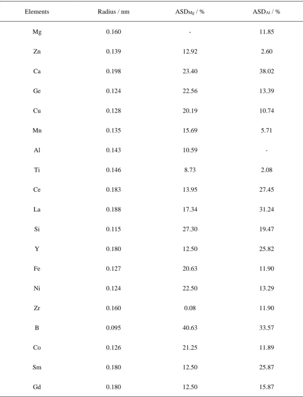
Table 2-3 Atomic radius of some common elements used in Mg alloys
Elements Radius / nm ASDMg / % ASDAl / %
Chapter 2 The microstructure, composition design and experiment methods of Mg-based alloy
Fig. 2.4 Projection of Mg- rich angle on liquid surface of ternary phase diagram of Mg-Ce-La [21].
Through the above analysis, the Ce and La were added to the Mg-based alloy in this research, and their ternary phase diagram with magnesium (Mg-Ce-La) is shown Fig. 2.4 [21].According to the ternary phase diagram, it is feasible to prepare the new Mg-RE (earth ) alloy by TRC process under the current casting conditions with the addition of rare-earth elements (Ce, La).
2.3 Preparation of Mg-based alloys with TRC
Chapter 2 The microstructure, composition design and experiment methods of Mg-based alloy
40
further grain growth, hence the cast sheet dipped into the tank could quickly solidify as exits at the rolls. The schematic diagram and practicality illustration of TRC are shown in Fig. 2.5 (a) and Fig. 2.5 (b), respectively.
Fig. 2.5 Schematic diagram (a) and practicality illustration (b) of TRC technique.
High purity of Mg ingot (99.99%), AZ31 ingot, Mg-10La and Mg-20Ce (in wt.%) were used as raw materials. The melting process was carried out in a resistance furnace under the protection of shielding gas (SF610% and CO2 90%). AZ31 sheets in this study were
Chapter 2 The microstructure, composition design and experiment methods of Mg-based alloy
Firstly, the Mg ingot and AZ31 ingot were placed into the resistance furnace, and the compound were heated up to 720 ℃ for 1.5 h to completely melted. Secondly, the Mg-10La and Mg-20Ce master alloys were added into the melt, then the melt temperature reduced to 700 ℃ and held for 1.5 h. It can be seen that AZ31 alloy sheets could be obtained in the first casting process, while Mg-RE alloy sheets could be obtained in the whole casting process. The final thickness of the sheets between 0.5 mm and 1.1 mm and the width of the strip rang is 25 mm to 50 mm.
2.4 Microscopic characterization
From the as-extruded Mg-based sheets, samples with a dimension of 10 × 10 × 1 mm3 were firstly grounded with SiC papers to1200 grid, and then by diamond pastes down to #0.25 μm grade. The microstructures of the polished surfaces were observed using a field emission scanning electron microscope (FE-SEM, JMS-6301, Tokyo, Japan) and the elements distribution maps were observed by electron probe micro-analysis (EPMA, JXA-8530F, Tokyo, Japan). The alloy phases were obtained by an X-ray diffractometer (XRD, D/Max 2500 PC, Tokyo, Japan). For TEM (HF-3300, Tokyo, Japan) analysis, the focused ion beam (FIB, JIB-4500, Tokyo, Japan) was used for preparation. During the original work of sample preparation, the alloy sheet was cut into 10 mm2 square shapes, and the thinned
section thickness was about 60 μm. Square metal sheets with an area of 1.5 mm2
were cut off from the as-cast sheet and stuck together on a Mo grid with some resin glue, and then the sample was further cut with FIB to obtain the ultimate sample thickness of 0.1 μm.
2.5 Preparation of TEM foils by focused ion beam (FIB) milling
Chapter 2 The microstructure, composition design and experiment methods of Mg-based alloy
42
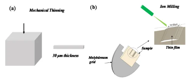
Fig. 2.6 From (a) to (b), the process flow for preparing TEM sample by mechanical thinning
and ion milling.
Beginning with 400 grit SiC paper and finishing with 1200 grint SiC paper, the individual cub segments were thinned with an automated parallel-plate polisher to a final thickness of approximately 30 μm (Fig. 2.6a). The samples were transferred and glued (resin glue) on a molybdenum grid, one at a time, to the ion mill for further thinning. The milling processes by FIB method was shown in Fig. 2.6 (b). The focus of sample was milled into stair-step shape initially, finally milled down to a thickness of 0.1 μm of thin film.
2.6 Electrochemical characterization
2.6.1 Introduction of Electrochemical Workstation
Electrochemical workstation is short for electrochemical measurement system. In this research field, it is mainly used in the study of corrosion or degradation behavior of Mg-based materials.
Chapter 2 The microstructure, composition design and experiment methods of Mg-based alloy
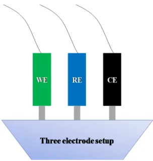
working electrode. The other circuit consists of a working electrode (WE) and a counter (CE) electrode, which transmits electrons to form a circuit. The connection mode of the three electrodes is shown in the Fig. 2.7.
Fig. 2.7 Model diagram of two connection modes of three-electrode operation.
Chapter 2 The microstructure, composition design and experiment methods of Mg-based alloy
44
One end of the working electrode is connected to the alloy sample in this study. The counter electrode and the working electrode form a series circuit. In addition, platinum or graphite materials are often used as counter electrodes. Common reference electrodes include saturated calomel electrode (SCE), Ag/AgCl electrode, reversible hydrogen electrode (RHE), Hg/HgO electrode, Hg/Hg2SO4 electrode,
etc. The diagram of the three electrodes as shown in the Fig. 2.8.
2.6.2 Tafel linear extrapolation
When a metal is immersed in a corrosive medium, the metal and the medium form a corrosive system, which is called the conjugate system. The metal is anodized and the depolarizer is reduced. Take metallic M in hydrochloric acid system as an example:
Anodic reaction: M-2e = M2+ Cathodic reaction: H++2e = H2
The current density of anode reaction is expressed as ia, and the speed of
cathode reaction is expressed as ib. When the system reaches stability, there will be
no net current accumulation in the system, and the system is at a stable potential Фc. According to Faraday's law, there is a strict one-to-one correspondence
between the current passing through the system and the amount of substances that react on the electrode, so the current density of the anodic and cathodic reaction can represent the corrosion rate of the metals. Therefore, the corrosion current of the metal is calculated to represent the corrosion rate of the metals.
Chapter 2 The microstructure, composition design and experiment methods of Mg-based alloy
2.6.3 Electrochemical impedance spectroscopy (EIS)
Typical electrochemical processes contain some basic components, such as double layers and Faraday reaction, which can be approximated by the following model as shown in Fig. 2.9. Internal resistance (RΩ): Internal resistance between
electrolyte and electrode. Double layer capacitance (Cd): From the electrolyte of
inactive ions, no chemical reaction occurred, and only change the charge distribution. Faraday impedance (Zf): From the active ions in the electrolyte, redox
reaction occurs and there is charge transfer.
Fig. 2.9 Electrochemical reaction model.
Electrochemical reaction equivalent circuit as shown in Fig. 2.10, where, Faraday process (Zf) is further divided into charge transfer resistance (Rct) and
Chapter 2 The microstructure, composition design and experiment methods of Mg-based alloy
46
It should be noted that for the same set of spectra, more than one circuit can be found to perform fitting analysis, so it is a highly subjective method to predict electrochemical reaction process by relying on equivalent circuit.
Fig. 2.10 Electrochemical reaction equivalent circuit diagram. Faraday process (Zf) is further
divided into charge transfer resistance (Rct) and warburg impedance (Zw).
2.7 Animals tests
In the present study, The Mg-based alloy as implants were investigated during a 16-week observation in rat femoral model. In addition, the evaluation of bone response might be influenced by bone remodeling result from sheet fixed model. This part mainly introduces the surgical methods of animal experiments.
2.7.1 Implant method
Chapter 2 The microstructure, composition design and experiment methods of Mg-based alloy
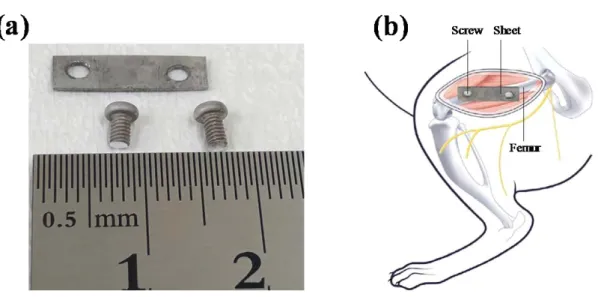
Fig. 2.11 Digital image showing implants before implantation (a); schematic diagram of cast-rolled or
Ti sheets and screws fixation device with rat femoral (b).
2.7.2 Experimental animals
All animal experiments were reviewed by the Saitama Institute of Technology Animal Care and Use Committee (Grant No. 2019-5). All procedures in this study were permitted by the Animal Institutional Review Board of Saitama Institute of Technology in accordance with the guidelines of the U.S. National Institutes of Health, and performed compliance with the Guiding Principles for the Care and Use of Animals in the Field of Physiological Sciences approved by the council of the Physiological Society of Japan. The experimental white rats (Wistar; male, 16 weeks of age, 340 ± 15 g) from Tokyo University Institute of Medicine were selected for the animal study. For micro-CT analysis and histological preparation, groups of fives animals for sheet implants were used for 16 weeks postoperatively respectively.
2.7.3 Surgical procedure
Chapter 2 The microstructure, composition design and experiment methods of Mg-based alloy
48
Fig. 2.12 Surgical procedure used to implant sheet implants: (a) marking indicating symbolic
and the skin incision to reveal the femur; (b) screw holes being created in rat femur using electric drill; (c) sheet was fixed inside by screws; (d) after the sheet was implanted; (e) the fascia was closed with monofilament; (f) surgical site fully closed and sutured.
Chapter 2 The microstructure, composition design and experiment methods of Mg-based alloy
hydrochloride (0.01 ~ 0.05 mg/kg; three times a day; one-week), carprofen (5 mg/kg; weekly; four weeks) and penicillin antibiotic (0.2 mg/kg; three times a day; one-week) were used to suppressed pain after the surgery.
2.8 Methods of in vivo test analysis
Micro-computed tomography (micro-CT) using a microtomographic imaging system (R.mCT2, Rigaku, Tokyo, Japan) at every two weeks evaluate device degradation and new bone formation. Image processing was performed by the 3D image processing software 3D Viewer and Volume Rendering Control. 3D volumes of the scanned devices were generated from acquired 2D lateral projections using Simple Viewer software (Fig. 2.13).
Fig. 2.13 2D images using Simple Viewer software and 3D images using 3D Viewer Volume
Chapter 2 The microstructure, composition design and experiment methods of Mg-based alloy
50
2.9 Concluding remarks
To overcome the disadvantage of rapid degradation of biological Mg alloy, a novel Mg-based alloy with amorphous/nanocrystalline structure is designed in this chapter.
According to the three experimental principles for evaluating the amorphous forming ability, and in combination with the selection of the glass forming alloy system, we selected the rare-earth elements (Ce, La) as the components of the new Mg-based alloy material. In addition, the experimental method of producing new Mg-based alloy by TRC process and the characterization method of microstructure are introduced in detail.
The corrosion performance of the new material in vitro can be evaluated by electrochemical experiments. Animal experiment program was designed, and the process of material implantation in animals was introduced, as well as the means of evaluating the effect of new material degradation on bone in vivo.
![Table 1-6 Density of Ti and its alloy; and some metallic implant alloys [10,11]](https://thumb-ap.123doks.com/thumbv2/123deta/10124377.1958347/16.892.209.759.630.875/table-density-ti-alloy-metallic-implant-alloys.webp)
![Fig. 1.7 Schematic diagram of the biocorrosion at magnesium/medium interface [15, 20]](https://thumb-ap.123doks.com/thumbv2/123deta/10124377.1958347/19.892.264.684.627.1026/fig-schematic-diagram-biocorrosion-magnesium-medium-interface.webp)
![Fig. 2.2 Schematic description of the effect of solidification rate on microstructure of alloy systems [6]](https://thumb-ap.123doks.com/thumbv2/123deta/10124377.1958347/40.892.281.675.289.542/fig-schematic-description-effect-solidification-microstructure-alloy-systems.webp)

![Fig. 2.4 Projection of Mg- rich angle on liquid surface of ternary phase diagram of Mg-Ce-La [21]](https://thumb-ap.123doks.com/thumbv2/123deta/10124377.1958347/46.892.267.683.98.484/fig-projection-angle-liquid-surface-ternary-phase-diagram.webp)