Higher-Order Structure Formation, Phase
Transition, and Distribution of Polymorphic
Crystals of Biodegradable Polymers Studied
Using Time-Resolved Infrared Spectroscopy,
X-ray Scattering, and High-Resolution Raman
Imaging Techniques
学位名
博士(理学)
学位授与機関
関西学院大学
学位授与番号
34504甲第645号
Higher-Order Structure Formation, Phase Transition, and
Distribution of Polymorphic Crystals of Biodegradable Polymers
Studied Using Time-Resolved Infrared Spectroscopy, X-ray
Scattering, and High-Resolution Raman Imaging Techniques
A Thesis for the Degree
of
Doctor of Science
Submitted to
School of Science and Technology
Kwansei-Gakuin University
By
Mengfan WANG
i
Contents
List of Symbols and Abbreviations ... I–II
General Introduction ... 1
1. Introduction of PHB and PBA ... 3
1.1 PHB and Its Crystallization Behavior ... 3
1.2 β-to-α Phase Transition Behavior and the Spherulite of PBA ... 6
2. Vibrational Spectroscopy-Based Chemical Imaging Technique and Its Application in Polymer Science ... 11
3. Outline of Each Chapter ... 13
References ... 16
Chapter 1: Higher-Order Structure Formation of a Poly(3-hydroxybutyrate) Film during Solvent Evaporation ABSTRACT ... 34
Introduction ... 35
Experimental Section ... 38
Results and Discussion ... 40
Conclusion ... 47
References ... 48
Chapter 2: Reinvestigation of the β-to-α Crystal Phase Transition of Poly(butylene adipate) by the Time-Resolved X-ray Scattering and FTIR Spectral Measurements in the Temperature-Jump Process ABSTRACT ... 66
Introduction ... 67
ii
Results and Discussion ... 72
Conclusion ... 77
References ... 78
Appendix 1 ... 90
Chapter 3: Distribution of Polymorphic Crystals in the Ring-Banded Spherulites of Poly(butylene adipate) Studied Using High-Resolution Raman Imaging ABSTRACT ... 94
Introduction ... 95
Experimental Section ... 98
Results and Discussion ... 101
Conclusion ... 109
References ... 110
Appendix 2 ... 126
Acknowledgments... 131
I
List of Symbols and Abbreviations
2D two dimensional
2θ diffraction/scattering angle of the X-ray measurement
AFM atomic force microscope
ATR-FTIR attenuated total reflection Fourier-transform infrared
CCD charge-coupled device
C=O carbonyl group
<d> averaged lamellar thickness of the stacked lamellar structure
D dichroic ratio
DSC differential scanning calorimetry
FTIR Fourier-transform infrared
GI-WAXD grazing incidence wide angle X-ray diffraction
HB(s) hydrogen bonding(s)
inter
Intermolecular interactions exists between PHB molecular chains and
chloroform molecules
inter, H highly ordered intermediate structure of PHB
inter, L less ordered intermediate structure of PHB
intra intramolecular interactions
LP long period of the stacked lamellar structure
MCT mercury-cadmium-telluride
II
NA numerical aperture of object lens
PBA poly(butylene adipate)
PHAs poly(hydroxyalkanoate)s
PHB poly(3-hydroxybutyrate)
POM polarized optical microscope
θc critical angle for total reflection
Q scattering invariant
S banding spacing of the ring-banded PBA spherulites
SAXS small-angle X-ray scattering
SEC solvent evaporation crystallization
Tc crystallization temperature
Tg glass transition temperature
Tm melting temperature
WAXD wide angle X-ray diffraction
XCrys weight fractions of crystalline structure
Xinter, H weight fractions of highly ordered intermediate structure
- 1 -
General Introduction
This thesis is mainly focused on investigating the crystal structure evolution and the distribution of polymorphic crystals within semicrystalline biodegradable polymers. Semicrystalline biodegradable polymers are very important kinds of polymer materials, which have been widely used in our daily life. For the real application of the biodegradable polymers, it is indubitable that the mechanical property and biodegradability are always the most important. These properties are highly related to the inner physical structure of the biodegradable polymers, such as crystal structures, crystal morphologies, crystal or amorphous phase distribution, etc. Therefore, the knowledge about these factors will help us better understanding and application of the biodegradable polymers.
A crystal structure, or a crystal phase, can form by a phase transition process from an amorphous phase or another crystal phase. Phase transition from the amorphous phase is usually called crystallization, and that from the crystal phase is named as crystal phase transition. Note that the phase transition behavior for polymeric materials is a multiple process, containing the evolution of complex hierarchical structures. Therefore, the research on the phase transition process is one of the most important topics of polymers. In the first and second chapters of this thesis, two typical semicrystalline biodegradable polymers, poly(3-hydroxybutyrate) (PHB) and poly(butylene adipate) (PBA) (also named poly(tetramethylene adipate) (PTMA)) were chosen as candidates to systemically investigate the crystallization process during solvent evaporation and crystal phase transition behavior by using time-dependent attenuated total reflection Fourier-transform infrared (ATR-FTIR) spectroscopy, FTIR spectroscopy, grazing incidence wide angle X-ray diffraction (GI-WAXD), and WAXD/small-angle X-ray scattering (SAXS).
The crystal morphologies, such as spherulitic morphologies and the distribution of the different phases within the polymeric material system can also affect the biodegradability. Since different morphologies and phases show different biodegradability as well. When PBA is isothermal melt-recrystallized at an ambient temperature, it will exhibit both ring-banded spherulite and polymorphic crystals, which makes the situation more complicated. To investigate
- 2 -
that, Raman imaging technique is a good choice, for the reason that Raman imaging combines the information not just from the morphologies but also the molecular structure. In the third chapter of the present thesis, the author studied and discussed in detail the distribution of the polymorphic crystals within the spherulite of PBA using Raman imaging.
The originality and novelty of this thesis can be described as follows:
(1) Up to now, studies on the crystallization of PHB have been mainly concerned with melt
crystallization process, but no related studies on solvent evaporation crystallization (SEC) process for PHB. Through the time-dependent ATR-FTIR spectra of PHB/chloroform solution during SEC process, the author found out that PHB/chloroform solution was in a homogeneous state at first. With the evaporation of chloroform, the separated PHB from the solution was in the mixture of intermediate and amorphous states, but no crystal structure formed due to the
presence of chloroform. Moreover, there was no C−H⋯O=C intramolecular interactions
within the intermediate structure of PHB. Subsequently, further evaporation induced a
transition from intermediate phase to crystal phase and the formation of C− H ⋯ O=C
intramolecular interactions within the latter. As the crystal structure developed, the intramolecular interaction become stronger due to the reduced intra-molecular distance within the lamella structure. Moreover, the time-dependent GI-WAXD profiles suggested the presence of two kinds of intermediate structures with different order (less ordered and highly ordered).
(2) The author have solved the long-term controversial problem about the mechanism of the
β-to-α phase transition of PBA through the techniques of the time-resolved FTIR measurement as
well as the simultaneous time-resolved WAXD/SAXS measurement in the quick and stable temperature jumping process, by which the time-dependent structural change has been traced quite clearly. The results indicated that the transformation from the β- to α-phase is not a solid-to-solid mechanism but occurs through the process of the melting of the β-phase into the amorphous phase followed by the subsequent recrystallization of the amorphous phase into the high temperature α-form.
(3) The relationship between ring-banded spherulites and polymorphic crystals behavior of PBA
have been constructed based on Raman spectroscopy and Raman imaging technique. The characteristic Raman bands for α, β-form crystals and the amorphous phase of PBA, which are
- 3 -
suitable for quantitative analysis, have been identified. Through Raman imaging, the α- and β-form crystals of PBA uniβ-formly distributed rather than alternate distributed as suggested by precious study within the ring-banded region and they grow together when the ring-banded PBA spherulites are formed.
1. Introduction of PHB and PBA
After the modern concept of polymers was proposed by Hermann Staudinger in 1920,1 the
polymeric materials have acted as an essential role in our daily life, since they hold wide range of properties. The polymeric materials can be divided into synthetic plastics and natural biopolymers, but they are all formed by many repeat subunits which are created through polymerization of the monomers. The traditional synthetic plastics which derived from petrochemicals, such as polyethylene (PE), polystyrene (PS), poly(vinyl chloride) (PVC), etc., have been popular for more than one century since they can displace traditional materials in the industries of packaging, piping, etc. However, with the big amount usage of the synthetic plastics, the serious challenges for the environment also become a worldwide problem. That is because these plastics are very stable and cannot be degraded naturally. Replacing the usage of traditional plastics by biodegradable polymers (or plastics) is a good idea to overcome the above problems; since the polymer chains in the biodegradable polymers can break down into small, stable end-products in physiological
environments.2 Moreover, some biodegradable polymers show not only good biocompatibility and
bioresorbable but also excellent mechanical properties, which make them very suitable to be used in biomedical field, such as surgical suture, wound dressings, tissue engineering scaffolds, etc. The above advantages make biodegradable polymers a promising kind of materials and have received
substantial interest in both fundamental research and technology in the recent several decades.2,3
1.1 PHB and Its Crystallization Behavior
Linear aliphatic polyesters, for example, PHB, is one of the most popular biodegradable materials. PHB, which belong to the poly(hydroxyalkanoate)s (PHAs) family, was first reported
to isolate from the bacterial cells and to characterize by Maurice Lemoigne in the 1920s.4 The
- 4 -
actually for all of the PHAs are that both of them having three-carbon backbone structure and one
of the hydrogen atoms in the 3 position is substituted by an alkyl group,4 and the different types
of alkyl groups in the 3 position play an important role in determine the physical properties of
PHAs5.
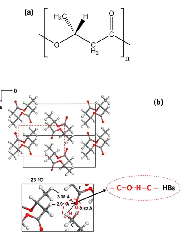
That alkyl group for PHB is a methyl group (−CH3), which can form a weak hydrogen
bonding (HB) with a carbonyl group (C=O) within the PHB crystals. The HBs have been supposed
to affect the thermal properties of the PHB crystals;6,7 which will be introduced in the following
paragraph. PHB can crystallize in two types of crystal modifications, the α and β forms. The chains
within the crystal structure of α-form is packed in an orthorhombic unit cell (P212121-D24) having
axes a=5.76 Å, b=13.2 Å and c(fiber period)=5.96 Å with left-handed 21 helical conformation;
8-10 while the polymer chains of β-form crystals with nearly planar zigzag conformation 11-13 packed
in an hexagonal unit cell12. Since PHB with the α-form crystals are most commonly used, which
can be easily produced by melt, cold and solution crystallization, it has been extensively
studied.6,7,14-29 PHB has a glass transition temperature (Tg) around 5 °C14 and a melting temperature
(Tm) around 178 °C15. Due to nearly perfect stereoregularity of its molecular chains, PHB can
achieve relative high crystallinity, which makes PHB products perform similar mechanical
properties to isotactic polypropylene (iPP).30
In 2004, Sato et al. firstly reported that C−H⋯O=C weak HBs existed between the C=O
groups in one helix and one of the C−H groups of CH3 groups in the adjacent helix within PHB
α-form crystals by temperature-dependent FTIR and WAXD experiments,6,7 the α-form crystal
structure of PHB with HBs is illustrated in Figure 1(b). They showed the following evidences: (1)
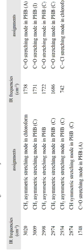
the IR band for CH3 asymmetric stretching vibration appeared at an unusual high wavenumber
(around 3009 cm-1), while the corresponding C=O stretching mode showed relative low frequency
shift to around 1731 cm-1;7 (2) the distance between the H atom of the CH3 group and the O atom
of the C=O group (2.63 Å) is shorter than their van der waals separation (2.72 Å);7,31 Recently,
Wang and Tashiro have further confirmed such HBs on the basis of accurate crystal structure
analysis and the normal mode calculations of the α-form PHB.29
For semicrystalline biodegradable polymers, like PHB, it is no doubted that the crystallization process is always the most important. Through controlling the crystal structures and the crystal morphologies, the crystallization can affect the final mechanical properties and biodegradability
- 5 -
for biodegradable polymers. Thus, the research topics about the crystallization is continuously popular throughout the development of the biodegradable polymers. The crystallization of polymer is a process that the polymer chains packed regularly into crystal region. Semicrystalline polymers consist of both crystalline and amorphous regions, since the usual length of the molecular chains is far greater than the size of the crystallites, one molecular chain is considered to pass through
many crystalline and noncrystalline regions successively. Lotz and Cheng et al.32,33 suggested that
every polymer chain should go through several selection processes on different length and time scales during crystallization. Therefore, the transformation from the entangled melt into the crystalline state will pass several steps, and each step will corresponds to a different state. Based
on a variety of evidence from experiments on several polymer systems, Strobl34 suggested that the
crystallization process from the amorphous state should passing over intermediate states (mesophase) before transforming to the lamellar crystallites. This novel crystallization model has
been widely proven and used in the recent research of the semicrystalline polymers.19,26,35-43
Due to its exceptional purity and low nucleation density,44,45 PHB is usually treated as a good
model for investigating polymer crystallization behavior. FTIR spectroscopy and X-ray analysis are very popular techniques to study the crystallization behavior of polymer materials for a long time since not only they can provide abundant information about the crystal structure evolution, but they are also easy to be performed. FTIR spectroscopy can tell us the molecular structure
change from a functional group level,46 for example, conformations and intermolecular interaction
change during crystallization. While X-ray analysis can give information from unit cell to lamellar periodical structure. Therefore, by combing the information from FTIR and X-ray, we can deeply understand the crystallization process.
Until now, by using FTIR,17,19,40,41,47,48 WAXD,26,49-52 and SAXS26,50-52 techniques, the
crystallization behavior of PHB and its copolymer have been extensively studied. Through time-dependent IR measurements combined with two-dimensional (2D) correlation analysis, Zhang et
al.19 investigated the isothermal melt-crystallization process of PHB. They found out that during
crystallization, the amorphous C=O stretching bands at 1747 and 1739 cm-1, the crystal band at
1731 cm-1 and another crystal band at 1728 cm-1 showed sequence change, and they suggested the
band at 1731 cm-1 might come from the intermediate structure. Recently, Suttiwijitpukdee et al.40
- 6 -
FTIR spectroscopy and synchrotron WAXD/SAXS measurements, respectively in detail.
It should be noted that, the studies on the crystallization of PHB have been mainly concerned
with isothermal melt-crystallization process at around 110 °C thus far, but there has few research
about the SEC process. Being different from crystallization from pure polymers system, such as
melt-crystallization, SEC is in general more complex,53-56 since the solvent molecules usually
exhibit complex interactions with polymer chains. Therefore, it is very necessary for us to
investigate the SEC of PHB. Moreover, as mentioned above, HBs also exist within the lamellar
structure of PHB, thus, study about the formation of HBs combining with the multiple crystal structure evolution during SEC (the molecular interaction exchange from polymer-solvent to polymer-polymer) is also very meaningful and importance.
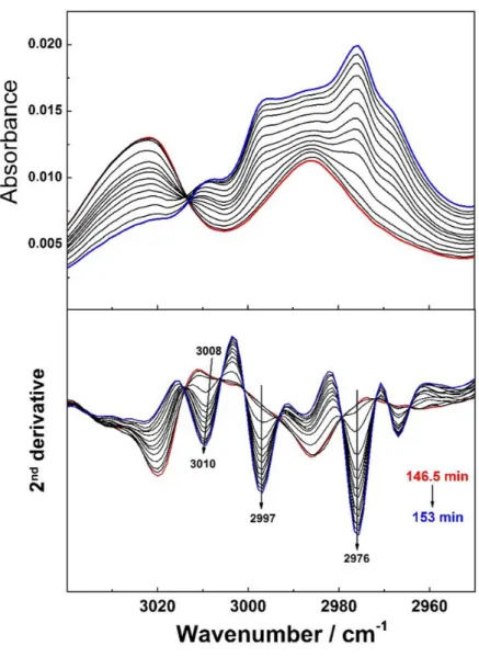
An SEC PHB film is formed in a few minutes at room temperature by using the solvent of chloroform, however, to ensure better signal-to-noise ratio (SNR), it usually takes at least 30 s to obtain one IR spectrum. Therefore, it is very difficult to measure SEC by normal FTIR. In Chapter 1 of this thesis, a glass tube was used to hold the PHB/chloroform solution, and the author successfully investigated the SEC of PHB by using time-resolved ATR-FTIR and GI-WAXD methods. The author had detected the detail SEC process from homogeneous solution to phase separation and finally formation the crystal structure. The transformation of the intermediate structure and the evolution of the HBs were discussed systemically.
1.2 β-to-α Phase Transition Behavior and the Spherulite of PBA
PBA is also an important kind of biodegradable linear aliphatic polyester, different from PHB,
PBA is petroleum-based polyester. PBA has shown its potential as it can be used in biomedical and ecofriendly materials. PBA is also an shared comonomer in poly(butylene
adipate-co-terephthalate) (PBAT, Ecoflex®) and 57poly(butylene succinate-co-adipate) (PBSA, Bionolle®),
which are known for flexible, toughness and good processability, and have been widely applied in cling wrap for food packing, water resistant coatings, plastic bags, drug encapsulation systems,
and so on.58,59 Recent years, more attention has been paid on PBA for its complicated and
interesting polymorphic crystalline structure, phase transition and crystal morphology.60-80
- 7 -
into two types of crystal modifications under different conditions. The crystal structure of PBA
was firstly studied by Fuller et al.81-83 and the lattice parameters were further identified later by
Minke et al.84, 85 through studied the PBA single crystal. The α-form is characterized by its chain
conformation of the planar zigzag type and these chains are packed in the monoclinic unit cell with the dimensions of a = 6.73 Å, b = 7.94 Å, c (fiber period) = 14.20 Å and β = 45.5°, while the β-form of the planar zigzag chain conβ-formation takes the orthorhombic unit cell with the dimensions
of a = 5.06 Å, b = 7.35 Å and c (fiber period) = 14.67 Å.84, 85 The crystal structures of PBA are
shown in Figure 2(b). It has been demonstrated that a film with α-form crystal structures has a higher degradation rate than that with form crystal structures, and that a film with both α- and
β-form crystal structures shows the slowest degradation rate.64
1.2.1 β-to-α
Phase Transition Behavior of PBA
In most of the cases, the β-form crystal structures for the linear aliphatic polyesters can be
achieved only by stretching the polymer film or solution spun-coating to obtain the planar zigzag polymer chain conformation, like β-form PHB and PLA; by normal crystallization condition, such
as solution-casting or isothermal melt-crystallization at Tc (crystallization temperature) higher than
Tg, α-form is mainly formed. However, in the case of PBA, it has been reported that the β-form
crystalline structure can be prepared by either stretching PBA film77 or just by simply isothermal
melt-crystallized at Tc that lower than 29 °C60 (The Tg of PBA is around -55 °C). Moreover, when
isothermal melt-crystallization at Tc that higher than 31 °C, α-form crystals of PBA are mainly
formed, while α- and β-form crystals are formed simultaneously at Tc that between 29 and 31 °C.
Such significantly temperature dependence of the PBA crystalline structure were firstly revealed
by Gan et al.60 In the same research, they also indicated that the α-phase of PBA can form through
non-isothermal crystallization with slow cooling rate of 1 °C/min from melt, while by fasters cooling (> 5 °C/min), β-phase is dominantly developed.
Spontaneous transformation from the β-phase to the α-phase of bulk PBA can occur slowly
by storing the specimen at room temperature for a week.84 Which suggests that α-phase is
thermodynamically more stable for the bulk PBA sample. This point was also proved by
Hoffman-- 8 Hoffman--
Weeks methods; the Tm0 value for the α-form is shown higher than that for the β-form, indicating
the α-form is a structure stable phase, while the β-form is a metastable phase.62
The mechanism of the β-to-α phase transition of PBA is still unclear until now, even though
some groups have already done some works about that. Gan et al.62 suggested this phase transition
is a solid-to-solid process accompanied by the lamellar thickening, which based on the results of
DSC, WAXD and SAXS. By time-resolved FTIR during annealing at 49 °C69 and temperature
dependent FTIR during melting process of the β-form crystal structure72, Yan et al.69 and Yang et
al.72, respectively also indicated the solid-solid phase transition. On the contrary, through FTIR
and WAXD measurements during heating process of the PBA/poly(4-vinylphenol) (PVPh) blends, Sun et al.86 found out that the T
m of α-phase PBA depression with the addition of the PVPh,
moreover, the β-to-α phase transition temperature showed parallel depression as well. They
suggested that this phase transition might be a microdomain melting and recrystallization process.
Li et al.76 based on the results of time-dependent FTIR of ultrathin PBA film during annealing at
45 and 47 °C, respectively, also suggested a melt-recrystallization phase transition process.
The controversial about the β-to-α phase transition of PBA is continuing since there is no direct evidence to demonstrate which is the really phase transition pathway. In general, the phase transition of the crystalline polymers usually occurs over a relatively wide temperature region; this is due to the crystallite size in the semicrystalline polymer distributes over a wide range and so the
melting point itself distributes correspondingly.87 Therefore, by the conventional temperature- or
time-dependent X-ray and vibrational spectroscopic measurements, it is sometimes difficult to judge the phase transition process, since temporal resolution limitation. Moreover, to investigate the phase transition, the evolution of the amorphous structure is also very important to trace at the same time.
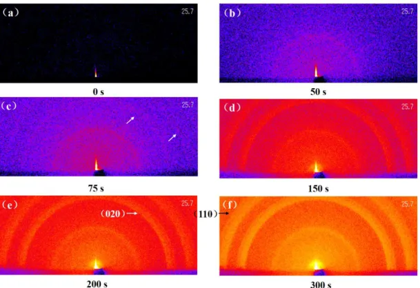
In Chapter 2 of the present thesis, a temperature-jump cell88 was used, the illustration is shown
in Figure 3. Based on the temperature-jump time-resolved measurement of the FTIR, WAXD and SAXS, the phase transition behavior of PBA has been systemically investigated. The author directly traced the sequentially-occurring change of the different phases, and found out that it is not a solid-to-solid phase transition but the combined phenomena of the melting of the β-phase followed by the recrystallization to the high temperature α-phase.
- 9 -
1.2.2 Spherulite of PBA
Spherulites of semicrystalline polymeric materials are spherically symmetric crystal colonies
which formation from viscous melts or solutions with enough supercooling degree ΔT (ΔT = Tm –
Tc).89-91 Spherulites are polycrystalline aggregates composed by highly anisometric crystallites
called subindividuals or subunits; through polarized optical microscopy (POM) observation, the spherulite will shows dark Maltese cross pattern (Figure 4(a)), this is due to the fact that all transparent crystals that are not cubic are birefringent. The morphologies and the growth rate of the spherulites are highly depending both on the polymeric materials themselves and the
crystallization conditions. For example, crystal structure,92-96 chiral of the polymer chains,97,98
molecular weight,99,100 crystallization temperature,101-103 film thickness,104,105 etc.
Among various morphologies, ring-banded spherulites have been attracting considerable attention for several decades. The ring-banded spherulite not only shows Maltese cross, but also shows alternately concentric light-dark rings by POM observation. The typical ring-banded spherulite is shown in Figure 4(b). Evidences show that the ring-banded pattern is originated from the continuous helicoidally twisted of the lamellar crystals along the growth direction of the
banded spherulite, with just a few exceptions.89,106,107 The mechanism of the lamellar twisting has
been considered over the decades, three major ideas have been proposed until now, they are: (1)
unbalanced surface stress effects of the lamellae;97,98,108 (2) isochiral giant screw dislocations along
the lamellar crystals;108,109 (3) response to the compositional or mechanical fields in the melt near
the interface that generated during the crystal growth process110. It should be noted that the present
theories are still not enough to release all of the ambiguities about the formation of the ring-banded spherulites, these theories will be improved to more perfect in the future with more deeply studying and understanding of this fascinating phenomenon.
The banding phenomenon is usually found in the spherulites of chiral polymers, such as
PHAs,98,111 PLA,112,113 poly(propylene oxide) (PPO),114 etc. However, some of the achiral
polymers, for example, PBA, can also crystallized into ring-banded spherulite under specific
conditions. It was firstly reported by Gan et al.60 at 2002 that when isothermal melt-crystallized
PBA in the temperature range of 30–33 °C, banded spherulites were formed, while when the crystallization temperature below 29 °C or above 34 °C, only ringless spherulites could form. What
- 10 -
more interesting they found was that the temperature region at which the ring-banded spherulites formed was just similar with the region that α- and β-form crystals of PBA can crystallized together, which has already mentioned in the Section 1.2.1. However, the ringless PBA spherulites only contain pure α- or β-form crystals.
Since this particular phenomenon, it is reasonable for people to wonder what the relationship is between ring-banded morphology and the polymorphic crystals. Thus, many studies have been
done about this “correlation” since 2002.64,65,67,71,73,74,115 Zhao et al.67 first investigated the banded
PBA spherulites based on the different biodegradation behaviors of the two crystal forms and the results of atomic force microscopy (AFM); they suggested that the banded spherulites are composed of alternating on and flat-on lamellae along the radial direction, and that the edge-on and flat-edge-on domains are composed of β- and α-form crystals, respectively. On the cedge-ontrary, the
following research by Woo et al.71 revealed that the ring-banded pattern had no relation with the
polymeric crystals, since they found the banded pattern can be composed by even singly α-form
crystals. More recently, Liu et al.73 researched the effect of the different molecular weights on the
morphologies of the PBA spherulite. Their results showed that either α or β crystals can form
regular ring-banded spherulites. The research by Wang et al.74 also suggested the similar result
through analyzed the crystallization behavior of PBA blended with structurally similar acrylic polymers.
Thus, the evidences from previous studies have shown that the mixture of the two crystal forms is not the fundamental reason for the formation of the ring-banded PBA spherulites. Even though, this particular phenomenon of PBA still left a lot of unresolved problem. For example, what is the truly reason that when PBA crystallized by itself, the ring-banded pattern only appear with the coexistence of the two crystal forms, but when the system is crystallized by just either one crystal form, only ringless spherulite can be detected and what is the formation process of the ring-banded PBA spherulite with polymeric crystals? What is more, the banded pattern which is formed by a single crystal form or by mixed crystal forms at the same crystallization temperature
appear very different band spacing,73 which means that the coexistence of the two crystal forms
may affect the ring-banded morphology. In addition, since the two crystal forms result in different mechanical properties and biodegradation behavior, their distribution will also affect the final performances of the PBA products. Accordingly, more systematically works about the relationship
- 11 -
between PBA spherulite and its polymeric crystals are still very necessary to be done.
In order to achieve that final goal, the first step for us to do should be finding out the distribution of polymeric crystals in the ring-banded spherulites of PBA. Until now, a vast majority of spherulite studies rely on POM. Interpretations of the POM images can provide important
information about the structures of spherulites;89 however, POM cannot give detailed information
on the internal structures. Recent years, vibrational spectroscopy-based chemical imaging technique, for instance, Raman or FTIR imaging technique, has showed its potential in the research of polymer science, since it combines the information from both morphology and molecular structure. Therefore high spatial resolution Raman imaging technique was used to investigate the distribution of polymeric crystals, as well as the molecular chains orientation within the ring-banded PBA spherulites for the first time. Moreover, since there is no Raman studies on PBA, particularly, the polymorphic crystals of PBA, have been reported thus far. Therefore, in present thesis, Raman spectroscopy and Raman imaging were used to explore the PBA spherulites for the first time. This part will be discussed in detail in Chapter 3 of the present thesis.
2. Vibrational Spectroscopy-Based Chemical Imaging Technique and Its
Application in Polymer Science
Vibrational spectroscopy-based chemical imaging technique, which has application in
various academic and industries fields, is a very powerful tool for solving real-world issues.116
Raman and FTIR imaging are the two typical branches of vibrational spectroscopy-based chemical imaging. Simply speaking, these imaging techniques are combining the advantages of digital imaging techniques and vibrational spectroscopy. The key points of chemical imaging based on Raman or FTIR spectroscopy are chemical specificity and the abundant information that stems from the full-range spectra.
Both of the IR and Raman spectroscopy are based on the fact that the chemical bonds between two or more atoms vibrate continually, such as stretching, bending, wagging, etc. When a “light”, or in other words, an electromagnetic radiation incident to a material, some of it will be absorbed when its frequency is resonant with the molecular vibration within the material. The vibrational energy of the molecular will be excited to a higher energy state by absorbing the light, and higher
- 12 -
energy state usually relaxes back to the lowest energy state quickly by releasing heat and/or light.116
Assuming that the groups of atoms in a molecule are not vibrationally coupled to the rest of the molecule, which means they have almost the same vibration frequencies in any molecule. Therefore, it is possible to associate a vibrational frequency with a particular chemical functional
group.116 The IR and Raman spectra are the absorbance and scattering spectra, respectively, and
the laser source of the IR and Raman are infrared light and visible laser, respectively. The basic
principle of the IR and Raman spectroscopy have been well published elsewhere.116
The semicrystalline polymer system is usually in spatially inhomogeneous state, for example, there should contain crystal and amorphous phases at the same time. If a polymer also shows polymorphic crystals behavior, the situation will become more complex. Since different phases will show different properties, such as mechanical property and biodegradability. Thus, fully understanding the distribution of different phases in the polymer system is very important to control and predict its final performance.
Since different phases or structures usually have their own molecule-specific band(s) in the IR or Raman spectrum. Then, the imaging instrument are used to collect IR or Raman spectra associate with the sample from every pixel of the region that is interested. The final chemical image is generated by calculating the relative value (intensity, intensity ratio, etc.) of the
molecule-specific band at each pixel, and then drawing these values in the Cartesian coordinates.116 The
schematic of chemical imaging technique is shown in Figure 5.
FTIR imaging has been extensively applied in the research of polymer science in the recent two decades since it can collect full IR image which containing hundreds or thousands of IR
spectra in a few minutes.28,41,42,96,113,117-127 For example, Siesler group have done a lot of work on
various of polymer blending system by using FTIR imaging;28,41,122-125 Hekima and Morikawa
investigated the inner molecular chains orientation within the stretched polymer fiber and different kinds of spherulites of polymeric materials by polarized FTIR imaging through their newly
proposed multipolarization calculation method.113,126,127 Using the same method, Hu and Tashiro
studied the phase transition behavior from form-II to form-I in the melt-grown spherulites of
isotactic polybutene-1 (it-PB-1) at 25 °C.96 Even though FTIR imaging technique has many
- 13 -
of IR imaging if the inhomogeneous part is very small.
Compared to FTIR imaging, Raman imaging can give much better spatial resolution (0.3 μm can be achieved), and it can also provide marked information about the molecular structure. Raman imaging instrument usually composed by a laser source, optical microscopy system, computer-controlled motorized sample stage and a CCD detector. Back-scattering is the most common mode for Raman imaging measurement, which means that during Raman experiment, the laser always focus on the surface or any another point along the thickness direction of the sample, and thus, only the thin laser focus plane of the sample will be measured. This feature makes Raman imaging can easily be used to measure the sample in not only two-dimensional (2D) plane but also three-dimensional (3D) space. Hence, Raman imaging has shown its powerful in recent decades and has been widely applied to investigate the distribution of different phase and structures in polymeric
materials.28,128-133 For example, Van Apeldorn et al.130 studied about the intracellular degradation
behavior and mechanism of the poly(lactic-co-glycolic acid) (PLGA) microsphere inside macrophages by using confocal Raman spectroscopy and imaging with spatial resolution of 1.5
μm; Chernenko et al.132 investigated the intracellular drug-delivery and degradation of the
biodegradable nanocarrier systems of poly(ε-caprolactone) (PCL) and PLGA;132 Huan et al.
researched about the phase behavior in poly(ethylene terephthalate)/high-density poly(ethylene) (PET/HDPE) polymer blends using high-spatial resolution Raman imaging.
In the third chapter of the present thesis, Raman imaging technique was chosen to investigate the complex distribution of inner physical structure within ring-banded PBA spherulites instead of FTIR imaging. That is mainly due to that spatial resolution of FTIR imaging is not enough to investigate such small band spacing of PBA spherulites efficiency.
3. Outline of Each Chapter
The outline of each chapter for the present thesis will be described as follows. This thesis consists of three chapters.
Chapter 1: this chapter describes the evolution of the intra-molecular interaction within PHB chains and inter-molecular interaction between PHB chains and chloroform molecules during SEC of PHB in a PHB/chloroform solution by using time-solved ATR-FTIR spectroscopy; and the
- 14 -
crystal structure formation of PHB during SEC by using time-solved GI-WAXD. From ATR-FTIR, it is found that the PHB/chloroform solution was in a homogeneous state at first, and within the solution, there contains the intermolecular interaction between C=O groups of the PHB chains and the C−H groups of chloroform. With the evaporation of chloroform, phase separation started since the solution concentration reached the saturation point, and PHB started to separate from the solution. The separated PHB was in the mixture of intermediate and amorphous states, but no
crystal structure formed due to the presence of chloroform. Moreover, no C=O…H−C interaction
within PHB was formed, which in other others, hydrogen bonding was not exist within the intermediate structure. Subsequently, further evaporation induced a transition from intermediate
to crystal structure and the formation of C=O…H−C intramolecular interactions within the latter.
As the crystal structure developed, the intramolecular interaction changed from weak to strong due to the reduced intra-molecular distance within the lamella structure. The results of the GI-WAXD, it is suggested the presence of two kinds of intermediate structures with different order (less ordered and highly ordered). During SEC, the intermediate structures formed firstly, subsequently transforming into a crystal structure.
Chapter 2: the mechanism of the β-to-α phase transition of PBA was systematically
investigated by using the techniques of the time-resolved measurements of the FTIR spectra as well as the simultaneous time-resolved WAXD/SAXS measurements in the quick and stable temperature-jump process. A majority of papers published so far reported that the phase transition from the β-form to the α-form occurs as the direct solid-to-solid process when the sample is heated up to the high temperature. However, the author found out that this phase transition was not a solid-to-solid phase transition but the melting of the β-phase into the amorphous phase and the subsequently occurred recrystallization of the amorphous phase into the α-form. The α-phase obtained by the melt-recrystallization of the original β-phase is different not only in the lamellar stacking structure but also in the degree of orderliness in the crystal lattice compared to the normal
α-phase as judged from the SAXS and WAXD data.
Chapter 3: in this chapter, the author discussed the polymorphic crystals and the molecular
chains orientation within the PBA spherulites in detail by using Raman spectroscopy and Raman imaging technique. Special attention has been paid to the so-called “ring-banded” PBA spherulites
- 15 -
characteristic Raman peaks for both α- and β-form crystal structures and the amorphous structure of PBA were observed for the first time. These peaks were employed to investigate the polymorphic crystal distribution through Raman imaging. It was found that the center and ring-banded regions contained both α- and β-form crystals, while the out-layer region contained only
α-form crystals. The α- and β-form crystals can nucleate and grow in the same temperature range
(31–33 °C), and the relative content of these two crystal forms within the ring-banded spherulites show temperature dependence. The higher isothermal melt-crystallization temperature, the higher content of the α-form crystals within the ring-banded PBA spherulites. The molecular chains within the PBA spherulites are oriented almost perpendicular to the spherulite growth direction. However, the ring-banded domains have different orientations about the substrate plane; the molecular chains orient perpendicular to the substrate plane in the flat-on domains and parallel to the substrate plane in the edge-on domains
- 16 -
References
1. Staudinger, H. Über polymerisation. Eur. J. Inorg. Chem. 1920, 53, 1073–1085.
2. Pan, P.; Inoue, Y. Polymorphism and isomorphism in biodegradable polyesters. Prog.
Polym. Sci. 2009, 34, 605–640.
3. Nair, L. S.; Laurencin, C. T. Biodegradable polymers as biomaterials. Prog. Polym. Sci.
2007, 32, 762–798.
4. Lenz, R. W.; Marchessault, R. H. Bacterial Polyesters: Biosynthesis, Biodegradable Plastics and Biotechnology. Biomacromolecules 2005, 6, 1–8.
5. Anderson, A. J.; Dawes, E. A. Occurrence, metabolism, metabolic role, and industrial uses of bacterial polyhydroxyalkanoates. Microbiol. Rev. 1990, 54, 450–472.
6. Sato, H.; Nakamura, M.; Padermshoke, A.; Yamaguchi, H.; Terauchi, H.; Ekgasit, S.; Noda, I.; Ozaki, Y. Thermal Behavior and Molecular Interaction of Poly(3-hydroxybutyrate-co-3-hydroxyhexanoate) Studied by Wide-Angle X-ray Diffraction. Macromolecules 2004, 37, 3763–3769.
7. Sato, H.; Murakami, R.; Padermshoke, A.; Hirose, F.; Senda, K.; Noda, I.; Ozaki, Y. Infrared Spectroscopy Studies of CH···O Hydrogen Bondings and Thermal Behavior of Biodegradable Poly(hydroxyalkanoate). Macromolecules 2004, 37, 7203–7213.
8. Cobntbekt, J.; Mabchessault, R. H. Physical properties of poly-β-hydroxybutyrate: IV. Conformational analysis and crystalline structure. J. Mol. Boil. 1972, 71, 735–756.
9. Yokouchi, M.; Chatani, Y.; Tadokoro, H.; Teranishi, K.; Tani, H. Structural studies of polyesters: 5. Molecular and crystal structures of optically active and racemic poly(β-hydroxybutyrate). Polymer 1973, 14, 267–272.
10. Bruckner, S.; Meille, S. V.; Malpezzi, L.; Cesaro, A.; Navarini, L.; Tombolini, R. The
Structure of Poly(D-(-)-β-hydroxybutyrate). A Refinement Based on the Rietveld Method.
Macromolecules 1988, 21, 967–972.
11. Yokouchi, M.; Chatani, Y.; Tadokoro, H.; Teranishi, K.; Tani, H. Structural studies of polyesters: 5. Molecular and crystal structures of optically active and racemic poly(β-hydroxybutyrate). Polymer 1973, 14, 267–272.
- 17 -
β Form in Poly(β-hydroxyalkanoates). Macromolecules 1990, 23, 5368–5370.
13. Lambeek, G.; VORENKAP, E. J.; Schouten, A. J. Structural Study of Langmuir-Blodgett Mono-and Multilayers of Poly(β-hydroxyburyrate). Macromolecules 1995, 28, 2023–2032. 14. Lageveen, R. G.; Huisman, G. W.; Preusting, H.; Ketelaar, P.; Eggink, G.; Witholt, B. Formation of polyesters by Pseudomonas oleovorans: effect of substrates on formation and composition of poly-(R)-3-hydroxyalkanoates and poly-(R)-3-hydroxyalkenoates. Appl.
Environ. Microb. 1988, 54, 2924–2932.
15. Abe, H.; Doi, Y.; Aoki, H.; Akehata, T. Solid-State Structures and Enzymatic Degradabilities for Melt-Crystallized Films of Copolymers of (R)-3-Hydroxybutyric Acid with Different Hydroxyalkanoic Acids. Macromolecules 1998, 31, 1791–1797.
16. Marchessault, R. H.; Kawada, J. PHB Lamellar Single Crystals: Origin of the Splintered Texture. Macromolecules 2004, 37, 7418–7420.
17. Padermshoke, A.; Sato, H.; Katsumoto, Y.; Ekgasit, S.; Noda, I.; Ozaki, Y. Crystallization behavior of poly(3-hydroxybutyrate-co-3-hydroxyhexanoate) studied by 2D IR correlation spectroscopy. Polymer 2004, 45, 7159–7165.
18. Padermshoke, A.; Katsumoto, Y.; Sato, H.; Ekgasit, S.; Noda, I.; Ozaki, Y. Surface melting and crystallization behavior of polyhydroxyalkanoates studied by attenuated total reflection infrared spectroscopy. Polymer 2004, 45, 6547–6554.
19. Zhang, J.; Sato, H.; Noda, I.; Ozaki, Y. Conformation Rearrangement and Molecular Dynamics of Poly(3-hydroxybutyrate) during the Melt-Crystallization Process Investigated by Infrared and Two-Dimensional Infrared Correlation Spectroscopy. Macromolecules
2005, 38, 4274–4281.
20. Furukawa, T.; Sato, H.; Murakami, R.; Zhang, J.; Noda, I.; Ochiai, S.; Ozaki, Y. Raman
microspectroscopy study of structure, dispersibility, and crystallinity of
poly(hydroxybutyrate)/poly(L-lactic acid) blends. Polymer 2006, 47, 3132–3140.
21. Sato, H.; Mori, K.; Murakami, R.; Ando, Y.; Takahashi, I.; Zhang, J.; Terauchi, H.; Hirose, F.; Senda, K.; Tashiro, K.; Noda, I.; Ozaki, Y. Crystal and Lamella Structure and C−H···O=C Hydrogen Bonding of Poly(3-hydroxyalkanoate) Studied by X-ray Diffraction and Infrared Spectroscopy. Macromolecules 2006, 39, 1525–1531.
- 18 -
and Isothermal Crystallization Kinetics of Poly(3-hydroxybutyrate) Investigated by Near-Infrared Spectroscopy. Macromolecules 2006, 39, 3841–3847.
23. Mori, K.; Mukoyama, S.; Zhang, Y.; Sato, H.; Ozaki, Y.; Terauchi, H.; Noda, I.; Takahashi, I. Crystalline Lamellae and Surface Morphology of Biodegradable Polyhydroxyalkanoate Thin Films: Thermal Behavior and Comparison between Poly(3-hydroxybutyrate-co-3-hydroxyhexanoate) and Poly(3-hydroxybutyrate). Macromolecules 2008, 41, 1713–1719. 24. Androsch, R. Surface structure of folded-chain crystals of poly(R-3-hydroxybutyrate) of
different chain length. Polymer 2008, 49, 4673–4679.
25. Sato, H.; Ando, Y.; Mitomo, H.; Ozaki, Y. Infrared Spectroscopy and X-ray Diffraction Studies of Thermal Behavior and Lamella Structures of Poly(3-hydroxybutyrate-co-3-hydroxyvalerate) (P(HB-co-HV)) with PHB-Type Crystal Structure and PHV-Type Crystal Structure. Macromolecules 2011, 44, 2829–2837.
26. Guo, L.; Spegazzini, N.; Sato, H.; Hashimoto, T.; Masunaga, H.; Sasaki, S.; Takata, M.; Ozaki, Y. Multistep Crystallization Process Involving Sequential Formations of Density Fluctuations, “Intermediate Structures”, and Lamellar Crystallites: Poly(3-hydroxybutyrate) As Investigated by Time-Resolved Synchrotron SAXS and WAXD. Macromolecules 2012, 45, 313–328.
27. Di Lorenzo, M. L.; Gazzano, M.; Righetti, M. C. The Role of the Rigid Amorphous Fraction on Cold Crystallization of Poly(3-hydroxybutyrate). Macromolecules 2012, 45, 5684–5691. 28. Unger, M.; Sato, H.; Ozaki, Y.; Fischer, D.; Siesler, H. W. Temperature-Dependent Fourier Transform Infrared Spectroscopy and Raman Mapping Spectroscopy of Phase-Separation
in a Poly(3-hydroxybutyrate)-Poly(L-lactic acid) Blend. Appl. Spectrosc. 2013, 67, 141–148.
29. Wang, H.; Tashiro, K. Reinvestigation of Crystal Structure and Intermolecular Interactions of Biodegradable Poly(3-Hydroxybutyrate) α-Form and the Prediction of Its Mechanical Property. Macromolecules 2016, 49, 581–594.
30. Satkowski, M. M.; Melik, D. H.; Autran, J. P.; Green, P. R.; Noda, I.; Schechtman, L. A., In Biopolymers; Steinbüchel, A., Doi, Y., Eds.; Wiley-VCH: Wienheim, Germany, 2001. 31. Sato, H.; Dybal, J.; Murakami, R.; Noda, I.; Ozaki, Y. Infrared and Raman spectroscopy and
quantum chemistry calculation studies of C–H⋯O hydrogen bondings and thermal behavior of biodegradable polyhydroxyalkanoate. J. Mole. Struct. 2005, 744–747: 35-46.
- 19 -
32. Lotz, B. What can polymer crystal structure tell about polymer crystallization processes?
Eur. Phys. J. E 2000, 3, 185–194.
33. Cheng, S.; Li, C. Y.; Zhu, L. Commentary on polymer crystallization: Selection rules in different length scales of a nucleation process. Eur. Phys. J. E 2000, 3, 195–197.
34. Strobl, G. From the melt via mesomorphic and granular crystalline layers to lamellar crystallites: A major route followed in polymer crystallization? Eur. Phys. J. E 2000, 3, 165– 183.
35. Strobl, G. Crystallization and melting of bulk polymers: new observations, conclusions and a thermodynamic scheme. Prog. Polym. Sci. 2006, 31, 398–442.
36. Qiu, J.; Wang, Z.; Yang, L.; Zhao, J.; Niu, Y.; Hsiao, B. S. Deformation-induced highly oriented and stable mesomorphic phase in quenched isotactic polypropylene. Polymer 2007, 48, 6934–6947.
37. Konishi, T.; Nishida, K.; Matsuba, G.; Kanaya, T. Mesomorphic Phase of Poly(butylene-2, 6-naphthalate). Macromolecules 2008, 41, 3157–3161.
38. Zhang, J.; Duan, Y.; Domb, A. J.; Ozaki, Y. PLLA Mesophase and Its Phase Transition Behavior in the PLLA−PEG−PLLA Copolymer As Revealed by Infrared Spectroscopy.
Macromolecules 2010, 43, 4240–4246.
39. Stoclet, G.; Seguela, R.; Lefebvre, J.; Rochas, C. New insights on the strain-induced
mesophase of poly(D, L-lactide): in situ WAXS and DSC study of the thermo-mechanical
stability. Macromolecules 2010, 43, 7228–7237.
40. Suttiwijitpukdee, N.; Sato, H.; Zhang, J.; Hashimoto, T. Effects of Intermolecular Hydrogen Bondings on Isothermal Crystallization Behavior of Polymer Blends of Cellulose Acetate Butyrate and Poly(3-hydroxybutyrate). Macromolecules 2011, 44, 3467–3477.
41. Unger, M.; Sato, H.; Ozaki, Y.; Siesler, H. W. Crystallization Behavior of Poly(3-hydroxybutyrate) (PHB), Poly(ε-caprolactone) (PCL) and Their Blend (50:50 wt.%) Studied by 2D FT-IR Correlation Spectroscopy. Macromol. Symp. 2011, 305, 90–100.
42. Lan, Q.; Li, Y. Mesophase-Mediated Crystallization of Poly(l-lactide): Deterministic Pathways to Nanostructured Morphology and Superstructure Control. Macromolecules
2016, 49, 7387–7399.
- 20 -
Low Temperatures by Low-Pressure CO2 That Provides Moderately Increased Molecular
Mobility. Macromolecules 2016, 49, 2262–2271.
44. Barham, P. J.; Keller, A.; Otun, E. L.; Holmes, P. A. Crystallization and morphology of a bacterial thermoplastic: poly-3-hydroxybutyrate. J. Mater. Sci. 1984, 19, 2781–2794. 45. Barham, P. J. Nucleation behaviour of poly-3-hydroxy-butyrate. J. Mater. Sci. 1984, 19,
3826–3834.
46. Bower, D. I.; Maddams, W. F. The vibrational spectroscopy of polymers; Cambridge University Press: Cambridge, 1992.
47. Zhang, J.; Sato, H.; Furukawa, T.; Tsuji, H.; Noda, I.; Ozaki, Y. Crystallization Behaviors
of Poly(3-hydroxybutyrate) and Poly(L-lactic acid) in Their Immiscible and Miscible Blends.
The J. Phys. Chem. B 2006, 110, 24463–24471.
48. Huang, H.; Guo, W.; Chen, H. In situ FTIR and generalized 2D IR correlation spectroscopic studies on the crystallization behavior of solution-cast PHB film. Anal. Bioanal. Chem. 2011, 400, 279–288.
49. Owen, A. J.; Heinzel, J.; Škrbić, Z; Divjaković, V. Crystallization and melting behaviour of PHB and PHB/HV copolymer. Polymer 1992, 33, 1563–1567.
50. Xing, P.; Dong, L.; An, Y.; Feng, Z.; Avella, M.; Martuscelli, E. Miscibility and
Crystallization of Poly(β-hydroxybutyrate) and Poly(p-vinylphenol) Blends.
Macromolecules 1997, 30, 2726–2733.
51. Liu, J.; Jungnickel, B. J. Crystallization and morphology of poly(vinylidene fluoride)/poly(3-hydroxybutyrate) blends. II. Morphology and crystallization kinetics by time resolved X-ray scattering. J. Polym. Sci. Part B: Polym. Phys. 2004, 42, 974–985. 52. Sato, H.; Suttiwijitpukdee, N.; Hashimoto, T.; Ozaki, Y. Simultaneous Synchrotron
SAXS/WAXD Study of Composition Fluctuations, Cold-Crystallization, and Melting in Biodegradable Polymer Blends of Cellulose Acetate Butyrate and Poly(3-hydroxybutyrate).
Macromolecules 2012, 45, 2783–2795.
53. Huang, H.; Hu, Z.; Chen, Y.; Zhang, F.; Gong, Y.; He, T.; Wu, C. Effects of Casting Solvents on the Formation of Inverted Phase in Block Copolymer Thin Films.
Macromolecules 2004, 37, 6523–6530.
- 21 -
Phase Transition in Solution-Cast Polystyrene-Poly(methyl methacrylate) Block Copolymer Thin Films. Macromolecules 2006, 39, 3369–3376.
55. Heinzer, M. J.; Han, S.; Pople, J. A.; Baird, D. G.; Martin, S. M. In Situ Measurement of Block Copolymer Ordering Kinetics during the Drying of Solution-Cast Films Using Small-Angle X-ray Scattering. Macromolecules 2012, 45, 3471–3479.
56. Heinzer, M. J.; Han, S.; Pople, J. A.; Baird, D. G.; Martin, S. M. In Situ Tracking of Microstructure Spacing and Ordered Domain Compression during the Drying of Solution-Cast Block Copolymer Films Using Small-Angle X-ray Scattering. Macromolecules 2012, 45, 3480–3486.
57. Siracusa, V.; Lotti, N.; Munari, A.; Dalla Rosa, M. Poly(butylene succinate) and poly(butylene succinate-co-adipate) for food packaging applications: Gas barrier properties after stressed treatments. Polym. Degrad. Stab. 2015, 119, 35–45.
58. Someya, Y.; Sugahara, Y.; Shibata, M. Nanocomposites based on poly(butylene adipate-co-terephthalate) and montmorillonite. J. Appl. Polym. Sci. 2005, 95, 386–392.
59. Brunner, C. T.; Baran, E. T.; Pinho, E. D.; Reis, R. L.; Neves, N. M. Performance of biodegradable microcapsules of poly(butylene succinate), poly(butylene succinate-co-adipate) and poly(butylene terephthalate-co-succinate-co-adipate) as drug encapsulation systems. Colloid.
Surface. B 2011, 84, 498–507.
60. Gan, Z.; Abe, H. Temperature-Induced Polymorphic Crystals of Poly (butylene adipate).
Macromol. Chem. Phys. 2002, 203, 2369–2374.
61. Pouget, E.; Almontassir, A.; Casas, M. T.; Puiggalí, J. On the Crystalline Structures of
Poly(tetramethylene adipate). Macromolecules 2003, 36, 698–705.
62. Gan, Z.; Kuwabara, K.; Abe, H.; Iwata, T.; Doi, Y. Metastability and Transformation of Polymorphic Crystals in Biodegradable Poly(butylene adipate). Biomacromolecules 2004, 5, 371–378.
63. Iwata, T.; Kobayashi, S.; Tabata, K.; Yonezawa, N.; Doi, Y. Crystal Structure, Thermal Behavior and Enzymatic Degradation of Poly(tetramethylene adipate) Solution-Grown Chain-Folded Lamellar Crystals. Macromol. Biosci. 2004, 4, 296–307.
64. Gan, Z.; Kuwabara, K.; Abe, H.; Iwata, T.; Doi, Y. The role of polymorphic crystal structure and morphology in enzymatic degradation of melt-crystallized poly(butylene adipate) films.
- 22 -
Polym. Degrad. Stab. 2005, 87, 191–199.
65. Wu, M. C.; Woo, E. M. Effects of α-form or β-form nuclei on polymorphic crystalline morphology of poly(butylene adipate). Polym. Int. 2005, 54, 1681–1688.
66. Noguchi, K.; Kondo, H.; Ichikawa, Y.; Okuyama, K.; Washiyama, J. Molecular and crystal structure of poly(tetramethylene adipate) α form based on synchrotron X-ray fiber diffraction. Polymer 2005, 46, 10823–10830.
67. Zhao, L.; Wang, X.; Li, L.; Gan, Z. Structural analysis of poly(butylene adipate) banded spherulites from their biodegradation behavior. Polymer 2007, 48, 6152–6161.
68. Woo, E. M.; Chang, C.; Wu, M. C. A new crystal morphology of straight-stalk dendrites in blends of poly(butylene adipate) with amorphous poly(vinyl acetate). Mater. Lett. 2007, 61, 3542–3546.
69. Yan, C.; Zhang, Y.; Hu, Y.; Ozaki, Y.; Shen, D.; Gan, Z.; Yan, S.; Takahashi, I. Melt Crystallization and Crystal Transition of Poly(butylene adipate) Revealed by Infrared Spectroscopy. J. Phys. Chem. B 2008, 112, 3311–3314.
70. Frömsdorf, A.; Woo, E. M.; Lee, L.; Chen, Y.; Förster, S. Atomic Force Microscopy Characterization and Interpretation of Thin-Film Poly(butylene adipate) Spherulites with Ring Bands. Macromol. Rapid Commun. 2008, 29, 1322–1328.
71. Woo, E. M.; Yen, K. C.; Wu, M. C. Analysis of multiple melting behavior of spherulites comprising ring-band shell/ringless core in polymorphic poly(butylene adipate). J. Polym.
Sci. Part B: Polym. Phys. 2008, 46, 892–899.
72. Yang, J.; Li, Z.; Pan, P.; Zhu, B.; Dong, T.; Inoue, Y. Temperature-dependent polymorphic crystalline structure and melting behavior of poly(butylene adipate) investigated by time-resolved FTIR spectroscopy. J. Polym. Sci. Part B: Polym. Phys. 2009, 47, 1997–2007. 73. Liu, J.; Ye, H.; Xu, J.; Guo, B. Formation of ring-banded spherulites of α and β modifications
in Poly(butylene adipate). Polymer 2011, 52, 4619–4630.
74. Wang, L.; Lugito, G.; Woo, E. M.; Wang, Y. Phase behavior, polymorphism and spherulite morphology in Poly(1, 4-butylene adipate) interacting with two structurally similar acrylic polymers. Polymer 2012, 53, 3815–3826.
75. Weng, M.; He, Y.; Qiu, Z. Effect of Uracil on the Isothermal Melt Crystallization Kinetics and Polymorphic Crystals Control of Biodegradable Poly(butylene adipate). Ind. Eng. Chem.
- 23 -
Res. 2012, 51, 13862–13868.
76. Li, Q.; Zhou, J.; Chai, L.; Memon, J.; Ren, Z.; Li, H.; Sun, X.; Yan, S. The effect of the poly(vinyl phenol) sublayer on the melting behavior of poly(butylene adipate) crystals.
Polym. Chem. 2014, 5, 4293–4303.
77. Song, Y.; Ye, H.; Xu, J.; Hou, K.; Zhou, Q.; Lu, G. Stretch-induced bidirectional polymorphic transformation of crystals in poly(butylene adipate). Polymer 2014, 55, 3054– 3061.
78. Yang, J.; Chen, Y.; Qin, S.; Liu, J.; Bi, C.; Liang, R.; Dong, T.; Feng, X. Effects of Cyanuric Acid on Crystallization Behavior, Polymorphism, and Phase Transition of Poly(butylene adipate). Ind. Eng. Chem. Res. 2015, 54, 8048–8055.
79. Mi, C.; Zhou, J.; Ren, Z.; Li, H.; Sun, X.; Yan, S. The phase transition behavior of poly(butylene adipate) in the nanoporous anodic alumina oxide. Polym. Chem. 2016, 7, 410– 417.
80. Sun, X.; Fang, Q.; Li, H.; Ren, Z.; Yan, S. Effect of Anodic Alumina Oxide Pore Diameter on the Crystallization of Poly(butylene adipate). Langmuir 2016, 32, 3269–3275.
81. Fuller, C. S.; Erickson, C. L. An X-ray study of some linear polyesters. J. Am. Chem. Soc.
1937, 59, 344–351.
82. Fuller, C. S.; Frosch, C. J. Further Investigation of the Chain Structure of Linear Polyesters.
J. Phys. Chem. 1939, 43, 323–334.
83. Fuller, C. S.; Frosch, C. J. X-ray investigation of the decamethylene series of polyesters. J.
Am. Chem. Soc. 1939, 61, 2575–2580.
84. Minke, R.; Blackwell, J. Polymorphic structures of poly(tetramethylene adipate). J.
Macromol. Sci., Part B: Phys. 1979, 16, 407–417.
85. Minke, R.; Blackwell, J. Single crystals of poly(tetramethylene adipate). J. Macromol. Sci.,
Part B: Phys. 1980, 18, 233–255.
86. Sun, X.; Pi, F.; Zhang, J.; Takahashi, I.; Wang, F.; Yan, S.; Ozaki, Y. Study on the Phase Transition Behavior of Poly(butylene adipate) in its Blends with Poly(vinyl phenol). J. Phys.
Chem. B 2011, 115, 1950–1957.
87. Ratri, P. J.; Tashiro, K. Phase-transition behavior of a crystalline polymer near the melting point: case studies of the ferroelectric phase transition of poly(vinylidene fluoride) and the
- 24 -
β-to-α transition of trans-1, 4-polyisoprene. Polym. J. 2013, 45, 1107–1114.
88. Tashiro, K., Measurement of the Physical Characteristics of Polymers by Vibrational Spectroscopy. Chalmers, J. M.; Griffiths, P. R., Chalmers, J. M.; Griffiths, P. R., Eds.;
Handbook of Vibrational spectroscopy; John Wiley & Sons, Ltd: Chichester, 2002.
89. Crist, B.; Schultz, J. M. Polymer spherulites: A critical review. Prog. Polym. Sci. 2016, 56, 1–63.
90. Shtukenberg, A. G.; Punin, Y. O.; Gunn, E.; Kahr, B. Spherulites. Chem. Rev. 2012, 112, 1805–1838.
91. Gránásy, L.; Pusztai, T.; Tegze, G.; Warren, J. A.; Douglas, J. F. Growth and form of spherulites. Phys. Rev. E 2005, 72, 011605.
92. Fu, Q.; Heck, B.; Strobl, G.; Thomann, Y. A Temperature-and Molar Mass-Dependent Change in the Crystallization Mechanism of Poly(1-butene): Transition From Chain-Folded to Chain-Extended Crystallization? Macromolecules 2001, 34, 2502–2511.
93. Yamashita, M.; Hoshino, A.; Kato, M. Isotactic poly(butane-1) trigonal crystal growth in the melt. J. Polym. Sci. Part B: Polym. Phys. 2007, 45, 684–697.
94. Yamashita, M. Regime II–III transition in isotactic polybutene-1 tetragonal crystal growth.
Polymer 2014, 55, 733-737.
95. Cavallo, D.; Gardella, L.; Portale, G.; Müller, A. J.; Alfonso, G. C. Kinetics of Cross-Nucleation in Isotactic Poly(1-butene). Macromolecules 2014, 47, 870–873.
96. Hu, J.; Tashiro, K. Time-Resolved Imaging of the Phase Transition in the Melt-Grown Spherulites of Isotactic Polybutene-1 as Detected by the Two-Dimensional Polarized IR Imaging Technique. J. Phys. Chem. B 2016, 120, 4689–4698.
97. Lotz, B.; Cheng, S. Z. D. A critical assessment of unbalanced surface stresses as the mechanical origin of twisting and scrolling of polymer crystals. Polymer 2005, 46, 577–610. 98. Ye, H.; Wang, J.; Tang, S.; Xu, J.; Feng, X.; Guo, B.; Xie, X.; Zhou, J.; Li, L.; Wu, Q.; Chen, G. Surface Stress Effects on the Bending Direction and Twisting Chirality of Lamellar Crystals of Chiral Polymer. Macromolecules 2010, 43, 5762–5770.
99. Umemoto, S.; Kobayashi, N.; Okui, N. Molecular weight dependence of crystal growth rate and its degree of supercooling effect. J. Macromol. Sci., Part B 2002, 41, 923–938.
- 25 -
growth rate in polymeric materials. Polymer 2005, 46, 8790–8795.
101. Barham, P. J.; Keller, A.; Otun, E. L.; Holmes, P. A. Crystallization and morphology of a bacterial thermoplastic: poly-3-hydroxybutyrate. J. Mater. Sci. 1984, 19, 2781–2794. 102. Woo, E. M.; Wu, P. L.; Wu, M. C.; Yan, K. C. Thermal Behavior of Ring-Band versus
Maltese-Cross Spherulites: Case of Monomorphic Poly(ethylene adipate). Macromol. Chem.
Phys. 2006, 207, 2232–2243.
103. Kajioka, H.; Taguchi, K.; Toda, A. Cellular Crystallization in Thin Melt Film of it-Poly(butene-1): An Implication to Spherulitic Growth From Bulk Melt. Macromolecules
2011, 44, 9239–9246.
104. Chen, M.; Chen, C.; Ke, K.; Ho, R. Regime crystallization and banded spherulite of poly(trimethylene terephthalate). J. Macromol. Sci., Part B 2002, 41, 1063–1078.
105. Nurkhamidah, S.; Woo, E. M. Unconventional Non-birefringent or Birefringent Concentric
Ring-Banded Spherulites in Poly(L-lactic acid) Thin Films. Macromol. Chem. Phys. 2013,
214, 673–680.
106. Woo, E. M.; Lugito, G. Origins of periodic bands in polymer spherulites. Eur. Polym. J.
2015, 71, 27–60.
107. Woo, E.; Lugito, G. Cracks in Polymer Spherulites: Phenomenological Mechanisms in Correlation with Ring Bands. Polymers 2016, 8, 329.
108. Keith, H. D.; Padden, F. J. Banding in Polyethylene and Other Spherulites. Macromolecules
1996, 29, 7776–7786.
109. Toda, A.; Okamura, M.; Taguchi, K.; Hikosaka, M.; Kajioka, H. Branching and Higher Order Structure in Banded Polyethylene Spherulites. Macromolecules 2008, 41, 2484–2493. 110. Schultz, J. M. Self-induced field model for crystal twisting in spherulites. Polymer 2003, 44,
433–441.
111. Wang, Z.; Li, Y.; Yang, J.; Gou, Q.; Wu, Y.; Wu, X.; Liu, P.; Gu, Q. Twisting of Lamellar Crystals in Poly(3-hydroxybutyrate-co-3-hydroxyvalerate) Ring-Banded Spherulites.
Macromolecules 2010, 43, 4441–4444.
112. Lan, Q.; Yu, J.; Zhang, J.; He, J. Direct formation of banded spherulites in poly(l-lactide)
from the glassy state: Unexpected synergistic role of chain structure and compressed CO2.
- 26 -
113. Hikima, Y.; Morikawa, J.; Hashimoto, T. Wavenumber Dependence of FT-IR Image of
Molecular Orientation in Banded Spherulites of Poly(3-hydroxybutyrate) and Poly(L-lactic
acid). Macromolecules 2013, 46, 1582–1590.
114. Beekmans, L.; Hempenius, M. A.; Vancso, G. J. Morphological development of melt crystallized poly(propylene oxide) by in situ AFM: formation of banded spherulites. Eur.
Polym. J. 2004, 40, 893–903.
115. Zhao, L.; Kong, J.; Tian, X.; Zhang, J.; Qin, S. Isothermal crystallization of poly(L-lactide) and poly(butylene adipate) crystalline/crystalline blends. Polym. J. 2014, 46, 323–329. 116. Sasic, S.; Ozaki, Y.; Eds.; Raman, infrared, and near-infrared chemical imaging; John
Wiley & Sons, Inc.: Hoboken, NJ, 2011.
117. Snively, C. M.; Koenig, J. L. Application of Real Time Mid-Infrared FTIR Imaging to Polymeric Systems. 1. Diffusion of Liquid Crystals into Polymers. Macromolecules 1998, 31, 3753–3755.
118. Snively, C. M.; Koenig, J. L. Fast FTIR imaging: A new tool for the study of semicrystalline polymer morphology. J. Polym. Sci. Part B: Polym. Phys. 1999, 37, 2353–2359.
119. Ribar, T.; Bhargava, R.; Koenig, J. L. FT-IR Imaging of Polymer Dissolution by Solvent Mixtures. 1. Solvents. Macromolecules 2000, 33, 8842–8849.
120. Ribar, T.; Koenig, J. L.; Bhargava, R. FTIR Imaging of Polymer Dissolution. 2. Solvent/Nonsolvent Mixtures. Macromolecules 2001, 34, 8340–8346.
121. Kazarian, S. G.; Chan, K. A. FTIR Imaging of Polymeric Materials Under High-Pressure Carbon Dioxide. Macromolecules 2004, 37, 579–584.
122. Vogel, C.; Wessel, E.; Siesler, H. W. FT-IR Spectroscopic Imaging of Anisotropic Poly(3-hydroxybutyrate)/Poly(lactic acid) Blends with Polarized Radiation. Macromolecules 2008, 41, 2975–2977.
123. Vogel, C.; Wessel, E.; Siesler, H. W. FT-IR Imaging Spectroscopy of Phase Separation in
Blends of Poly(3-hydroxybutyrate) with Poly(L-lactic acid) and Poly(ε-caprolactone).
Biomacromolecules 2008, 9, 523–527.
124. Unger, M.; Morita, S.; Sato, H.; Ozaki, Y.; Siesler, H. W. Variable-Temperature Fourier Transform Infrared Spectroscopic Investigations of Poly(3-Hydroxyalkanoates) and Perturbation-Correlation Moving-Window Two-Dimensional Correlation Analysis. Part I: