東 京 工 科 大 学 博 士 学 位 論 文
Inhibitory effect of thymoquinone against amyloid beta and synuclein-induced neurotoxicities in rat primary and human induced
pluripotent stem cells-derived neurons
平成 27 年 1 月
ALHIBSHI AMANI HASAN A
1
Contents
1. Introduction
1.1 Alzheimer’s disease………3
1.2 Amyloid beta peptide………...3
1.3 Amyloid beta formation and neurotoxicity………...4
1.4 Alpha Synuclein………7
1.5 Human induced pluripotent stem cells.……….8
1.6 Nigella Sativa………10
1.7 Thymoquinone……….13
1.8 Outline of this thesis………15
2. Inhibitory effect of thymoquinone against amyloid beta-induced neurotoxicity in rat primary and hiPSC-derived cholinergic neurons
2.1 Introduction………..162.2 Materials and methods….……….19
2.2.1 Aβ1-42 and TQ Preparation ………..19
2.2.2 Neurons viability………...…..20
2.2.3 Caspase-3 and -7 activities………...…...22
2.2.4 Mitochondrial membrane potential……….24
2.2.5 Glutathione levels………26
2.2.6 ROS generation………...28
2.2.7 Synaptic vesicles recycling ………30
2.2.8 Spontaneous firing activity……….33
2.2.9 Aβ1-42 aggregation……….……….36
2.3 Results………...37
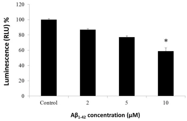
2.3.1 Effect of Aβ1-42 on hippocampal neurons viability……….38
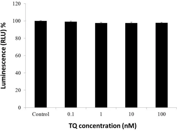
2.3.2 Effect of TQ on hippocampal neurons viability……….……….39
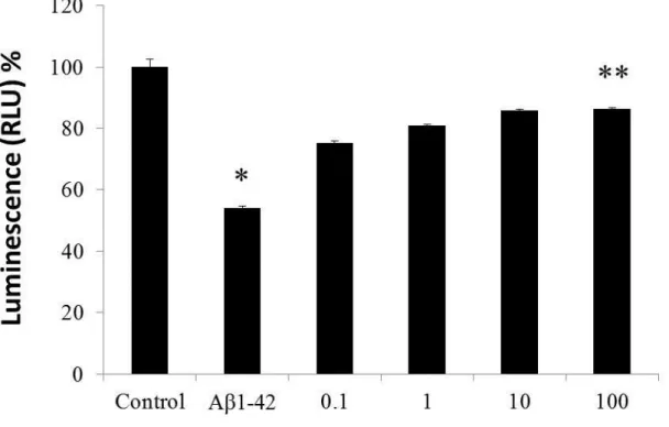
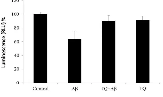
2.3.3 Effect of TQ against Aβ1-42 –induced cell death……….40
2.3.4 Effect of TQ against Aβ1–42-induced caspase 3/7 activities………42
2.3.5 Effect of TQ against Aβ1-42-induced MMP loss………..43
2
2.3.6 Effect of TQ against Aβ1–42-induced GSH level reduction………...44
2.3.7 Effect of TQ against Aβ1-42-induced ROS generation………....45
2.3.8 Effect of TQ against Aβ1-42-induced synapse damage………47
2.3.9 Effect of TQ on Aβ1-42-induced spontaneous firing activity inhibition…50 2.3.10 Effect of TQ on Aβ1–42 aggregation in vitro….………..54
2.4 Discussion………55
3. Inhibitory effect of thymoquinone against alpha synuclein-induced synapse damage in rat primary and hiPSC-derived neurons
3.1 Introduction……….603.2 Materials and methods……….62
3.2.1 Preparation of αSN and P123H-βSN………..……..62
3.2.2 Synaptic protein expression level………..………...62
3.2.3 Synaptic vesicle recycling………..…………..63
3.2.4 Synaptic vesicle recycling (P123H-βSN)………..….………..64
3.2.5 Spontaneous firing activity………..……….66
3.3 Results………..………..…68
3.3.1 Effect of TQ against αSN-induced synaptic protein inhibition………….……..68
3.3.2 Effect of TQ against αSN-induced synaptic vesicle recycling inhibition………73
3.3.3 Effect of TQ against P123H-βSN-induced synaptic vesicle recycling inhibition……...………..………..74
3.3.4 Effect of TQ against αSN-induced spontaneous firing activity inhibition……75
3.4 Discussion………77
4. Summary………..79
3
Chapter 1 Introduction
1.1 Alzheimer’s disease (AD)
Progressive mental deterioration in old age has been recognized and described throughout history. However, it was not until 1906 when Dr. Alois Alzheimer, a German physician, specifically identified a collection of brain cell abnormalities as a disease.
One of Dr. Alzheimer’s patients died after years of severe memory problems, confusion and difficulty understanding questions. Upon her death, while performing a brain autopsy, the doctor noted dense deposits surrounding the nerve cells (neuritic plaques).
Inside the nerve cells he observed twisted bands of fibers (neurofibrillary tangles).
Today, this degenerative brain disorder bears his name, and when found during an autopsy, these plaques and tangles mean a definite diagnosis of Alzheimer's disease (AD). Since its discovery more than 100 years ago, there have been many scientific breakthroughs in AD research.
In the 1960s, scientists discovered a link between cognitive decline and the number of plaques and tangles in the brain. The medical community then formally recognized Alzheimer’s as a disease and not a normal part of aging. In the 1970s, scientists made great strides in understanding the human body as a whole, and AD emerged as a significant area of research interest. This increased attention led in the 1990s to important discoveries and a better understanding of complex nerve cells in the brains of AD patients. More research was done on AD susceptibility genes, and several drugs were approved to treat the cognitive symptoms of the disease. Specific genes related to both the early-onset and late-onset forms of AD have been identified, but genetic risk factors alone do not fully explain its causes, so researchers are actively exploring environment and lifestyle to learn what role they might play in the development of this disease.
1.2 Amyloid beta peptide (Aβ)
One of the hallmarks of AD is the accumulation of amyloid plaques between
neurons in the brain. Amyloid is a general term for protein fragments that the body
produces normally. Amyloid beta (Aβ) is a peptide of 36–43 amino acids that is
4
processed from the amyloid precursor protein (APP). Aβ is the main component of amyloid plaques found in the brains of patients with AD. In a healthy brain, these protein fragments are broken down and eliminated. In AD, the fragments accumulate to form hard, insoluble plaques.
1.3 Amyloid beta formation and neurotoxicity
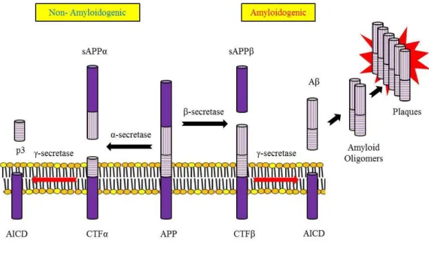
APP is an integral membrane protein expressed in many tissues and concentrated in the synapses of neurons. Its primary function is not known, though it has been implicated as a regulator of synapse formation [1], neural plasticity [2] and iron export [3]. APP can be processed in different ways by different sets of enzymes one pathway leads to amyloid plaque formation (amyloidogenic), while another does not (non-amyloidogenic). Usually about 90% of APP enters the non-amyloidogenic pathway, and 10% the amyloidogenic one, but these ratios can change due to mutations, environmental factors, as well as the age of the individual. Cleavage products from both these pathways may play important roles in neural development and function (Fig.1-1).
In the non-plaque-forming pathway, APP is cleaved first by α-secretase a proteases in the secretase family to yield a soluble N-terminal fragment (sAPPα) and a C-terminal fragment (CTFα). sAPPα may be involved in the enhancement of synaptogenesis, neurite outgrowth and neuronal survival, and are considered to be neuroprotective. CTFα is retained in the membrane, where it is acted upon by presenilin-containing γ-secretase to yield a soluble N-terminal fragment (p3) and a membrane-bound C-terminal fragment (AICD, or APP intracellular domain). AICD may be involved in nuclear signalling via transcriptional regulation as well as axonal transport through its ability to associate with a host of different proteins.
In the plaque-forming pathway, APP is cleaved first by a different enzyme, β-secretase (a transmembrane aspartic protease), yielding a soluble N-terminal fragment (sAPPβ) and a membrane-bound C-terminal fragment (CTFβ). This cut is made closer to the N-terminal end of APP than with α-secretase, making CTFβ longer than CTFα.
CTFβ is then acted upon by γ-secretase (as occurred in the previous pathway), yielding
a membrane-bound C-terminal fragment (AICD) the same as before, and a soluble
N-terminal fragment Aβ that is longer than p3. The γ-secretase can generate a number
of isoforms of 36-43 amino acid residues in length. The most common isoforms are
Aβ
40and Aβ
42; The Aβ
40form is the more common of the two, but Aβ
42is the more
fibrillogenic and is thus associated with disease states. Mutations in APP associated with
early-onset Alzheimer's have been noted to increase the relative production of Aβ
42, and
5
thus one suggested avenue of Alzheimer's therapy involves modulating the activity of β and γ secretases to produce mainly Aβ
40[4]. γ-Secretase is a large multi-subunit complex whose components have not yet been fully characterized, but includes presenilin, whose gene has been identified as a major genetic risk factor for Alzheimer's [5]. The progressive accumulation of Aβ
1-42(Fig.1-2) aggregates is widely believed to be fundamental to the initial development of neurodegenerative pathology and to trigger a cascade of events such as, lipid peroxidation in brain cell membranes [6], mitochondrial dysfunction through free radical generation, and resultant oxidative stress [7], synaptic plasticity impairment [8], and chronic inflammatory reactions [9].
Importantly, Aβ oligomers have been demonstrated to potentiate synaptic loss and
inhibit hippocampal Long-term potentiation (LTP) [10]. It has been also suggested that
neurotoxic amyloid aggregates may lead to synaptic dysfunction and eventually
synaptic loss [11], leading finally to neuronal death [12].
6
Figure 1-1. APP Processing: α-secretase and γ-secretase produce non-plaque forming p3, while β-secretase and γ-secretase produce amyloid plaque-forming Aβ
Figure 1-2. Primary sequence of Aβ
1-427
1.4 Alpha Synuclein (αSN)
Synucleins (SNs) are small (113-143 amino acids) proteins, highly conserved among vertebrates [13], with up to 87% homology between the human and the zebra finch sequence [14-17]. Three different genes, α, β and γ, encode αSN, βSN, and γSN, respectively. SNs were first described in 1988 when a novel protein was isolated in the electrical organ of
Torpedo Californicaand in rat brain [14]. The protein was named synuclein because of its synaptic and nuclear localization. A second intrinsic peptide component of human AD amyloid, named ‘non-amyloid-beta-component precursor protein’ (NACP), belonging to the SN family, was reported in 1993 [15]. Later, in 1995, data were presented documenting the presence of αSN in the telencephalic song control circuit of the zebra finch [17]. αSN was subsequently found in Parkinson’s Disease (PD) neuropathologic hallmarks, named Lewy Bodies (LBs) and Lewy neurites, and in cytoplasmic inclusions of patients affected by Dementia with Lewy Bodies (DLBs) and multiple system atrophy [18-25]. αSN-positive-LBs have also been found in LBs variant of AD, familial AD and Down’s syndrome [26, 27]. Because of the presence of αSN deposits, all these neurodegenerative disorders are now grouped under the name of synucleinopathies [28].
αSN has received much attention because of the discovery that a mutation of the
protein is involved in familial PD [29]. αSN is expressed throughout the brain: the
molecular layer of cerebellum, hippocampus, amygdala, retina, neocortex, nucleus
accumbens, striatum and olfactory mucosa. The primary sequence of αSN consists of
140 residues. The mechanism(s) by which αSN plays a role in the pathogenesis of
synucleinopathies is (are) not yet clear. This is due, at least in part, to the fact that the
physiologic function of αSN still remains unknown. Numerous studies strongly support
the idea that αSN plays a role in neurotransmission. Indeed, the protein is located in
proximity to synaptic vesicles in presynaptic terminals [30] where it co-localizes with
βSN [31]. Interestingly, a small fraction of αSN is associated with vesicular membranes
[32]. Moreover, recent studies, examining structural synaptic changes after treatment of
cultured hippocampal neurons with an antisense oligonucleotide, a manipulation that
reduces αSN expression, have demonstrated a marked selective reduction of the
presynaptic vesicle distal pool size in treated neurons [33, 34]. These findings are
interpreted as support for a role of αSN in interaction and regulation of vesicles storage
and turnover that might be compromised in synucleinopathies [33, 34].
8
1.5 Human induced pluripotent stem cells (hiPSC)
Human Induced Pluripotent Stem Cells (hiPSC) are a type of pluripotent stem cell that can be generated directly from adult cells. The iPSC technology was pioneered by Shinya Yamanaka’s lab in Kyoto, Japan, who showed in 2006 that the introduction of four specific transcription factors could convert adult cells into pluripotent stem cells [35]. He was awarded the 2012 Nobel Prize along with Sir John Gurdon "for the discovery that mature cells can be reprogrammed to become pluripotent.
hiPSC hold great promise in the field of regenerative medicine. Because they can propagate indefinitely, as well as give rise to every other cell type in the body (such as neurons, heart, pancreatic, and liver cells), they represent a single source of cells that could be used to replace those lost to damage or disease. An important subject of future research in this area involves the use of human iPSC-based strategies to study the morphology and electrophysiological behavior of neurons from patients with synucleinopathies and compare them with those of healthy cells. There are many potential causes for the failed translation of drug discovery from levels of molecular and animal models to human therapeutics. In particular, the success of preclinical phases of drug development is based on animal models [36]. Furthermore, less than 10% of the compounds that enter the clinical phase of testing reach the stage of market approval;
the estimated cost of the entire drug development process is 1.2–1.7 billion US$ per drug [36-38].
Drug discovery/development platforms using hiPSC- based disease models could
be useful in filling the gap between animal models and clinical trials. Cell lines and
animal models contribute to the exploration of disease mechanisms and drug
development for various diseases. However, the animal models do not always
demonstrate the same phenotypes as those seen in humans [39]. For instance, in mice
the type and/or distribution of cardiac ion channels are different from those in humans,
demonstrating a relatively shorter duration of action potential and higher heart rate (600
bpm) [40]. An in vitro analysis of human cardiomyocytes is therefore critical to an
understanding of the mechanism of genetics-related arrhythmias (irregular heartbeat) in
humans [40]. Also, compounds that demonstrate significant benefit in animal models
may fail to show effectiveness in clinical trials in humans [39, 41, 42]. The use of
transgenic mice of mutant superoxide dismutase (SOD1), a gene found to be associated
with amyotrophic lateral sclerosis (a specific disorder that involves the death of
neurons) [43], enabled the identification of several compounds that relieve the disease
phenotype, including vitamin E and creatine [44-46]. However, when these compounds
9
were tested in humans, no clinical improvements were observed [44-46]. The toxicity of
compounds is sometimes missed in cell lines and animal models because specific
interactions with human biological processes cannot be recapitulated in these systems
[36] Also, the use of animal models for toxicity assays may be ethically problematic, the
animals may be expensive to purchase and maintain, and the process may be difficult to
automate [36]. Clearly, different drug screening models that complement these systems
and represent the human condition with high fidelity are required [47]. hiPSC are
expected to fulfill these requirements and are amenable to the demands of drug
development.
10
1.6 Nigella Sativa
.
Historical and current studies and surveys indicate that the Eastern region of the Mediterranean has been distinguished throughout the generations with a rich inventory of natural medicinal herbs [48]. Among the promising medicinal plants, Nigella Sativa (also known as the black seed or black cumin), a dicotyledon of the Ranunculaceae family, has been used for medicinal purposes for centuries, both as a herb and pressed into oil, in Asia, Middle East and Africa. It has been traditionally used for a variety of conditions and treatments related to respiratory health, stomach and intestinal health, kidney and liver function, circulatory and immune system support, and for general well-being [49-51]. In Islam, it is regarded as one of the greatest forms of healing medicine available. The Islamic prophet Muhammad once stated that the black seed can heal every disease except death. Black cumin seeds were found in the tomb of Egyptian Pharaoh Tutankhamun, who ruled Egypt from 1332 BC to 1323 BC. He was the son of Akhenaten and Nefertiti and his tomb is the most complete Egyptian tomb ever discovered. The Egyptians supposedly put the seeds in his tomb so that he may have excellent health in the afterlife [52].
The black cumin herb goes by many different names. For example, in old Latin it
is called as ‘Panacea’ meaning ‘cure all’ while in Arabic it is termed as ‘Habbat Al
Baraka’ translated as ‘Seeds of blessing’. The plant belongs to the
Ranunculaceaefamily of flowering plants and genus of about 14 species including
Nigella arvensis, Nigella ciliaris, Nigella damascene, Nigella hispanica, Nigella integrifolia, Nigella nigellastrum, Nigella orientalis and Nigella sativa, respectively. Among these, Nigella sativa is the species most exhaustively investigated for therapeutic purposes althoughother species have also been implicated for therapeutic uses [53]. The species grow to
20-30 cm tall, with finely divided leaves wherein the leaf segments are narrowly linear
to threadlike. The flowers are white, yellow, pink, pale blue or pale purple, with 5-10
petals. The fruit is a capsule composed of several united follicles, each containing
numerous seeds. The parts of the plant most commonly used for the therapeutic
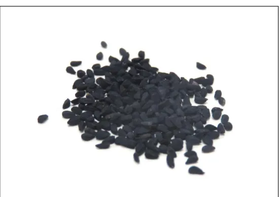
purposes in the “Alternative Medicinal” systems are the seeds (Fig.1-3), which are
contained in an inflated capsule formed from the united follicles containing
considerable amount of oil having pungent and bitter taste. Commonly the seeds are
used primarily as a spice and food preservative. In folk medicinal practices they are
ingested with food or mixed with honey and are primarily used as galactogogues and
anthelmintic agents. The seeds have also been used as diuretics, anti-hypertensive,
muscle relaxants and as immunity enhancers in immune compromised people. There are
11
reports that the oil from the seeds can be used to treat dermatitis topically [54]. Several
beneficial pharmacological effects have been attributed to various crude or purified
components of these seeds including antihistaminic [55], antihypertensive [56],
hypoglycemic [57], antifungal [58], anti-inflammatory [59], along with significant
anti-neoplastic activities [60]. These studies collectively provide early indication that
further development of agents derived from
Nigella sativa seeds could be useful inmodern medicine.
12
Figure 1-3. Nigella Sativa Seeds
13
1.7 Thymoquinone (TQ)
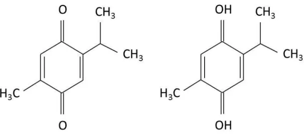
The chemical composition of the black Nigella sativa seed is diverse and contains amino acids, proteins, carbohydrates, fixed and volatile oils, alkaloids, saponins and many other compounds. Thin Layer Chromatography (TLC) screening of the oil samples showed the presence of four main components, viz. thymoquinone, carvacrol, tanethole and 4-terpineol, which demonstrated respectable radical scavenging property.
These four constituents and the essential oil possessed variable antioxidant activity when tested in the 2,2′-diphenyl-p-picrylhydrazyl (DPPH) assay for non-specific hydrogen atom or electron donation. The oil samples showed variable antioxidant activity which was ascribed mainly to the variable composition of these constituents [61]. According to a previous study [62], the bioactive constituents of the volatile oil of black seed (54%) were identified showing that Thymoquinone or, in short, TQ (Fig.1-4) was the main active constituent of volatile oil of the black seed although it is accompanied by other analogous compounds such as Thymol and Thymoquinone dimer named as Dithymoquinone.
TQ has a variety of beneficial properties including analgesic and
anti-inflammatory actions [63, 64], neuroprotection [65], and suppression of oxidative
stress-induced neuropathy [66]. Moreover, it has been reported that TQ prevents
oxidative injury in hepatocytes induced by carbon tetrachloride [67]. TQ also caused
morphological improvements and prevented neurodegeneration against chronic toluene
exposure [68], and showed significant anti-anxiety like activity through possible
modulation of NO and GABA [69], and suppress nuclear factor kappa B (NF-KB)
activation in brain and spinal cord [70]. Additionally, TQ exerts inhibitory effects on the
cell proliferation in many types of cancer cells, including breast and ovarian
adenocarcinoma and on angiogenesis and tumor growth at low dosages by blocking
tumor angiogenesis [71, 72].
14
Figure 1-4. Chemical structure of Thymoquinone and Thymohydroquinone
15
1.8 Outline of this thesis
The present study was designed to investigate first as will be indicated in the
second chapter of this thesis the possible protective effect of TQ against Aβ
1-42-induced
neurotoxicity using rat primary and hiPSC-derived cholinergic neurons. We investigated
cell viability, caspase 3/7 activities, mitochondria membrane potential (MMP),
Glutathione (GSH) levels, reactive oxygen species (ROS) generation, synaptic activity,
spontaneous firing activity, and the effect of TQ on the aggregation state of Aβ
1-42. In
the third chapter of this thesis we investigated the possible protective effect of TQ
against alpha synuclein (αSN)-induced synaptic damage using both rat hippocampal and
hiPSC-derived neurons. We investigated synaptic protein (synaptophysin) levels,
synaptic activity, and spontaneous firing activity. In the fourth chapter of this thesis we
summarized our studies and the suggested mechanism for TQ protective effects.
16
Chapter 2
Inhibitory effect of thymoquinone against amyloid beta-induced neurotoxicity in rat primary and hiPSC-derived cholinergic neurons
2.1 Introduction
AD is the most common form of dementia in the elderly. It is a slowly progressive neurodegenerative disorder of the central nervous system, characterized by profound impairment of cognitive function and memory [73]. The Aβ peptide was initially identified and biochemically characterized in 1984 [74] as a peptide that aggregated and was deposited outside neurons in the brain tissue of Alzheimer’s patients, leading to the formation of neuritic plaques in the AD brain. The presence of these neuritic plaques is the major pathological hallmark of AD. The Aβ peptide, a principal component of these plaques, is thought to play a central role in AD and is regarded as the causative agent in development of the disease. Soon after the identification of Aβ as the major component of amyloid plaques in AD, reports of both toxicity and trophic activity appeared [75]. The toxicity was reported to be related to the aggregation state of Aβ, with aggregation being required for toxicity [76, 77]. Aβ aggregate-mediated toxicity has been documented in vitro and in vivo.
Initial reports in 1994 showed that elevated oxidative stress, one of the early pathological events of AD, was mediated by hydrogen peroxide (H
2O
2) produced through the reduction of metal ions by Aβ peptides [78]. Furthermore, Aβ aggregate-mediated toxicity impairs synaptic function, which leads to the progressive memory loss and cognitive failure associated with AD which has been also well documented. Synaptic dysfunction is triggered by changes in synaptic structure and neurochemicals induced by oligomerized Aβ rather than amyloid plaques [79].
In addition to oxidative damage and synaptic failure, Aβ aggregates can induce
mitochondrial dysfunction, which is another pathological hallmark of AD. The
alteration of synaptic mitochondria, as a result of the buildup of Aβ, may underlie the
synaptic pathology in AD [80]. Reported mitochondrialmal functions as a consequence
of membrane localized Aβ include the inhibition of protein transport into mitochondria,
17
the disruption of the electron transport chain leading to impaired glucose utilization in neurons, and mitochondrial damage due to an increase in ROS production [81].
In recent years, the cholinergic system of neurons has been a main focus of research in aging and neural degradation, specifically as it relates to AD [82]. In addition, it is known that the dysfunction and loss of basal forebrain cholinergic neurons and their cortical projections are among the earliest pathological events in AD [83]. In presenile (early onset), and in the advanced stages of late-onset AD, a severe loss of cortical cholinergic innervation has extensively been documented [84]. The brain of an individual with AD exhibits extracellular plaques of aggregated Aβ, intracellular neurofibrillary tangles that contain hyperphosphorylated tau protein and a profound loss of basal forebrain cholinergic neurons that innervate the hippocampus and the neocortex.
Aβ accumulation may trigger or contribute to the process of this neurodegeneration [85].
It has been hypothesized that Aβ peptides induce neurodegenerative changes at cholinergic terminals [86], while other studies revealed that oligomeric Aβ causes cell death [87], encourage apoptosis by physically piercing the cell membrane, causes neurotoxic cascade and neurodegeneration that leads to AD [88, 89]. As previously reported by many studies, both AD and mild cognitive impairment brains have significantly decreased levels of antioxidant enzymes, making the brain more vulnerable to Aβ induced toxic effects [90]. Oxidative stress is also evident in AD brain by marked levels of protein, lipid, DNA, and RNA oxidation, neuronal dysfunction and death [91, 92]. Consequently, antioxidants have long been considered as an approach to slow down AD progression.
As an established historical and religion-based remedy for a wide range of health
problems,
Nigella sativa is one of the herbal medicines that is being activelyinvestigated and is thus gaining worldwide recognition [93]. The seeds of Nigella sativa
L., commonly known as black seed or black cumin, has been used for medicinalpurposes for centuries, both as herb and pressed into oil, in Asia, Middle East, and
Africa. It has been traditionally used for various conditions and treatments related to
respiratory health, stomach and intestinal health, kidney and liver function, circulatory
and immune system support, and for general well-being [51]. Reviews have reported
Nigella sativa as having antioxidant and neuroprotective effects in addition to manyother therapeutic effects, such as antitumor, immune potentiation, anti-inflammatory,
and antimicrobial [50, 68, 94]. Consuming antioxidant nutrients, such as Nigella sativa,
could be one of the promising health strategies to help prevent the oxidative damage to
cells, particularly in the brain regions, which are related to memory functions [95]. The
seeds contain fixed and essential oils, proteins, alkaloids, and saponin. Much of the
18
biological activity of the seeds has been shown to be due to TQ, the major component of essential oil, which is also present in the fixed oil [49].
TQ is known to be the active principle responsible for many of the seed antioxidant and anti-inflammatory effects [96], which is frequently used in herbal medicine. TQ seems promising due to its many biological effects, which include antioxidant, anti-inflammatory, anticancer, and neuroprotection characteristics [97, 64, 65, 98] that may be beneficial in the management of AD. TQ was reported to counteract the induced oxidative stress in rats’ brain tissue by reducing the levels of peroxidation, and enhancing the activities of enzymatic and non-enzymatic antioxidants, and protects PC12 cells against cytotoxic agents via attenuation of oxidative stress [99, 100]. A study demonstrated that TQ has a protective role against ethanol-induced neuronal apoptosis in primary rat cortical neurons [101], and suppression of oxidative stress-induced neuropathy [66]. TQ also causes morphological improvements and prevents neurodegeneration by chronic toluene exposure [67], shows significant anti-anxiety-like activity through possible modulation of nitric oxide
(NO) and brain GABA content [68], and suppresses nuclear factor kappa B (NF-KB) activation in brain and spinal cord [69].
Searching for new compounds that slow down or stop the progression of Aβ is an important target of research in central nervous system drug development. And identifying a way to reduce the Aβ induced neurotoxicity would be beneficial for AD treatment. Although the precise mechanisms underlying Aβ induced neurotoxicity remains obscure, several lines of evidence suggest that they are associated with oxidative stress-dependent apoptosis [102]. Therefore, antioxidants are considered as potential drug candidates for amyloid hypothesis based therapy.
Accordingly, this study is designed to evaluate the possible protective effects of TQ against Aβ
1-42induced neuronal toxicity using embryonic rat primary hippocampal and cortical neurons and hiPSC-derived cholinergic neurons. First, cell viability was assessed using the CellTiter-Glo assay. Then, we evaluated apoptosis measuring caspase-3, -7 activations. Next, we measured mitochondrial membrane potential (MMP) using Rhodamine 123, glutathione (GSH) level using GSH-Glo Glutathione assay, the production of ROS using DCFH-DA and ROS-Glo H
2O
2assays . Because synaptic degeneration is one of the mechanisms underlying the neurotoxicity of Aβ, we investigated the role of TQ and Aβ
1-42on synaptic vesicle recycling using FM1–43 dye, and on spontaneous firing activity using a multielectrode array system. To further clarify our results, we investigated the role of TQ in the cellular defense against Aβ
1-42using a thioflavin T binding assay.
19
2.2 Materials and methods
2.2.1 Aβ
1-42and TQ Preparation
1mM stock solutions of Amyloid β-Protein 1-42 (Peptide Institute, INC), molecular weight (4514.0), was made by dissolving the content of Aβ1-42 in the vial in 120μl Dimethyl Sulphoxide (DMSO, Wako) and store it at -20°C. 10mM stock solution of Thymoquinone (Sigma-Aldrich), molecular weight (164.20), was prepared by dissolving 1.642 mg of TQ in a solution of 0.3 ml DMSO and 0.7 ml culture medium.
Final concentrations of TQ were prepared in culture medium. Cultures were treated in
every experiment with a freshly prepared TQ.
20
2.2.2 Neurons viability
Brain atrophy caused by neuronal loss is a prominent pathological feature of AD.
Aβ, the major component of senile plaques, is considered to play a central role in neuronal cell death. In addition the removal of the toxic Aβ, direct suppression of neuronal loss is an essential part of AD treatment; however, no such neuroprotective therapies have been developed. Excess amount of Aβ evokes multiple cytotoxic mechanisms, involving increase of the intracellular Ca
2+level, oxidative stress, and receptor-mediated activation of cell-death cascades. Such diversity in cytotoxic mechanisms induced by Aβ clearly indicates a complex nature of the AD-related neuronal cell death [103].
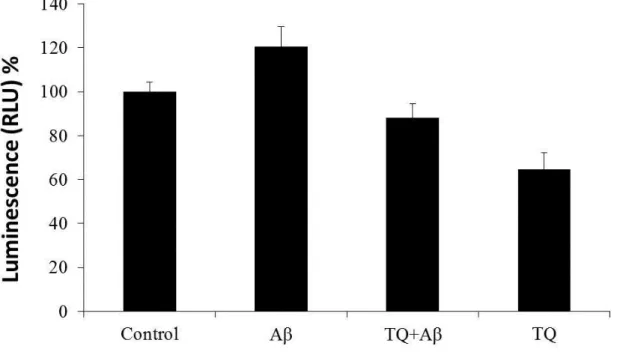
In order to investigate the protective effect of TQ against Aβ
1-42induced neurotoxicity, we first investigated its effect against Aβ
1-42induced cell death. We used CellTiter-Glo Luminescent Cell Viability Assay, a homogenous method for determining the number of viable cells in a culture based on quantitation of ATP, which signals the presence of metabolically active cells. The CellTiter-Glo Luminescent Cell Viability assay uses luciferase as the detection enzyme because of the absence of endogenous luciferase activity in mammalian cells. The Luciferase used in the CellTiter-Glo Luminescent Cell Viability Assay kit generates a stable, glow-type signal that has a half-life of greater than four hours. Luciferase enzyme requires ATP in order to generate light. Metabolically active cells produce ATP as energy for respiration and other vital processes. After an equal volume of CellTiter-Glo Reagent is added to the cell culture, luminescence is measured. Light signal is proportional to the amount of ATP present which correlates with the number of viable cells present. The CellTiter-Glo assay was used to determine the effect of various concentrations of Aβ
1-42on the cell viability and the protective effect of TQ on the cell death.
CellTiter-Glo (Promega); reagent was prepared by transferring the contents of one
bottle of CellTiter-Glo Buffer to one bottle of CellTiter-Glo Substrate, mixed by
inversion until the substrate is thoroughly dissolved. Reconstituted reagent was used on
the same day it is prepared or stored at -20°C. Primary embryonic Wistar rat
hippocampal neurons were prepared and harvested on a Poly-
D-lysine (PDL, 0.1 mg/ml
in deionized water, MW150,000 - 300,000, Sigma–Aldrich) coated 96-microwell plate
(Nunc) in Neurobasal Medium, density at 2×10
4cell/well. Plates were incubated at
37°C, 5% CO
2. Aβ
1-42was applied to cultures on 13 DIV in a series of concentrations (2,
5, 10 μM) and cell survival was assessed 72 hours later. To investigate the effect of TQ
on the survival rate of hippocampal neurons, cultures were treated with TQ (0.1, 1,
21
10,100 nM) on culture day 13 for 72 hours. Finally, to investigate the neuroprotective potential of TQ against Aβ
1-42toxicity, cultures were treated with TQ and Aβ
1-42simultaneously for 72 hours.
hiPSC-derived cholinergic neurons (RCESDA001, ReproCell) were cultured at a
density of 3.0× 10
4cell/well in 96-microwell plates, treated with Aβ
1–42(5 μM) with or
without TQ (100 nM) for 48 hours on culture day 13. On experiment day, the plate and
contents were equilibrated to room temperature for approximately 30 minutes. An equal
volume to the culture medium of CellTiter-Glo Reagent was added, and mixed gently
for 2 minutes, then incubated at room temperature for 10 minutes to allow luminescent
signal to stabilize. Microplate reader (TECAN) was used for measuring Relative Light
Units (RLU) values.
22
2.2.3 Caspase-3 and -7 activities
Caspases (cysteinyl aspartate-specific proteases) are a family of cysteine proteases that play essential roles in apoptosis (programmed cell death), necrosis, and inflammation. Caspases are essential in cells for apoptosis in development and most other stages of adult life, and have been termed "executioner" proteins for their roles in the cell. Some caspases are also required in the immune system for the maturation of lymphocytes. Failure of apoptosis is one of the main contributions to tumor development and autoimmune diseases; this, coupled with the unwanted apoptosis that occurs with ischemia or AD, has stimulated interest in caspases as potential therapeutic targets since they were discovered in the mid-1990s. Caspases have been broadly classified by their known roles in apoptosis (caspase-3, -6, -7, -8, and -9 in mammals), and in inflammation (caspase-1, -4, -5, -12 in humans and caspase- 1, -11, and -12 in mice). Caspases involved in apoptosis have been sub classified by their mechanism of action and are either initiator caspases (caspase- 8 and -9) or executioner caspases (caspase-3, -6, and -7) [104].
Caspase-3 is a caspase protein that plays a central role in the execution-phase of cell apoptosis. A previous study revealed that comparing to other proteinases;
caspase-3 is the primary activator of apoptotic DNA fragmentation [105] which is the best recognized biochemical event of apoptosis [106, 107]. This result was consistent with another study that demonstrated that caspases-3 and -7, but not caspases-6 and -8, inactivate recombinant DFF45, releasing active DFF40 which lead to chromatin condensation and DNA fragmentation and therefore apoptosis [108]. Neuronal death in a variety of neurodegenerative diseases, including AD, has been associated with deregulated caspase activation [109]. However, several lines of evidence suggest that the role of caspases in AD may involve more than just action as cellular executioners driven by upstream disease processes. Caspase-mediated cleavage of APP has been reported [110], as has Aβ peptide induced apoptosis by the activation of caspase-3 [111, 112].
Under normal circumstances, caspases recognize tetra-peptide sequences on their substrates and hydrolyze peptide bonds after aspartic acid residues. Caspase 3 and caspase 7 share similar substrate specificity by recognizing tetra-peptide sequence DEVDG (Asp-Glu-Val-Asp-Gly) with cleavage occurring on the carboxy side of the second aspartic acid residue (between D and G) [113, 114]. The C-terminal Asp is absolutely required while variations at other three positions can be tolerated [115].
Caspase substrate specificity has been widely used in caspase based inhibitor and drug
23
design [116].
In this experiment we investigated the effect of Aβ
1-42and TQ on caspase-3 and -7 activates using Caspase-Glo 3/7 Assay (Promega), a homogeneous, luminescent assay that measures caspase-3 and -7 activities. The assay provides a luminogenic caspase-3/7 substrate, which contains the tetra-peptide sequence “DEVD”, which is preferentially recognized by caspase-3 and -7 in a reagent optimized for caspase activity, luciferase activity and cell lysis. Adding a single Caspase-Glo 3/7 Reagent results in cell lysis, followed by caspase cleavage of the substrate and generation of a “glow-type”
luminescent signal, produced by luciferase. Luminescence is proportional to the amount of caspase activity present.
hiPSC-derived cholinergic neurons were treated with Aβ
1–42(5 μM) with or
without TQ (100 nM) for 48 hours on culture day 13. On the day of the experiment,
Caspase-Glo3/7 Buffer and lyophilized Caspase-Glo 3/7 Substrate were equilibrated to
room temperature. The contents of the Caspase-Glo 3/7 Buffer bottle were transferred
into the amber bottle containing Caspase-Glo 3/7 Substrate, mixed by swirling or
inverting the contents until the substrate was thoroughly dissolved to form the
Caspase-Glo 3/7 Reagent. Cell culture plate was removed from the incubator and plate
was allowed to equilibrate to room temperature for 10 minutes. 100 μl from
Caspase-Glo 3/7 Reagent was added to each well. The contents of wells were gently
mixed using a plate shaker at 300–500rpm for 30 seconds, and then incubated at room
temperature for 1 hour. Luminescence was measured using a microplate reader
(TECAN).
24
2.2.4 Mitochondrial membrane potential
Biochemical and morphological alterations of mitochondria may play an important role in the pathogenesis of AD. Particularly, mitochondrial dysfunction is a hallmark of Aβ-induced neuronal toxicity in AD. Studies of postmortem brains from AD patients and transgenic mouse models of AD suggest that oxidative damage, induced by Aβ, is associated with mitochondria early in AD progression [81]. Evidence from AD postmortem brain as well as cellular and animal AD models showed that Aβ triggers mitochondrial dysfunction through a number of pathways such as impairment of oxidative phosphorylation, elevation of ROS production, alteration of mitochondrial dynamics, and interaction with mitochondrial proteins [117]. A decline in protein import seems to precede increased ROS and decreased MMP suggesting a gradual failure of mitochondria [117]. Moreover, Aβ interacts with mitochondrial matrix components inducing an improper mitochondrial complex function leads to a decreased MMP of the organelle [118] and impairing ATP formation [119]. Previous data demonstrated mitochondrial dysfunction through reduction of MMP in a novel triple transgenic AD mouse model [120]. Cationic probes appear to exhibit a potential-dependent interaction with the native mitochondria of living cells and may reflect intercellular variations in MMP based on variations in the intensity of mitochondria-associated fluorescence [121].
Rhodamine123 (Rh123) is a cationic cell-membrane permeable fluorescent dye that selectively localizes in mitochondria of viable cells. Rh123 also is positively charged at physiological pH and does not appear to have cytotoxic effects. In screening a number of Rhodamine compounds it has become apparent that only those that are positively charged at physiological pH-namely, 123, 6G, and 3B-are able to stain mitochondria specifically whereas uncharged Rhodamines (such as B, 19, 110, and 116) and the negatively charged compound fluorescein do not. These results suggest that an attraction of cationic Rhodamine molecules by the relatively high negative electric potential across the mitochondrial membrane may be the basis for the selective staining of mitochondria by Rh123 in living cells [56].
Therefore, in this study we used Rh123 to investigate the effect of Aβ
1-42and TQ
on MMP in hippocampal neurons. In this experiment primary hippocampal neurons
were cultured on a PDL coated 96-microwell plate (density at 2×10
4cell/well) for 13
days in MACS Neuro Medium, and incubated at 37°C, 5% CO
2. Rhodamine123
(Sigma-Aldrich), molecular weight (380), stock solution of (1 μg/ml) was made by
dissolving the whole content into 400 μl DMSO, covered with aluminum foil and kept
25
in -20°C. On the experiment day final (1 nM/ml) solution was made in Phosphate buffer
saline (PBS). Cultures were treated with Aβ
1-42(10 μM) and TQ (100 nM) on culture13
DIV for 72 hours. On experiment day, after the medium was removed, the cells were
incubated with Rh123 (1 nM) by adding 100 μl in each well for 15 minutes at 37°C, and
then the cells were washed with PBS to remove the extracellular Rh123. The cells were
then suspended in PBS. The fluorescence intensity (relative fluorescence unit) was
monitored using TECAN microplate reader with excitation at 485 nm and emission at
535 nm.
26
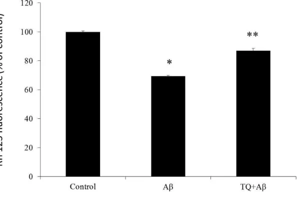
2.2.5 Glutathione levels
GSH (L-gamma-glutamyl-L-cysteinylglycine) is a tripeptide present in large quantities in all mammal cells and in small amounts extracellularly [122], being mainly located in the cytosol, mitochondria, and endoplasmic reticulum [123]. It plays a very important role in many biological processes involved in organism homeostasis, most notably, in neutralizing the free radicals that produce ROS (due to its great antioxidant activity) [124-127], since oxidation is a basic process in the genesis of neurodegenerative disorders [128].Glutathione exists in the thiol-reduced (GSH) and disulfide-oxidized (GSSG) forms [129]. GSH is the most important component of the antioxidant mechanism of the brain [130]. An increase in cellular GSH concentration makes the neurons more resistant to cytotoxic injuries [131-133]. The progressive decrease of GSH levels resulting from aging and related illnesses is of great interest for investigators [134]. The decrease found with aging is linked to an increase of ROS [135]. There is scientific evidence of a relationship between aging and a number of neurodegenerative processes due to the excessive production of free radicals and the imbalance between the oxidant species and antioxidant defenses [136, 137]. GSH levels are decreased in diseases with oxidative stress -including AD- and with age [138].
Oxidative stress, the imbalance between antioxidants and ROS, has been increasingly implicated as an important causative factor in various neurodegenerative diseases including AD [139].
Aβ has been shown to be a source of free radical oxidative stress that may lead to
neurodegeneration. Aβ, perhaps in concert with bound redox metal ions, initiates free
radical processes resulting in protein oxidation, lipid peroxidation, ROS formation,
cellular dysfunction leading to calcium ion accumulation and subsequent neuronal death
[140]. A previous study showed the potential role of free radical toxicity and the
damage of cholinergic neurons in the Aβ-treated mice as there was an increased lipid
peroxidation by elevated malondialdehyde – a reactive species and a marker of
oxidative stress- and decrease of GSH levels [141]. Neuronal cell dysfunction and
oxidative cell death caused by Aβ contribute to the pathogenesis of AD [142] According
to the oxidative stress hypothesis of AD, the Aβ inserts into the neuronal membrane
bilayer and generates oxygen-dependent free radicals and then causes the lipid
peroxidation and protein oxidation. The loss of membrane integrity leads to cellular
dysfunction, such as loss of Ca
2+homeostasis, disruption of signal pathways and
activation of nuclear transcription factors and apoptotic pathways. The neuronal death is
the ultimate consequence of these cellular dysfunctions [143].
27
In this study we investigated the effect of Aβ
1-42and TQ on oxidative stress by assessing GSH levels in hiPSC-derived cholinergic neurons. Oxidative stress was assessed through measurement of the GSH using the GSH-Glo
TMGlutathione assay (Promega). The GSH-Glo™ Glutathione assay is a luminescence-based assay for the detection and quantification of GSH. The assay is based on the conversion of a luciferin derivative into luciferin in the presence of GSH, catalyzed by glutathione S-transferase (GST). The signal generated in a coupled reaction with firefly luciferase is proportional to the amount of GSH present in the sample.
In this experiment hiPSC-derived cholinergic neurons were treated with Aβ
1–42(5
μM) with or without TQ (100 nM) for 48 hours on culture day 13. On experiment day
the culture medium was removed from the wells and 100 μM form the prepared
GSH-Glo reagent was added to each well and then, the contents were mixed briefly
using a plate shaker and incubated for 30 minutes at room temperature. Then, 100 μM
of reconstituted luciferin detection reagent was added to each well. The contents were
mixed briefly using a plate shaker. Plate was incubated for 15 minutes and
luminescence signals were measured using a microplate reader (TECAN).
28
2.2.6 ROS generation
AD brain is under extensive oxidative stress as measured by protein oxidation and lipid peroxidation. Aβ
1-42has been shown to induce protein oxidation and lipid peroxidation in vitro and in vivo [144]. A study demonstrated that both oligomers and fibrils induced ROS production inside the cells [145]. Moreover, the incubation of cortical neurons with low concentrations of soluble Aβ
1-40results in an early time-dependent increase in the ROS production, preceding both microtubule perturbation and caspase activation [146]. Additionally, increasing data suggest that age-related oxidative stress contributes to degenerative changes in basal forebrain cholinergic systems as levels of Aβ increased [147]. It has been reported that Aβ impairs mitochondrial redox activity and increases the generation of ROS [148].
Several studies also suggest that Aβ-induced oxidative stress leads to apoptotic neuronal cell death that can be inhibited by antioxidants [149]. Taken together, these findings support the central role of Aβ
1-42in the pathogenesis of AD as a mediator of oxidative stress.
Numerous studies demonstrated that TQ protect against oxidative stress through its antioxidant properties. Treatment with TQ increased resistance to oxidative stress and proved to be beneficial in restoring declined superoxide dismutases (SOD) and catalase due to ischemia insult. Moreover, TQ significantly improved antioxidant status and reduced lipid peroxidation in rat hippocampus [150]. Furthermore, TQ have inhibitory effects against lipid peroxidation process during cerebral ischemia-reperfusion injury in rat hippocampus [151]. In addition, TQ protected liver and kidney tissues of diabetic rats from peroxidative changes through increase in the levels of antioxidant enzymes and low molecular weight antioxidants in TQ treated diabetic rats [152]. Additionally, TQ supplementation prevents the development of diethylnitrosamine -induced initiation of liver cancer by decreasing oxidative stress and preserving both the activity and mRNA expression of antioxidant enzymes [153].
To examine whether the protective effect of TQ on the toxicity of Aβ
1-42is
mediated by antioxidant ability, first the level of intracellular ROS was determined in
rat cortical neurons by the change of the fluorescent probe Dichlorofluorescin diacetate
(DCFH-DA). When apply to intact cells, DCFH-DA readily diffuses through the cell
membrane and is hydrolyzed enzymatically by intracellular esterases to non-fluorescent
DCFH. In the presence of ROS, DCFH is oxidized to highly fluorescent
dichlorofluorescin (DCF). DCF Fluorescent intensity is proportional to the amount of
ROS formed intracellulary.
29
In this experiment primary cortical neurons were cultured in a PDL coated 96-microwell plate (density at 2×10
4cell/well) for 13 days in MACS Neuro Medium, incubated at 37°C, 5% CO
2. Dichlorofluorescin diacetate (DCFH-DA; Sigma-Aldrich, molecular weight: 487.29) was prepared by dissolving 0.0097 g in 2ml DMSO to make a 100 μM stock solution. Cultures were treated with Aβ
1-42(10 μM) and TQ (100 nM) on culture day 13. After the medium was removed, the cells were incubated with 100 μM DCFH-DA for 30 minutes at 37°C, and the cells were washed with PBS to remove the extracellular DCFH–DA. The cells were then suspended in PBS and the fluorescence signals were determined using a TECAN microplate reader at the excitation wavelength of 485 nm and emission wavelength of 535 nm [154].
Next, the effect of Aβ
1-42and TQ on intracellular ROS level was determined in hiPSC-derived cholinergic neurons by ROS
TMH
2O
2assay (Promega). H
2O
2is a reactive oxygen species that is measured in cells as a marker of oxidative stress. It is also measured as a marker of enzyme activities that either consume or produce H
2O
2. It is desirable to screen chemical compounds for their capacity to alter H
2O
2levels in cultured cells or for their effects on H
2O
2levels in enzyme reactions. The assay determines ROS level by measuring H
2O
2concentration. An H
2O
2Substrate is employed that reacts directly with H
2O
2to generate a luciferin precursor. Upon addition of ROS-Glo™ Detection Reagent containing Ultra-Glo™ Recombinant Luciferase and D-Cysteine, the precursor is converted to luciferin by the D-Cysteine, and the produced luciferin reacts with Ultra-Glo™ Recombinant Luciferase to generate a luminescent signal that is proportional to H
2O
2concentration.
hiPSC-derived cholinergic neurons were treated with Aβ
1–42(5 μM) with or without TQ (100 nM) for 48hours on culture day 13. After 42 hours of treatment, H
2O
2Substrate solution was prepared and added to the cells. Then, the plate was incubated at 37 °C for 6 hours. 100 μl of ROS-Glo™ Detection Solution was added to each well.
The plate was incubated for 20 minutes at room temperature. Relative luminescence
units were recorded using a microplate reader (TECAN).
30
2.2.7 Synaptic vesicles recycling
Communication between neurons involves the fusion of a subpopulation of synaptic vesicles with the cell membrane at a specialized region of the nerve terminal called the active zone or release site, thereby releasing their contents (exocytosis or synaptic vesicle fusion). The released synaptic vesicles are then retrieved from the plasma membrane (endocytosis or synaptic vesicle retrieval), refilled with fresh neurotransmitters, and trafficked back to rejoin the existing pool of 200–500 vesicles to wait to do it all again. The whole process is often called synaptic vesicle recycling.
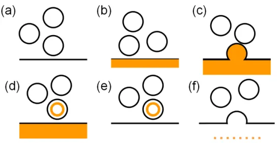
Styryl dyes are typically used in the same manner to investigate synaptic vesicle recycling [155]. When incubated with neuronal preparations, the dyes insert into the outer leaflet of the plasma membrane and are internalized during vesicle retrieval after stimulation of exocytosis. The remaining external dye can be washed quickly away, leaving fluorescently labeled synaptic vesicles inside the nerve terminals (Fig.2-1).
There is a range of styryl dyes available. The more hydrophilic probes FM1-43 (Fig.2-2) and FM2-10 are most widely used, as they possess a short half-time (t1/2) in membrane, allowing non-internalized dye to be washed off neurons quickly. This is an important consideration for reliable quantification of exocytosis because the dye has to dissociate fully from the vesicle membrane upon fusion. Styryl dyes allow visualization of the fusion of synaptic vesicles with the plasma membrane in nerve terminals by fluorescent imaging of regions loaded with previously accumulated dye, followed by monitoring of a stimulated dye release. Styryl dyes can also reveal a locus of action of proteins or drugs in synaptic vesicle recycling, often at steps previously assumed to be unaffected. For example, the addition of Aβ
1-42oligomers affected αSN protein–induced inhibition of synaptic vesicle recycling. Thus, pre-mixing αSN with Aβ
1-42oligomers enhanced αSN -induced inhibition of FM1-43 uptake into synapses [156]. Also the exocytosis of FM1-43 was significantly reduced by chronic application (24h) of 500nM Aβ [157].
Therefore, using FM1-43 dye, we investigated the protective effect of TQ against Aβ
1-42induced synaptic damage. In this experiment, primary cortical neurons were cultured on a PDL coated 24-microwell plate (IWAKI) in Neurobasal medium, density at 3.36×10
5cell/well, incubated at 37°C, 5% CO
2. On culture day 13 cortical neurons were treated with Aβ
1-42(2 μM) only or with TQ (100 nM) simultaneously for 72 hours.
FM1-43 (Molecular Probes) was prepared by dissolving 100μg from the dye in 100μl
methanol and stored at -20°C. Artificial cerebrospinal fluid (ACSF) is commonly used
when sampling from brain interstitial fluid. This solution closely matches the electrolyte
31
concentrations of CSF. It is prepared from high purity water and analytical grade reagents: NaCl 126 mM, KCl 90 mM, KH
2PO
41.3 mM, Glucose 10 mM, NaHCO
32.6 mM, MgSO
41.4 mM, CaCl
24.2 mM. On experiment day, HEPES 10 mM was added to prepared ACSF solution; pH was adjusted to 7.4, and then kept at 4°C. ACSF and FM1-43 (1 μg/ml) solution was made by adding 1 μl from the dye to each 1ml of ACSF and warmed in water bath at 37°C. Culture medium was removed from each well and treated cultures were incubated at 37°C with ASCF/FM1-43 solution for 10 minutes, washed 5 times in ice cold PBS and solubilised in methanol at 1×10
6neurons/ml.
Soluble extracts were transferred into black 96-microwell plate. The fluorescence intensity was determined using a TECAN microplate reader at the excitation wavelength of 485 nm and emission wavelength of 612 nm.
hiPSC-derived cholinergic neurons were treated with Aβ
1–42(5 μM) with or
without TQ (100 nM) for 72 hours on culture day 13. On experiment day, the culture
medium was removed from the treated samples and the prepared ACSF/FM1-43
solution added to each well and incubated for 5 minutes at 37°C. The dye solution was
removed from the dish and the samples were washed twice with cold PBS, suspend in
PBS. Fluorescent signal was measured; excitation at 480 nm, emission at 612 nm using
a microplate reader (TECAN).
32
Figure 2-1. Typical FM dye experiment. (a) Synaptic vesicles near the plasma membrane. (b) FM dye is added, binds to the outer membrane, and becomes fluorescent. (c) The preparation is stimulated and a vesicle fuses with the plasma membrane exposing the luminal membrane to the FM dye. (d) The vesicle is endocytosed with FM dye inside. (e) The FM dye is washed out of the bath and labelled vesicle is imaged. (f) The preparation is stimulated again in a dye-free medium and vesicles exocytosis is measured as dye leaves the fusing vesicle.
Figure 2-2. Labeling of synaptic vesicles recycling. Note that the FM1-43 fluorescence is concentrated at sites where neurites cross over and interact,
indicating the sites of active synaptic vesicle recycling.
33
2.2.8 Spontaneous firing activity
Many fields of neuroscience research investigating long term processes share a common problem. Although numerous methods are available to assess parameters like cell vitality, neurite outgrowth or the expression of proteins, long term monitoring of electrophysiological properties is often not possible. Traditional electrophysiological
in-vitro techniques are generally limited to recording periods of a few hours at most.However, by combining substrate integrated microelectrode arrays (MEA) with cell and tissue culture techniques, this limitation can be overcome [158]. Since its introduction in 1972 [159], MEA technology and the related culture methods for electrophysiological cell and tissue assays have been vastly improved. MEA is an electrochemical biosensor developed to detect the action potential in the extracellular microenvironment of cells. On an MEA, a thin metallic film is fabricated between a substrate of glass or silicon and a passivation layer with several electrode sites exposed for sensing the extracellular field potential changes generated by the objective cells (Fig.2-3). When spreading on the microelectrodes, cultured cells adhere to the substrate.
But there is still a minute volume of electrolyte between the cells and the microelectrodes; thus, a solid-liquid interface on the electrode surfaces is formed. The electrochemical properties of the interface are the basis of the sensing mechanism of MEA [160]. Thus, MEA electrodes facilitate non-invasive stimulations and recordings from cells and networks, enabling repeated recordings from the same cells over extended periods of time.
Neurons create ion currents through their membranes when excited, causing a
change in voltage both inside and outside the cell. When recording, the electrodes on
MEA transduce the change in voltage from the environment carried by ions into
currents carried by electrons (electronic currents). When stimulating, electrodes
transduce electronic currents into ionic currents through the media. This triggers the
voltage-gated ion channels on the membranes of the excitable cells, causing the cell to
depolarize and trigger an action potential. MEA have been in use to study multiple
aspects of electrically excitable cells. In particular, MEA have been applied to explore
the pharmacological and toxicological effects of numerous compounds on spontaneous
activity of neuronal and cardiac cell networks. The MEA system enables simultaneous
extracellular recordings from as many as 64 sites in the network in real time, increasing
spatial resolution and thereby providing a robust measure of network activity. Neuronal
network grown on MEA responds to neuroactive compound sensitively with changes in
their native, spontaneous activity patterns. This altered activity is often substance- and
34
concentration-specific. The influence includes direct metabolic effects, specific synaptic effects, transmission effects that stop action potential propagation and generic mebrance effects mediated through non-synaptic Ca
2+or K
+channels or by the generation of new channels [160].
AD patients show alterations in both neuronal network oscillations and the cognitive processes associated to them. Related to this clinical observation, it has been found that Aβ could lead to differential changes in neural network activity (even overexcitation) by forming aggregates with different sizes that produce differential effects on network activity [161]. Aβ was also found to inhibit spontaneous network activity in the olfactory bulb of mice and rats [162]. Using MEA a study demonstrated that at all measured concentrations, Aβ completely abolishes spontaneous spiking activity. The same study showed that MEA can be used to reliably detect functional effects of low doses of Aβ (100 nM) as well as screen for the rescue effect of the Aβ oligomerization inhibitor, curcumin [163]. Moreover, it has been well established that neurons cultivated on MEA form networks where extracellular action potentials or spikes can be monitored noninvasively to quantify the functional effects of neuroactive compounds [164].
Therefore, we used the MEA system (ALPHA MED SCIENTIFIC, Japan) to evaluate the effect of TQ and Aβ
1-42on the action potentials of hippocampal neurons and hiPSC-derived cholinergic neurons. MEA probes (MED-P530A, ALPHA MED SCIENTIFIC, Japan) were PDL-coated for 24 hours at 37°C. After rinsing PDL, the MEA probe was dipped into solid state 2.25% (w/v) agarose (BM Bio, Japan) and spread using a spin-coater (MIKASA CO. LTD) at 2500 rpm for 20 seconds to form a 5μm thick agarose layer. To obtain the best recording results, the portion of agarose layer upon each electrode was etched by spot heating using a 1064-nm infrared laser to form micro chambers [165,166]. Hippocampal neurons were cultured at a density of 1×10
6neurons/mm
2. The MEA probe was placed in sterile-petri dish, and then incubated at 37 °C, 5% CO
2. After 24 hours. On culture day 13, cultures were exposed to A
1–42(2
M) or to A
1–42and TQ (100 nM). Measurements were performed before applying TQ and Aβ
1-42for 24 hours and directly after applying TQ and Aβ
1-42for 72 hours using Mobius software. In every hour the measurements were obtained for 5 minutes and the firing frequency average for the 5 minutes measured was calculated.
hiPSC-derived cholinergic neurons were plated on the MEA probes (MED-P515A,
Alpha MED Scientific) seeded at 1.0 × 10
6cells/cm
2. Recordings were obtained for 10
minutes at four times per day using MED64 software (Alpha MED Scientific). Cultures
were treated with Aβ
1-42(5 μM) with or without TQ (100 nM) on culture day 13. Firing
35
analyses were performed for number of firings, which obtained at 64 electrodes.
36
)LJXUH0LFURHOHFWURGHDUUD\SUREHZLWKPLFURHOHFWURGHV HPEDGHGLQWKHERWWRPRIWKHGLVKUDWKLSSRFDPSDOQHXURQV',9