O O O P P P T T T I I I M M M I I I Z Z Z A A A T T T I I I O O O N N N O O O F F F S S S T T T E E E A A A D D D Y Y Y - -S - S S T T T A A A T T T E E E V V V I I I S S S U U U A A A L L L
R R R E ES E S S P P P O O O N N N S S S E ES E S S
F F F O OR O R R R R R O OB O B BU U U S S S T T T B B B R R R A AI A I I N N N - - - C C C O OM O M MP P P U U U T T T E E E R R R I I I N N N T T T E ER E R R F FA F A AC C C E ES E S S
P P P H H H . .D . D D . . . T T T H H H E E E S S S I I I S S S
Hovagim Bakardjian
A Dissertation
Submitted in partial fulfillment of The requirements for the degree of
Doctor of Philosophy
Department of Electronic and Information Engineering Tokyo University of Agriculture and Technology
2010
A BSTRACT
The emergence of successful Brain-Computer Interfaces (BCI) that can assist the healthy and aid the disabled users, has introduced new limitless possibilities for wider use of brain technology in society. Yet, existing BCI designs are still unable to fulfill such high expectations due to restricted reliability, insufficient usability, and inadequate understanding of the underlying brain mechanisms.
This work aimed to address directly the above problems by presenting and optimizing two new multi-command BCI designs based on Steady-State Visual Evoked Potential (SSVEP) brain responses. SSVEP brain activity is elicited in precise synchrony with observed flickering visual patterns. The main goal of the work in this thesis was to optimize in new ways the information transfer rates and the reliability of multi-command SSVEP- based BCI systems. This was accomplished using two approaches: (1) Stimulus optimization to achieve maximal cortical response (frequency, size, movement, contents) for further use in BCI, and (2) Development of sensitive adaptive algorithms to extract weak target oscillations from ‘noisy’ EEG input signals immediately after SSVEP onset, based on the estimation of single-trial narrowband-energy changes (online) as well as on new phase-locking- and wavelet-energy variability measures (offline).
To understand in detail the properties of the SSVEP stimuli for BCI, EEG experiments were performed with very small checkerboard patterns. Challenging small-sized SSVEP stimuli were introduced to allow more flexible BCI designs through taking up as little visual estate as possible, even at the cost of reduced SSVEP signal strength. The frequency- response curve for these small SSVEP patterns was studied for 32 frequencies between 5 and 84Hz. Understanding the SSVEP frequency characteristics for small checkerboards allowed the pinpoint definition of optimal parameters and limitations of the investigated checkerboard-based SSVEP-BCI paradigm, as well as better control of inter-subject variability. The SSVEP time dynamics for 8, 14 and 28Hz stimulation and three sensor locations (occipital, parieto-temporal and frontal) was investigated in a further series of experiments, aiming to establish the time-delay limitations of the SSVEP brain response as captured by the online BCI. The knowledge acquired from these neurophysiological experiments was utilized for the optimal design of an 8-command SSVEP-based BCI system. A novel dynamic feedback paradigm was created which featured a set of eight small checkerboard patterns assembled in a very tight but simultaneously moving 2-D spatial configuration, fixed around a main object, and controlled by the user’s intent. Due to this
design visual field occlusion was minimized, and long-term fatigue was reduced substantially by scaling down the demands on the visual system, and on the attention to the overall control task. BCI performance was measured and evaluated for three locations on the scalp (occipital, parieto-temporal and frontal), two of which included hairless areas for easy sensor application. In a further set of experiments, another type of SSVEP stimuli was investigated, flickering affective-face video sequences, in order to study the hypothesis that emotional-face content would be able to boost the SSVEP signals. Short happy and angry face videos were presented flickering at 5 different rates (5.6-12Hz) to eight experimental subjects, as well as three control stimuli - the blurred versions of the affective faces, and a reversing checkerboard. In an offline analysis of their basic properties, brain activity changes associated with SSVEP onset were evaluated using two measures - a new measure for phase locking value variability (PLVV) which was sensitive to inherent phase-reset changes in cortex at stimulation onset, and a more classical estimate of the wavelet energy variability. This approach using engaging SSVEP stimulus contents was further applied in a second 8- command SSVEP-BCI design which employed the same band energy evaluation measures as before for comparison. Separate BCI sessions were performed using affective face videos, their blurred versions, and checkerboards as SSVEP stimuli, and the information transfer rates were measured for each experimental condition. The BCI system was designed allow self-paced control of a multi-joint robotic arm, especially suitable for disabled and elderly users.
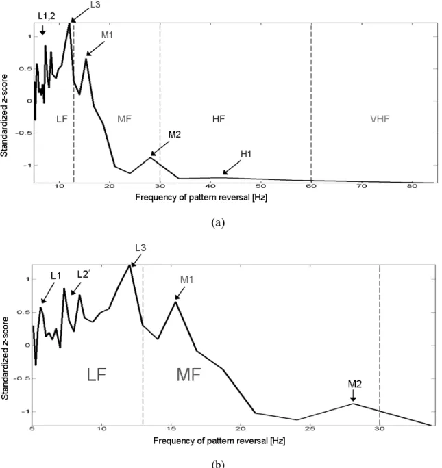
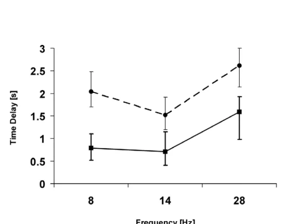
The results of the offline SSVEP signal analysis, normalized for all subjects, showed that the 5.6 – 15.3Hz small-pattern reversal stimulation evoked the strongest responses, peaking at ~12Hz, and exhibiting weaker local maxima at 5.6 Hz, 8 Hz (7.6-8.8 Hz), 12 Hz, 15.3 Hz, and 28 Hz. The shortest SSVEP onset delays were observed starting at ~0.5s for the medium-frequency 14Hz reversal. The first SSVEP maxima, which are captured by online BCI algorithms, were measured offline at 1.5-2.5s after onset. Overall, the long-term SSVEP dynamics was highly non-stationary, especially in the high-frequency range. In the online BCI evaluation experiments, an information transfer rate of 50 bits/min was achieved at occipital sensor locations (mean success rate of 98 %, mean time delay 3.4s). The BCI performance deteriorated with increasing distance from the visual cortex (down to 74% for frontal sensor locations), as also inter-subject variability increased. Notably, in this BCI design, all flickering very small stimuli conformed strictly to international guidelines for prevention of photic- and pattern-induced seizures. Furthermore, novel affective-face video contents in the flicker stimulation evoked significantly stronger SSVEP responses than their corresponding emotionally-neutral blurred versions, as well as the checkerboards. No significant differences in the occipital SSVEP responses were detected due to emotional valence (happiness vs. anger), and there were also no significant differences between the SSVEP responses of individual subjects for all experimental conditions. The frequency response curve measured for affective-face SSVEP stimuli indicated that the SSVEP activity
was strongest for 10Hz flicker when averaged over all stimulus types. Although the PLVV measure was more sensitive than the wavelet energy, an adaptive combination of both phase- lock and narrow-band energy measures would be recommended in future asynchronous SSVEP-BCI implementations for best results in terms of signal sensitivity, elimination of competing brain transients in the critical SSVEP frequency bands, and lower inter-subject variability. Online SSVEP-BCI evaluation of the effect of affective face video sequences for 10 Hz flicker demonstrated that BCI delays were ~20 % shorter for affective-face stimuli than for blurred-face and neutral flicker (2.7s vs. 3.4s; success rate 99% vs. 98%). When using affective-face stimuli, a higher mean BCI information transfer rate (ITR) was achieved (64 bits/min vs. 50 bits/min for neutral stimuli). All subjects reported higher level of attention, interest and motivation for affective-face stimuli, which resulted in higher resistance to fatigue after long-term SSVEP exposure.
Taken together, these findings point to new directions in BCI research by using unobtrusively small SSVEP stimuli, emotional-face video sequences, and new analysis methods to optimize substantially the SSVEP-BCI performance and enhance the user experience. Future BCI designs using a battery of SSVEP extraction algorithms, such as the time-resolved phase synchrony and energy variability measures demonstrated here, may lead to even more robust and reliable SSVEP brain response estimates in unsupervised clinical and home settings, as well as for other challenging brain-technology tasks in service of society.
To my family, for their unconditional love.
A CKNOWLEDGEMENTS
I wish to thank my thesis advisor Dr. Toshihisa Tanaka for his encouragement and scientific discussions.
Also, I would like to express my thanks to Dr. Andrzej Cichocki, the Head of the Laboratory for Advanced Brain Signal processing (LABSP), Brain Science Institute (BSI), RIKEN, where the work described in this thesis was done.
Parts of the BCI analysis system were implemented in a team work with Dr. Pablo Martinez.
T ABLE OF C ONTENTS
PAGE
Abstract . . . ii
Dedication . . . v
Acknowledgements . . . vi
Table of Contents . . . vii
List of Figures . . . xiii
List of Tables . . . xvi
Abbreviations . . . .. . . xvii
1. INTRODUCTION . . . 1
1.1. The Ideal Brain-Computer Interface (BCI). . . 1
1.2. Existing BCI Problems: Why is the Ideal BCI Difficult to Achieve? . . . 3
1.3. Existing BCI Problems: Using SSVEP-BCI . . . 4
1.4. Research Objectives Addressed in This Thesis . . . 7
1.5. Summary of Achievements in this Thesis . . . 9
1.6. Organization of the Thesis . . . 11
2. OVERVIEW OF PREVIOUS RESEARCH . . . 13
2.1. Brain-Computer Interfaces (BCIs) . . . 13
2.1.1. Why are Brain-Computer Interfaces necessary? . . . 13
2.1.2. Brain-Computer Interfaces – Definition, Application and Requirements . . . . 13
2.1.3. BCI Approaches and Types . . . 15
2.1.4. Invasive BCI . . . 20
2.1.5. Non-Invasive BCI . . . 21
2.1.5.1. Motor-imagery based BCI . . . 22
2.1.5.2. Slow cortical potentials based BCI . . . 23
2.1.5.3. P300 potentials based BCI . . . 24
2.1.5.4. Non-motor cognitive tasks (mental tasks) based BCI . . . 24
2.2. Steady-State Visual Potentials (SSVEP) . . . 26
2.2.1. Cortical Frequency Responses to SSVEP Stimulation . . . 26
2.2.2. Time Dynamics of the SSVEP Response . . . 30
2.2.3. Additional Parameters of the SSVEP Response . . . 31
2.2.4. Activation of Brain Areas During the SSVEP Response . . . 32
2.2.5. Photosensitive Epilepsy and Its Prevention . . . 34
2.3. SSVEP-Based BCI Approaches . . . 37
2.3.1. Designs of SSVEP-Based BCI Systems . . . 37
2.3.2. Signal Processing Methods in SSVEP-Based BCI Systems . . . 40
2.3.3. Can effective SSVEP BCI control be achieved without dependence on eye movements? . . . 41
2.4. Affective Processes in the Brain (Emotions) and Connection to SSVEP Research . . . 44
2.4.1. Models of the Emotional Continuum and classification Schemes . . . 44
2.4.2. Emotion-Cognition Interactions . . . 46
2.4.3. Emotions – Cultural Influences and Gender . . . 47
2.4.4. Empathy . . . 49
2.4.5. Neuroanatomical Correlates of Emotion . . . 50
2.4.6. Affective Picture Processing . . . 54
2.4.7. Emergence of Affective SSVEP . . . 55
3. OPTIMIZATION OF SSVEP RESPONSES USING SMALL CHECKERBOARD STIMULI . . . 59
3.1. Objectives– SSVEP Using Small Stimuli . . . 59
3.2. Methods . . . 60
3.2.1. Experimental Subjects . . . 60
3.2.2. EEG Data Acquisition – EEG system . . . 61
3.2.3. SSVEP Stimuli and Display . . . 62
3.2.4. Data Pre-processing - Artifact Rejection Using ICA/BSS . . . 63
3.2.5. Methods - The SSVEP Frequency Response of the Brain . . . 65
3.2.6. Methods - The Time Dynamics of the SSVEP Response . . . 66
3.2.7. Statistical Evaluations . . . 68
3.3. Results . . . 69
3.3.1. SSVEP Frequency Response of the Brain . . . 69
3.3.2. Time Dynamics of the SSVEP Response - SSVEP Onset- and 1st Peak Delays . . . 72
3.3.3. Effects of Sensor Locations on SSVEP Properties . . . 76
3.3.4. Time-Frequency, Area, and Cross-Cumulant Measures for SSVEP Evaluation. . . 79
4. ONLINE MULTI-COMMAND SSVEP-BASED BCI USING SMALL CHECKERBOARD STIMULI . . . 83
4.1. Objectives – SSVEP-Based BCI . . . 83
4.2. Methods . . . 84
4.2.1. Experimental Subjects . . . 84
4.2.2. Basic Structure of the Multi-command SSVEP-Based BCI . . . 84
4.2.3. EEG Data Collection – SSVEP-BCI . . . 85
4.2.4. BCI-SSVEP Stimulation and Real-Time Neurofeedback Module . . . 87
4.2.5. Online BCI Data Analysis Module (Workflow) . . . 91
4.2.6. Three Modes of Operation of the SSVEP-Based BCI System . . . 98
4.2.7. Computation of Information transfer rates (ITR) for Evaluation of the BCI Performance . . . 99
4.3. Results . . . 101
5. BRAIN RESPONSES TO FLICKERING EMOTIONAL FACE VIDEO STIMULI . . . 105
5.1. Objectives . . . 105
5.2. Methods . . . 106
5.2.1. Experimental Subjects . . . 106
5.2.2. EEG Data Acquisition and Collection . . . 106
5.2.3. Stimuli and Display – SSVEP Using Emotional Face Video Stimuli . . . 107
5.2.4. Data Pre-processing and ICA/BSS-based Artifact Removal . . . 110
5.2.5. SSVEP Onset Detection Using Phase-Locking Value Reset and Wavelet Energy Variability Measures . . . 110
5.2.6. Statistical Evaluations . . . 112
5.3. Results . . . 113
6. ENHANCED MULTI-COMMAND SSVEP-BCI
USING FLICKERING EMOTIONAL FACE VIDEO STIMULI . . . 119
6.1. Objectives . . . 119
6.2. Methods . . . .. . . 120
6.2.1. Experimental Subjects . . . 120
6.2.2. EEG Data Acquisition and Collection . . . 120
6.2.3. SSVEP-BCI Stimulation - SSVEP Using Emotional-Face Video Stimuli . . 120
6.2.4. Online SSVEP-BCI Analysis and User-Interface Modules . . . 123
6.3. Results . . . 123
6.3.1. Multi-command SSVEP-BCI Using Emotional-Face Video Stimuli . . . 123
6.3.2. Overall Behavioral Measures for the User Emotional Experience . . . 125
7. CONCLUSIONS . . . 128
7.1. Conclusions – Properties of SSVEP Using Small Checkerboard Stimuli . . . 129
7.2. Conclusions - Multi-command SSVEP-BCI SSVEP Using Small Checkerboard Stimuli . . . 132
7.3. Conclusions – Properties of SSVEP Using Emotional-Face Video Stimuli . . . 135
7.4. Conclusions – Multi-command SSVEP-BCI SSVEP Using Emotional-Face Video Stimuli . . . 137
7.5. Remaining Open Problems . . . 138
7.5.1. Open Problems - Steady-State Visual Evoked Potentials (SSVEP) . . . 139
7.5.2. Open Problems - SSVEP-Based Brain-Computer Interfaces . . . 140
7.5.3. Open Problems – Affective-face video SSVEP and BCI
Based on Stimuli with Emotional or Cognitive Content . . . 141
7.6. Possible Future Directions for SSVEP-BCI . . . 145
7.7. A Few Final Words . . . 146
APPENDIX A – Independent Component Analysis (ICA) . . . 147
A.1. Introduction . . . 147
A.2. Independent Component Analysis (ICA) . . . 148
A.3. Algorithms for ICA . . . 149
A.4. Deflation and filtered reconstruction of multidimensional data . . . 151
A.5. The AMUSE algorithm for ICA/BSS (Chapters 4, 6) . . . 151
A.6. The SOBI algorithm for ICA/BSS (Chapter 3) . . . 154
A.7. The UNICA algorithm for ICA/BSS (Chapter 5) . . . 154
Bibliography . . . 156
Complete List of Publications . . . 178
L IST OF F IGURES
FIGURE PAGE
1. Diagram of the organization of this thesis . . . 12 2. Basic block diagram of a Brain-Computer Interface (BCI) system . . . 14 3. SSVEP brain responses to fast repetitive 14Hz stimulation
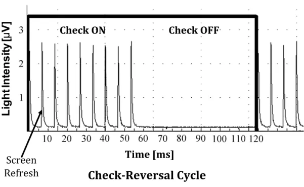
using a small checkerboard . . . 33 4. Time course of the actual light emitted by a CRT display
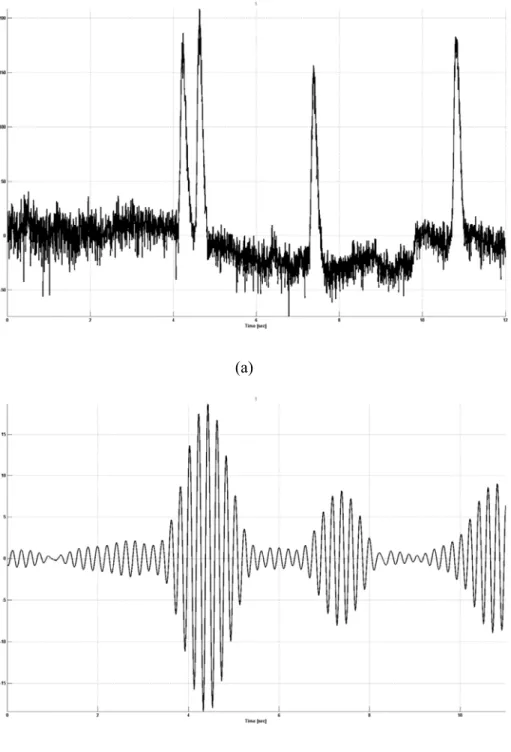
during checkerboard reversal (8.4 Hz) . . . 63 5. Negative impact of ocular artifacts on the SSVEP signal . . . 64 6. Single-trial SSVEP response to 14-Hz pattern stimulation . . . 68 7. Frequency response curve of the occipital brain
for small-checkerboard SSVEP stimuli . . . 70 8. Averaged 5-cycle waveforms for the 8 Hz, 14 Hz and 28 Hz SSVEP
brain responses (small checkerboards) . . . 73 9. SSVEP response delays for 8 Hz, 14 Hz, and 28 Hz stimulation
with small patterns for SSVEP onsets and SSVEP 1st peaks . . . 75 10. Topographic patterns of activation at the first peak of the 14Hz
SSVEP envelope (a single-trial example) . . . 76 11. Dynamics of single-trial responses at an optimal occipital location
for 14 Hz stimulation and all 6 trials (single subject) . . . 77 12. Dynamics of single-trial responses at a left parieto-temporal location
for 14 Hz stimulation and all 6 trials (single subject). . . 78
13. Dynamics of single-trial responses at a frontal location for 14 Hz stimulation and all 6 trials (single subject). . . 78
14. Wavelet-based time-frequency decomposition of non-stationary single-trial SSVEP for 14 Hz stimulation . . . 80
15. Properties of SSVEP responses at occipital, left parieto-temporal and frontal sensor locations measured for 8 Hz, 14 Hz, and 28 Hz stimulation using small reversing checkerboards on a black background . . . 82
16. Active electrode locations used in the SSVEP-based BCI design . . . 86
17. SSVEP-based BCI designs with multiple independent commands . . . 89
18. An experimental subject during evaluation of the multi-command SSVEP-based BCI system with moving stimulation design . . . 90
19. Block diagram of all modules of the online 8-command BCI system . . . 92
20. Online ICA/BSS-based artifact removal . . . 93
21. Block diagram of the SSVEP-BCI online analysis module . . . 95
22. An online bank of Butterworth filters to extract SSVEP power for all 8 stimulation frequency bands and 6 EEG channels . . . 96
23. Running comparison of the extracted SSVEP band features for all 8 BCI commands in steps of 1/8 s . . . 102
24. Time course of the actual light emitted by a LCD display during checkerboard reversal of 12Hz . . . 108
25. Affective-face video SSVEP stimuli . . . 109
26. Cortical changes at the onset of affective-face SSVEP and neutral SSVEP . . . 115
27. Frequency response curve of the occipital brain for affective-face SSVEP and neutral SSVEP . . . 116
28. Statistically significant SSVEP onset changes due to emotional-face
contents of the flicker stimuli . . . 117
29. No significant SSVEP differences due to inter-subject variability and emotion type/valence (positive-negative emotions) . . . 117
30. Brain activation during affective-face and neutral SSVEP stimulation at 8.5Hz . . . 118
31. Affective-face BCI interface with 8 commands . . . 121
32. Neutral 8-command SSVEP-BCI stimulus designs using reversing checkerboard-type patterns . . . 121
33. Multi-command BCI platform based on affective-face SSVEP - user control of a multi-joint robotic arm device (iARM) . . . 122
34. BCI command delays for affective-face video and neutral SSVEP . . . 125
35. Behavioral measures (self-estimate) in affective-face SSVEP experiments . . . 127
36. A proposed model of emotion-vision interactions . . . 143
37. Block diagram of the ICA/BSS artifact rejection procedure . . . 149
L IST OF T ABLES
TABLE PAGE
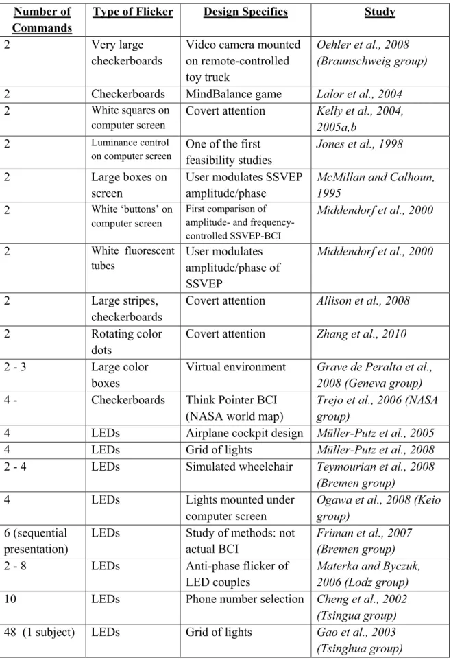
1. Comparison of SSVEP-BCI approaches in this thesis and previous research . . . 6
2. BCI experimental approaches – Advantages and problems . . . 19
3. Summary of Invasive BCI studies reviewed in this chapter . . . 21
4. Summary of Non-Invasive BCI studies reviewed in this chapter . . . 22
5. Summary of Steady-State Visual Evoked Potential (SSVEP) studies reviewed in this chapter . . . 36
6. Summary of SSVEP-BCI studies reviewed in this chapter . . . 43
7. Summary of emotion studies reviewed in this chapter . . . 53
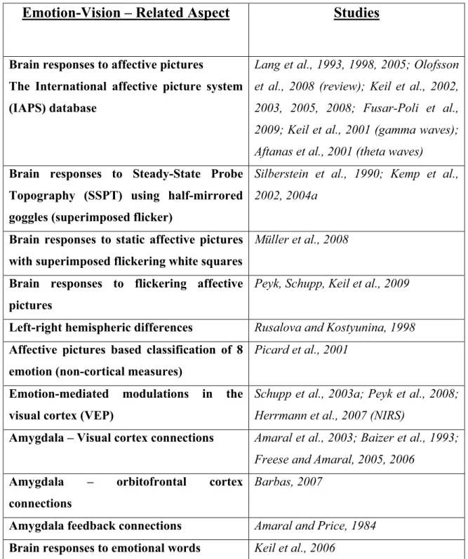
8. Summary of affective vision studies reviewed in this chapter . . . 58
9. Main peak response frequencies and z-scores to SSVEP stimulation . . . 72
10. SSVEP onset- and first-peak delays of the brain response for three frequencies in the low-, medium- and high-frequency range . . . 75
11. Performance summary for SSVEP-based BCI with 8 commands, and comparison of the effects of electrode placement over three different brain locations – occipital, left-parieto-temporal, and frontal forehead) . . . 103
12. Individual performance of the SSVEP-based BCI system for each subject including the effects of electrode placement over 3 different brain locations . . . 104
A BBREVIATIONS
BCI: Brain-Computer Interface
BMI: Brain-Machine Interface
CMS: Common Mode Sense (used by Biosemi EEG system)
CRT: Cathode Ray Tube
DRL: Driven Right Leg (used by Biosemi EEG system)
EEG: Electroencephalogram / electroencephalography
EMG: Electromyogram / electromyography
FC: Frontal Cortex
fMRI: Functional Magnetic Resonance Imaging
ITR: Information Transfer Rate
LCD: Liquid Crystal Display
LDA: Linear Discriminant Analysis
LED: Light-Emitting Diode
LPTC: Lateral Parieto-Temporal Cortex
MEG: Magnetoencephalogram / magnetoencephalography
OC: Occipital Cortex
PET: Positron Emission Tomography
PLV: Phase-Locking Value
PLVV: Phase-Locking Value Variability
SPLVL: Single-trial Phase-Locking Value
SSVEP: Steady-State Visual Evoked Potentials
SWT: Single-trial Wavelet Transform energy
TCP/IP: Transmission Control Protocol/Internet Protocol
VEP: Visual Evoked Potentials
WT: Wavelet Transform
WTV: Wavelet Transform (Energy) Variability
Chapter 1:
I NTRODUCTION
This thesis is a multidisciplinary effort, bringing together neuroscience, information science and engineering. The work described here aims mainly to help find new ways to improve quality of life using robust and adaptive brain technology. Knowledge from three separate research areas was utilized – Brain-Computer Interfaces (BCI), Steady-State Visual Potentials (SSVEP). and affective neuroscience (emotions).
“The most beautiful thing we can experience is the mysterious. It is the source of all true art and all science.” This quote from the German-born physicist Albert Einstein (1879 -
1955) relates closely to my long-term fascination with unraveling the endless mysteries of the brain, the most complex organ in the human body, in service of society, and of my fellow human beings.
1.1. The Ideal Brain-Computer Interface (BCI)
Brain-Computer Interfaces (BCI) are useful tools created with the ultimate goal of assisting the disabled, and increasing the productivity of the healthy members of society by using brain technology in daily as well as professional tasks. A BCI in operation has the simple yet very challenging job to recognize reliably from the measured brain-activity the intent of the user as requested commands and to execute the predefined tasks associated with these commands.
As explained in detail in the overview Chapter 2, there are several types of BCI. The most important BCI taxonomy is based on the way the brain activity is generated or modified consciously by the user in order to enable identification. BCIs can be based, for example, on visual potentials, motor imagery, P300 potentials, slow cortical potentials, and various mental tasks. Another useful BCI classification distinguishes between invasive and non-invasive brain signal measurements.
Regardless of the utilized paradigm and design, there are a number of basic desirable attributes (see also Table 2). An ‘ideal’ BCI system should include the following target features:
1) Maximal information transfer rates (same or better than human natural capability) 2) Maximal number of available independent BCI commands
3) Minimal command delays
4) Maximal operating reliability (³ 99%) 5) No user training necessary
6) No system training necessary
7) The inherent inter-subject / inter-session variability does not interfere with operations
8) Minimal user effort: Minimal demands to all attentional / sensory systems of the brain (user is free to perform other tasks)
9) Minimal mental fatigue after long-term operations
10) Maximal resistance to distractions, passing emotions, and neurological disorders 11) Friendly / attractive user interface
12) BCI system is mobile (no bulky equipment)
13) Emergency OFF switch is available and maximally reliable
14) Minimal preparation time ; no need for electro-gel (e.g. dry/remote electrodes)
15) Minimal operating costs.
1.2. Existing BCI Problems: Why is the Ideal BCI Difficult
to Achieve?
In spite of the maturing stage of development in the Brain-Computer Interfaces research field, even the most sophisticated existing BCI designs still suffer from substantial problems. Almost none of the ‘ideal’ target features listed in Chapter 1.1 have been achieved completely and in a consistent way. Regardless of the paradigm type and implementation, non-invasive BCI systems (in general) are still short of the desired characteristics due to several important factors:
1) Imperfect signal feature extraction and classification results in restricted reliability of the BCI commands, especially for more than 2 commands
2) Low brain signal-to-noise ratios in single trials result in long time delays between user intent onset and BCI command recognition
3) Inadequate understanding of the underlying brain mechanisms resulting in undesirable high inter-subject signal variability and low number of BCI commands
4) Inherent low-sensitivity shortcomings of the electroencephalographic mode of recording
5) User training and system classifier training necessary (depending on paradigm) which results in long delays before an optimal performance can be achieved 6) Substantial attentional demands in order to evoke stronger brain activity: Users
need to concentrate 100% on operating the BCI system, leaving them no room for
other useful tasks. Continuous attention results in fatigue after a few hours of operation. Minimal resistance to distractions
7) Application of EEG (electroencephalographic) electrodes needs specialized knowledge and time, as in most cases (currently) electro-gel is used, which dry up after several hours of operation. Hair needs to be washed of the electro-gel after the end of operations.
8) Equipment is bulky and expensive, usability is very limited.
1.3. Existing BCI Problems: Using SSVEP-BCI
The experimental designs and results presented in this thesis are based on the conscious control of the brain responses to rapidly changing visual stimuli. Steady-State Visual Evoked Potential (SSVEP) brain activity is elicited in precise synchrony with observed flickering visual patterns (see a review in Chapter 2.2). If two or more patterns oscillate at different frequencies on a computer screen, a user can choose which one to attend so that a corresponding BCI command is recognized. The SSVEP-BCI approach has been recognized as having a number of advantages and able to overcome many of the drawbacks of other BCI paradigms (see a review in Chapter 2.3). In spite of that promising assessment, however, the existing SSVEP-BCI systems haven’t been able to realize their full potential mainly due to inherent difficulties with low SSVEP signal-to-noise ratios in the single trial measurements typical for BCI (as measured using EEG). This has prevented a much needed increase in the number of BCI commands, decrease in the size of SSVEP stimuli, and recording from suboptimal but more convenient non-occipital brain areas.
Prior to this work, existing SSVEP-BCI systems based on computer-generated stimuli (see Table 1) used mostly 2-4 large reversing checkerboard patterns in order to evoke
sufficiently strong, measurable SSVEP responses (Lalor et al., 2004; Trejo et al., 2006; Oehler et al., 2008). These checkerboard stimuli covered up most of the useful visual area on the screen, substantially limiting the possible BCI applications and increasing user fatigue. In addition, the SSVEP frequencies in these studies were pre-selected in a relatively arbitrary way. The resulting BCI command delays were not studied systematically in order to understand the underlying signal dynamics, and to increase substantially the information transfer rates.
To summarize, the most important problems in SSVEP-based BCI systems prior to this work were:
1) Usage of too large on-screen SSVEP stimuli to evoke stronger brain responses, which obscure useful visual space (visual stimuli need to be very small)
2) Fixed usage of uniform white or large checkerboard SSVEP stimuli (other types of visual stimuli may be more appropriate)
3) Fixed flicker frequencies (optimal frequencies need to be determined for each type of SSVEP stimuli)
4) Stimuli are located far away from each other (e.g. on the edges of the screen) prompting larger eye movements (increased BCI delays, fatigues, artifacts)
5) Small number of SSVEP-BCI commands
6) FFT-based signal processing with subjective parametrization of the weights of the harmonic frequencies for each flicker
7) No insight into the mechanisms of SSVEP and BCI delays, which prevents optimization
8) Fixed data acquisition from occipital sensor locations only (no insight for BCI performance at other brain sites).
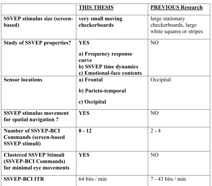
Table 1. Comparison of SSVEP-BCI approaches in this thesis and previous research
THIS THESIS PREVIOUS Research
SSVEP stimulus size (screen- based)
very small moving checkerboards
large stationary checkerboards, large white squares or stripes Study of SSVEP properties? YES
a) Frequency response curve
b) SSVEP time dynamics c) Emotional-face contents
NO
Sensor locations a) Frontal
b) Parieto-temporal c) Occipital
Occipital
SSVEP stimulus movement for spatial navigation ?
YES NO
Number of SSVEP-BCI Commands (screen-based SSVEP stimuli)
8 - 12 2 - 4
Clustered SSVEP Stimuli (SSVEP-BCI Commands) for minimal eye movements
YES NO
SSVEP-BCI ITR 64 bits / min 7 - 43 bits / min
1.4. Research Objectives Addressed in This Thesis
The work presented in this thesis aimed to address directly the existing problems described above. New approaches were necessary to remove a number of essential obstacles to the creation of practical unsupervised BCI paradigms able to work outside of the laboratory. As a result, the following research goals were pursued in this work:
1) Using very small visual patterns to minimize visual occlusion while evoking measurable SSVEP responses at different frequencies (freeing up essential screen space for application purposes) (Chapter 3, 4) 2) Using SSVEP patterns in close proximity to each other without brain
response interference (Chapter 4)
3) Using a high number of commands without brain response interference (Chapter 4)
4) Optimal selection of SSVEP stimulation frequencies based on objectively measured EEG data (frequency response curve) (Chapter 3)
5) Understanding the time dynamics of the SSVEP response to small stimuli as used for BCI (time delay limitations of BCI commands) (Chapter 3)
6) Measuring reliable SSVEP responses from fast moving flickering/reversing visual patterns, in a novel dynamic paradigm for pattern SSVEP BCI (control of a moving object) (Chapter 4)
7) Achieving the highest possible BCI information transfer rates and robustness for the proposed paradigms (Chapter 4)
8) Using sub-optimal temporal or pre-frontal sensor locations on the head for new SSVEP-BCI designs with dry electrodes (without the need for application of EEG gel) (Chapter 3, 4)
9) Effect of using emotional-face SSVEP stimuli instead of checkerboards in BCI applications (Chapter 5, 6).
These research objectives were pursued using two general directions:
1) Stimulus optimization to achieve maximal cortical response for further use in BCI (frequency, size, movement, and contents of SSVEP stimuli)
2) Development of sensitive adaptive algorithms to extract weak target SSVEP oscillations from ‘noisy’ EEG input signals, both offline for detailed analysis, and online for SSVEP-BCI evaluation.
Table 1 presents a concise comparison between the new approaches presented here and previous research.
In addition, the Brain-Computer Interface design and implementation described in this thesis aimed to comply fully to the 4 basic criteria for a BCI (Pfurtscheller et al., 2010b):
1) The BCI device must use directly brain signals
2) The BCI device must be capable of real-time signal processing
3) The BCI device must allow the user to modulate intentionally at least one brain signal feature through goal-directed behavior
4) The BCI device must give the user feedback information.
Overall, the research goals described above were the focus of an effort to use new knowledge in order to design, optimize experimentally and evaluate the performance of a novel eight-command SSVEP-based BCI system as described in the following chapters.
1.5. Summary of Achievements in this Thesis
The work in this thesis aimed mainly at the substantial optimization of cortical visual responses and signal processing algorithms, as well as achieving superior multi-command SSVEP-BCI information transfer rates and reliability. The following major conclusions were reached:
1) Very small checkerboard SSVEP stimuli were shown to elicit measurable and reliable single-trial brain responses
2) The SSVEP frequency response curve (5.1 Hz - 84 Hz) for small stimuli was studied, and the optimal SSVEP brain response range was determined to be 5.6 Hz – 15.3 Hz (12 Hz maximum)
3) The onset of single-trial SSVEP responses was shown to be detectable between 1.5 s and 2.5 s (using band oscillation envelope measures)
4) SSVEP dynamics (onset delays, first peaks, stationarity) were strongly dependent on the stimulation frequency. When 3 stimulation frequencies were compared (8, 14 and 28 Hz), the 14 Hz responses exhibited fastest onsets and maximal signal stability
5) The feasibility of practical SSVEP-BCI designs using dry electrodes (without electro-gel) was established by evaluating SSVEP responses from 3 distinct sensor locations (occipital, parieto-temporal and frontal). While occipital visual responses were optimal as expected, the parieto-temporal and frontal locations also allowed the detection of SSVEP responses, although with reduced strength, stationarity and reproducibility
6) A new SSVEP-BCI system with 8 independent commands, using small checkerboard stimuli flickering in the optimal range, reached a mean information transfer rate (ITR) of 50 bits/min, with success rate of 98% and command delay of 3.4 ± 0.7 s for occipital sensor locations
7) SSVEP-BCI performance was evaluated also for parieto-temporal and frontal sensor locations, and deteriorated with increasing distance from the visual cortex (81% success rate and 4s delay for parieto-temporal sensor locations and 74% success rate and 4.3s delay for frontal electrodes)
8) No user training was necessary for this SSVEP-BCI system
9) Very close proximity (clustering) of multiple small SSVEP stimuli was used successfully for the first time in the SSVEP-BCI design
10) A moving block of multiple small SSVEP stimuli was used successfully for the first time in the SSVEP-BCI design (a novel dynamic feedback paradigm)
11) Due to the small size and close clustering of the checkerboard stimuli in the SSVEP-BCI design the visual field occlusion was substantially minimized, and long-term user fatigue was reduced
12) Flickering emotional face video sequences were shown to elicit superior SSVEP responses than checkerboard- and blurred stimuli
13) The Affective-face SSVEP frequency response curve (5 Hz - 12 Hz) was studied, and the optimal brain response for this type of SSVEP stimuli was 10 Hz
14) A new single-trial phase-locking value variability (PLVV) measure was used in the offline SSVEP analysis, and was shown to be more sensitive to SSVEP onset than a comparable band-energy based measure – wavelet-transformed energy variability (WTV)
15) A new Affective-face SSVEP-BCI design was evaluated for 10 Hz flicker and was shown to be superior to the small-checkerboard design (mean BCI delay 2.7s vs. 3.4s; mean success rate 99% vs. 98%; mean ITR 64 bits/min vs. 50 bits/min). 16) BCI users reported substantially higher motivation, attention, and less long-term
fatigue when using the Affective-face SSVEP-BCI design as compared to neutral checkerboard- and blurred-face SSVEP-BCI stimuli
17) The higher reliability and stronger brain responses allowed users of the Affective- face SSVEP-BCI system to control successfully a multi-joint robotic arm with complex movements with minimal online errors. A multi-command BCI system controlling reliably a robotic arm has substantial advantages for disabled members of society.
1.6. Organization of the Thesis
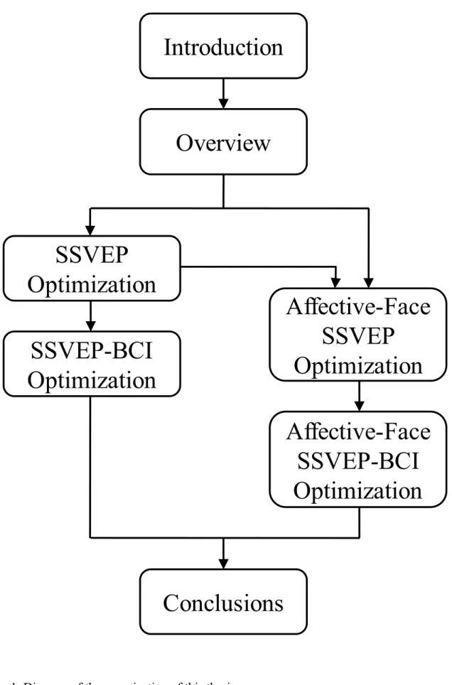
This thesis (Fig. 1) focuses on an integrated approach to the optimization of SSVEP- based Brain-Computer Interfaces (Fig. 2). This was accomplished, first, by investigating the brain responses to continuous visual flicker stimulation with very small checkerboards (Chapter 3), as well as with flickering emotional face videos (Chapter 5). Second, the results were applied for designing two optimized SSVEP-BCI paradigms using the same stimuli (Chapter 4 and Chapter 6). In this work, several novel concepts were introduced and explored in order to enable new types of efficient and user-friendly Brain-Computer Interfaces.
Fig. 1. Diagram of the organization of this thesis
Introduction
Overview
SSVEP
Optimization
SSVEP-BCI
Optimization
Affective-Face
SSVEP
Optimization
Affective-Face
SSVEP-BCI
Optimization
Conclusions
Chapter 2:
OVERVIEW OF PREVIOUS RESEARCH
2.1. Brain-Computer Interfaces (BCIs)
2.1.1. Why are Brain-Computer Interfaces necessary?
Today's advances in technology and signal processing algorithms are giving brain researchers a new edge, allowing us to go one crucial step further, from understanding brain functions, to actually using this knowledge to change the world. Brainwave-based technologies such as Brain-Computer Interfaces (BCI), when made sufficiently user-friendly, reliable, and affordable, will help mankind not only to conquer crippling disabilities through neuroprosthetics and rehabilitation, but also to improve precision of control for vehicles and robots in hostile environments such as space, to let people live in intelligent e-homes, to integrate new electronic body enhancements whenever necessary, and to play and communicate in novel ways.
2.1.2. Brain-Computer Interfaces – Definition, Application and Requirements
A Brain-Computer Interface (BCI) is any brain-to-machine communication system, which is able to interpret and execute voluntary brain commands, with no dependence on the normal executive pathways of the body such as muscles and peripheral nerves (Wolpaw et al., 2000).
Fig.2. Basic block diagram of a Brain-Computer Interface (BCI) system (Bakardjian, 2004)
BCI technology has the potential to find wide acceptance in society. Possible practical applications of BCI are:
1) Mobility assistance for the elderly and disabled; recreation (brain control of wheelchairs, robots and vehicles for self-reliance and self-service)
2) Remote control of devices (hazardous environments - space, fire; telerobotics) 3) Communications, creative work (email, documents)
4) Virtual reality creations (multi-player games, role play, navigation) 5) Neuroprosthesis and rehabilitation (replace or restore function of limbs) 6) Remote monitor of attention in pilots or drivers (Vaughan et al., 2003) 7) Brain-based art creation (Vaughan et al., 2003)
8) Neural enhancements control in a healthy human body such as additional artificial limbs, memory chip implants (there may be ethical issues)
BCI is a particularly challenging task from the point of view of biosignal processing, because often single-trial analysis use dense electrode arrays to compensate for low signal-to- noise ratios due to the unavailability of inter-trial statistics. In addition to the single trials, online BCI systems need to overcome a further limitation in spatial resolution, with only a few electrodes available in order to make such devices easier to handle. From the point of view of the necessary requirements, an optimal BCI system should exhibit the following characteristics:
1) Maximal number of commands (maximal choice / degrees of freedom)
2) Maximal accuracy of command recognition (high noise and artifact resistance) 3) Maximal speed of command transfer (stimulus rate, information content)
4) Maximal comfort for users (sensors, environment, ease of control = low mental load)
5) Maximal tolerance to fatigue and shifting emotional states
6) Maximal tolerance to distractions (external stimuli, passing thoughts, memories) 7) Maximal tolerance to neurological disorders
8) Maximal tolerance to inter-subject variability 9) Minimal training (both for user and machine) 10) Minimal cost of operation
11) Maximal robustness: ON/OFF/IDLE commands; recycle or cancel commands 12) Maximally attractive user interface (for enhanced motivation).
2.1.3. BCI Approaches and Types
In 1967, Edmond Dewan (Dewan, 1967) described what seems to be one of the first accounts of a BCI communication. In his experiments, subjects controlled voluntarily the
amplitude of their own alpha EEG waves which allowed a computer program to convert these changes and send corresponding Morse code messages. In 1973, Jacques Vidal, a computer science researcher at UCLA, described the working principles of a direct brain-computer interface (BCI) (Vidal, 1973) and four years later, he published the results of a successful online BCI implementation with 4 commands (Vidal, 1977). Vidal used visual evoked potentials (VEP) elicited by brief illumination of a checkerboard to estimate with over 90% success rate which one of the four preset gaze positions around the pattern was attended, in order to control a moving object in a maze. The data processing approach in his study was already built around the current online BCI architecture, including data acquisition, artifact rejection, preprocessing, feature selection, and classification modules.
The fundamental goal of BCI is to identify each user intention from its corresponding brain pattern in near-real-time. However, current recording techniques are unable to deliver unique patterns for each user intention for usage as unlimited commands. In practice, using a limited number of sensors results in overlapping of the measurable characteristics of the available brain patterns, which need to be controlled / separated using specific tasks for the user to perform during the BCI session.
The prevalent recording technique for BCI is electroencephalography (EEG). Current brain signal processing approaches distinguish between ‘spontaneous’ and ‘evoked’ EEG. Spontaneous EEG refers to the measurement of continuous brain waves, including the delta (up to 4Hz), theta (4-8Hz), alpha (8-12Hz), beta (12-30Hz), and gamma (30-100+Hz) waves, while evoked EEG represents brain potentials with limited duration which are recorded in response to specific stimuli, such as visual, auditory, somatosensory, or olfactory. BCI paradigms can be based on both spontaneous and evoked brain signals, for example, motor- imagery BCI using modulation of spontaneous ‘mu’ and ‘beta’ waves, or SSVEP-BCI (steady-state visual evoked potentials) using periodically evoked visual responses.
Currently, BCI tasks can be classified into two main categories:
1) Internally-supported – Voluntary generation of specific mental state patterns (usually based on modification of spontaneous brain activity)
The mental state BCI paradigms could be either cue-, or internally-paced, and are usually based on conscious control of naturally-occurring brain waves, such as mu-, beta- and alpha, slow potentials, or multi-unit neuronal activity.
There are two subtypes of mental load BCI tasks, both of which use response modification by feedback (Curran and Stokes, 2003):
1.1) Cognitive load (using specifically-defined imagination tasks like limb movement, grasping, mental arithmetic) (Pfurtscheller et al., 2006; Penny et al., 2000)
1.2) ‘Operant conditioning’ (no specific cognitive targets, “just do it” principle) (Wolpaw et al., 2000; Bensafi et al., 2003)
2) Externally-supported – Voluntary modification of the normal responses to external stimulation (usually based on modification of evoked brain activity)
2.1) Using selective attention to choose a target stimulus corresponding to a BCI command which evokes a specific recognizable brain response (Bakardjian et al., 2010; Gao et al., 2003; Farwell and Donchin, 1988). Examples for such attention-driven BCI tasks are:
2.1.1) Cognitive load (using specifically-defined imagination tasks like limb movement, grasping, mental arithmetic) (Pfurtscheller et al., 2006; Penny et al., 2000)
2.1.2) P300 oddball response (Farwell and Donchin, 1988) 2.1.3) SSVEP (steady-state visual evoked potentials) (Sutter,
1984; Gao et al., 2003; Müller-Putz et al., 2005)
2.1.4) SSSEP (steady-state somatosensory evoked potentials) (Müller-Putz et al., 2006)
2.2) Using voluntary modification to external stimulus responses in order to indicate a BCI command. An example is a 2-command BCI task based on self- controlled visual priming in which the subjects imagined (or not) observing smooth natural visual motion while actually viewing discrete apparent motion (‘jumping’) bar stimuli (Bakardjian, 2003).
Internally-supported mental-state tasks have the advantage that they do not usually require external stimulation equipment (except for the feedback of the results to the user). However, the complexity of the involved natural brain processes can be very high so that the reliability and number of commands is currently limited in mental-state paradigms. In order to create Brain-Computer Interfaces with enhanced information transfer rates for both the internally- and externally-supported BCI task paradigms, strict controls are imposed by using well-known cognitive imagery processes (such as motor imagery), and strong phase-locked sensory responses which are reliably detectable in single trials (such as SSVEP).
Table 2. BCI experimental approaches – Advantages and problems
Attribute
Externally-Driven (stimulated responses)Internally-Driven (mental states)
Large Number of Commands YES NO
High Reliability of Commands YES NO
(in most cases)
High Information Transfer Rates YES NO
Short User Tuning (before 1stusage) YES NO
Short Training YES NO
Self-Paced YES / NO YES / NO
Short Command Delays (±1sec) NO / YES YES / NO
ON/OFF Switch YES YES
(in most cases)
No Stimulation Equipment Necessary NO YES
No Sensory Engagement Necessary NO YES
Resistant to High Subject Variability YES NO
Resistant to Mental Fatigue NO NO
Resistant to Passing Emotions YES (in most cases)
NO (in most cases)
Resistant to Distraction NO
(in most cases) NO
Resistant to Neurological Disorders YES
(in most cases) Unknown Attractive User/Neurofeedback Interface YES
(possible)
YES (possible)
Comfortable for User NO
(in most cases) YES
EXAMPLES for BCI Approaches
SSVEP (steady-state) (Sutter 1992; MacMillan 2000; Gao 2002)
P300 oddball response (Donchin 2000)
SCP (slow cortical potentials) (Kübler 2001)
Visual motion response (Bakardjian 2003)
Motor imagery (Pfurtscheller 1994)
Mental states (Millán 2003) Cognitive load (specific imagination tasks): (Pfurtscheller 2004; Penny 2000)
Operant conditioning’ (“just do it”): (Humphrey
1970;Birbaumer 1999; Wolpaw 2000; Nicolelis 2004)
Apart from the BCI task dichotomy, another BCI classification is possible from the point of view of brain data acquisition, as there are two principal approaches (Vaughan et al., 2003):
1) invasive recording of individual or local neuronal activities (surgery necessary) 2) non-invasive measurement reflecting only large-scale neuronal activations (EEG, NIRS, time-resolved fMRI).
2.1.4. Invasive BCI
Neuronal spikes due to action potentials are measured using arrays of miniature electrodes implanted in the brain tissue. From 1996, Kennedy’s group at Neural Signals Inc started implanting “locked-in” paralyzed human users with their special cone electrodes with hollow tips for growth of the neural tissue (Kennedy et al., 1998). One of their first subjects equipped with invasive BCI technology, was able to control a cursor on a computer screen and spell about three letters per minute (Kennedy et al., 2000).
Further invasive BCI studies have demonstrated increasingly complex usage of direct neuronal activity information, such as remote control of a TV set or a computer cursor by Donoghue’s BrainGate device (Donoghue, 2002), or the operation of a robotic gripper arm controlled entirely by brain signals optimized by visual feedback (Taylor et al., 2002; Carmena et al., 2003; Chapin et al, 1999). In a less invasive procedure, local field potentials reflecting the activity of groups of neurons can be recorded from subdural or epidural locations under the scull to control, for example, a computer cursor (Schalk et al., 2008).
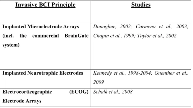
Table 3. Summary of Invasive BCI studies reviewed in this chapter
Invasive BCI Principle Studies
Implanted Microelectrode Arrays (incl. the commercial BrainGate system)
Donoghue, 2002; Carmena et al., 2003; Chapin et al., 1999; Taylor et al., 2002
Implanted Neurotrophic Electrodes Kennedy et al., 1998-2004; Guenther et al., 2009
Electrocorticographic (ECOG) Electrode Arrays
Schalk et al., 2008
2.1.5. Non-Invasive BCI
Success in non-invasive BCI has been also impressive in recent years, although arguably less spectacular due to the substantial challenges posed by the recording of large- scale synchronized activity often originating from multiple neuronal populations behind the layers of the cerebrospinal fluid, meninges, skull bone and scalp skin. The non-invasive approach offers promise for a wide range of immediate real-life applications, both for healthy and disabled users.
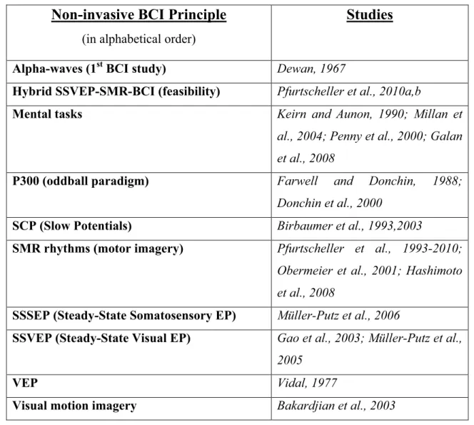
Table 4. Summary of Non-Invasive BCI studies reviewed in this chapter (see Table 6 for further SSVEP-BCI examples)
Non-invasive BCI Principle
(in alphabetical order)
Studies
Alpha-waves (1stBCI study) Dewan, 1967
Hybrid SSVEP-SMR-BCI (feasibility) Pfurtscheller et al., 2010a,b
Mental tasks Keirn and Aunon, 1990; Millan et
al., 2004; Penny et al., 2000; Galan et al., 2008
P300 (oddball paradigm) Farwell and Donchin, 1988;
Donchin et al., 2000
SCP (Slow Potentials) Birbaumer et al., 1993,2003
SMR rhythms (motor imagery) Pfurtscheller et al., 1993-2010; Obermeier et al., 2001; Hashimoto et al., 2008
SSSEP (Steady-State Somatosensory EP) Müller-Putz et al., 2006
SSVEP (Steady-State Visual EP) Gao et al., 2003; Müller-Putz et al., 2005
VEP Vidal, 1977
Visual motion imagery Bakardjian et al., 2003
2.1.5.1. Motor-imagery based BCI
Most of the BCI researchers to date use motor imagery tasks, which allow detection of changes in the brain’s sensorimotor rhythm (SMR) in preparation for motor tasks (muscle movements). Motor-imagery BCIs use ‘cognitive load’ self-regulation type tasks (as described above), transmitting predictable commands due to the ease of detection and speed
of the SNR changes, even though the number of commands and BCI reliability may be relatively limited. Pfurtscheller’s group (Graz-BCI) was among the first research labs to actively develop and explore this BCI mode (Pfurtscheller et al., 1993). The introduction of the concepts of Event-related Desynchronization (ERD) and Event-related Synchronization (ERS) revealing non-phase-locked events in the frequency domain (Pfurtscheller et al., 2003) helped advance this paradigm to a point where a five-class BCI was reported (Obermeier et al., 2001).
Using this approach, Pfurtscheller’s group was also able to demonstrate the successful artificial stimulation of the hand grasp function in a tetraplegic patient with direct brain control (Pfurtscheller et al., 2003). Beta- and mu-wave activity from surface EEG data was recorded and classified in real time while the tetraplegic subject imagined foot movement, after which the output signal directed a functional electrical stimulation (FES) device to help the paralyzed hand grasp a cylinder.
Recently, a team of researchers at Keio University demonstrated that within the 3-D virtual reality web environment Second Life a disabled person was able to communicate from his home with a student logged in 16 km away and to walk his avatar to him by using only his brain waves (Hashimoto et al., 2008). The system used 3 electrodes to identify hand and foot imagery and to show the feasibility of virtual contacts for disabled people.
2.1.5.2. Slow cortical potentials based BCI
Slow cortical potentials (SCP) for BCI control were pioneered by the Birbaumer group at Tübingen (Birbaumer et al., 1999; Birbaumer et al., 2003). They used SCP as an
‘operant conditioning’ type of BCI, and called it a Though-Translation Device (TTD). In their initial implementation, the users controlled a spelling device which two ALS patients
learned to use at a rate of two characters per minute (after two months of training). In another setup, subjects tried to move a ball on the screen toward a target. Both healthy users and
‘locked-in’ patients were able to gain control of the system through their own strategies, including imagery.
2.1.5.3. P300 potentials based BCI
Donchin and Farwell were among the first researchers to design a BCI device based on the detection of the well-known P300 ‘target’/’surprise’ brain wave (Farwell and Donchin, 1988; Donchin et al., 2000). In their design, a grid containing the letters of the alphabet and some other functions flashed randomly each character, while the user concentrated on the desired letter. When the target was flashed, a P300 wave indicated a selection. This principle allows high reliability, however, often it is necessary to use multiple trials to achieve selection, and the increased attention efforts may (arguably) cause fatigue faster than in some other BCI paradigms.
2.1.5.4. Non-motor cognitive tasks (mental tasks) based BCI
Keirn and Aunon (Keirn and Aunon, 1990) demonstrated that it is possible to distinguish between five specific mental tasks using the delta (0-3 Hz), theta (4-7 Hz), alpha (8-13 Hz), and beta (14-20 Hz) frequency bands, and a Bayes quadratic classifier.
Even though for this type of BCI a large number of commands is theoretically possible, it is still very challenging due to the high rate of command misclassifications. Recently, the most commonly used features are mental rotation or mental arithmetic. A study by Penny (Penny et al., 2000) utilized a pattern classifier with parameter uncertainty while
the user’s mental arithmetic task was to subtract sevens successively from a three digit number.
In recognition that a more flexible approach is necessary, later studies used a mixed paradigm in which both motor imagery and another mental task were involved. In one such study, subjects controlled the movement of a miniature robot in a small model house by first selecting and working with three of the following tasks: relaxation, left hand motor imagery, right hand motor imagery, visualizing a spinning cube, performing elementary subtractions by a fixed number (for example, 64–3=61, 61–3=58, an so on), and concatenating related words (Millan et al., 2004).
For wheelchair control, a recent BCI design used a combination of left-hand movement imagery, rest, and a word association task in which the subjects had to find mentally words starting with the same letter (Galan et al., 2008).
Since BCI is a single-trial approach, spatial-domain information can also be utilized in addition to the time- and frequency-domains to increase the efficiency of the brain pattern recognition. One such successful technique, which is increasingly popular in many recent studies, is the Common Spatial Patterns (CSP) algorithm (Ramoser et al., 2000). Another method to use multichannel data for pattern separation is Independent Component Analysis (ICA) / Blind Source Separation (BSS) (Cichocki and Amari, 2003). Yet, online implementation for ICA/BSS has been feasible so far only for some very fast BSS algorithms (Martinez et al., 2007). A general survey of the most common BCI classification approaches, as well as guidelines how to choose a classifier, can be found in (Lotte et al., 2007). (Bashashati et al., 2007) offer a complimentary overview of the algorithms used in the various signal processing stages of recent BCI systems.
The Brain-Computer Interfaces are a very rapidly developing and promising platform, which, depending on progress in contactless sensors, has the potential to become a gateway for brain technology to enter everyday life, and change society.
BCI approaches using Steady-State Visual Evoked Potentials (SSVEP) have the capability to satisfy many of the strict requirements of an optimal BCI. In this stimulus- driven type of BCI, each command is assigned to a separate visual stimulus, as multiple lights or patterns flicker or reverse at slightly different frequencies. The BCI system recognizes the synchronized brain responses to the specific SSVEP stimulus which the user is currently directing his/her attention to.
2.2. Steady-State Visual Potentials (SSVEP)
Steady-State Visual Evoked Potentials (SSVEP) are brain responses that are precisely synchronized with fast (e.g. more than 4Hz) repetitive external visual stimulation such as flashes, reversing patterns or luminance-modulated images. SSVEP responses can be measured within narrow frequency bands (such as ±0.1 Hz) around the visual stimulation frequency, or using other signal processing methods that exploit the specific characteristics of the SSVEP signal, such as rhythmicity and synchronization. The strongest responses occur in the primary visual (striate) cortex, although other brain areas are also activated in varying degrees.
2.2.1. Cortical Frequency Responses to SSVEP Stimulation
David Regan was one of the early researchers to study extensively the various properties of the steady-state evoked potentials in human adults. In a series of studies he
found three distinct frequency response regions which he termed low-frequency (LF), medium-frequency (MF) and high-frequency (HF) region (Regan, 1977; Milner et al., 1972; Regan, 1989). For large unpatterned flicker (with very low spatial frequency), the maximal mean response was produced by ~10 Hz stimulation in the LF region, while the peak in the MF region (13-25 Hz) was found at a flicker rate of about 18 Hz, and the HF (40-60 Hz) local maximum was between 45 and 50 Hz (Regan, 1968). The three peak amplitudes in the LF, MF and HF regions were in an inverse relationship with frequency, as brain responses declined at faster flicker rates.
The results for small-check reversals with 0.2° arc check size (high spatial frequency) showed stronger preference for low-frequency stimulation, peaking at about 7 Hz, while SSVEP responses for larger checks with 0.7° arc size were more distributed and more similar to unpatterned flicker. Both for small and larger checks, SSVEP amplitudes were measured only up to 20 Hz and no conclusions were offered for higher frequencies. Regan hypothesized that responses to stimuli with very high spatial frequencies have low temporal frequency preference, while increasing the check size leads to a mixture of the (different) pattern-specific and flash-specific mechanisms (Regan and Richards, 1973).
Pastor (Pastor et al., 2003) studied the EEG responses to 50ms white strobe flashes using 14 frequencies ranging from 5 to 60 Hz. They found the amplitude of the maximal EEG response at 15 Hz for occipital areas and at 25 Hz for frontal areas. The study presented also measurements of the regional cerebral blood flow using positron emission tomography (PET) during visual stimulation at 5, 10, 15, 25, and 40 Hz. Their PET results showed activation of the primary visual cortex with the maximum at 5 Hz when comparing the five tested stimulation frequencies. Earlier PET research had found the strongest response peak in the occipital cortex in the LF region at 7-8 Hz using both goggles with a grid of embedded red