【症例】坐骨静脈遺残を伴い痕跡的大腿静脈を起源とする類上皮血管内皮腫の 1 例
4
0
0
全文
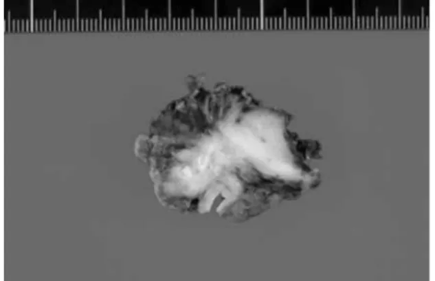
(2) 388. 日血外会誌 15巻 3 号. SMA. Fig. 1. Computed tomography scan of pelvis showing enhanced groin mass adjacent to the femoral artery and calcification of the femoral vein.. Fig. 3. Descending phlemogram demonstrating regurgitation of the deep femoral vein and PSV. PSV: persistent sciatic vein. の一部と外腸骨∼大腿動・静脈を含めen blocに切除し. Fig. 2. Preoperative venogram demonstrating the stenotic common femoral artery and hypervascuralization representative of the position of the tumor mass.. Fig. 4. The tumor from the groin has a pale, fibrous cut surface with dense calcification.. 性で,類上皮血管内皮腫と診断された.. た.動脈を人工血管で再建したが,大腿静脈は遺残坐. 考 察. 骨静脈が開存していたため再建せず,中枢,末梢断端 は閉鎖した.. EHEは非常に稀な腫瘍で,1982年にWeissとEnzinger 1). 術後グラフト造影:外腸骨∼大腿動脈の再建は良好. が軟部組織に発生する特異な組織像を呈する血管内皮. で,遺残坐骨静脈も温存されていた.. 細胞由来の腫瘍を報告したことに始まる.現在では肺. 摘出標本 (Fig. 4):割面は線維性皮膜で被われ,乳白. 原発のIVBAT(intravascular bronchioalveolar tumor)2),. 色の軟骨様を呈した.大腿静脈は石灰化し,閉塞して. Ishakら3)の肝原発,Tsuneyoshiら4)の骨原発も発生部位が. いた.. 異なるだけで,本質的には同じものと考えられている.. 病理所見(Fig. 5a, b):腫瘍細胞は空胞を有する類円. また,縦隔,脳,リンパ節,脾臓などにも原発すること. 形で管腔構造や索状配列を示した.一部に骨形成や少. が知られており,理論的には血管内皮細胞があるとこ. 数の細胞分裂像を認め,免疫染色でCD31,factor VIII陽. ろにはどこにでも発生しうる.. 24.
(3) 2006年 4 月. Fig. 5. 389. 久貝:類上皮血管内皮腫. (a) Photomicrographs of epithelioid hemangioendothelioma showing cordlike pattern and vacuoles representing primitive lumina, some containing erythrocytes (H&E, ×200). (b) Endothelial cells in tumor are immunoreactive for CD31 (immunoperioxidase stain; ×2).. a. b. 判定には今後さらなる症例の蓄積が必要である.. 1.血管発生の類上皮血管内皮腫 現在まで,EHEは肺・肝・軟部組織などからの発生が. 2.坐骨静脈遺残. 5). 多いものの,動・静脈など血管発生の報告は少ない .. 坐骨静脈遺残はKlippel-Trenaunary症候群 (KTS) に合併. Charetteら6)は2001年までに発表された英論文33例を集. することが多く,単独奇形で存在するのは稀である10, 11).. 計し,経験豊富な血管外科医でも一生に 1 例しか経験. 自験例のように慢性静脈弁不全または下肢静脈瘤を合. しないだろうと述べている.腫瘍の約50%は大型∼中. 併し,うっ滞性皮膚炎の原因になるため本疾患を念頭. .. に置く必要がある11, 12).自験例はこれまで下腿腫脹など. 性差はなく,年齢は広範囲に及ぶが比較的若年者に発. の深部静脈閉塞の症状もなく,また12年前の静脈造影. 生している.肉眼的には境界明瞭,乳白色で軟骨様の. で大腿静脈は全く造影されず,側副血行路の発達もな. 透明感を示す.組織像では腫瘍細胞は類円形で,胞巣. い.当時腫瘍が存在していたとは考えにくく,大腿静. 状の塊を作ったり,索状配列を示す.細胞質の大きな. 脈の閉塞と腫瘍は無関係と考えられる.むしろ下肢静. 空胞が特徴的で,粘液染色は陰性である.また,石灰. 脈発生の過程で大腿静脈は退縮し,坐骨静脈が遺残し. 化,骨化を伴う.通常,核分裂像は見られないが,と. た.そして,その痕跡的な大腿静脈を起源に自験例は. きに核の異型性が顕著となり,細胞密度が高くなると. 発生したと推察される.本腫瘍は大腿静脈に由来する. 細胞分裂が観察される.10高感度視野で 6 以上の分裂. ことが多いが,萎縮遺残した静脈からも発生すること. 型の血管から発生し,とくに大腿静脈由来が多い. 1, 7). 5). 像が見られるのは予後不良の徴候である .免疫組織学. が示唆された.. 的に第 8 因子関連抗原,CD31,CD34など血管内皮細. 結 語. 胞のマーカーが陽性となる.進行が比較的遅いことか ら,血管肉腫と血管腫の中間悪性度として扱われる. 本邦初と考えられる坐骨静脈遺残と類上皮血管内皮. が,術後再発や転移も認められる.本腫瘍の13%が局. 腫の極めて興味深い組合せを経験した.本腫瘍は大腿. 所再発し,30%に遠隔転移が認められ,5 年生存率は80. 静脈に由来することが多いが,坐骨静脈遺残により萎. %弱と決して良くない 5, 8, 9).化学療法,放射線療法は無. 縮遺残した大腿静脈からも発生することが示唆され. 効であり,唯一外科的完全切除のみが有効である.. た.. EHEは症例自体が少なく,最善の治療法の選択,予後. 25.
(4) 390. 日血外会誌 15巻 3 号. 文 献. 1100-1103, 2001.. 1) Weiss, S. W. and Enzinger, F. M.: Epithelioid hemangioen-. 7) Kempsom, R. L., Fletcher, C. D. M., Evans, H. L., et al.:. dothelioma: a vascular tumor often mistaken for a carci-. Tumors of the soft tissues. Atlas of Tumor Pathology, 3rd. noma. Cancer, 50: 970-981, 1982.. Ser., Fascicle 30, Washington DC, 1996, Armed Forces,. 2) Dail, D. H., Liebow, A. A., Gmelich, J. T., et al.: Intravas-. Institute of Pathology, pp. 307-368.. cular, bronchiolar, and alveolar tumor of the lung (IVBAT):. 8) Mentzel, T., Beham, A., Calonje, E., et al.: Epithelioid. an analysis of twenty cases of a peculiar sclerosing endo-. hemangioendothelioma of skin and soft tissues: clinico-. thelial tumor. Cancer, 51: 452-464, 1983.. pathologic and immunohistochemical study of 30 cases.. 3) Ishak, K. G., Sesterhenn, I. A., Goodman, M. Z. D., et al.:. Am. J. Surg. Pathol., 21: 363-374, 1997.. Epithelioid hemangioendothelioma of the liver: a clinico-. 9) Delin, A., Johansson, G. and Silfverswärd, C.: Vascular. pathologic and follow-up study of 32 cases. Hum. Pathol.,. tumours in occlusive disease of the iliac-femoral vessels.. 15: 839-852, 1984.. Eur. J. Vasc. Surg., 4: 539-542, 1990.. 4) Tsuneyoshi, M., Dorfman, H. D. and Bauer, T. W.: Epithe-. 10) Cherry, K. J., Gloviczki, P. and Stanson, A. W.: Persistent. lioid hemangioendothelioma of bone: a clinicopathologic,. sciatic vein: diagnosis and treatment of a rare condition. J. Vasc. Surg., 23: 490-497, 1996.. ultrastructural, and immunohistochemical study. Am. J.. 11) Parry, D. J., Aldoori, M. I., Hammond, R. J., et al.: Persistent. Surg. Pathol., 10: 754-764, 1986. 5) Enzinger, F. M. and Weiss, S. W.: Hemangioendothelioma:. sciatic vessels, varicose veins, and lower limb hypertrophy:. vascular tumors of intermediate malignancy. Soft Tissue. an unusual case or discrete clinical syndrome? J. Vasc. Surg., 36: 396-400, 2002.. Tumors, Enzinger, F. M. ed., St Louis, 1995, Mosby, pp.. 12) Trigaux, J. P. F., Vanbeers, B. E., Delchambre, F. E., et al.:. 627-640. 6) Charette, S., Nehler, M. R., Whitehill, T. A., et al.: Epithe-. Sciatic venous drainage demonstrated by varicography in. lioid hemangioendothelioma of the common femoral vein:. patients with a patent deep venous system. Cardiovasc.. case report and review of the literature. J. Vasc. Surg., 33:. Intervent. Radiol., 12: 103-106, 1989.. Epithelioid Hemangioendothelioma of the Remnant Femoral Vein Associated with a Persistent Sciatic Vein Tadao Kugai Department of Cardiovascular Surgery, Prefectural Okinawa Naha Hospital Key words: Epithelioid hemangioendothelioma, Femoral vein, Persistent sciatic vein. Epithelioid hemangioendothelioma (EHE) is an extremely rare vascular tumor of intermediate or “ borderline” malignancy. Generally it develops from the soft tissue, the lung or the liver and occasionally is known to metastasize. We report a case of EHE of a remnant and atrophic femoral vein associated with a persistent sciatic vein (PSV), that developed in a 69-year-old woman. She complained of a solitary painful mass on the right groin. She underwent complete resection of the tumor and the segments of the iliac-femoral artery and vein involved. The defect of the iliacfemoral artery was reconstructed with a prosthetic graft, but the iliac-femoral vein was not reconstructed because of the patency of the PSV. The pathological examination revealed EHE with low mitoses and bone formation. Radiation and chemotherapy do not improve survival or prevent recurrence, so that the need for complete excision is emphasized. To the best of our knowledge, this is the first case report of EHE arising from the femoral vein in such an unique situation in Japan. (Jpn. J. Vasc. Surg., 15: 387-390, 2006) 26.
(5)
図
関連したドキュメント
仙骨の右側,ほぼ岬角の高さの所で右内外腸骨静脈
The angle of inclination was calculated in the actual range of the disk diameter, the radius of the disk edge, the distance between the central plane of two successive disks and
10例中2例(症例7,8)に内胸動脈のstringsignを 認めた.症例7は47歳男性,LMTの75%狭窄に対し
An idea to use frequency-domain methods and certain pseudodifferential operators for parametrization of control systems of more general systems is pointed
Standard domino tableaux have already been considered by many authors [33], [6], [34], [8], [1], but, to the best of our knowledge, the expression of the
Correspondence should be addressed to Salah Badraoui, [email protected] Received 11 July 2009; Accepted 5 January 2010.. Academic Editor:
An example of a database state in the lextensive category of finite sets, for the EA sketch of our school data specification is provided by any database which models the
We show that a discrete fixed point theorem of Eilenberg is equivalent to the restriction of the contraction principle to the class of non-Archimedean bounded metric spaces.. We
