管束の肥厚が役立つ.スリガラス濃度のみを示す結節では,境界の明瞭性やノッチの所見が悪性を示唆する所見として 重要である.結節の性状は,既存構造や気腫性変化により影響を受けるため,結節の辺縁や内部構造だけでなく,結節
周囲や肺野全体の所見に注意することも重要である.症状の有無など臨床情報も鑑別に役立つ場合がある.(肺癌.
2002;42:686-697)
索引用語 ━━ 高分解能 CT,見落とし,肺胞上皮置換型,肺結節,スリガラス濃度
CT Diagnosis of Lung Cancer: The Detection
and Differentiation of Small Pulmonary Nodules
Tsuneo Matsumoto
1ABSTRACT ━━ We presented typical and atypical CT findings of small lung cancers and benign lesions. Endobron-chial lung cancers and located centrally may be missed on CT, as well as small lung cancers near the threshold for the detectability . A potential contributor to overlooked lung cancer is a satisfaction-of-search error . Well-defined and smooth edges are typical of benign lesions. Although some malignant nodules such as endobronchial tumors show well-defined and slightly lobulated edges, benign patterns of calcification, presence of fat, absence of significant lung nodule enhancement and no connection between vessels and the nodule at CT are strongly predictive of a benign nature. Most nodules with irregular or spiculated edges are malignant. However, some benign nodules such as organizing pneumonia show these edges, and the presence of satellite lesions, thickening of bronchovascular bundles and a concave contour are suggestive of a benign nature. Well-defined margins and notches are suggestive of a malignant lesion in nodules with only ground-glass attenuation. Note that some lung cancers in emphysema patients show findings similar to focal organ-izing pneumonia, i.e. a spindle-shaped or trapezoidal lesion adjacent to the pleura or along the bronchovascular bundle and evidence of satellite lesions. The CT findings around the nodules and in the whole lung field and the clinical infor-mation of present illness or past history are sometimes important in the differentiation of small pulmonary nodules. (JJLC. 2002;42:686-697)
KEY WORDS ━━ HRCT, Observer error, Replacement of alveolar lining cell, Nodule, Ground-glass attenuation
1山口大学医学部構造制御病態学(放射線医学)講座. 1Department of Radiology , Yamaguchi University School of
Medicine, Japan.
はじめに
3 cm以下の肺結節の CT による存在診断と鑑別診断 の基本的な事項,ピットフォールや検討すべき所見につ いて解説する.3 次元画像や矢状断,冠状断の再構成像か らの診断は他稿で述べられるため,本稿では水平断での 所見について述べる.肺癌の存在診断
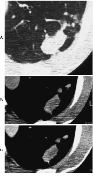
CTの濃度分解能は 0.5% の X 線吸収差を識別可能で ある.断層像でもあり,単純写真の肺癌検出能に比べ明 らかに良好である.50 mAs 以下の低線量 CT での集検で 発見された肺癌のうち単純写真で発見できるのは 23∼ 28% とされている.1-3 CTでも,単純写真と同様に見逃しが起こる可能性が ある.White ら4 は,CT で見逃された 14 例の検討を行 い,その理由として,気管支内あるいは血管の大きい肺 門周囲に存在する場合,非典型的な形を呈している場合 や部分容積減少に伴う場合をあげ,また単純写真と異な り下葉に多いとしている.他に大きな異常がある場合, い わ ゆ る satisfaction-of-search observer error に つ い て も指摘している.手術可能肺癌において,同時性多発肺 癌の頻度は決して少なくないことが報告されており,5,6 ひとつの肺癌が見つかっても肺門や下葉など見落としが 起こりやすい領域には注意が必要である(Figure 1).肺結節の質的診断
肺結節の診断は,組織学的あるいは細菌学的な確定診 断によるが,経過観察で縮小すること,あるいは軟部組 織濃度を示す結節では 2 年以上大きさや性状に変化がなFigure 1. Multiple lung cancers. Conventional and high-resolution CT show a small nodule with ground-glass attenuation in the left upper lobe(A)and a nodule with soft-tissue attenuation in the right lower lung (B). One of the reasons for failure to detect a lung cancer is major distracting findings present elsewhere in the thorax(satisfaction-of-search observer error). The nodules located immediately adjacent to the me-diastinum or hilum may be overlooked.
A
いことから良性と診断できる.CT をはじめとする画像 診断による肺結節の良性と悪性の鑑別診断は,生検や手 術が必要であるか,内科的治療を行うのか,経過観察や 放置で良いのかを決定する目的で行われる.そこで,正 しい鑑別診断は,迅速な治療方針の決定とともに,不要 な生検・治療や経過観察を減少させることになる. CTによる質的診断は,結節の組成を判断するための 結節の濃度や造影効果を見る方法と,結節の進展様式や 周囲の正常構造の反応を判断するための辺縁や内部構造 の解析を行う方法とがある. 1.結節濃度からの鑑別診断 結節濃度を判断する鑑別診断において,良性病変とし て信頼性の高い所見として,境界明瞭な結節で,脂肪濃 度の存在(過誤腫)と中心部や結節全体をしめる良性パ ターンの石灰化がある.また,生検の禁忌である血管性 病変は,心臓や大血管と同程度の造影効果を示すことに より CT で診断が可能である.過誤腫のうち thin-section CT上脂肪濃度が確認できるのは 50% 以下である.7 内 部濃度の解析には部分容積減少の影響を考慮する必要が ある.1 cm以下の結節に 10 mm 厚の CT で内部濃度を検 討しても石灰化の検出は十分ではない(Figure 2). 血管性病変や石灰化結節を除いた肺結節における造影 効果による良性と悪性の鑑別について,Swensen ら8 の 多施設共同研究をおこなった.3 mm 厚の CT で 300 mg! mlのヨード造影剤を使うと造影 4 分後までに,悪性結節 の 98% で 15 HU 以上の造影効果が認められ,それ以下 の造影効果しか認めない結節は良性病変である可能性が 非常に高いことを示した(Figure 3).しかし,良性結節 の 40% 以上に 15 HU 以上の造影効果 が 認 め ら れ て い る.ヨード過敏症の可能性があることは informed con-sentにおいて重要なことである. 2.高分解能 CT(HRCT)を用いた内部構造や辺縁の解析 による鑑別診断 Siegelmannら9 は,CT 診断において結節の辺縁を 4 つに分類し,各々の良性悪性の頻度を示している.spicu-lationを伴なった非常に不規則な辺縁を示した結節では 94.3% が,不規則な凹凸や軽度の spiculation を示した結 節で 86.6% が悪性であり,境界明瞭で辺縁平滑な結節の うち 78.8% が良性であった.一方,最も多かった中等度 平滑とした結節では,良性 42.3%,悪性 57.7% と辺縁の 性状のみでは鑑別が困難な症例が多いことを示した.し かし,その後の検討では,内部構造や辺縁の解析におい て HRCT の所見は病理像をよく反映するとされる.10,11 わが国では,HRCT における内部構造や辺縁の種々の所 見を用いて鑑別診断が行われている.その所見は,主に 従来の断層写真で検討された所見が用いられているた め,所見の意義は多くの症例での検討に基づいている. Table 1に良悪の鑑別に使用される HRCT 所見と,我々 の施設で確定診断された 3 cm 以下の肺結節におけるそ の出現頻度を示す.12 多くの所見において良性と悪性の
Figure 2. Partial volume effect. Calcification at the center of the lobulated nodule, which is shown on thin-section CT images with a 2 mm slice-thickness(A), is not confirmed on the CT image with 10 mm slice-thickness (B)due to the partial volume effect.
Table 1. Significance of HRCT findings for malignant nodules in a study including 90 malignant and 58 benign nodules ≦ 3 cm
PPV Accuracy Frequency HRCT Findings P value Benign Malignant 0.723 0.742 0.804 0.755 0.864 0.775 0.738 0.795 0.709 0.688 0.716 0.696 0.490 0.585 0.760 0.682 0.540 0.659 0.682 0.639 0.513 0.694 < 0.01 = 0.09 < 0.01 < 0.01 < 0.01 < 0.01 < 0.01 < 0.01 < 0.01 = 0.17 < 0.01 0.483 0.138 0.155 0.448 0.138 0.155 0.376 0.259 0.431 0.259 0.500 0.811 0.233 0.411 0.889 0.567 0.344 0.622 0.644 0.678 0.367 0.811 irregular with GGA multiple multiple presence presence presence presence presence presence absence Shape Density Subsegment
Connection with vessel Convergence Air bronchogram Pleural tag Notch Spiculation Bubble-like a. Satellite lesion
GGA: ground-glass attenuation, Bubble-like a.: bubble-like appearance, PPV: positive predictive value.
間で出現頻度に有意差が認められる.しかし個々の所見 の正診率や positive predictive value は必ずしも高い と いえず,診断にはこれらの組み合せが必要で,多くの所 見が認められると確信度が高くなる.
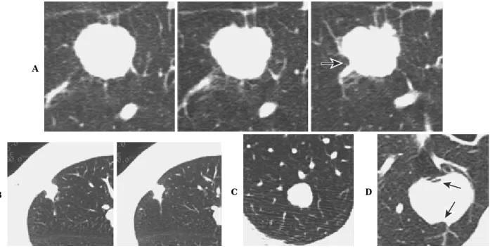
進展様式の違いから肺癌の内部や辺縁部の HRCT 所 見は大きく二つに分けられる.ひとつは,Acinar adeno-carcinoma, Solid adenocarcinoma with mucin forma-tion,Squamous cell carcinoma,Small and Large cell car-cinomaや転移のように,肺胞構造を破壊性にあるいは肺 胞腔を埋めるように増殖するために,軟部組織濃度で境 界明瞭なタイプである.もう一つは,Bronchioloalveolar carcinoma,Adenocarcinoma with mixed subtypes(pre-dominantly BAC pattern)で代表される辺縁部で肺胞上 皮を置換しながら増殖するため辺縁部では細かい spicu-lation(スピクラ)やスリガラス濃度を示すタイプである. 進 展 様 式 の 違 い か ら,所 見 の 組 み 合 せ に よ る 典 型 的 HRCTパターンも異なり,鑑別にあがる良性病変の特徴 的 HRCT 所見も異なる. 1)軟部組織濃度を示し,境界明瞭な結節の鑑別診断 (非置換型肺癌との鑑別) 破壊性あるいは充填性に進展するタイプの肺癌の典型 的な HRCT 所見は,軟部組織濃度を示し,境界明瞭で辺 縁に凹凸がある不整形で,肺動脈や気管支の関与が認め られ,小葉と無関係に広がるため静脈を越えて進展して いる.大きさや浸潤の程度により,気管支の閉塞,気管 支透亮像の存在13,spiculation や胸膜嵌入を伴う(Fig-ure 4A). 一方,軟部組織濃度を示す境界明瞭な良性結節のうち, 炎症性病変や肉芽腫性病変では,直線状の境界あるいは, 隣接肺の病巣方向への突出14,15 や散布巣を認める(Fig-ure 4B)ことが典型的である.また過誤腫などの良性腫瘍 では辺縁平滑で円形あるいは楕円形で血管の関与がない Figure 3. Lung nodule enhancement . HRCT shows
a smooth and well-defined nodule(A). The density of the nodule on contrast-enhanced CT(C)is the same as that on unenhanced CT(B). This nodule is strongly suggestive of a benign nature.
A
B
Figure 4. Nodules with soft-tissue attenuation and well-defined borders. A. Squamous cell carcinoma. B. Tuberculoma. Cand D. Hamartoma Typical HRCT findings of a malignant nodule with an expansive and destructive pattern are some ir-regular undulations, slight spiculation, and notching(arrow)(A). Typical HRCT findings of a benign nodule are polygonal shape, satellite lesions(B), rounded shape, and no vascular connections(C).In many benign nodules, the locations of the hollow are different from those where vessels or bronchi contact with the nodule(arrows)even if the nodule shows some irregular undulation and contact with the vessels or bronchi(D).
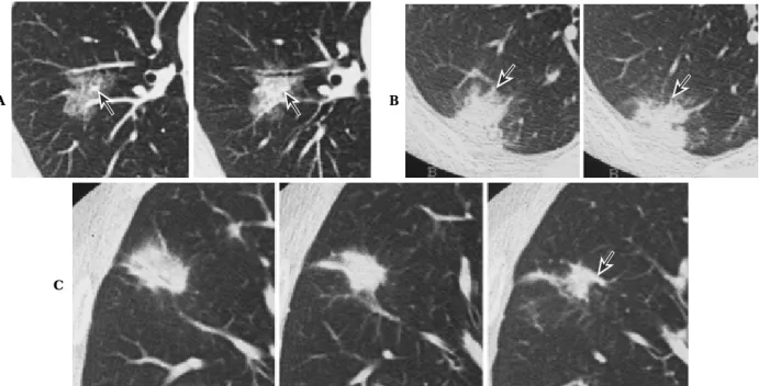
Figure 5. Strand opacification projecting from the nodule. A. Tuberculoma. B. Intrapulmonary lymph node. C. Small cell lung cancer The smooth thickening of the bronchovascular bundles, interlobular septum or distal vessels, which con-tact with the nodule, is observed in a benign inflammatory nodule such as tuberculoma(A)or an intrapulmonary lymph node(B). Note that the nodule does not contain notching or fine spiculation. The long strand opacities are irregular and accompany HRCT findings of fine spiculation or notching in a malignant nodule(C).
C D
B
A B
ことが特徴である(Figure 4C). 良性結節が凹凸を示し血管と接した場合には,くびれ は血管や気管支と接しない部位で認められることが多く (Figure 4D).一方,悪性結節のくびれ(notch)は,血管 や気管支により進展 が 阻 止 さ れ て で き る 場 合 が 多 い (Figure 4A). 結節につながる線状陰影(spiculation など)は,結核 腫などの炎症病変では,平滑で長く,気管支,血管や小 葉間隔壁の肥厚として認識できることも多い(Figure 5 A).また肺内リンパ節も小葉間リンパ装置との関連か ら,円形,多角形の結節から小葉間隔壁の肥厚による長 い線状陰影を認める場合がある(Figure 5B).いずれも fine spiculaといわれる短い spiculation が少なく,平滑 な弧状あるいは直線状の辺縁であることが特徴である. 一方悪性結節では,長い棘状突起がある場合には,通常 fine spiculaも認められる.末梢性の小細胞癌では気管支 粘膜下を進展し,気管支血管束の肥厚所見をとるが,そ の辺縁は不規則である(Figure 5C).中枢側に血管の関与 がある場合,多臓器からの転移も考慮に入れるべきであ る.16,17 良性病変の特徴である,辺縁平滑で境界明瞭な軟部組 織濃度を示す肺癌として,腺管腺癌やカルチノイドのよ うな気管支内腫瘍がある.気管支の途絶と末梢支配領域 の透過性亢進や小葉中心性陰影の存在が気管支内腫瘍を 示唆する所見である(Figure 6). また,多角形や隣接肺の病巣方向への突出という良性 病変の特徴は,既存構造である葉間胸膜や気腫性!胞に 接した結節の場合には良性の根拠とならない.肺気腫患 者の肺癌でも同様に多角形や扁平な形を示すことが多い (Figure 7). 2)辺縁部に多数の spiculation やスリガラス濃度を示 す結節の鑑別診断(置換型肺癌との鑑別) 辺縁部で肺胞上皮を置換しながら進展する肺癌では, 内部に気管支透亮像を認め,細かい spiculation や結節辺 縁部にスリガラス濃度(ground-glass attenuation; GGA) を示すことが特徴である.内部の線維化の程度に応じて, 多数の血管の関与,集束像や胸膜嵌入が認められる.小 葉と関係のない広がりをするため静脈の関与が認められ る.(Figure 8A,B).器質化肺炎や限局性線維化巣でも同 様な所見を示すが,良性を示唆する所見として,静脈の 巻き込みを認めないこと,18 気管支血管束に沿った広が りが鑑別に有用である(Figure 8C). 器質化肺炎の特徴として,Kohno ら19 は,胸膜に広く 接している,気管支血管束に沿って位置する,扁平ある Figure 6. Round or oval malignant nodules. A. Carcinoid tumor. B. Acinar adenocarcinoma Endobronchial malignant tumors show rounded and well-defined nodules. The obstruction of bronchi(arrows), and satellite lesions or surrounding hyperlucent areas in the periphery of the nodule are observed.
A
いは紡錘状,多角形を示し,不整な形で散布巣,気管支 透亮像や胸膜陥入を認めることが多いとした(Figure 9). 肺気腫に合併した肺癌は,CT 上肺気腫を認めない患 者の肺癌に比べ,既存構造に進展を阻止され,形は扁平 で,長い棘状突起を示し,気管支透亮像よりも bubble-like appearanceを示すことが多いという特徴がある.ま た,周囲に既存の気道病変を反映して小葉中心性陰影(散 布巣)を認めることも多い(Figure 10).20 このため,肺 気腫例では肺癌と器質化肺炎の鑑別が困難な症例が多く 認められ,初期には 1∼2 ヶ月間隔での経過観察が必要で ある.一方,CT 上既存肺に肺気腫がない場合には上述し た器質化肺炎の所見は肺癌との鑑別に有用な所見であ る.そこで結節影の画像診断において既存肺の状態を検 討することが必要である.肺気腫例では結節周囲のスリ ガラス影の存在が肺癌の確信度を高くする. 上述したように軟部組織濃度を示し,境界明瞭な肺癌 には,倍加時間が短いとされる組織型が多い.一方,鑑 別の対象となる良性疾患では良性腫瘍や血管性病変など 経時的変化がほとんどない疾患が多いため retrospective にみれば,以前の画像でその存在が確認できる場合が多 い.また我々の検討12 では,2∼3 cm の結節影を示した 吸収遅延性肺炎や器質化肺炎の多くは,発見前数ヵ月以 内に一時的な咳嗽や喀痰,発熱の呼吸器症状を呈してい た.一方既存肺に肺気腫などの慢性気道疾患を認めない 場合,辺縁に spiculation やスリガラス影を伴う 3 cm 以 下の肺癌例では症状を呈することは少ない.このように 一回の画像のみに頼ることなく,臨床症状や以前の画像 との対比をあわせて診断することも鑑別診断を行う上で 非常に重要である. CTのみで発見されるような小結節で,スリガラス濃 度のみあるいは辺縁部がスリガラス濃度を示し内部に充 実部分を持つタイプは,炎症の治癒過程の場合があるが, 経過観察で消退しない場合は腺癌あるいは異型腺腫様過 形成の可能性が高い.境界が明瞭でスリガラス濃度が高 くなるほど腺癌の可能性は高く,notch が認められると より確信度は高まる.境界不明瞭あるいは円形性に乏し いほど良性病変を示唆する(Figure 11). 3)1 cm 以下の良性結節の HRCT 像 CTで発見される 1 cm 以下の結節は,良性結節のこと が多く,通常 5 mm 以下では経過観察,5∼10 mm では経 過観察あるいは生検が勧められている.経過観察を長く Figure 7. Polygonal malignant nodule. A. Adenocarcinoma with mixed subtypes.
B.Adenocarcinoma with mixed subtypes in a pulmonary emphysema patient. The malignant nodules may show a polygonal shape if they are adjacent to an interlobar fissure(A)or dilated air spaces(B).
Figure 8. Solitary pulmonary nodules with ground-glass attenuation or fine spiculation at the periphery. A and B. Ade-nocarcinoma with mixed subtypes(predominantly BAC pattern)C. Delayed-resolution pneumonia A vein running be-tween two subsegments is observed within the nodule in malignancy(A and B). However, it is observed at the border of the nodule in benign lesions(C).
Figure 9. Benign nodules . A-C. Organizing pneumonias. D. Aspergillosis. Relatively broad pleural-based lesions(A, B), location along the bronchovasucular bundle(B, D), and with somewhat flat, spindle-shaped or trapezoidal shapes(A-D) are often helpful in differentiating focal organizing pneumonias from lung cancer.
A B
C
A B C
続けるときの被検者の精神的,経済的負担や悪性病変で あったときの診断の遅延を考えるとできるだけ早く,生 検し確定診断するか経過観察を終了することを考えるべ きである.そのためには,1 cm 以下の肺癌では特徴的所 見を示さないことが多いとされており,良性結節の特徴 を知っておくことも必要である. 我々の施設で thin-section CT が施行された,石灰化の ない 1 cm 以下の良性 159 結節について,HRCT所見を以 下のように分類しその頻度を検討した.スリガラス濃度 のみを示す結節は除き,同時期に確定された 37 悪性結節 (20 転移を含む)を対象とした.21 各 HRCT 所見の良性
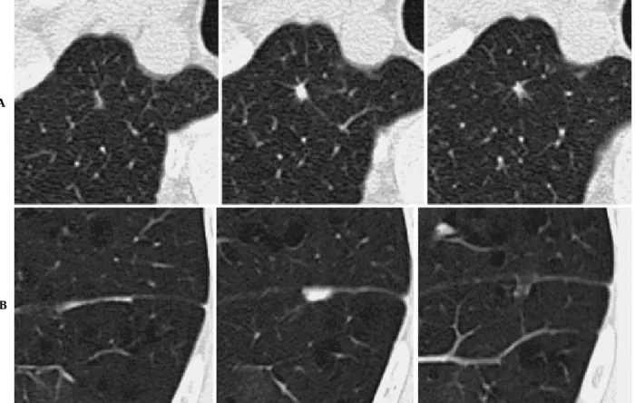
結節における頻度と positive predictive value をみると, 5 mm以下,散布巣があること,小葉間隔壁や気管支血管 束の肥厚があることは,高頻度でかつ良性の特異性が高 い所見であり,境界明瞭,平滑,円形・楕円形,血管の 関与の所見は,良性結節でよく認められる所見であった が,悪性結節にも比較的高頻度に認められた.ノッチや fine spiculationは 1 cm 以下の良性結節で認められるこ とは少なく,悪性を疑う所見であった.これらの所見を 組み合せてその頻度や特異性を見てみると,胸膜から離 れて存在し,境界の明瞭不明瞭にかかわらず,散布巣, 気管支血管束や小葉間隔壁の肥厚を伴う結節が全良性結 節の約半数で認められた(Figure 12).胸膜から離れて存 在し,多角形の結節(Figure 13A)が 25%,扁平な形の 結節と,胸膜に接する多角形あるいは半円形の結節 (Fig-ure 13B)がそれぞれ約 20% の頻度であった.以上の所見 を示す悪性結節はほとんどなかった.血管の関与がない 円形あるいは楕円形の結節は良性結節(Figure 14A)の 12 %と頻度は少なく,また,血管の関与があり気管支血管 束や小葉間隔壁肥厚を伴わない円形結節(Figure 14B)や spiculationか notch のある不整形の結節の良性での頻 度は 10% 以下であり,一方,それぞれ悪性結節(Figure 14C)の 25% ずつがこの所見を示した. a)1 cm 以下で散布巣や気管支血管束・小葉間隔壁の 肥厚を伴う結節,多角形や扁平な結節,胸膜に接した半 円形の結節は肺気腫など慢性気道性疾患がなければ良性 の典型的所見と考えられる.b)血管の関与がないある いは血管の関与があるにもかかわらず周囲に散布巣や気 管支血管束,小葉間隔壁の肥厚がない円形,楕円形の良 性結節の頻度は比較的少なく,低分化な肺癌や転移との 鑑別のため短期間の経過観察を行い,大きさの変化を確 認する必要がある.c)fine spiculation や notch を伴う結 節では悪性の可能性が十分あり注意深い経過観察あるい は生検が必要である.ただし以上の方針を決定する前に 可能な限り,以前の画像と対比することが必要である. スリガラス濃度を示す結節については前述したとお り,炎症の治癒過程である場合を除き AAH あるいは腺 癌の可能性が高いが,手術による摘出が妥当かどうかに ついては明らかでない.
Figure 10. Lung cancers in pulmonary emphysema patients. A-C. Adenocarcinoma with mixed subtypes. Lung cancers with pulmonary emphysema tend to contain long spiculation and!or pleural tags, and straight or concave margins and thus form trapezoidal shapes because collagen fibers proliferate in interlobular septa or perivascular or peribronchial in-terstitium, and tumor growth is interrupted by thick inin-terstitium, bullae or dilated air spaces.
まとめ
正確な画像診断は,被検者を適切な治療へ迅速に導く
とともに,無駄な侵襲や時間,また経済的・精神的負担 を省くことができる.従来から行われた画像所見と最終 診断との解析結果から,画像のみで,悪性,良性と確信 Figure 11. Small nodules with ground-glass attenuation. A. BAC(Noguchi A). B. Atypical adenomatous hyperplasia(AAH). C.Benign nodule(unclassified)BAC shows a well-defined and rounded contour with notching(A). AAH shows an ill-defined and rounded contour(B). A benign nodule shows an ill-defined and irregular contour(C).
Figure 12. Benign nodules with soft tissue density less than 1 cm in diameter. A-C. Benign nodules HRCT images show the nodule with satellite lesions and!or thickening of interlobular septa or bronchovascular bundles(A-C). This HRCT pattern was most frequent in benign nodules smaller than 1 cm.
A B C
A B
Figure 13. Benign nodules with soft tissue density less than 1 cm in diameter. A and B. Benign nodules HRCT images show a polygonal nodule(A)and a semicircular nodule broadly contacting the pleura(B). These HRCT patterns were fre-quently observed in benign nodules.
Figure 14. Small nodules with soft tissue density less than 1 cm in diameter. A. Benign nodule. B. Benign nodule. C. Ma-lignant nodule(metastasis)HRCT images show a rounded or oval nodule without vascular connections or satellite lesions (A). This finding was relatively infrequent in benign nodules, and also rare in malignant nodules. HRCT images show rounded or oval nodules with vascular connection and without thickening of BVB or ILS(B and C). This HRCT pattern was infrequent in benign nodules and was not infrequent in the malignant nodules including metastasis.
B
A
度高く診断できるものもある.画像診断を行うためには, 良性・悪性結節の典型像を把握し,結節自体の性状を検 討するとともに,周囲との関係,既存肺の状態など CT 像に現されている所見全てを検討することも必要であ る.さらに,以前の画像との対比,臨床症状などを加味 することで,確信度を上げていくことが可能になる. REFERENCES
1.Kaneko M, Eguchi K, Ohmatsu H, et al. Peripheral lung cancer: screening and detection with low-dose spiral CT versus radiography. Radiology. 1996;201:798-802. 2.Henchke CI, McCauley DI, Yankelevits DF, et al. Early
lung cancer action project: overall design and findings from baseline screening. Lancet. 1999;354:99-105. 3.Sone S, Li F, Yang ZG, et al. Characteristics of lung
can-cers invisible on conventional chest radiography and de-tected by population based using spiral CT. Br J Radiol . 2000;73:137-145.
4.White CS, Romney BM, Mason AC, et al. Primary carci-noma of the lung overlooked at CT: Analysis of findings in 14 patients. Radiology. 1996;199:109-115.
5.Zwirewich CV, Miller RR, M!ller NL. Multicentric adeno-u carcinoma of the lung: CT-pathologic correlation. Radiol-ogy.1990;176:185-190.
6. Suzuki K, Takahashi K , Yoshida J , et al . Synchronous double primary lung carcinomas associated with multiple atypical adenomatous hyperplasia . Lung Cancer . 1998; 19:131-139.
7.Siegelmann SS, Khouri NF, Scott WW Jr, et al. Pulmonary hamartoma: CT findings. Radiology. 1986;160:313-317. 8.Swensen SJ, Viggano RW, Midthun DE, et al. Lung nodule
enhancement at CT: multicenter study. Radiology . 2000; 214:73-80.
9.Siegelmann SS, Khouri NF, Leo FP, et al. Solitary pulmo-nary nodules: CT assessment . Radiology . 1986;160:307-312.
10.Kuriyama K, Tateishi R, Doi O, et al. CT-pathologic corre-lation in small peripheral lung cancers . AJR . 1987;149: 1139-1143.
11.Zwirewich CV, Vedal S, Miller RR, et al. Solitary pulmo-nary nodule: high-resolution CT and radiologic-patho-logic correlation. Radiology. 1991;179:469-476.
12.松本常男,三浦剛史,田中伸幸,他.HRCT による肺野小 結節の鑑別診断における副所見や臨床情報の有用性.肺 癌.2000;40:413.
13.松本満臣,堀越浩幸,茂木孝夫,他.境界明瞭・辺縁平滑 な肺結節の CT 診断.臨放.1993;38:75-83.
14.Furuya K, Murayama S, Soeda H, et al. New classification of small pulmonary nodules by margin characteristics on high-resolution CT. Acta Radiol . 1999;40:496-504.
15.尾下文浩,江口研二,宮 敏路,他.肺野限局性炎症性病
変の thin-section CT 像―切除肺との 対 比―.日 医 放 会 誌.1989;49:1525-1533.
16. Meziane MA , Hruban RH , Zerhouni EA , et al . High-resolution CT of the lung parenchyma with pathologic correlation. Radiographics. 1988;8:27-54.
17.Milne ENC, Zerhouni EA. Blood supply of pulmonary me-tastases. J Thorac Imaging. 1987;2:15-23.
18.清水邦彦,山田耕三,野田和正,他.CT で発見されるよ うな 10 mm 以下の肺野微少病変の thin-section CT 画像 による解析,CT 画像と病理像の対比.臨放.1998;43:9-18.
19. Kohno N , Ikezoe J , Johkoh T , et al . Focal organizing pneumonia: CT appearance. Radiology. 1993;189:119-123. 20.Matsumoto T, Miura G, Tanaka N, et al. Peripheral lung cancer in patients with and without pulmonary emphy-sema: Comparison of CT findings of nodules, surrounding lungs, and growth pattern. Radiology. 1999;213(suppl): 558.
21.松本常男,三浦剛史,田中伸幸,他.1 cm 以下の良性結 節性病変の CT 像.肺癌.2001;41:400.