Paraneoplastic Hypocalcemia Developed in Gastric Cancer
Accompanied by Osteoblastic Metastasis
Jun Okazaki
1, Naoki Muguruma
1, Shinji Kitamura
1, Tetsuo Kimura
1, Koichi Okamoto
1,
Hiroshi Miyamoto
1, Kazuhiro Kishi
2, Yoshimi Bando
3, Takeshi Kondo
4, Itsuro Endo
4,
Masahiro Abe
4and Tetsuji Takayama
1Abstract
Paraneoplastic syndromes are generally defined as clinical disorders associated with malignant diseases, and hypocalcemia associated with cancer is a rare condition. A woman in her 60s was referred to our hospital for the further examination of massive ascites due to carcinoma of unknown primary origin. She complained of numbness around her lips, and marked hypocalcemia of 5.0 mg/dL was noted. After two courses of che-motherapy, computed tomography showed a decrease in the ascites, and her serum calcium level increased. Although hypocalcemia is a very rare condition in patients with gastric cancer, serum calcium values should be evaluated when neurological symptoms are observed.
Key words: hypocalcemia, gastric cancer, bone metastasis
(Intern Med 56: 1345-1349, 2017) (DOI: 10.2169/internalmedicine.56.8545)
Introduction
Paraneoplastic syndromes are generally defined as clinical disorders associated with malignant diseases and are not di-rectly related to the physical effects of the primary or metas-tatic tumors (1, 2). Among the symptoms or laboratory dis-orders that have been reported relating to this broad spec-trum of conditions, hypercalcemia is one of the most com-mon paraneoplastic syndromes, occurring in up to 30% of cancer patients (3). In contrast, hypocalcemia is a rare con-dition that is sometimes observed in the advanced stage of prostatic cancer due to abnormal calcium influx in develop-ing bone metastasis. To date, however, cases of severe hy-pocalcemia associated with gastric cancer have not been re-ported. This report describes a very rare case of persistent hypocalcemia that developed in a patient with advanced gas-tric cancer and bone metastasis.
Case Report
A woman in her 60s visited a hospital because of increas-ing abdominal pain. She underwent esophagogastroduodeno-scopy (EGD) and abdominal CT in which findings were un-remarkable. Four months later, she developed massive as-cites and an abdominal puncture was performed. A cytologi-cal examination of the ascites showed no malignancy, and positron emission tomography (PET)-CT demonstrated no abnormal accumulation of 18F-fluorodeoxyglucose. Diagnos-tic laparoscopy was carried out as a further examination, and multiple disseminated nodules were revealed in the ab-dominal cavity. Histopathologically, the nodules were diag-nosed as adenocarcinoma. Eventually, the patient was re-ferred to our hospital for the further examination of the pri-mary lesion and therapy. When she visited our hospital, she complained of numbness around her lips. Her medical his-tory consisted of a post-thyroidectomy state due to thyroid cancer in her 50s, myoma of the uterus, and ovarian cyst.
1Department of Gastroenterology and Oncology, Tokushima University Graduate School of Biomedical Sciences, Japan,2Department of
Gastro-enterology, National Hospital Organization Higashi Tokushima Hospital, Japan,3Division of Pathology, Tokushima University Hospital, Japan
and4Department of Hematology, Endocrinology and Metabolism, Tokushima University Graduate School of Biomedical Sciences, Japan
Received for publication November 12, 2016; Accepted for publication January 5, 2017 Correspondence to Dr. Naoki Muguruma, [email protected]
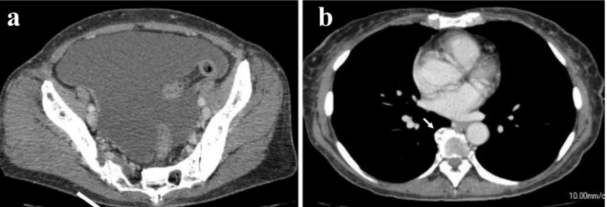
Figure 1. Contrast-enhanced CT showed massive ascites (a) and an abnormal bone formation ad-jacent to the thoracic spine (b, arrow).
a
b
Figure 2. Bone scintigraphy demonstrated the increased up-take of 99m-technetium diffusely throughout the skeleton,
show-ing a ’beautiful bone scan’.
She was not a smoker or alcohol drinker. She had no fever or abdominal pain. In a general examination, her blood pres-sure and heart rate were normal. No lymphadenopathy was noted. Her abdomen was slightly distended due to ascites, but there was no tenderness. Paresthesia was noted in the extremities and lips. The laboratory data (complete blood count, chemistry, urinalysis, tumor markers, and coagula-tion) showed mild anemia (11.8 g/dL of hemoglobin), and the tumor markers carcinoembryonic antigen (CEA) and CA 19-9 were elevated to 8.3 ng/mL and 4,510 U/mL, respec-tively. The lactate dehydrogenase (LDH) and alkaline phos-phatase (ALP) were also elevated to 339 IU/L and 9,668 IU/ L, respectively. Marked hypocalcemia of 5.0 mg/dL was also noted, and the corrected calcium value was 5.9 mg/dL by the correction formula applying 3.1 g/dL of serum
albu-considered to have caused her hypocalcemia.
Given the above findings, the patient was admitted imme-diately to our hospital due to severe hypocalcemia. Contrast-enhanced CT demonstrated massive ascites (Fig. 1a), but there were no lung or liver tumors. It also revealed an ab-normal bone formation adjacent to the thoracic spine (Fig. 1b arrow). Bone scintigraphy using technetium 99m (99mTc) showed an increased uptake of the radio-labeled agent diffusely throughout the skeleton: cervical-thoracic-lumbar spine, pelvis, femurs, skull and shoulders. There was no increased uptake in the kidneys (Fig. 2). Additional en-docrinological studies were performed, and the following re-sults were recorded: inorganic phosphorus (IP) 6.0 mg/dL (normal range: 2.4-4.6 mg/dL), magnesium 2.7 mg/dL (1.6-2.3 mg/dL), intact parathyroid hormone (PTH) 78 pg/mL (10-65 pg/mL), bone-specific alkaline phosphate 848.0 μg/L (3.8-22.6 μg/L), carboxy-terminal telopeptide of type I colla-gen (ICTP) 42.5 ng/mL (<4.5 ng/mL), 1,25-(OH)2 vitamin D 148 pg/mL (20-60 pg/mL), urine N-terminal telopeptide 731 nmolBCE/nmolCr (14.3-89.0 nmolBCE/nmolCr), urine calcium 12.8 mg/gCr, and maximum tubular reabsorption of phosphate per glomerular filtration rate (GFR) (TmP/GFR) 6.0 mg/dL. Based on these findings, she was diagnosed with cancerous peritonitis and diffuse bone metastasis derived from cancer of unknown primary (CUP). The prolonged hy-pocalcemia was considered a paraneoplastic syndrome, pre-sumably due to the exhaustion of calcium by diffuse os-teoblastic bone metastasis.
Treatment with intravenous administration of paclitaxel and carboplatin was started in accordance with the therapeu-tic guidelines for CUP. After two courses of chemotherapy, a CT scan showed a gradual decrease in the ascites as well as a decrease in the CEA. The serum calcium level was also increased (Fig. 3). During chemotherapy, she complained of abdominal discomfort, and EGD was performed. A slightly depressed pale lesion with fold convergence was found in the upper body (Fig. 4a-c). A histopathological examination of a biopsy specimen from the lesion revealed a poorly
dif-Figure 3. Clinical course of the case. The serum calcium level was increased and the CEA level de-creased due to chemotherapy.
1μg 4μg 6μg 1,200mg 1,800mg 2,400mg 3,200mg 4,000mg 4,800mg 40mL 80mL 120mL 80mL Vit.D/day (p.o.) Calcium L-aspartic acid/day (p.o.) 8.5%Calcium gluconate/day (i.v.) 0 1 2 3 4 5 6 7 8 9 0 1 2 3 4 5 6 7 8 9 Corrected Ca CEA 2014.10 Corrected Ca (mg/dL) CEA (ng/mL) 2014.9 Carboplatin 6mg/GFR+25 Paclitaxel 175mg/m2 Carboplatin 6mg/GFR+25 Paclitaxel 175mg/m2 TC Therapy tri-weekly
Figure 4. EGD demonstrated a slightly depressed pale lesion with fold convergence (a). In images with indigocarmine spray (b), encroachment and swelling of the converging folds were enhanced, and narrow band imaging with magnification (c) showed irregular microvessels on the surface of the le-sion. Histopathology of a biopsy specimen showed a poorly differentiated tubular adenocarcinoma (d).
a
b
Table. Cases of Osteoblastic Metastases Due to Primary Gastric Cancer. Age Sex Histology Endoscopic
appearance
Serum
calcium Prognosis Reference 21 M sig Erosion 7.8 mg/dL 2 months 16 65 M diffuse Linitis plastica 9.1 mg/dL - 15
45 F sig Micro-nodular - - 14
71 M sig Small erosion - - 13
41 F por Slight depression - 8.5 months 12 64 M por/sig Slight depression - - 12 40 F sig Folds thickening 7.9 mg/dL 4 months 11
60 M sig - normal - 10
64 M por/sig Borrmann-III - 3 months 9 57 M mod Tiny erosion 8.6 mg/dL 3 months 8 36 M - Ulceration 10 mg/dL 1 month 7 50 M - Linitis plastica 9.2mg/cent 2 weeks 6 69 F por Small depression 5.0 mEq/L Alive Our case
diffuse: diffuse pattern adenocarcinoma, mod: moderately differentiated adenocarcinoma, por: poorly differentiated adenocarcinoma, sig: signet ring cell carcinoma, -: data were not shown
ules. Ultimately, this patient was diagnosed with primary gastric cancer with abdominal dissemination and bone me-tastases. The chemotherapy regimen was changed to S-1 plus docetaxel in accordance with the therapeutic strategy for gastric cancer. Oral calcium intake was discontinued af-ter six months of administration, but the serum level of cal-cium remained stable. She has been receiving chemotherapy as an outpatient and is in stable condition with normal se-rum calcium levels.
Discussion
Bone metastases often occur during the clinical course of prostate cancer, breast cancer, and lung cancer. However, the incidence of bone metastases in gastric cancer is relatively low, ranging from 1.0% to 20.0% (4), and osteoblastic me-tastases are extremely rare. Osteoblastic meme-tastases are asso-ciated with higher serum levels of alkaline phosphatase and hypocalcemia (5). Hypocalcemia occurs more frequently in patients with osteoblastic metastases (28%) than in those with osteolytic metastases (11%) (5). Osteoblastic metasta-ses from gastric cancer have been reported only in 12 cametasta-ses from 1,977 until now (Table) (6-16). Among these reported cases, only 2 showed hypocalcemia, and their serum calcium values were 7.8 mg/dL and 7.9 mg/dL, respectively (11, 16), while our case showed severe and intractable hypocalcemia (6.0 mg/dL).
In our case, the patient underwent thyroidectomy previ-ously, which may have caused potential hypoparathyroidism and impaired the regulation of serum calcium. The condition also showed the characteristic diffuse uptake of the radioiso-tope throughout the skeleton that is called a ‘beautiful bone scan’ in bone scintigraphy terminology. It is speculated that increasing new bone formation caused the excessive calcium uptake from the blood, regardless of the continuous supply
cemia in this case. Although the serum phosphate was slightly increased, it was likely due to the effect of tumor lysis resulting from the extensive bone metastasis.
Among gastric cancer cases accompanied by osteoblastic metastases, 80% (10 out of 11) were poorly differentiated adenocarcinoma or signet-ring cell carcinoma (Table). In bone metastasis of cancer cells, various molecular mecha-nisms seem to be active and involve, for example, insulin-like growth factor (IGF), transforming growth factor-beta (TGF-β), bone morphogenetic proteins (BMP), and platelet-derived growth factor (PDGF) (17). However, whether or not these molecules cause hypocalcemia is unclear. In our case, no definite key molecule was identified, so it might be possible that other osteoblastic factors are associated with the patient’s marked osteoblastic bone metastasis and severe hypocalcemia. The prognosis for gastric cancer with os-teoblastic bone metastases is generally poor (18). However, our case has remained alive for more than two years since the initial diagnosis without disease progression, probably owing to aggressive chemotherapy.
In conclusion, we experienced a case of prolonged hy-pocalcemia that developed in a patient with advanced gastric cancer and bone metastasis. Although hypocalcemia is a very rare condition in patients with gastric cancer, serum calcium should be evaluated when neurological symptoms are observed.
The authors state that they have no Conflict of Interest (COI).
References
1. Kanaji N, Watanabe N, Kita N, et al. Paraneoplastic syndromes
associated with lung cancer. World J Clin Oncol 5: 197-223, 2014.
2. Spiro SG, Gould MK, Colice GL; American College of Chest
Physicians. Initial evaluation of the patient with lung cancer: symptoms, signs, laboratory tests, and paraneoplastic syndromes:
Chest 132 (3 Suppl): 149S-160S, 2007.
3. Farolfi A, Ferrario C, Aquilina M, et al. Paraneoplastic hypocalcemia-induced heart failure in advanced breast cancer: A case report and literature review. Oncol Lett 10: 773-777, 2015.
4. Yoshikawa K, Kitaoka H. Bone metastasis of gastric cancer. Jpn J
Surg 13: 173-176, 1983.
5. Raskin P, McClain CJ, Medsger TA Jr. Hypocalcemia associated
with metastatic bone disease. A retrospective study. Arch Intern Med 132: 539-543, 1973.
6. Banerjee AK, Banerjee K, Bhattacharya B. Osteosclerotic bony
metastases from carcinoma stomach. J Indian Med Assoc 68: 38-39, 1977.
7. Carstens SA, Resnick D. Diffuse sclerotic skeletal metastases as
an initial feature of gastric carcinoma. Arch Intern Med 140: 1666-1668, 1980.
8. Uchida T, Shikata T, Shimizu SI, et al. Gonadotropin and alkaline
phosphatase producing occult gastric carcinoma with widespread metastasis of generalized bone. Cancer 48: 140-150, 1981.
9. Mohandas KM, Swaroop VS, Krishnamurthy S, et al. Unusual
bone metastasis as the initial symptom of gastric cancer-a report of four cases. Indian J Cancer 30: 146-150, 1993.
10. Narváez JA, Narváez J, Clavaguera MT, Juanola X, Valls C, Fiter
J. Bone and skeletal muscle metastases from gastric adenocarci-noma: unusual radiographic, CT and scintigraphic features. Eur Radiol 8: 1366-1369, 1998.
11. Chung YS, Choi TY, Ha CY, et al. An unusual case of osteoblastic
metastasis from gastric carcinoma. Yonsei Med J 43: 377-380, 2002.
12. Koyama M, Koizumi M. Two cases of diffuse osteoblastic
metas-tases from early type gastric cancer. Clin Nucl Med 32: 545-547, 2007.
13. Kang SH, Kim JI, Moon HS, et al. Overt bone marrow metastasis
from early gastric cancer. Endoscopy 40 (Suppl 2): E34-E35, 2008.
14. Anagnostopoulos G, Sakorafas GH, Kostopoulos P, Margantinis G,
Tsiakos S, Pavlakis G. Early (mucosal) gastric cancer with syn-chronous osteosclerotic bone metastases: a case report. Eur J Can-cer Care 19: 554-557, 2010.
15. Santos VM, Vieira TA, Marinho CS, Loures TP, Brandao BB,
Botan RN. Infiltrating gastric adenocarcinoma with disseminated osteoblastic metastases. An Sist Sanit Navar 36: 153-157, 2013.
16. Ermi! F, Erkan ME, Besir FH, Oktay M, Kutlucan A, Aydın Y.
Osteoblastic metastasis from signet ring cell gastric cancer in a young male. Turk J Gastroenterol 25 (Suppl 1): 284-286, 2014.
17. Iguchi H. Recent aspects for disseminated carcinomatosis of the
bone marrow associated with gastric cancer: What has been done for the past, and what will be needed in future? World J Gastroen-terol 21: 12249-12260, 2015.
18. Kim HS, Yi SY, Jun HJ, et al. Clinical outcome of gastric cancer
patients with bone marrow metastases. Oncology 73: 192-197, 2007.
The Internal Medicine is an Open Access article distributed under the Creative Commons Attribution-NonCommercial-NoDerivatives 4.0 International License. To view the details of this license, please visit (https://creativecommons.org/licenses/ by-nc-nd/4.0/).
Ⓒ 2017 The Japanese Society of Internal Medicine http://www.naika.or.jp/imonline/index.html